Large-Scale Identification of Pathogenicity Factors in Bartonella by Signature-Tagged Mutagenesis
←
→
Page content transcription
If your browser does not render page correctly, please read the page content below
Large-Scale Identification of Pathogenicity Factors

                       in Bartonella

        by Signature-Tagged Mutagenesis

                    Inauguraldissertation

                                zur

         Erlangung der Würde eines Doktors der Philosophie

                           vorgelegt der

          Philosophisch-Naturwissenschaftlichen Fakultät

                       der Universität Basel

                               von

                    Luis-Henri Saenz Dürrmeier

                    aus Ingolstadt, Deutschland

                           Basel, 2005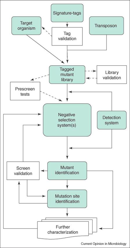
Genehmigt von der Philosophisch-Naturwissenschaftlichen Fakultät

auf Antrag von

Prof. Christoph Dehio

Prof. Urs Jenal

Prof. Guy Cornelis

(Dissertationskomitee)

Basel, den 05.04.2005

                                                           Dekan: Prof. Hans-Jakob Wirz
FÜR SIMONE
STATEMENT TO MY PHD THESIS

This work was carried out from March 2001 to April 2005 in the group of Prof. Christoph

Dehio in the Division of Molecular Microbiology at the Biozentrum of the University of

Basel. My PhD thesis committee consisted of:

Prof. Christoph Dehio

Prof. Urs Jenal

Prof. Guy Cornelis

My PhD thesis is written in a cumulative format. It consists of the published review article

about the signature-tagged mutagenesis method used for my work and the manuscript of a

research article about my work. The chapter “Concluding Remarks” summarizes the major

findings, the chapter “Perspective” discusses the continuation of the projects started based on

my work.
TABLE OF CONTENTS

TABLE OF CONTENTS

STM REVIEW ...................................................................................................................... 1
             Introduction ............................................................................................................ 1
             Modular structure of the STM approach ................................................................ 1
             Recent advances in STM........................................................................................ 2
             Conclusions ............................................................................................................ 7
             Acknowledgements ................................................................................................ 7
             References and recommended reading................................................................... 7
STM MANUSCRIPT............................................................................................................ 9
             Abstract .................................................................................................................. 10
             Introduction ............................................................................................................ 11
             Results .................................................................................................................... 14
             Discussion .............................................................................................................. 18
             Material and methods ............................................................................................. 30
             Acknowledgements ................................................................................................ 34
             References .............................................................................................................. 35
             Figures.................................................................................................................... 41
             Tables ..................................................................................................................... 45
CONCLUDING REMARKS ................................................................................................ 53
PERSPECTIVE..................................................................................................................... 56
REFERENCES...................................................................................................................... 59
ACKNOWLEDGEMENTS .................................................................................................. 60
CURRICULUM VITAE ....................................................................................................... 62
Signature-tagged mutagenesis: technical advances in a negative
selection method for virulence gene identification
Henri L Saenz and Christoph Dehio
Signature-tagged mutagenesis (STM) is a powerful negative short individual DNA sequences) inserted in the trans-
selection method, predominantly used to identify the genes of a posons to mark mutants individually. Mutants that carry
pathogen that are required for the successful colonization of an distinct signature-tags are pooled and injected into the
animal host. Since its first description a decade ago, STM has animal host to test in parallel for their survival. This is
been applied to screen a vast amount of transposon insertion advantageous as it minimizes both the work-load and the
mutants in 31 bacterial species. This has led to the number of animals required.
identification of over 1700 bacterial genes that are involved in
virulence. Despite the preservation of the basic design, the Owing to its frequent application, STM has been
STM method has been developed further owing to recent reviewed extensively in recent years [2–7]. Review arti-
advances including different designs of the signature-tags and cles have compared technical variations in STM studies
profound changes in the mode of detection. These advances [5], have presented limitations of the STM approach
promoted substantially the application range and versatility of [2,3], and have summarized the results of STM studies
the STM method. until 2000 [4] and 2001 [6]. In this review, we highlight
the modular structure of this powerful negative selection
Addresses method and focus on the technical advances since 2002.
Division of Molecular Microbiology, Biozentrum, University of Basel,
Klingelbergstrasse 70, 4056 Basel, Switzerland
Modular structure of the signature-tagged
Corresponding author: Dehio, Christoph (christoph.dehio@unibas.ch) mutagenesis approach
For the purpose of this review, we present STM as a
flowchart of interconnected modules, as depicted in
Current Opinion in Microbiology 2005, 8:1–8 Figure 2. The original STM approach described by
This review comes from a themed issue on Hensel et al. [1] was designed to detect new virulence
Genomics genes of the target organism Salmonella typhimurium in a
Edited by Gerhard Gottschalk and Stephan C murine model of typhoid fever. To this end, miniTn5
Schuster
transposons that contained signature-tags composed of
random sequences of 40 bp were randomly inserted into
1369-5274/$ – see front matter the bacterial genome, yielding a tagged mutant library.
# 2005 Elsevier Ltd. All rights reserved. To validate the suitability of individual tags for detection
DOI 10.1016/j.mib.2005.08.013
within a pool of differently tagged mutants, single
mutants were pooled and used for test hybridizations.
Therefore, the tags of a pool were polymerase chain
reaction (PCR)-amplified with universal tag primers,
Introduction radioactively labeled, and hybridized on membranes
The availability of complete genome sequences for most spotted with DNA from the corresponding mutants. Only
bacterial pathogens increased substantially the number of mutants with clear tag hybridization signals were
genes with unknown function. Genome-wide approaches included in the subsequent selection process. Input pools
to functionally characterize these genes in the process of of 96 mutants were subjected to a negative selection
infection have become of great importance. Gene-disrup- system, in this case a mouse infection model. The corre-
tion strategies, such as random transposon mutagenesis, sponding output pools recovered after selection were
produce insertion mutants that can be tested for attenu- grown on complex medium and their tags amplified
ated virulence (e.g. in an animal infection model). The and labeled for detection. A weak or absent hybridization
isolation of attenuated mutants thus leads to the identi- signal from the output pool compared to the input pool
fication of genes or operons that are required for survival identified attenuated mutants (Figure 3a). These mutants
in the infected host. Before the invention of signature- were tested by different means (e.g. for competition with
tagged mutagenesis (STM) ten years ago by David Hol- wild-type bacteria in mixed infections) to validate the
den and co-workers [1], these mutants had to be screened screening results. Identification of the mutation site by
one by one; however, STM combines the power of cloning and sequencing revealed known virulence genes,
insertional mutagenesis and negative selection with a but also genes previously unrelated to virulence and those
detection system, which allows one to identify individual with unknown function. Most strikingly, further charac-
attenuated mutants from a complex mutant pool terization of selected mutants led to the discovery of a
(Figure 1). To this end, STM uses signature-tags (i.e. novel Salmonella pathogenicity island (SPI-2) [8].
www.sciencedirect.com Current Opinion in Microbiology 2005, 8:1–8
COMICR 2932 Genomics
Figure 1
Comparison of standard random transposon mutagenesis (RTM) and signature-tagged mutagenesis (STM), displaying similarities and differences
between these two methods.
Since this initial STM study, numerous STM screens changes in the different modules of these STM studies
have followed a similar protocol. Modifications within (Table 2).
individual modules have increased the versatility of the
STM method. Some target organisms, such as Neisseria Target organism
meningitidis, are refractory to transposon mutagenesis, Although most STM studies examine pathogen–host
leading Sun et al. [9] to use in vitro mutagenesis and interactions, the method is not limited to this application.
homologous recombination to assemble the tagged One recent study investigated the symbiont–host inter-
mutant library. Other STM studies have used two dif- action for Xenorhabdus nematophila in its nematode host
ferent negative selection systems [10] or have re-screened Steinernema carpocapsae [14] and another studied the
to validate their initial screen results by constructing new commensal–host interaction for Campylobacter jejuni in
pools with attenuated mutants and submitting them to a chicken [15].
second screen under the same or similar conditions as in
the initial screen [11]. In addition, profound changes have Transposon
been made to some of the modules of the STM screen. Most studies to date have applied the miniTn5 transpo-
For tag validation, Mei et al. [12] introduced pre-selection son system [16], which was used in the original STM
of tags that showed reproducible detection and no cross- screen [1] for tag-delivery and mutation of the chosen
reactivity. Each tagged transposon could be subsequently target organism. This system works in g-Proteobacteria,
used separately to generate a large amount of tagged among others, but as host factors are required for trans-
mutants. Many STM studies adopted this procedure or position and owing to target DNA composition, some
directly used the pre-selected tags from previous studies, bacteria are (nearly) refractory to random mutagenesis by
facilitating the establishment of the method for the Tn5-derived transposons. For this reason, several recent
specific needs of the study. Also, the way in which studies in Streptococcus pneumoniae, N. meningitidis and C.
mutants are detected has changed profoundly from the jejuni [15,17,18] applied transposons from the mariner
original STM methodology. Lehoux et al. [13] introduced family, such as magellan2 or Himar1 [19]. The activity of
PCR detection instead of hybridization (Figure 3b). these transposons is not dependent on host factors and
thus they are applicable to a broad variety of organisms,
Recent advances in signature-tagged and only the respective transposase is needed for in vitro
mutagenesis transposition [20]. The high frequency of transposition
In recent years, many new STM studies have been and the low insertion-site specificity render these trans-
carried out (Table 1). We summarize the major technical posons ideal for random transposon mutagenesis [21].
Current Opinion in Microbiology 2005, 8:1–8 www.sciencedirect.comRecent technical advances in STM Saenz and Dehio 3
Figure 2 of 7 bp that is variable. By use of tag-specific PCR primers
together with a flanking generic primer, every tagged
mutant can be detected with a specific PCR reaction.
The common features of tags that are optimized for
hybridization or PCR are their constant size and their
variable sequence, which are required to discriminate
different tags. In size-marker tags, the variable size
enables tag discrimination, whereas their sequence is
not relevant. Walsh and Cepko [24] first used size-marker
tags, but in a totally different context. Two studies on
group A Streptococcus and Staphylococcus aureus adapted
this tag design for STM [25,26]. To construct size-
marker tags, Benton et al. [26] cloned 100–600 bp frag-
ments of unrelated DNA in a mutagenesis vector. As
these studies used different tag design and an alternative
mode of detection, they called their techniques poly-
morphic-tag-lengths-transposon-mutagenesis [25] and
size-marker identification technology [26]. Neverthe-
less, these techniques represent variations of STM.
Tagged mutant library
Benton et al. [26] employed pre-tagging of the target
organism before mutagenesis. Therefore, they integrated
the size-marker tags by site-directed mutagenesis in the
S. aureus genome without impairing virulence [26].
Thus, the random mutagenesis is independent from
the tagging step. The tag-insertion site must be chosen
carefully and even extensive testing does not exclude a
changed in vivo behavior of the target organism.
In all STM studies, a library of tagged mutants is
assembled. During this assembly, bacteria are cultivated
on plates or in liquid medium. Therefore, mutants with
transposon insertions in essential genes for growth are
excluded. By choosing defined culture conditions, specific
mutants can additionally be excluded, for example auxo-
trophic mutants by the use of minimal medium [27,28].
Not associated with virulence per se, specific auxotrophies
indicate the nutritional abundance or limitation inside
different host niches. However, this exclusion procedure
enables exact tailoring of the desired mutant library.
Flowchart overview of the different modules and their interconnectivity
in an STM screen. The core components of the STM method are shown in Negative selection system(s)
grey/green boxes and the optional components are presented in white
boxes. The modules are discussed in more detail in the main text.
The negative selection system is the central module in
the STM technique. In vivo systems have a high selection
pressure and are, therefore, the screening system of
Tags choice. This is reflected by their broad application in
The original STM approach applied signature tags with STM studies. Additionally, the use of genetically mod-
40 bp random sequence for hybridization-detection. To ified (e.g. knockout or transgenic) animals extends the
allow hybridization-detection on a high-density oligonu- versatility, as shown in the study of the counter-immune
cleotide array chip, Karlyshev et al. [22] used double-tags strategy of Mycobacterium tuberculosis in immunodeficient
that had two variable regions of 20 bp (see module ‘detec- mice [29]. In the absence of an adequate in vivo system, in
tion system’). Lehoux et al. [13] pioneered detection by vitro cell culture systems can represent an alternative.
PCR, presenting a totally new tag design in their STM However, in vitro results do not necessarily mirror in vivo
study of Pseudomonas aeruginosa lung infection [23]. The behavior; for example, a comparison of Klebsiella pneumo-
tags used contained 13 bp of invariant region and a stretch niae mutants obtained by in vivo (mice) and in vitro
www.sciencedirect.com Current Opinion in Microbiology 2005, 8:1–84 Genomics
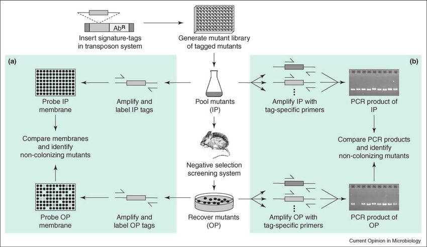
Figure 3
Comparison of two signature-tag detection methods in STM. (a) The original STM method, as developed by Hensel et al. [1], in which mutants
were detected by hybridization. From the mutant library, duplicate hybridization membranes are prepared (colony blots, dot blots with PCR
product or dot blots with plasmid DNA). Differently tagged mutants are pooled and an aliquot is used for the preparation of the input pool (IP)
hybridization probe. The mutant pool is subjected to the negative selection screening system (e.g. an animal model of infection) and mutants that
survive this screen are recovered for preparation of the output pool (OP) hybridization probe. PCR amplification and labeling yields the input and
output pool probes used to hybridize on the previously prepared membranes. The output and the input pool membranes are then compared.
Mutants that fail to be recovered from the negative selection screen produce a signal on the input pool membrane, but not on the output pool
membrane. (b) STM with PCR detection, as developed by Lehoux et al. [13]. After pooling of differently tagged mutants from the mutant library,
an aliquot from the input pool is used for detection of the individual tags by amplification with tag-specific primers. For each tag, one PCR
reaction is prepared. The mutant pool is subjected to the negative selection screening system and mutants that survive this screen are recovered
for detection of the individual tags of the output pool by amplification with tag-specific primers. Mutants that fail to be recovered from the
negative selection screen are identified as producing a PCR product with the input pool as template, but not with the output pool as template.
(intestine cells) selection revealed a minimal overlap of fied tags from the output pool on a membrane and then
only one gene required in both conditions [30]. STM can probing this with an amplified and labeled probe that
also be applied in the absence of an animal or cell culture contains all tags used for pool generation. The advantage
model as the only requirement is a negative selection of this change is that the number of hybridizations neces-
system. Geoffroy et al. [18] tested N. meningitidis for sary to screen the mutant library in more than one screen-
factors required for serum resistance in a cell-free system. ing system is substantially reduced because pools of tags
recovered from more than one screening system can be
Detection system analyzed simultaneously [31].
As outlined previously (Figure 3), hybridization and PCR
are two different detection methods. A special type of Many recent STM studies have used PCR detection,
detection by hybridization is the high-density oligonu- which was established by Lehoux et al. [13] and then
cleotide array technique, in which the presence or applied in their recent P. aeruginosa-STM study [23]. The
absence of 192 individual tag sequences that correspond basic principles of this method are depicted in Figure 3b.
to 96 double-tags can be detected separately [22]. When The tags are amplified individually with a tag-specific
an STM study includes several negative selection primer and a generic primer in a constant region flanking
screens, the multi-screening approach of Struve et al. the tag. The presence of a PCR product of one tag in the
[31] reduces work-load. This study describes a reversion input pool and absence of a PCR product for the same tag
of a dot blot hybridization protocol by spotting the ampli- in the output pool shows the loss of the specific tagged
Current Opinion in Microbiology 2005, 8:1–8 www.sciencedirect.comRecent technical advances in STM Saenz and Dehio 5
Table 1
STM studies in bacteria (2002 to present).
Species a Route of Host Niche c Mutants (attenuated Pool Reference
infection b versus screened) size
Numbers %
S. e. Typhimurium ip Mice Spleen 40/1152 3.5 96 [1]
S. e. Typhimurium Oral Calves, chicken GI tract 84/1045 8.0 95 [35]
S. e. Choleraesuis Oral, ip Swine GI tract, spleen 3/45 6.7 45 [36]
Escherichia coli (UPEC) tu Mice Urinary tract 19/2049 0.9 46 [37]
E. coli (EHEC) Oral Calves GI tract 79/1900 4.2 95 [27]
E. coli (EHEC) Oral Calves GI tract 62/570 10.9 95 [38]
E. coli (APEC) it Chicken Spleen 30/1800 1.7 90 [39]
Vibrio cholerae Oral Mice Small intestine 251/9600 2.6 96 [33]
Yersinia pestis sc Mice Spleen 16/300 5.3 20 [34]
Citrobacter rodentium Oral Mice Colon 14/576 2.4 24 [40]
Klebsiella pneumoniae Oral Mice, cell culture Colon, intestinal cells 29/2200 1.3 48 [30]
K. pneumoniae Oral, tu Mice Colon, bladder 19/1440 1.3 48 [31]
Proteus mirabilis tu Mice Bladder 32/2088 1.5 47 [41]
Xenorhabdus nematophila Contact Nematode Intestinal vesicle 15/3000 0.5 48 [14]
Pasteurella multocida ip, im Mice, chicken Blood 15/420 3.6 42 [42]
Actinobacillus it Swine Lung 105/2064 5.1 48 [43]
pleuropneumoniae
Haemophilus influenzae ip Rats Blood 24/1632 1.5 24 [44]
Pseudomonas aeruginosa it Rats Lung 13/1056 1.2 11 [23]
P. aeruginosa it Rats Lung 160/7968 2.0 72 [32]
Brucella melitensis ip Mice Spleen 36/1152 3.1 96 [45]
Neisseria meningitides Serum resistance 37/4548 0.8 44-48 [18]
Burkholderia pseudomallei ip Mice Spleen 1/96 1.0 96 [46]
Burkholderia cenocepacia it Rats Lung 102/2627 3.9 37 [28]
Campylobacter jejuni Oral Chicken Cecum 29/1550 1.9 74-82 [15]
Helicobacter pylori Oral Mongolian gerbils Stomach 252/960 26.3 24 [47]
Staphylococcus aureus ip Mice Spleen 24/6300 0.4 50 [26]
Streptococcus pneumoniae in Mice Lung 387/6149 6.3 63/40 [17]
Streptococcus group A sc Mice Spleen 1/21 4.8 21 [25]
(GAS)
Mycobacterium tuberculosis iv Mice (knockout) Lung, spleen, liver 3/48 6.3 48 [29]
Mycobacterium marinum ip Goldfish Liver 40/1008 4.0 48-53 [48]
Mycobacterium bovis sc Guinea pigs Spleen 15/1215 1.2 45 [49]
a
APEC, avian pathogenic E. coli; EHEC, enterohemorrhagic E. coli; S. e., Salmonella enterica; UPEC, uropathogenic E. coli.
b
im, intramuscular; in, intranasal; ip, intraperitoneal; it, intratracheal; iv, intravenous; sc, subcutaneous; tu, transurethral.
c
GI, gastrointestinal.
mutant during the negative selection screen. An elegant lished by Geoffroy et al. [18], aimed to map the transposon
way in which to screen 72 mutants simultaneously with a insertion sites of all mutants tested in their STM screen to
tag-pool size of 24 Lehoux-tags was shown by Potvin et al. answer this question. Despite the knowledge of mutational
[32]. They used three transposons that differed only in coverage, this comprehensive mapping is not a preferential
the incorporated selection markers. For detection, this strategy owing to the high time and labor consumption.
study employed multiplex PCR, using three selection
marker-specific primers instead of one generic primer. Further characterization
However, unlike hybridization, PCR detection does not The STM method can be reused for testing randomly
allow different attenuation levels to be distinguished. chosen or deliberately selected mutants in pools. Merrell
This limitation led Hunt et al. [28] to introduce real-time et al. [33] subjected a pool of selected Vibrio cholerae
PCR detection. Real-time PCR allows the relative quan- intestinal colonization-attenuated mutants, denominated
tification of template DNA, in this case of the different virulence-attenuated pool (VAP), to a cell-free assay for
mutants represented in a pool [28]. acid shock. In addition, further analyses can comprise
testing of mutants individually for their specific pheno-
Mutation site identification type. Thereby, STM has the advantage that the output of
To date, one of the major shortcomings of STM has been the selection are interesting mutant strains, which can be
the inability to detect whether a certain gene is dispensable used directly for further investigation, for example to
during negative selection or whether it is simply not evaluate possible vaccination targets in the human patho-
present in the mutant library. Only one approach, pub- gen Yersinia pestis [34].
www.sciencedirect.com Current Opinion in Microbiology 2005, 8:1–86 Genomics
Table 2
Technical details of the STM studies presented in Table 1.
Species a Transposon Tags Detection Mutation site Special features d Reference
system b identification c
S. e. Typhimurium miniTn5 Hensel-tags rh (cb) c, s First STM screen [1]
S. e. Typhimurium miniTn5 Hensel-tags rh (dbp) c, s Two negative [35]
selection systems
S. e. Choleraesuis miniTn5 Hensel-tags rh (cb) c, s [36]
Escherichia coli (UPEC) miniTn5 Hensel-tags nrh (dbpl) Arbitrary PCR / c, s [37]
E. coli (EHEC) miniTn5 Hensel-tags rh (cb) c, s Auxotrophic exclusion [27]
E. coli (EHEC) miniTn5 Hensel-tags rh (cb) c, s [38]
E. coli (APEC) miniTn5 Hensel-tags nrh (dbp) Arbitrary PCR, s [39]
Vibrio cholerae miniTn5 Hensel-tags nrh (dbpl) c, s Virulence-attenuated [33]
pools
Yersinia pestis miniTn5 Hensel-tags rh (dbpl) Single primer Vaccine candidate [34]
PCR, s testing
Citrobacter rodentium miniTn5 Hensel-tags rh (dbpl) c, s [40]
Klebsiella pneumoniae miniTn5 Hensel-tags rh (cb) c, s Two negative [30]
selection systems
K. pneumoniae miniTn5 Hensel-tags nrh (dbp) c, s Multi-screening STM [31]
Proteus mirabilis miniTn5 Hensel-tags nrh (dbpl) Arbitrary PCR / c, s [41]
Xenorhabdus nematophila miniTn5 Hensel-tags rh (dbpl) c, s First symbiosis STM [14]
Pasteurella multocida Tn916 Hensel-tags nrh (dbp) Inverse PCR, s [42]
Actinobacillus mini Tn10 Hensel-tags rh (cb) c, s Vaccine candidate [43]
pleuropneumoniae testing
Haemophilus influenzae Tn1545 Hensel-tags rh (cb) Arbitrary PCR, s [44]
Pseudomonas aeruginosa miniTn5 Lehoux-tags PCR c, s First application of [23]
PCR detection
P. aeruginosa miniTn5 Lehoux-tags PCR (multiplex) c, s Multiplex PCR detection [32]
Brucella melitensis miniTn5 Hensel-tags rh (dbp) Arbitrary PCR / Sequel to [50] [45]
inverse PCR, s
Neisseria meningitidis Himar1 Hensel-tags rh (cb) Ligation-mediated First cell-free screen [18]
PCR, s
Burkholderia pseudomallei miniTn5 Hensel-tags rh (cb) c, s [46]
Burkholderia cenocepacia pTnMod Lehoux-tags PCR (real-time) Self-cloning, s Real-time PCR [28]
detection, auxotrophic
exclusion
Campylobacter jejuni Himar1 Hensel-tags nrh (dbp) c, s First commensal STM [15]
derivative
Helicobacter pylori TnMax5 Hensel-like PCR c, s [47]
tags (20 bp)
Staphylococcus aureus Tn551 and Size-marker PCR (real-time) Inverse PCR, s SMIT, pre-tagging [26]
Tn917lac tags
Streptococcus pneumoniae magellan2 Hensel-tags nrh (dbp) Arbitrary PCR, s Two-stage STM [17]
Streptococcus group A IS256 Size-marker tags PCR Self-cloning, s PTTM [25]
(GAS)
Mycobacterium tuberculosis Tn5370 Hensel-tags rh (cb) Inverse PCR, s Knockout mice as [29]
model
Mycobacterium marinum phasmid Hensel-tags rh (dbpl) c, s [48]
Mycobacterium bovis illegitimate Hensel-tags rh (sbpl) c, s [49]
recombination
a
APEC, avian pathogenic E. coli; EHEC, enterohemorrhagic E. coli; S. e., Salmonella enterica; UPEC, uropathogenic E. coli.
b
cb, colony blot; dbp, dot blot with PCR product; dbpl, dot blot with plasmid DNA; hdh, high-density hybridization on chip; nrh, non-radioactive
labeling and hybridization; rh, radioactive labeling and hybridization; sbpl, Southern blot with plasmid DNA.
c
c, cloning; s, sequencing.
d
PTTM, polymorphic-tag-length-transposon-mutagenesis; SMIT, size marker identification technology.
Current Opinion in Microbiology 2005, 8:1–8 www.sciencedirect.comRecent technical advances in STM Saenz and Dehio 7
Conclusions 12. Mei JM, Nourbakhsh F, Ford CW, Holden DW: Identification of
Staphylococcus aureus virulence genes in a murine model of
One decade after its first description, STM has become a bacteraemia using signature-tagged mutagenesis. Mol
genetic method widely used for the in vivo identification Microbiol 1997, 26:399-407.
of virulence traits in pathogenic bacteria. Recent techni- 13. Lehoux DE, Sanschagrin F, Levesque RC: Defined
oligonucleotide tag pools and PCR screening in signature-
cal advances, mainly in the choice and combination of tagged mutagenesis of essential genes from bacteria.
negative selection system(s) and in the choice of detec- Biotechniques 1999, 26:473-478, 480.
tion systems, have broadened its applicability and versa- 14. Heungens K, Cowles CE, Goodrich-Blair H: Identification of
tility. The STM method is an invaluable tool to provide a Xenorhabdus nematophila genes required for mutualistic
colonization of Steinernema carpocapsae nematodes.
better understanding of microbial behavior in vivo. The Mol Microbiol 2002, 45:1337-1353.
use of other genome-scale techniques such as in vivo
15. Hendrixson DR, DiRita VJ: Identification of Campylobacter
expression technology, microarray analysis, genome ana- jejuni genes involved in commensal colonization of the chick
lysis and mapping by in vitro transposon mutagenesis, or gastrointestinal tract. Mol Microbiol 2004, 52:471-484.
First description of an STM study that investigates the commensal–host
transposon site hybridization complement the lessons relationship. Using a model of chick gastrointestinal colonization, the
learned by STM. authors indicate a role of motility for persistence of bacteria in the gut.
Furthermore, they discuss differences and similarities with pathogen-
STM screens and one symbiosis-STM screen [14].
Acknowledgements
We thank Anja Seubert and Gunnar Schroeder for stimulating 16. de Lorenzo V, Herrero M, Jakubzik U, Timmis KN: Mini-Tn5
discussions and for helpful comments on the manuscript. Research in transposon derivatives for insertion mutagenesis, promoter
our laboratory is supported by the grant 3100-061777 from the Swiss probing, and chromosomal insertion of cloned DNA in
National Science Foundation (to CD) and a grant from the International Gram-negative eubacteria. J Bacteriol 1990, 172:6568-6572.
Research Scholars Program Infectious Diseases and Parasitology of the 17. Hava DL, Camilli A: Large-scale identification of serotype 4
Howard Hughes Medical Institute. Streptococcus pneumoniae virulence factors. Mol Microbiol
2002, 45:1389-1406.
References and recommended reading 18. Geoffroy MC, Floquet S, Metais A, Nassif X, Pelicic V: Large-scale
Papers of particular interest, published within the annual period of analysis of the meningococcus genome by gene disruption:
review, have been highlighted as: resistance to complement-mediated lysis. Genome Res 2003,
13:391-398.
of special interest
of outstanding interest 19. Plasterk RH, Izsvak Z, Ivics Z: Resident aliens: the Tc1/mariner
superfamily of transposable elements. Trends Genet 1999,
15:326-332.
1. Hensel M, Shea JE, Gleeson C, Jones MD, Dalton E, Holden DW:
Simultaneous identification of bacterial virulence genes by 20. Lampe DJ, Churchill ME, Robertson HM: A purified mariner
negative selection. Science 1995, 269:400-403. transposase is sufficient to mediate transposition in vitro.
EMBO J 1996, 15:5470-5479.
2. Chiang SL, Mekalanos JJ, Holden DW: In vivo genetic analysis of
bacterial virulence. Annu Rev Microbiol 1999, 53:129-154. 21. Doak TG, Doerder FP, Jahn CL, Herrick G: A proposed
superfamily of transposase genes: transposon-like elements
3. Perry RD: Signature-tagged mutagenesis and the hunt for in ciliated protozoa and a common ‘‘D35E’’ motif. Proc Natl
virulence factors. Trends Microbiol 1999, 7:385-388 (and Acad Sci USA 1994, 91:942-946.
discussion 7:388-389).
22. Karlyshev AV, Oyston PC, Williams K, Clark GC, Titball RW,
4. Shea JE, Santangelo JD, Feldman RG: Signature-tagged Winzeler EA, Wren BW: Application of high-density array-based
mutagenesis in the identification of virulence genes in signature-tagged mutagenesis to discover novel Yersinia
pathogens. Curr Opin Microbiol 2000, 3:451-458. virulence-associated genes. Infect Immun 2001, 69:7810-7819.
5. Lehoux DE, Levesque RC: Detection of genes essential in 23. Lehoux DE, Sanschagrin F, Levesque RC: Identification of in vivo
specific niches by signature-tagged mutagenesis. Curr Opin essential genes from Pseudomonas aeruginosa by PCR-
Biotechnol 2000, 11:434-439. based signature-tagged mutagenesis. FEMS Microbiol Lett
6. Mecsas J: Use of signature-tagged mutagenesis in 2002, 210:73-80.
pathogenesis studies. Curr Opin Microbiol 2002, 5:33-37. 24. Walsh C, Cepko CL: Widespread dispersion of neuronal clones
7. Lehoux DE, Sanschagrin F, Kukavica-Ibrulj I, Potvin E, across functional regions of the cerebral cortex. Science 1992,
Levesque RC: Identification of novel pathogenicity genes by 255:434-440.
PCR signature-tagged mutagenesis and related technologies. 25. Hidalgo-Grass C, Ravins M, Dan-Goor M, Jaffe J, Moses AE,
Methods Mol Biol 2004, 266:289-304. Hanski E: A locus of group A Streptococcus involved in
8. Shea JE, Hensel M, Gleeson C, Holden DW: Identification of a invasive disease and DNA transfer. Mol Microbiol 2002,
virulence locus encoding a second type III secretion system 46:87-99.
in Salmonella typhimurium. Proc Natl Acad Sci USA 1996,
26. Benton BM, Zhang JP, Bond S, Pope C, Christian T, Lee L,
93:2593-2597.
Winterberg KM, Schmid MB, Buysse JM: Large-scale
9. Sun YH, Bakshi S, Chalmers R, Tang CM: Functional genomics identification of genes required for full virulence of
of Neisseria meningitidis pathogenesis. Nat Med 2000, Staphylococcus aureus. J Bacteriol 2004, 186:8478-8489.
6:1269-1273. The authors describe a variation of STM, which they denominate size-
marker identification technology. It describes pre-tagging bacteria with
10. Coulter SN, Schwan WR, Ng EY, Langhorne MH, Ritchie HD, tags of different sizes, subsequent mutagenesis independent of the
Westbrock-Wadman S, Hufnagle WO, Folger KR, Bayer AS, Stover tagging procedure, and detection by multiplex PCR. Interestingly, the
CK: Staphylococcus aureus genetic loci impacting growth and three S. aureus-STM studies published to date overlap only in one gene
survival in multiple infection environments. Mol Microbiol 1998, (asd).
30:393-404.
27. Dziva F, van Diemen PM, Stevens MP, Smith AJ, Wallis TS:
11. Darwin AJ, Miller VL: Identification of Yersinia enterocolitica Identification of Escherichia coli O157: H7 genes influencing
genes affecting survival in an animal host using signature- colonization of the bovine gastrointestinal tract using
tagged transposon mutagenesis. Mol Microbiol 1999, signature-tagged mutagenesis. Microbiology 2004,
32:51-62. 150:3631-3645.
www.sciencedirect.com Current Opinion in Microbiology 2005, 8:1–88 Genomics
28. Hunt TA, Kooi C, Sokol PA, Valvano MA: Identification of 40. Mundy R, Pickard D, Wilson RK, Simmons CP, Dougan G,
Burkholderia cenocepacia genes required for bacterial Frankel G: Identification of a novel type IV pilus gene cluster
survival in vivo. Infect Immun 2004, 72:4010-4022. required for gastrointestinal colonization of Citrobacter
rodentium. Mol Microbiol 2003, 48:795-809.
29. Hisert KB, Kirksey MA, Gomez JE, Sousa AO, Cox JS, Jacobs WR
Jr, Nathan CF, McKinney JD: Identification of Mycobacterium 41. Burall LS, Harro JM, Li X, Lockatell CV, Himpsl SD,
tuberculosis counterimmune (cim) mutants in Hebel JR, Johnson DE, Mobley HL: Proteus mirabilis genes
immunodeficient mice by differential screening. Infect Immun that contribute to pathogenesis of urinary tract
2004, 72:5315-5321. infection: identification of 25 signature-tagged mutants
attenuated at least 100-fold. Infect Immun 2004,
30. Maroncle N, Balestrino D, Rich C, Forestier C: Identification of 72:2922-2938.
Klebsiella pneumoniae genes involved in intestinal
colonization and adhesion using signature-tagged 42. Harper M, Boyce JD, Wilkie IW, Adler B: Signature-tagged
mutagenesis. Infect Immun 2002, 70:4729-4734. mutagenesis of Pasteurella multocida identifies mutants
displaying differential virulence characteristics in mice and
31. Struve C, Forestier C, Krogfelt KA: Application of a novel chickens. Infect Immun 2003, 71:5440-5446.
multi-screening signature-tagged mutagenesis assay for
identification of Klebsiella pneumoniae genes essential in 43. Sheehan BJ, Bosse JT, Beddek AJ, Rycroft AN, Kroll JS,
colonization and infection. Microbiology 2003, 149:167-176. Langford PR: Identification of Actinobacillus
pleuropneumoniae genes important for survival
32. Potvin E, Lehoux DE, Kukavica-Ibrulj I, Richard KL, Sanschagrin F, during infection in its natural host. Infect Immun 2003,
Lau GW, Levesque RC: In vivo functional genomics of 71:3960-3970.
Pseudomonas aeruginosa for high-throughput screening of
new virulence factors and antibacterial targets. Environ 44. Herbert MA, Hayes S, Deadman ME, Tang CM, Hood DW,
Microbiol 2003, 5:1294-1308. Moxon ER: Signature tagged mutagenesis of Haemophilus
influenzae identifies genes required for in vivo survival.
33. Merrell DS, Hava DL, Camilli A: Identification of novel factors Microb Pathog 2002, 33:211-223.
involved in colonization and acid tolerance of Vibrio cholerae.
Mol Microbiol 2002, 43:1471-1491. 45. Lestrate P, Dricot A, Delrue RM, Lambert C, Martinelli V,
De Bolle X, Letesson JJ, Tibor A: Attenuated signature-tagged
34. Flashner Y, Mamroud E, Tidhar A, Ber R, Aftalion M, Gur D, mutagenesis mutants of Brucella melitensis identified during
Lazar S, Zvi A, Bino T, Ariel N et al.: Generation of Yersinia pestis the acute phase of infection in mice. Infect Immun 2003,
attenuated strains by signature-tagged mutagenesis in search 71:7053-7060.
of novel vaccine candidates. Infect Immun 2004, 72:908-915.
The authors evaluate the efficacy of one attenuated STM mutant for 46. Atkins T, Prior R, Mack K, Russell P, Nelson M, Prior J, Ellis J,
vaccine development. They show that a mutant in the pcm gene, involved Oyston PC, Dougan G, Titball RW: Characterisation of an
in bacterial stress response, is a more efficient live cellular vaccine than acapsular mutant of Burkholderia pseudomallei identified by
the current one (EV76). signature tagged mutagenesis. J Med Microbiol 2002,
51:539-547.
35. Morgan E, Campbell JD, Rowe SC, Bispham J, Stevens MP,
Bowen AJ, Barrow PA, Maskell DJ, Wallis TS: Identification of 47. Kavermann H, Burns BP, Angermuller K, Odenbreit S, Fischer W,
host-specific colonization factors of Salmonella enterica Melchers K, Haas R: Identification and characterization of
serovar Typhimurium. Mol Microbiol 2004, 54:994-1010. Helicobacter pylori genes essential for gastric colonization.
J Exp Med 2003, 197:813-822.
36. Lichtensteiger CA, Vimr ER: Systemic and enteric colonization
of pigs by a hilA signature-tagged mutant of Salmonella 48. Ruley KM, Ansede JH, Pritchett CL, Talaat AM, Reimschuessel R,
choleraesuis. Microb Pathog 2003, 34:149-154. Trucksis M: Identification of Mycobacterium marinum
virulence genes using signature-tagged mutagenesis and the
37. Bahrani-Mougeot FK, Buckles EL, Lockatell CV, Hebel JR, goldfish model of mycobacterial pathogenesis. FEMS
Johnson DE, Tang CM, Donnenberg MS: Type 1 fimbriae and Microbiol Lett 2004, 232:75-81.
extracellular polysaccharides are preeminent uropathogenic
Escherichia coli virulence determinants in the murine urinary 49. Collins DM, Skou B, White S, Bassett S, Collins L, For R,
tract. Mol Microbiol 2002, 45:1079-1093. Hurr K, Hotter G, de Lisle GW: Generation of attenuated
Mycobacterium bovis strains by signature-tagged
38. van Diemen PM, Dziva F, Stevens MP, Wallis TS: Identification of mutagenesis for discovery of novel vaccine candidates.
enterohemorrhagic Escherichia coli O26:H- genes required Infect Immun 2005, 73:2379-2386.
for intestinal colonization in calves. Infect Immun 2005,
73:1735-1743. 50. Lestrate P, Delrue RM, Danese I, Didembourg C, Taminiau B,
Mertens P, De Bolle X, Tibor A, Tang CM, Letesson JJ:
39. Li G, Laturnus C, Ewers C, Wieler LH: Identification of genes Identification and characterization of in vivo attenuated
required for avian Escherichia coli septicemia by signature- mutants of Brucella melitensis. Mol Microbiol 2000,
tagged mutagenesis. Infect Immun 2005, 73:2818-2827. 38:543-551.
Current Opinion in Microbiology 2005, 8:1–8 www.sciencedirect.comSTM MANUSCRIPT
Manuscript in preparation
Functional genomics of Bartonella pathogenesis:
Large-scale signature-tagged mutagenesis reveals a high number of genes
required for infection of the mammalian host
Saenz H. L.1, Stoeckli M. C.1, Vayssier-Taussat M.2, Lanz C.3, Schuster S. C.3, Dehio C.1,*
1
Division of Molecular Microbiology, Biozentrum, University of Basel, Klingelbergstrasse 70,
4056 Basel, Switzerland,
2
ENVA, UMR 956, Microbiologie-Immunologie, 94 700 Maisons-Alfort, France,
3
Max-Planck-Institute for Developmental Biology, 72076 Tuebingen, Germany.
Running title: STM screen for pathogenicity factors in Bartonella
*Corresponding author: Prof. Christoph Dehio
Division of Molecular Microbilogy
Biozentrum, University of Basel
Klingelberstrasse 70
CH-4056 Basel, Switzerland
Tel. +41-61-267-2140
Fax: +41-61-267-2118
E-mail: christoph.dehio@unibas.ch
Author contributions: H.L.S., C.D. designed research; H.L.S., M.C.S., M.V., C.L. performed
research; S.C.S. contributed analytic tools; H.L.S., C.D. analyzed data; and H.L.S. wrote the
paper
9STM MANUSCRIPT - Abstract
ABSTRACT
Bartonellae are bacterial pathogens uniquely adapted to cause intraerythrocytic infection in
their mammalian reservoir hosts. In the case of human-specific Bartonella bacilliformis and
Bartonella quintana, the intraerythrocytic bacteremia leads to the clinical manifestations of
Oroya fever and trench fever, respectively. Here, we adapted large-scale signature-tagged
mutagenesis (STM) for the first time to Bartonella, allowing us to screen for pathogenicity
factors required for infection of the mammalian reservoir host in vivo. A total of 3084 STM
mutants of rat-specific B. tribocorum were screened in a rat infection model for these criteria.
After two rounds of screening, 130 mutants showed severe attenuation compared to wild-type
B. tribocorum. We mapped the transposon insertion sites of these mutants to 80 different
genes, and categorized them according to their putative function. Besides already described
pathogenicity factors responsible for interaction with the host, like the two type IV secretion
systems VirB-D4 and Trw, we discovered factors previously unlinked to pathogenesis. These
belong to diverse functional classes, like transport, gene-expression regulation, cell envelope
integrity, or metabolism. A quarter of the identified genes are (conserved) hypothetical coding
for novel pathogenicity factors. We have used an additional PCR-screening approach on the
entire mutant library to test for the level of mutational saturation and to identify non-essential
genes in a pathogenicity island encoding 18 gene products related to the process of type IV
secretion.
10STM MANUSCRIPT - Introduction
INTRODUCTION
Bartonellae are small, fastidious, pleomorphic, Gram-negative rods, which are pathogenic for
a wide range of mammalian hosts. The genus Bartonella currently comprises 20 species that
are highly adapted to their mammalian reservoir hosts. Of these 20 species, 8 have been
associated with human disease (20). The three major human pathogens are the human-specific
Bartonella bacilliformis and Bartonella quintana and the cat-specific Bartonella henselae,
where humans appear as incidental hosts. The common theme of Bartonella infections in the
reservoir host is the long-lasting intraerythrocytic bacteremia. The course of infection is most
frequently asymptomatic, but can also lead to severe clinical manifestations like Oroya fever
in the case of B. bacilliformis or trench fever in the case of B. quintana. Both in the incidental
and the reservoir host, Bartonella interacts also with endothelial cells which in a
immunocompromized individual can cause vasoproliferative lesions like verruga peruana in
the case of B. bacilliformis or bacillary angiomatosis in the case of B. quintana and B.
henselae (10).
Recently, Schulein et al. developed an animal model for erythrocyte colonization of
Bartonella in the reservoir host (73). After injection of rat-specific Bartonella tribocorum in
the tail vein of rats, bacteria are rapidly cleared from the circulating blood. Plate-grown
bartonellae obviously are not able to directly invade erythrocytes and are not detected in the
blood on the first days of infection. Thus, they first have to interact with a yet not
experimentally identified primary niche, where upon invasion they become competent for the
subsequent hemotropic stage (73), a process including transcriptional reprogramming. So,
colonization of erythrocytes and persistence therein is the endpoint of a complex series of
bacterium-host interactions. To understand such a complex pathogenesis including interaction
with and invasion into different host cell types, identification of involved pathogenicity
factors of the bacteria are of prime significance. Here, pathogenicity is defined in a broad
sense, i.e. the ability of a microorganism to breach barriers in the host and to thrive either
hidden from or in the face of the host immune defense. Intraerythrocytic bacteremia is the
hallmark of Bartonella infection in the reservoir host and is responsible for the disease
symptoms, elicited by the human-pathogenic bartonellae mentioned above. So, bacteremia
can be used as a read-out, whether mutant bacteria are still able to colonize the
intraerythocytic niche. Thus, abacteremic mutants then by definition carry a mutation in a
gene coning for a pathogenicity factor. Our current knowledge about these pathogenicity
factors of Bartonella is summarized in the following paragraph. Using the B. tribocorum-rat
11STM MANUSCRIPT - Introduction
infection model, Schulein and Dehio (71) described mutants in components of the VirB-D4
type IV secretion system (T4SS) to be abacteremic, proving these components to be the first
bona fide pathogenicity factors in Bartonella. Seubert et al. discovered a second T4SS, the
Trw system, and also could show its essential role as pathogenicity trait in the rat-infection
model (74). Very recently, Riess et al. identified a surface-expressed, afimbrial adhesin of B.
henselae designated as Bartonella adhesin A (BadA), formerly known as “type IV pilus” (3)
to be a major pathogenicity factor (61). A family of variably expressed outer-membrane
proteins (VompA-D) in B. quintana, orthologous to BadA, supports these findings (91).
Further possible pathogenicity factors of Bartonella have been reviewed recently (20)
and include (i) a yet not clearly characterized proteinaceous angiogenic factor, found in B.
bacilliformis, B. quintana, and B. henselae, (ii) a secreted bacterial factor called deformin of
B. bacilliformis and B. henselae, involved in the deformation of erythrocyte membranes, (iii)
the flagella of B. bacilliformis, facilitating erythrocyte invasion, (iv) unusual
lipopolysaccharide with low endotoxic activity of B. henselae and B. quintana, enabling the
interaction with endotoxin-sensitive endothelial cells, (v) hemin-binding proteins (HbpA-E of
B. quintana and HbpA-D of B. henselae), involved in iron acquisition, (vi) the surface-
exposed proteinaceous hemolysin of B. bacilliformis, responsible for hemolysis during Oroya
fever, (vii) the inducible Bartonella autotransporter (Iba) family, specifically upregulated
during endothelial infection by B. henselae in vitro and transiently activated during rat
infection by B. tribocorum in vivo, (viii) the two gene products IalA and IalB of the invasion-
associated locus (ial) mediating erythrocyte invasion by B. bacilliformis in vitro, and (ix)
multiple outer membrane proteins of B. henselae demonstrated to bind to endothelial cells in
vitro (20).
The sequencing of two Bartonella genomes, namely of B. quintana and B. henselae,
published by Alsmark et al. in 2004 (1), opened the way for whole-genome approaches to
comprehensively study pathogenicity factors in Bartonella. Cellular models can be applied to
study aspects of the versatile bacterium-host interactions, but in vivo studies better depict the
complex natural situation. A powerful technology for in vivo screening is signature-tagged
mutagenesis (STM) (30), which allows simultaneous screening of pools of transposon
mutants for loss of pathogenicity in an animal-infection model. Thus, factors identified by
STM are essential for colonization in the tested model, a unique feature of STM compared to
other in vivo screening techniques. The application of this technique identified many new
pathogenicity factors in a broad spectrum of pathogenic bacteria and fungi (recently reviewed
by Mecsas [49]). In STM, the transposon insertion mutants are individually marked with a
12STM MANUSCRIPT - Introduction
specific signature-tag, i.e. a short variable double-stranded DNA sequence. In pools of
mutants, the presence or absence of a single mutant can be tested by detection of this tag.
Originally, Hensel et al. used DNA hybridization as detection method (30), but more recently,
Lehoux et al. described mutant detection by PCR as a simpler and more rapid detection
technique (43), which could be even automated for high-throughput screening (42). Pools of
mutants can be screened in an animal model, cell culture, or any other negative selection
screen. The presence of an individual mutant in the pool before selection and its absence
thereafter identifies the mutant as a non-colonizing mutant candidate. Since STM uses mutant
pools, where different mutants influence each other, some STM screens employed a
rescreening of the non-colonizing candidates in newly assembled pools to discriminate
between fully attenuated mutants, which are attenuated in every pool tested, and partially
attenuated mutants, which are present or not depending on the pool composition. Darwin and
Miller (18) first described this two-stage STM.
Here, we established large-scale STM for Bartonella to identify pathogenicity factors
of B. tribocorum essential for inducing intraerythrocytic bacteremia during infection of the
laboratory rat as the mammalian reservoir host. By screening 3084 transpositional mutants for
an abacteremic phenotype, we discovered novel pathogenicity factor as well as factors
previously unlinked to pathogenesis and re-discovered known pathogenicity factors, like the
two T4SS VirB-D4 and Trw. We have used an additional PCR-screening approach on the
entire mutant library to test for the level of mutational saturation and to identify non-essential
genes in a pathogenicity island encoding 18 gene products related to the process of type IV
secretion.
13STM MANUSCRIPT - Results
RESULTS
Construction and testing of the transposon vector
The transposon vector pHS003 contains an oriT for conjugative transfer, the Himar1
transposon, carrying a kanamycin resistance marker, and a hyperactive transposase (Figure 1).
This suicide vector construct was transferred into B. tribocorum via conjugation and
transconjugants were tested for transposon insertions, leading to kanamycin-resistant colonies.
The frequency of transposition events (transconjugants divided by B. tribocorum recipients)
was 2.4x10⁻⁴. With specific PCR reactions on single colonies, we could amplify a part of the
transposon, but no PCR product could be obtained with primer pairs derived from the vector
backbone, indicating insertion of the transposon into the genome of B. tribocorum and
contradicting a potential integration of the whole plasmid (data not shown). Southern analysis
(Figure 2) confirmed this indication and showed that in each of the 13 randomly chosen
mutants, the transposon inserted in a single copy in distinct sites of the chromosome,
suggesting random transposon distribution.
Construction of the STM mutant library
A mixture of signature-tags was produced by PCR using degenerate oligonucleotide
templates, ligated into transposon vector pHS006 (derivative of pHS003, see “Material and
Methods”), and transformed into Escherichia coli NovaBlue. The central variable sequence of
the tag allows the potential generation of more than 1022 different variants. Ninety-six
ampicillin/kanamycin-resistant colonies were picked (E. coli NovaBlue pHS006tagn with n =
001-096) and the inserted tag was sequenced. None of the plasmids contained identical tag
sequences (data not shown). We chose, based on reproducible PCR detection results, 36
plasmids to construct the STM mutant library, and transferred the tagged transposon vectors
individually to B. tribocorum via conjugation. The overall frequency of transposition events
of 36 individual conjugation assays presented as 2.3x10⁻⁴. From each conjugation assay, we
selected 96 single kanamycin-resistant colonies. Using this procedure, we assembled an STM
mutant library with 3456 mutants.
Screening of the mutant library
The first 16 mutant pools consisted of 19 differently tagged mutants, the remaining 80 mutant
pools of 36 differently tagged mutants. Each mutant pool was used to inoculate two rats (=
input pool, see also “Material and Methods”) and peripheral blood was drawn on days 7 and
14STM MANUSCRIPT - Results
14 postinfection. Bacteria were recovered from the blood by freeze-lysation and grown on
plates (=output pool). To identify abacteremic mutant candidates, present in the input pool
and missing in the output pool of both rats on both time points investigated, we compared the
individual tag-specific PCR signals in input and output pool. Of 3184 mutants tested, 100 had
to be excluded due to missing input pool signal. One tag (tag047) was excluded from all pools
due to constantly weak PCR detection results. Of the remaining 3084 mutants tested, 359
were abacteremic mutant candidates. To confirm this abacteremic phenotype, we reassembled
the 359 strains into 18 pools of 34 mutants. For keeping the pool size constant at 34 mutants
per pool, we tested some strains repeatedly. In this rescreen, the pools were used to inoculate
four rats, two like in the primary screen and two competitively mixed with a pool of 5 mutants
that showed wild-type behavior in the primary screen and all contained the same tag. This
pool was called the tagged wild-type-like pool. Since the mutants in this pool showed wild-
type behavior in the screen and carried mainly intergenic transposon insertions (data not
shown), we assumed that this pool should be competitive like wild-type bacteria. The rescreen
revealed 130 abacteremic mutants of the 359 candidates, which corresponds to 4% of the
3084 mutants tested. In total, these 130 abacteremic mutants were tested absent from the
blood of at least 6 rats (Table 1) on two time points, where bacteremia peaks with wild-type
B. tribocorum. All abacteremic mutants have a calculated in-pool competitive index (CI, see
“Material and Methods”) lower than 0.00001. The in vitro growth in all abacteremic mutants
was comparable to wild-type bacteria.
Interestingly, 47 strains could not be detected from the competition pool, where more wild-
type like bacteria were present, but they could be recovered from the mutant pools. This
indicates that wild-type like strains outcompeted these otherwise bacteremic strains, which
will be called outcompeted mutants in this work.
Mapping of the transposon insertion site
We determined the insertion site of the transposon for 314 of the 359 abacteremic mutant
candidates by direct sequencing out of the transposon into the genome of B. tribocorum. The
remaining 45 mutants gave ambiguous sequencing results, suggesting that these mutants
might carry multiple transposon insertions. This finding is supported by Southern analysis,
where 20 selected mutants showed unique single bands, whereas two suspected double
insertion mutants showed also two bands (data not shown). The 130 abacteremic mutants
represent transposition events into 80 different open reading frames (ORFs). With the help of
the (not yet fully assembled) genome sequence of B. tribocorum (of approx. 2.7 Mb)
15STM MANUSCRIPT - Results
(S. C. Schuster, P. Engel, H. L. Saenz, M. C. Stoeckli, C. Lanz, G. Raddatz, and C. Dehio,
unpublished results) and the two published Bartonella genomes of B. quintana (1.6 Mb) and
B. henselae (1.9 Mb) (1), we annotated the affected 80 ORFs, whose predicted products fall
into a wide variety of functional classes (Table 1). Remarkably, various multiple hits were
found within the trw and virB gene clusters, coding for the two T4SS known to be essential
for rat infection (71, 74).
Categorization of abacteremic mutants
Of the 130 abacteremic mutants, displayed in Table 1, 24 strains carried transposon insertions
in intergenic regions. This localization could affect transcriptional termination of the upstream
gene, or more likely, disrupt the promoter or regulator region of the downstream gene, or
disrupt an operon. Additionally, the transposon could have hit a small not yet discovered
ORF, a sequence encoding tRNA or a small regulatory RNA. We included these intergenic
mutants in the classification as affecting the downstream gene, if the insertion was not more
than 500 bp upstream of a gene and nearer to the downstream gene than to the upstream gene
and if there was no indication for a functional small ORF, tRNA gene (predicted with
tRNAscan-SE), or a sequence coding for a small regulatory RNA. This issue would
nevertheless need further experimental exploration. Thirteen (of thirteen analyzed) intergenic
hits could be classified by these criteria. Besides the intergenic hits, 106 transposon insertions
occurred within 80 ORFs. These insertions can be divided into 84 hits in 64 genes with
predicted putative function, 14 hits in 12 conserved hypothetical genes (CH) with no
predicted function, 2 hits in 2 phage-specific genes (not present in B. henselae or B.
quintana), and 6 hits in 2 Bartonella-specific genes. Figure 3 shows the functional
classification of the affected genes. Genes with products involved in adhesion and invasion or
transport over the bacterial membranes and gene-expression regulation constituted more than
half of the total hit genes (35 and 14 of 80, respectively), highlighting the importance of these
processes in pathogenicity. Twelve genes with products involved in the cellular metabolism,
like energy metabolism, amino acid or cofactor biosynthesis, and carbon metabolism were
affected. The individual mutants are listed in Table 1 with their assigned classification, their
putative assigned function, their orthologs in B. henselae and B. quintana (if present), and the
number of animals, in which they were tested.
16You can also read


