SARS-COV-2, ACE2, AND HYDROXYCHLOROQUINE: CARDIOVASCULAR COMPLICATIONS, THERAPEUTICS, AND CLINICAL READOUTS IN THE CURRENT SETTINGS - MDPI
←
→
Page content transcription
If your browser does not render page correctly, please read the page content below
pathogens
Review
SARS-CoV-2, ACE2, and Hydroxychloroquine:
Cardiovascular Complications, Therapeutics,
and Clinical Readouts in the Current Settings
Rajkumar Singh Kalra 1, *,† , Dhanendra Tomar 2, *,† , Avtar Singh Meena 3 and
Ramesh Kandimalla 4,5
 1   AIST-INDIA DAILAB, DBT-AIST International Center for Translational & Environmental
     Research (DAICENTER), National Institute of Advanced Industrial Science & Technology (AIST),
     Higashi 1-1-1, Tsukuba 305 8565, Japan
 2   Center for Translational Medicine, Lewis Katz School of Medicine, Temple University,
     Philadelphia, PA 19140, USA
 3   CSIR-Centre for Cellular and Molecular Biology (CCMB), Habsiguda, Uppal Road,
     Hyderabad 500 007, Telangana State, India; avtar.ccmb@yahoo.com
 4   Applied Biology, CSIR-Indian Institute of Chemical Technology (IICT), Uppal Road, Tarnaka,
     Hyderabad 500007, Telangana State, India; ramesh.kandimalla@iict.res.in
 5   Department of Biochemistry, Kakatiya Medical College, Warangal 506007, Telangana State, India
 *   Correspondence: raj-singh@aist.go.jp (R.S.K.); dhtomar@temple.edu (D.T.)
 †   Equal contribution.

 Received: 1 June 2020; Accepted: 5 July 2020; Published: 7 July 2020

 Abstract: The rapidly evolving coronavirus disease 2019 (COVID-19, caused by severe acute
 respiratory syndrome coronavirus 2- SARS-CoV-2), has greatly burdened the global healthcare
 system and led it into crisis in several countries. Lack of targeted therapeutics led to the idea of
 repurposing broad-spectrum drugs for viral intervention. In vitro analyses of hydroxychloroquine
 (HCQ)’s anecdotal benefits prompted its widespread clinical repurposing globally. Reports of
 emerging cardiovascular complications due to its clinical prescription are revealing the crucial role
 of angiotensin-converting enzyme 2 (ACE2), which serves as a target receptor for SARS-CoV-2.
 In the present settings, a clear understanding of these targets, their functional aspects and
 physiological impact on cardiovascular function are critical. In an up-to-date format, we shed
 light on HCQ’s anecdotal function in stalling SARS-CoV-2 replication and immunomodulatory
 activities. While starting with the crucial role of ACE2, we here discuss the impact of HCQ on systemic
 cardiovascular function, its associated risks, and the scope of HCQ-based regimes in current clinical
 settings. Citing the extent of HCQ efficacy, the key considerations and recommendations for the use
 of HCQ in clinics are further discussed. Taken together, this review provides crucial insights into the
 role of ACE2 in SARS-CoV-2-led cardiovascular activity, and concurrently assesses the efficacy of
 HCQ in contemporary clinical settings.

 Keywords: SARS-CoV-2; COVID-19; ACE2; hydroxychloroquine; cardiovascular system;
 cardiovascular disease (CVD); therapeutics

1. Introduction
    A new type of pneumonia outbreak surfaced in December 2019 in Wuhan, Hubei province,
China, which was caused by a novel coronavirus, viz., severe acute respiratory syndrome
coronavirus (SARS-CoV)-2 [1]. The pandemic disease, named coronavirus disease 2019 (COVID-19),
had 5,867,727 confirmed cases by 29th May 2020 and resulted in 362,238 deaths globally, as sourced

Pathogens 2020, 9, 546; doi:10.3390/pathogens9070546                        www.mdpi.com/journal/pathogens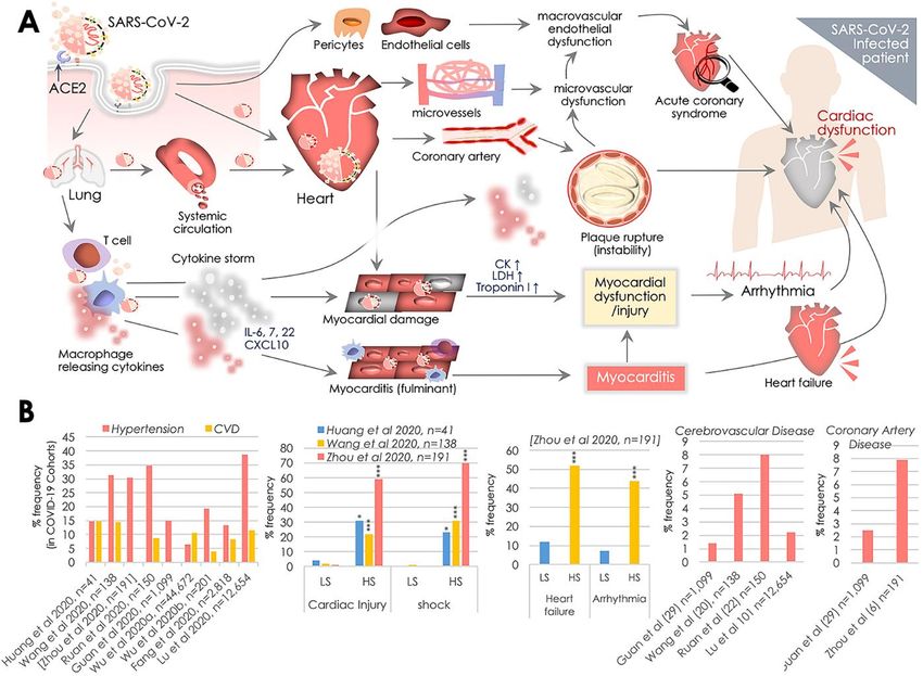
Pathogens 2020, 9, 546                                                                               2 of 35

by the Coronavirus Resource Center, John Hopkins University (JHU) (https://coronavirus.jhu.
edu/). SARS-CoV-2 shares 82% genomic similarity with the other SARS-CoVs, while two
other bat-SARS-CoV-like viruses (retrieved from Rhinolophus sinicus, Zhoushan, China), viz.,
bat-SL-CoVZC45 and bat-SL-CoVZXC21 were found to have >89% similarity [1]. To date, SARS-CoV-2
has crossed all continental boundaries, and presently Europe and North America have been its major
epicenters. The COVID-19 symptoms are comparable to those produced by SARS-CoV and Middle
East respiratory syndrome (MERS). However, the earliest estimate showed its lower (2%) fatality
rate, while about ~ 20% of COVID-19 patients had developed severe conditions [2]. SARS-CoV-2
tropism to the lungs/respiratory system is prominent, in which it infects the lung cells and causes
interstitial pneumonitis that may lead to developing acute respiratory distress syndrome (ARDS)
and manifestations related to the cardiovascular (CV) system causing multiple organ failure [3–8].
Amongst severe COVID-19 patients, 23% of cases had cardiac injuries [9] and, therefore, highlighted
this as a common feature that promotes disease severity. Of note, elevated levels of creatinine kinase
(CK; >200U/L) in 13% of COVID-19 patients in the general cohort, where most of these lacked any
cytokine storm-induced systemic inflammatory response, further affirmed the association of COVID-19
with cardiovascular complications [2]. The common CV complications reported in COVID-19 patients
include arrhythmia, myocardial injury (marked by higher troponin I (hs-cTnI) and CK levels) and
myocarditis, acute myocardial infarction, acute heart failure and cardiomyopathy, and disseminated
intravascular coagulation (DIC) [3,4,10,11]. Although the association of SARS-CoV-2 infection with
these manifestations is now known, preexisting CV comorbidities could further contribute to COVID-19
severity and mortality [3,4,10,11]. The earliest report describing a meta-analysis of the COVID-19
clinical cohort revealed a strikingly high existing prevalence of hypertension and cardiovascular
disease (CVD) in hospitalized patients, that made them prone to require critical care [10]. COVID-19
patients with CVDs were found to have a relatively five-fold higher mortality risk as compared to the
patients with no CVD background [4].
      SARS-CoV-2 interacts with an ACE (Angiotensin-converting enzyme) homolog, viz.,
transmembrane angiotensin-converting enzyme 2 (ACE2) to enter border-line host cells including type
II pneumocytes, perivascular pericytes, macrophages, and cardiac cardiomyocytes [12,13]. ACE2 is a
carboxy-monopeptidase and an essential component of the renin-angiotensin system (RAS), where it
critically participates in maintaining normal CV functions while its dysregulation, observed in multiple
CVDs, includes hypertension, myocarditis, and heart failure [14]. Expression of ACE2 on pericytes and
cardiomyocytes brought heart and CV tissues to potential risk for SARS-CoV-2 infection, and therefore
explained a higher prevalence of CV complications in COVID-19 patients. With the evolving COVID-19
pandemic situation, tremendous pressure and a lack of targeted anti-viral or vaccine prompted
researchers and clinicians to consider all available therapeutic options. In this context, the two
aminoquinolines, viz., Chloroquine (CQ) and Hydroxychloroquine (HCQ, a less-toxic derivative of
CQ) were repurposed widely as therapeutic options for COVID-19. In multiple reports earlier, CQ was
shown to be effective in inhibiting SARS-CoV viral replication in vitro [15–17]. This evidence prompted
an early assessment of CQ and HCQ efficacies against SARS-CoV-2 [18–20], where in post-SARS-CoV-2
infection HCQ was found to impair viral replication more effectively than CQ [18]. These preliminary
in vitro findings pave the way to assess the therapeutic application of HCQ in clinical studies [21–25].
As of May 29, 2020, searching with “COVID” and “Hydroxychloroquine” terms, 206 clinical trials
including that of the National Institutes of Health (NIH) are in progress to assess the therapeutic utility
of HCQ globally (details available at https://clinicaltrials.gov/ct2/home). HCQ’s anecdotal repurposing
is now being extensively exercised in clinics worldwide. However, in the light of SARS-CoV-2 infection,
ACE2 function, and emerging CV challenges, we lacked a clear understanding of HCQ’s pharmacology,
mode of action, benefits, and inevitable risks for COVID-19 patients. In this review, we provide insights
into the crucial part ACE2 plays in SARS-CoV-2 infection and its significance in systemic cardiovascular
function and reviewed the impact of HCQ on SARS-CoV-2 replication and immunomodulatory
activities. Taking readouts from clinical COVID-19 studies so far, we reviewed cardiovascular risk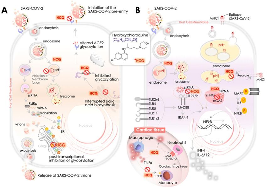
Pathogens 2020, 9, 546                                                                                3 of 35

and the benefits of HCQ in current clinical settings. We further brief on key considerations in HCQ
repurposing and its future perspectives.

2. SARS-CoV-2, ACE2, and Cardiovascular Challenges
      SARS-CoV-2 is a non-segmented, single-stranded (ss), positive (+) sense RNA virus [26]. It belongs
to the family of enveloped RNA beta-coronavirus. Out of seven known species of beta-coronavirus, only
three (SARS, MERS, and COVID-19) cause potentially fatal human disease. SARS-CoV-2 produces a
50–200 nanometers virion that is constituted by four structural proteins, viz., the S (spike), E (envelope),
M (membrane), and N (nucleocapsid), wherein the N protein is aligned with its RNA genome, while
the S, M, and E proteins collectively constitute the viral envelope [27]. The S protein at the SARS-CoV-2
envelop resembles a spike projection that serves as a tool for it to enter the host cell [28]. Phylogenetic
analysis revealed 99% similarity of S protein comparing SARS-CoV-2 and SARS-CoV [29] and therefore
reaffirmed the evidence that SARS-CoV-2 exploits the same ACE2 receptor [1] that originally served as
a functional receptor for SARS-CoV [30].
      ACE2 is present in alveolar epithelial cells and frequently localized at the cell membrane of
enterocytes (intestine), pericytes, cardiomyocytes, and macrophages [12,13,31]. ACE2 at the surface of
pericytes and cardiomyocytes serves a vital activity of the RAS by maintaining normal CV functions
by catalyzing the Ang (angiotensin) I and II [14]. SARS-CoV-20 s S protein primarily binds to the
ACE2 of alveolar epithelial cells in the respiratory tissues that enable its further access to the systemic
circulation, reaching cardiomyocytes in the heart and pericytes and endothelial cells in the macro-vessels
(Figure 1A). Endocytosis-driven internalization of ACE2 on the membrane of cardiomyocytes, pericytes,
and endothelial cells by SARS-CoV-2 results in omitting ACE2 from the cell surface and potentially
raises the risk of CV complications in COVID-19 patients [32]. The loss of ACE2 carboxypeptidase
function was earlier shown to compromise cardiac function [33]. A higher ACE2 level in patients with
existing CVD and/or hypertension was also suggested to increase the susceptibility to SARS-CoV-2
infection [34]. In light of this information, clinical readouts from six studies, including 1527 COVID-19
patients, revealed 17.1%, 16.4%, and 9.7% prevalence of hypertension, cardiac & cerebrovascular
disease, and diabetes, respectively [10]. Prevalence of these CVD comorbidities was found to be higher
in patients requiring ICU than the non-ICU patient groups. Analyses of mortalities in a cohort of
44,672 COVID-19 patients from Wuhan, China also showed 10.5%, 7.3%, and 6% mortalities in patients
having CVD, diabetes, and hypertension, respectively, significantly greater than the overall mortality
rate (2.3%) for COVID-19 patients [4]. To date, nine clinical studies from China [2,4–8,35–37] have
comprehensively assessed CV comorbidities in COVID-19 patient cohorts and yielded similar clinical
results (Figure 1B). However, disparities in testing, standardization and options for standard procedure
in clinical studies from China [6,8,38,39] [2,37,40] and elsewhere [40] have impacted the quantitative
clinical outcomes. To assess the cardiovascular outcomes of SARS-CoV-2 infection in a recent report,
Liu et al. reported a significantly higher level of circulating Ang II in COVID-19 patients than the
controls; circulating Ang II in levels COVID-19 patients also correlated well with viral load [41]. Of note,
these results were consistent with reduced ACE2 activity. They again underlined the crucial role of
RAS in COVID-19 disease and reaffirmed the focus on the cardio-protective function of ACE2, where
an alteration in its activity may substantially impact the cardiovascular outcomes [33,34]. Therefore,
in light of these reports, ACE2 has gained recognition as a key and central target in COVID-19 pathology
and associated CV complications. Taking note of SARS-CoV-2 infection severity, here we review the
frequent clinical cardiovascular complications observed in COVID-19 patients and further shed light
on the potential involvement of ACE2 activity.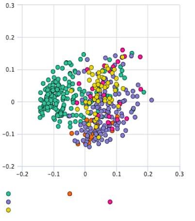
risk of systemic diseases, e.g., hypotension or hypoxia, then a specific cardiac dysfunction. In the
    given context, the role of the ‘cytokine storm’ elicited by SARS-CoV-2 immunoreactivity appears to
    be a key mediator [34]. Aberrant expressions of a variety of cytokines are evident in severely ill
    COVID-19 patients, while elevated plasma interleukin-6 (IL-6) levels were seen in patients with
    cardiac injury [43] (Figure 1A). Given the fact that ACE2 levels are present at the cell surface and in
Pathogens 2020, 9,in
    circulation    546
                     the CV system, direct SARS-CoV-2’s cardiomyocyte infection is suggested to be a 4 of 35
    definite possibility [44].

     Figure
        Figure1. 1.SARS-CoV-2,
                     SARS-CoV-2, angiotensin         converting
                                     angiotensin converting          enzyme
                                                                  enzyme       2 (ACE2),
                                                                           2 (ACE2),         and cardiovascular
                                                                                       and cardiovascular              complications.
                                                                                                               complications.
     (A)(A)
          Transmembrane
             TransmembraneACE2            receptorfacilitates
                                 ACE2 receptor        facilitates   SARS-CoV-2
                                                                 SARS-CoV-2     entryentry   to host
                                                                                       to host         cell primarily
                                                                                                cell primarily           in the lungs,
                                                                                                                 in the lungs,
     andand  then
          then   thethevascular
                        vascularsystem,
                                  system, postulating
                                             postulatingcardiovascular
                                                             cardiovascular complications
                                                                                 complicationsby causing   inflammation
                                                                                                   by causing              and
                                                                                                                  inflammation     and
        myocardial
     myocardial        dysfunction. SARS-CoV-2
                     dysfunction.     SARS-CoV-2 access accesstoto
                                                                 thethe
                                                                      systemic
                                                                        systemiccirculation  via the
                                                                                    circulation   vialungs   potentiates
                                                                                                        the lungs         heart heart
                                                                                                                    potentiates
        infection,while
     infection,      whileits
                            itsdirect
                                direct infection
                                        infection ofofassociated
                                                        associated  pericytes  andand
                                                                       pericytes    endothelial   cells may
                                                                                         endothelial     cellscause
                                                                                                                may vascular
                                                                                                                      cause vascular
        endothelial dysfunction. Cardiac SARS-CoV-2 infection causes micro-vessel dysfunction, and
     endothelial dysfunction. Cardiac SARS-CoV-2 infection causes micro-vessel dysfunction, and elevated
        elevated immunoreactivity disrupts atherosclerotic plaques leading to the progression of the acute
     immunoreactivity disrupts atherosclerotic plaques leading to the progression of the acute coronary
        coronary syndromes. SARS-CoV-2 infection of alveolar pneumocytes (type II) cells progressively
     syndromes. SARS-CoV-2 infection of alveolar pneumocytes (type II) cells progressively develops
        develops the systemic inflammation and elevated immunoreactivity that eventually produces the
     the‘cytokine
          systemic     inflammation
                     storm’,  marked byand      elevated
                                           elevated          immunoreactivity
                                                       IL-6, IL-7, IL-22, and CXCL10  thatcytokine
                                                                                            eventually     produces
                                                                                                     levels.            the ‘cytokine
                                                                                                             It potentiates  T-
     storm’,   marked
        cell and          by elevated
                    macrophage             IL-6,infiltrating
                                    activation    IL-7, IL-22,     and CXCL10
                                                               infected   myocardialcytokine
                                                                                        tissues levels.
                                                                                                 and may  It potentiates
                                                                                                              produce severeT-cell and
     macrophage
        cardiac damageactivation   infiltrating infected
                             and myocarditis,      leading myocardial       tissues
                                                              to heart failure.       and may
                                                                                  Cytokine       produce
                                                                                              storm   may severe
                                                                                                             further cardiac
                                                                                                                      increasedamage
     and myocarditis, leading to heart failure. Cytokine storm may further increase damage of cardiac
     monocytes causing myocardial dysfunction and subsequent development of arrhythmia. These events
     cumulatively produce cardiac dysfunction. (B) Manifestation (%) of cardiovascular complications in
     hospitalized COVID-19 patients reported in key clinical studies exhibiting comorbidities including
     hypertension, cardiovascular disease (CVD), cerebrovascular disease, coronary artery disease and rate
     of cardiac injury, shock, heart failure, and arrhythmia in low (LS), and high severity (HS) patient groups.
     p values indicate *** (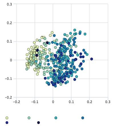
Pathogens 2020, 9, 546                                                                              5 of 35

study reported 23% cases of congestive cardiac failure in COVID-19 patients among all in-hospital
Chinese patients (Figure 1B). Markedly, these incidences in deceased and survivor cases were found to
be 52% and 12%, respectively [7]. A lack of information on the possible association of cTnl increase
with pre-existing CV complications limits our ability to predict causalities. However, an increase in
cTnl levels was found to depict poor prognosis in other systemic diseases. Therefore, such associations
of cTnl levels with prognosis is likely to predict the risk of systemic diseases, e.g., hypotension or
hypoxia, then a specific cardiac dysfunction. In the given context, the role of the ‘cytokine storm’
elicited by SARS-CoV-2 immunoreactivity appears to be a key mediator [34]. Aberrant expressions of a
variety of cytokines are evident in severely ill COVID-19 patients, while elevated plasma interleukin-6
(IL-6) levels were seen in patients with cardiac injury [43] (Figure 1A). Given the fact that ACE2 levels
are present at the cell surface and in circulation in the CV system, direct SARS-CoV-20 s cardiomyocyte
infection is suggested to be a definite possibility [44].

2.2. Cardiac Arrhythmia
     Manifestations of viral infections are frequently seen to be associated with myocardial inflammation,
metabolic dysfunction, and modulation of the sympathetic nervous system that all serve as key factors
in causing the cardiac arrhythmia. A meta-analysis of 138 COVID-19 patients cohort reported 16.7%
incidences of developing arrhythmia in patients, which in terms of serious complications came second
after ARDS [8]. Another cohort comparing patients based on their admittance to ICU revealed
strikingly higher (44%) cases of arrhythmia in ICU admitted patients, while in the non-ICU admitted
patients group it remained at 4% [39] (Figure 1B). These findings postulated the role of systemic
inflammation and elevated immunoreactivity produced by cytokine storm that may damage cardiac
monocytes causing myocardial dysfunction and subsequent development of arrhythmia (Figure 1A).
The internalization of ACE2 by SARS-CoV-2 served as a key event that led to the altered RAS system,
which was postulated earlier to cause pro-inflammatory and pro-oxidant activities [32].

2.3. Myocarditis
     Acute viral infections are known to cause cardiac injury and acute myocarditis. A recent report
by the National Health Commission, China, showed the infiltration of mononuclear cells and the
onset of monocyte necrosis in cardiac muscle autopsy specimens. Along these lines, other findings
concerning fulminant myocarditis indicate the possibility of myocarditis in COVID-19 patients as
a cause of acute cardiac injury [43,45]. Despite the findings of these reports and individual clinical
cases [43,45], we presently lack any information on the underlying mechanism, its prevalence, and
clinical importance, and therefore this emphasizes the need for detailed clinical analyses. However,
the earliest reports suggested that fulminant myocarditis may potentially be a clinical manifestation
of SARS-CoV-2 infections of cardiomyocytes [43,45], postulated to be caused by elevated IL-6, IL-7,
IL-22, and CXCL10 cytokine levels produced as a result of cytokine storm (Figure 1A). ACE2-led
SARS-CoV-2 infection of alveolar pneumocytes (type II) cells has been suggested to trigger the onset
of systemic inflammation and elevated immunoreactivity leading to a ‘cytokine storm’, that may
essentially potentiate T-cell and macrophage activation infiltrating infected myocardial tissues and
resulting in cardiac damage and myocarditis (Figure 1A). However, a detailed assessment of these
events is needed to further confirm the acquisition of systemic myocarditis in COVID-19 patients.

2.4. Acute Coronary Disease (ACD) and Ischemia
     Most clinical studies so far lack any insights into ACD in COVID-19 patients; however, it is
suggested that it impacts on destabilizing coronary plaques in COVID-19 patients [7,46,47]. Of
note, the role of the systemic inflammatory response is implicated primarily in destabilizing
atherosclerotic plaques [48], which further supports pro-inflammatory and pro-oxidative consequences
of SARS-CoV-2-led ACE2 loss in COVID-19 patients (Figure 1A,B). More specifically, COVID-19
patients with heart failure are at higher risk of acute events or ischemic syndrome.
Pathogens 2020, 9, 546                                                                            6 of 35

2.5. Disseminated Intravascular Coagulation (DIC)
      Incidences of pulmonary embolism (PE) and subsequent disseminated intravascular coagulation
(DIC) are linked with coronavirus infection, as COVID-19 patients demonstrate a hypercoagulable
state, marked by prolonged prothrombin time, elevated D-dimer level and fibrin split. Of note, 71.4%
of non-survivor patients were found to have DIC [49]. COVID-19 patients characteristically also had
vast pulmonary embolism features [50]. Importantly, increase in D-dimer in COVID-19 patients was
suggested to predict adverse survival outcome, for instance, a study of a retrospective cohort showed
that increased D-dimer levels (>1 g/L) were able to closely predict in-hospital mortality [7]. However,
the mechanistic basis of these features of SARS-CoV-2 infection is yet to be elucidated, while new
knowledge of pro-inflammatory/oxidant activities in these syndromes could further shed light on the
underlying role of ACE2 function in SARS-CoV-2 pathogenesis.

2.6. Immune Function in Cardiovascular Complications
     After respiratory infection, the immune response is the second most exploited system in COVID-19
patients, and this has severe implications for the cardiovascular system. Firstly, Huang et al. reported
elevated systemic IL-2, IL-6, IL-7, C-X-C motif chemokine 10 (CXCL10), chemokine (C-C motif) ligand
2 (CCL2), tumor necrosis factor-α (TNFα), and granulocyte colony-stimulating factor (G-CSF) levels in
COVID-19 patients [6]. The elevated levels of systemic cytokines shared clinical features with cytokine
release syndrome (CRS) [13] that may substantially contribute to COVID-19 severity. The above
systemic immune response resembles cytokine profiles raised in hemophagocytic lympho-histiocytosis
(HLH) syndromes [51]. Sorting of the immune cell population in COVID-19 patients revealed
the presence of hyperactivated T-cells with high fractions of HLA-DR+, CCR6+ Th17 CD4+ and
CD38+ CD8+/CD4+ T-cells. This emphasized the role of hyperactivated T-cells, which may partly
be associated with severe immune injury [13]. Furthermore, elevated levels of circulating IL-6 in
a cohort of 150 patients in a recent retrospective study were found to be predictive of mortality in
hospitalized COVID-19 patients [5]. Of note, the role of IL-6 has been earlier primarily implicated
in CV complications, including atherosclerosis and coronary heart disease, and with increasing the
risk of cardiac inflammation and morbidity [52,53]. Therefore, the prevalence of systemic cytokine
response/CRS or cytokine storm in clinical COVID-19 patients significantly raises an obvious risk of
cardiovascular complications (Figure 1A).

3. ACE2 Receptor and Its Significance in Systemic Cardiovascular Function
     ACE2 comprises an 805-amino acid (aa; Mr 110,000 glycoprotein) long endothelium-bound
carboxy-mono-peptidase that consists of a 17-aa N-terminal peptide (catalytic domain-oriented
extracellularly) and a C-terminal anchor integrated into the membrane. ACE2 is catalytically a zinc
metalloprotease and the only homolog of ACE known in humans [54]. ACE2 is part of the RAS
that plays a crucial function in maintaining normal cardiovascular functions, while dysfunction in
RAS contributes to CVDs, including hypertension, myocarditis, coronary heart disease, and heart
failure [14]. RAS is constituted by a set of catalytic enzymes that includes angiotensinogen, renin,
Ang II, Ang II receptors (AT1R and AT2R), and ACE [55]. Among these, ACE2 has a crucial role to
play by catalyzing Ang II to Ang (1–7) or Ang I to Ang (1–9) [56]. ACE2 can access substrate/peptide
in the circulation, and it is known for its circulatory presence and catalytic function in the blood and
body fluid. Given its carboxy-monopeptidase activity, ACE2 primarily trims the COOH-terminal
phenylalanine residue from Ang II [57]. ACE2-led trimming of Ang II to Ang (1–7) is a significant event
in the RAS, since the role of Ang II is critically implicated in producing hypertension by promoting
vasoconstriction, fibrosis, Na+ retention, and pro-inflammation and pro-oxidant activities. At the same
time, elevated levels of Ang (1–7) peptide inhibits the Ang II/AT1R axis and induces anti-inflammatory,
anti-oxidant, anti-fibrotic, and vasodilatory activities (Figure 2A) [56,58]. Therefore, ACE2 activity
switches on the processing of Ang II in the classical RAS system and loss of ACE2 or its function
influenza viruses viz., H1N1, H5N1, and H7N9 [62–64].
Mechanistically, the intracellular entry of SARS-CoV-2 in host cells is facilitated by binding of
its S (spike) protein’s receptor binding region with the ACE2 extracellular domain, at a high affinity
(15 nM) [65]. Prior to their binding, the host cell serine protease, viz., TMPRSS2S, processes cleavage
of S protein down the dibasic Arg sites and yields to the S1 and S2 subunits. S protein cleavage is a
Pathogens 2020, 9, 546 7 of 35
crucial step which enables S2-led membrane fusion and ACE2-mediated SARS-CoV-2 internalization
by endocytosis [66,67] in the type II pneumocytes, pericytes, or cardiomyocytes [12,13]. Structural
couldanalyses
put thesuggest that S protein of SARS-CoV-2 has a receptor-binding domain (RBD) to interact with
RAS system to an overall higher Ang II level [58].These cardioprotective activities of
human ACE2, wherein 441Leu, 472Phe, 479Gln, 480Ser, 487Asn, and 491Tyr residues of S protein
ACE2 are regulated through the Ang I (1–9)/AT2R and Ang I (1–7)/MasR axes [55].On the contrary,
were predicted to have a critical role in its binding [68]. Higher virulence of SARS-CoV-2 than SARS-
ACE CoVdegrades Ang (1–7) and forms ANG II that results in promoting inflammation, fibrosis, and
was shown to reflect a higher affinity of S1 protein for ACE2 [69]. SARS-CoV-2 binding of ACE2
high at
blood
the membrane(Figure
pressure 2). The roleloss
and its subsequent of ACE2 was alsoimpact
by endocytosis implicated
RAS andin the hydrolysis
change of apelin
the overall Ang and
des-arginine bradykinin (des-Arg1-BK) apelin peptides, wherein des-Arg1-BK was shown
II:Ang (1–7) ratio (enriching cardio-inflammatory Ang II, and decreasing cardio-protective Ang (1-7) to have a
pro-inflammatory
levels), eventuallyfunction via stimulating
exacerbating the B1
cardiac injury by receptor [59] (Figure
SARS-CoV-2. However, 2).the
Besides
extent its critical
of CV role in
tissue
the CVdamage dueACE2
system, to the presence
was earlierof SARS-CoV-2
discoveredintothe
becirculation has not
a key binding been precisely
receptor analyzed as
for SARS-CoV yetNL63
and
[6,70]. ACE2
(HCoVNL63) is a primary established
coronaviruses routerecently
[30,60], while of Ang IIitmetabolism that generates
was identified Ang (1–7) in the
to be a SARS-CoV-2 heart, [61].
receptor
and therefore its loss is frequently seen as compromising systemic cardiovascular function
ACE2 is also shown to play a key role in acute respiratory/lung injury caused by influenza viruses viz., [32,71–
73], wherein hypertension, inflammation, vasoconstriction, and oxidative activities have been the
H1N1, H5N1, and H7N9 [62–64].
common CV complications [32,60,74,75].
Figure
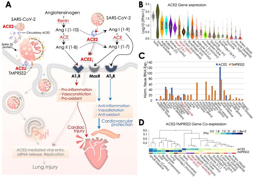
Figure 2. 2.SARS-CoV-2
SARS-CoV-2 pathology
pathologyand ACE2-led
and ACE2-ledregulation of cardiovascular
regulation function offunction
of cardiovascular the Renin-
of the
Angiotensin System (RAS). A. Schematic diagram illustrating the central role of ACE2 in SARS-CoV-
Renin-Angiotensin System (RAS). (A) Schematic diagram illustrating the central role of ACE2 in
2 recognition and the differential regulation of the RAS system for cardiovascular protection or
SARS-CoV-2 recognition and the differential regulation of the RAS system for cardiovascular protection
cardiac injury. SARS-CoV-2 spike (S) protein undergoes priming by the TMPRSS2, a host cell
or cardiac injury. SARS-CoV-2 spike (S) protein undergoes priming by the TMPRSS2, a host cell
membrane protease, and it subsequently binds to ACE2 infecting the host cell. In the RAS system,
membrane
ACE2 protease, andMasR,
activity with it subsequently
and AT2R binds to ACE2
receptors infecting
provides the host cell.
cardiovascular In the RAS
protection. system, aACE2
In contrast,
activity with MasR, and AT2R receptors provides cardiovascular protection. In contrast, a reduced
ACE2 activity as a result of its binding to SARS-CoV-2 and engulfment into the cell may elevate ACE
activity and Ang II levels that essentially potentiates cardiac damage/injury. (B) ACE2 gene expression
data of ACE2 retrieved from Genotype-Tissue Expression (GTEx) showing its expression across human
tissues, wherein heart tissues are marked in red at x-axis. Expression values are shown in the log10
scale for TPM (Transcripts Per Million) unit. (C,D) ACE2 and TMPRSS2 mRNA levels retrieved from
Human Protein Atlas (HPA; C) and Genotype-Tissue Expression (GTEx; D) showing their co-expression
across various human tissues; heart tissues are marked in red at x-axis.
Mechanistically, the intracellular entry of SARS-CoV-2 in host cells is facilitated by binding of
its S (spike) protein’s receptor binding region with the ACE2 extracellular domain, at a high affinity
(15 nM) [65]. Prior to their binding, the host cell serine protease, viz., TMPRSS2S, processes cleavage
of S protein down the dibasic Arg sites and yields to the S1 and S2 subunits. S protein cleavage is aPathogens 2020, 9, 546 8 of 35
crucial step which enables S2-led membrane fusion and ACE2-mediated SARS-CoV-2 internalization
by endocytosis [66,67] in the type II pneumocytes, pericytes, or cardiomyocytes [12,13]. Structural
analyses suggest that S protein of SARS-CoV-2 has a receptor-binding domain (RBD) to interact with
human ACE2, wherein 441Leu, 472Phe, 479Gln, 480Ser, 487Asn, and 491Tyr residues of S protein were
predicted to have a critical role in its binding [68]. Higher virulence of SARS-CoV-2 than SARS-CoV
was shown to reflect a higher affinity of S1 protein for ACE2 [69]. SARS-CoV-2 binding of ACE2 at
the membrane and its subsequent loss by endocytosis impact RAS and change the overall Ang II:Ang
(1–7) ratio (enriching cardio-inflammatory Ang II, and decreasing cardio-protective Ang (1-7) levels),
eventually exacerbating cardiac injury by SARS-CoV-2. However, the extent of CV tissue damage
due to the presence of SARS-CoV-2 in the circulation has not been precisely analyzed as yet [6,70].
ACE2 is a primary established route of Ang II metabolism that generates Ang (1–7) in the heart, and
therefore its loss is frequently seen as compromising systemic cardiovascular function [32,71–73],
wherein hypertension, inflammation, vasoconstriction, and oxidative activities have been the common
CV complications [32,60,74,75].
Given the fact that ACE2 circulating levels are endogenously deficient, its adequacy in preventing
viral dissemination by sequestering SARS-CoV-2 in circulation was questioned [54], yet its protective
effects against hypertension, myocardial hypertrophy, inflammation, and fibrosis were evident [76].
Also, amid these speculations [54,77], a proposed clinical trial (NCT04287686) to study the infusion of
recombinant ACE2 to restrict viral infection was subsequently withdrawn. However, a recent report
testing the potential of clinical-grade human recombinant soluble ACE2 (hrsACE2) in engineered
human tissues showed an effective SARS-CoV-2 inhibition by a factor of 1000–5000. Therefore, soluble
ACE2 might be the key therapeutic alternative restricting SARS-CoV-2 infection at early stage [78].
An alternative strategy using ACE2-specific antibodies to target membrane-aligned and soluble ACE2
was earlier found to be effective against SARS-CoV infection [79]. ACE2, inhibition of TMPRSS2
in the murine models also limited coronavirus infection and improved survival [80,81]. Presently,
two ongoing clinical trials (NCT04321096 & NCT04338906) are testing the efficacy of TMPRSS2
inhibition by camostat mesilate to evaluate its benefits against COVID-19.
TMPRSS2 is critical in the host recognition of SARS-CoV-2, and therefore for the S protein it
serves as a cofactor for ACE2-mediated viral entry into the host cell [12]. Hence, TMPRSS2 and
ACE2 co-expression is crucial to facilitate an optimum SARS-CoV-2 infection. To get an insight
into the above possibility, we firstly surveyed the ACE2 transcript expression at Genotype-Tissue
Expression (GTEx) that revealed its enriched expression, particularly in testis, small intestine, adipose
and heart/cardiac tissues (Figure 2B). This primarily explains the vulnerability of these tissues to
SARS-CoV-2 infection. Of note, ACE2 transcript levels in the heart were higher than in the lungs
which could help to explain the higher vulnerability of SARS-CoV-2 infection in the CV system and
the prevalence of CV complications in COVID-19 patients. Similar observations were obtained in a
recent report [44]. However, our survey of ACE2 and TMPRSS2 transcript co-expression at Human
Protein Atlas (HPA; http://www.proteinatlas.org) further revealed a lesser co-occurrence of their
expressions in the heart, while a strong correlation of their co-expression was observed in intestinal
(colon, duodenum, small intestine) and renal tissues [82] (Figure 2C). Of note, a moderate expression
of ACE2 and its co-expression with TMPRSS2 in the kidney/renal tissues also underlines association of
SARS-CoV-2 infection with renal injury, which is frequently seen in COVID-19 patients. In critically ill
patients admitted to ICU, 0.5–29% incidences of acute kidney injury have been reported [2,6–9,38],
and, thus, kidney injury was also recognized as a key feature of disease severity and correlated
negatively for patient survival [7]. Interestingly, enzymatically active/secretory tissues (pancreas,
prostate, salivary gland, seminal vesicle, stomach, and thyroid) were more enriched with TMPRSS2
expression. A much similar co-expression pattern of ACE2 and TMPRSS2 transcript was observed
in the GTEx database (Figure 2D). Of note, in both analyses, a higher expression of TMPRSS2 was
more evident in the lungs than the heart. However, the functional significance of such disparity in its
expression warrants further investigation. Results of these surveys overlapped in parts with findingsPathogens 2020, 9, 546 9 of 35
of an earlier report that analyzed co-expression of ACE2 and TMPRSS2, along with HAT in influenza
and SARS-coronavirus, and reported their evident co-expression in respiratory, gastro-intestinal and
cardiovascular tissues [83]. This evidence postulates that ACE2 and concomitant TMPRSS2 expression
in pericytes and cardiomyocytes could essentially potentiate chances of SARS-CoV-2 infection in the
systemic CV system and also explain the higher prevalence of CV issues in COVID-19 patients.
4. SARS-CoV-2, ACE2, Hydroxychloroquine and beyond: Preventive and Therapeutic Aspects
The ongoing COVID-19 pandemic prompted the urgent need to develop targeted therapeutic
strategies and exercise all options of repurposing conventional drugs as a viable solution, considering
their known pharmacological aspects and benefits. Efforts identified CQ and HCQ as two therapeutic
drugs potentially useful in preventing COVID-19. The earliest reports analyzing the effects of CQ and
HCQ in vitro against SARS-CoV-2 revealed their inhibitory antiviral activities [19,84,85]. Besides their
earlier known antimalarial activity [86], these aminoquinoline analogs provide broad-spectrum benefits
against types of bacterial, fungal, and viral infections [87–89]. Given its inexpensive cost, less toxicity,
good tolerance, and immunomodulatory activities in patients [90], early reports in the last decade
explored the repurposing of CQ against human immunodeficiency virus (HIV) and other viruses
causing inflammation [91]. Therapeutic benefits of CQ’s anti-inflammation and immunomodulatory
activities were also explored in autoimmune diseases [92].
Keyaerts et al. in 2004, first demonstrated the antiviral activity of CQ against SARS coronavirus [16],
while further reports revealed an inhibitory function of CQ on HCoV- 229E replication in epithelial
lung cell in vitro [93,94]. Keyaerts et al. in 2009 also showed that infection of HCoV-O43 coronavirus
in newborn mice could be treated by medication of CQ through the mother’s milk [95]. With growing
concerns about the toxicity of CQ medication in humans, HCQ, a less toxic derivative of CQ quinine
analog, was subsequently opted for and used in clinical studies broadly. While keeping SARS-CoV-2
and ACE2 in focus, in the following sections we discuss the pharmacology of HCQ, its benefits
in vitro and clinical settings, and what impact HCQ has upon SARS-CoV-2 replication and underlying
immunomodulatory activities.
4.1. HCQ Pharmacology, In Vitro and Clinical Outcomes
The HCQ parental molecule, i.e., quinine, was first extracted from the Cinchona, a native tree
to Peru, and primarily used for its benefits against malaria [96]. Later, an amine acidotrophic
form of quinine, viz., CQ, was synthesized in Germany in 1934 as a natural substitute. CQ and
its 4-aminoquinoline derivative, viz., HCQ, are both weak bases and share a common molecular
family. Addition of a hydroxyl group at the CQ side-chain terminal end (β- hydroxylation of
the N-ethyl substituent) forms HCQ. It is usually administered orally in the form of HCQ sulfate,
while its pharmacokinetics are similar to those of CQ, which include its swift absorption in the
gastro-intestine and its renal release. HCQ, being a positively charged base, stays in a protonated form.
However, un-protonated HCQ can access the intracellular organelles/compartments, where it becomes
protonated, which, in turn, increases the localized pH. This mechanism explains the accumulation
of CQ/HCQ within acidic organelles, e.g., endosome, lysosomes, and the Golgi vesicles [97], and
thus becomes one aspect of its pharmacokinetic activity. This fact may also explain its 200–700-times
higher accumulation in splenic, hepatic, renal, and heart and lung than in plasma [98]. With its quick
absorption, HCQ achieves its maximum concentration in serum in 2–3.5 h, while the half-life of its
clearance was 22–45 days [99].
Multiple in vitro studies performed earlier on SARS-CoV, exhibiting the benefits of CQ or HCQ
against viral replication, provided an early hope for the potential repurposing of CQ/HCQ against
COVID-19. Firstly, Keyaerts et al. in 2004 demonstrated that sub-toxic CQ concentration (8.8 +/− 1.2 µM,
much lower than the CC50 (261.3 +/− 14.5 µM)) could effectively reduce the SARS-CoV replication rate
in Vero E6 (kidney epithelial; source-African green monkey) cells by 50% [16]. In another report, Vincent
et al. showed that, at 10µM, CQ concentration effectively inhibited SARS-CoV viral replication in VeroPathogens 2020, 9, 546 10 of 35
E6 [15]. These inhibitory effects of CQ treatment were effective in pre- or post-SARS-CoV infected cells
and therefore hinted at its prophylactic and therapeutic applications [15]. Biot et al. also observed a
similar finding with CQ and HCQ; however, they reported that CQ causes more potent inhibition of
viral replication [17]. These reports pitched the anecdotal benefits of CQ/HCQ for their repurposing
against the ongoing COVID-19 pandemic and prompted researchers to evaluate their activity against
SARS-CoV-2. In the earliest efforts, Yao et al. analyzed the antiviral activities of HCQ and CQ
on SARS-CoV-2 in Vero cell lines [18]. HCQ and CQ both showed SARS-CoV-2 inhibitory activity;
however, in contrast to the study by Biot et al., which show better efficacy of CQ against SARS-CoV [17],
Yao et al. showed that HCQ (EC50 = 0.72 µM) has much higher antiviral potency than CQ (EC50 =
5.47 µM) for SARS-CoV-2 [18]. In a post-infection treatment regime, HC impaired viral replication
more effectively. While analyses of prophylactic activity also indicated the greater efficacy of HCQ
(EC50-5.85Mm) than CQ (EC50-18.01Mm) in 48 h, an extended treatment suggested the production of
a more significant anti-viral effect [18]. To determine a potential clinical regime for HCQ, Yao et al.
enrolled physiology-based pharmacokinetic modeling and considered multiple parameters, including
drug administration route, its physiological assimilation (i.e., intestinal absorption and accessibility
to lung tissue), and biochemical activities. This report also discussed simulated concentrations of
lung fluid but lacked inclusion of all details used in the model [18]. Based on physiology-based
pharmacokinetic modeling, Yao et al. proposed a treatment regime that included an initial dose of
400 mg HCQ twice a day and a continuation of 200 mg dose twice daily for the next four days, which
turned out to be a key outcome of this study. However, the study lacked a 95% confidence interval
value for the estimated EC50 dose, which suggests that the aforementioned dose regime needs to
be adopted with caution to avoid inaccuracy in treatments [18]. Another report by Liu et al., using
a similar antiviral regime at four multiplicities of infection, revealed the efficacy of CQ and HCQ
in inhibiting SARS-CoV-2 viral replication at all four tested infection regimes [85]. Although they
suggested a more robust potency of CQ than HCQ, this was only found to be significant at 0.01 and
0.2 multiplicities of infection. Importantly, using immunofluorescence-based co-localization assay,
they analyzed entry of SARS-CoV-2 virion into the endosome-lysosome proteolysis pathway and
found an accumulation of more virions at early endosomes and lesser at endolysosomes in CQ and
HCQ treated cells in comparison to untreated viral infected control cells [85]. Using a similar Vero E6
cell system, Wang et al. further showed that a combination of remdesivir (EC50 = 0.77 µM) and CQ
(EC50 = 1.13 µM) could effectively control viral infection in vitro [19]. This study enrolled SARS-CoV-2
at a multiplicity of infection (MOI) of 0.05 and pre-treated Vero E6 cells with 0.01, 0.05, 0.1, 0.5, 1, 5, and
10 µM CQ for 1 h. Regarding the antiviral activities of remdesivir and CQ, Wang et al. suggested their
inclusion in clinical therapeutic regimes against SARS-CoV-2 [19]. In another combinatorial approach,
Andreani et al. showed a synergistic effect of CQ and Azithromycin (AZM) against SARS-CoV-2 [100].
Using concentrations of 1, 2 or 5 µM CQ along with 5 or 10 µM AZM and multiplicity of infection
(MOI) at 0.25 they showed that 5 µM CQ treatment in combination with 10 and 5 µM AZ led relatively
to 97.5% and 99.1% viral inhibition respectively [100]. The details of these in vitro studies testing
efficacies of CQ and HCQ against SARS-CoV-2 are provided in Table 1, where the 2004 report of
Keyaerts et al. is taken as reference.
Initial findings of HCQ/CQ antiviral activity from in vitro studies [18,19,85,100] raised an early
clinical interest in testing the efficacy of HCQ/CQ in the clinic for COVID-19 treatment. An interim
analysis from China, including more than 100 COVID-19 patients, showed the superiority of CQ
treatment compared to the control group [24]. Although this report provided no details of enrolled
patients, their clinical features including the benefits of HQ in improving lung imaging, viral shedding,
and in shortening disease course were discussed. One key takeaway from this study was the
recommended CQ dose (500 mg twice daily -b.i.d.) for the next ten days for patients exhibiting mild,
moderate or severe symptoms [24].Pathogens 2020, 9, 546 11 of 35
Table 1. Pre-clinical readouts from the key in vitro studies investigating therapeutic efficacy of HCQ against SARS-CoV-2. HCQ, Hydroxychloroquine; EC50, Effective
concentration; AZM, Azithromycin.
Investigation/References Cell Systems Drug, Concentration, and Assay Time (h) Study Control Key Findings/Comments
-HCQ showed better SARS-CoV-2 inhibitory
CQ and HCQ
Vero E6 cell (Origin-African activity than CQ.
Yao et al. 2020 0.032, 0.16, 0.80, 4, 20, & 100 µM -
green Monkey) -An extended incubation period may produce
2h
greater anti-viral effect
-HCQ inhibited the steps including infection/entry
CQ and HCQ
and post-infection
Liu et al. 2020 Vero E6 Cells 0.068, 0.21, 0.62, 1.85, 5.56, 16.67, and 50 µM PBS (Phosphate buffer saline)
-At the higher viral replication rate, anti-viral
1h
efficacy of HCQ found to be lesser than of CQ
-HCQ inhibited the viral activity at low µM conc.
CQ and others *
(effective conc. EC50 = 1.13 µM)
Wang et al. 2020 Vero E6 Cells 0.01, 0.05, 0.1, 0.5, 1, 5, and 10 µM DMSO
-CQ effectively inhibited SARS-CoV-2 infection
1h
in vitro
Combination of hydroxychloroquine and
CQ- 1, 2 or 5 µM associated with 5 or 10 µM for
Andreani et al. 2020 Vero E6 cells - azithromycin has a synergistic effect in vitro on
azithromycin.
SARS-CoV-2 at concentrations
-CQ potently inhibits SARS-CoV activity at a lesser
CQ (8.8 ± 1.2 µM) concentration than its cytostatic
Keyaerts et al. 2004 (*Earliest
Vero E6 cell 0, 0.8, 4, 20, & 100 µM - activity (261.3 ± 14.5 µM)
report from the SARS-CoV)
8 h to 3 days -Addition of CQ even after 5 h of SARS-CoV
infection could yet be inhibitory activePathogens 2020, 9, 546 12 of 35
A comprehensive review of available clinical data so far on the prophylactic and therapeutic use of
HCQ/CQ against COVID-19 in human cohorts included nine clinical studies and two case series/reports,
as summarized in the Table 2. In the earliest report, Chen et al. analyzed the efficacy of HCQ in a
small-size (30 inpatients) randomized controlled trial in Shanghai, China [21]. When comparing the
clinical outcome of HCQ to the standard of care, they found no statistically significant differences in
virus clearance in control (93%, p > 0.05) and HCQ (87%) group by day 7. Also, no difference in the
clinical symptoms, including the fever, its duration, and any alteration in lung features was observed
in 400 mg HCQ treated patients for five days. Although the admitted patients in control and HCQ
groups had symptoms for ~ 6 and 7 days respectively, no detail of COVID-19 severity in the enrolled
patients was reported. In mid-March 2020, Gautret et al., in an open-label, non-randomized clinical trial,
reported that HCQ causes significant viral clearance at day 6 from the nasopharynx of treated patients
(60%) as compared to control (15%) [22]. A faster viral clearance in patients who were given HCQ
and Azithromycin (AZM) was reported and hinted at the synergistic effect of the two drugs. Given its
non-randomized, unequal settings, and exclusion of six patients from analyses [22], the clinical outcome
of this study was criticized. Another report from this group with a non-randomized cohort of 80 patients
(with ~5 days symptoms) treated with HCQ and AZM revealed that 93% treated patients were negative
of SARS-CoV-2 in just 4.5 days of treatment, as validated using reverse transcription-polymerase chain
reaction (RT-PCR) of nasopharynx samples [25]. An absence of a comparison arm in this analysis
compromised the clinical outcome of the report. Malina et al., another group from France, testing the
combination of HCQ and AZM in a prospective, open-label study showed that, out of ten patients,
only two exhibited viral load reduction by day six, and they therefore doubted the clinical outcome of
studies published by Gautret et al. [22]. In another report from China, Chen et al. using HCQ for a mild
symptomatic COVID-19 patient cohort showed faster clearance of cough and fever in HCQ treated
patients than control [101]. However, this report had several limitations, including the exclusion
of patients for unclear reasons, delivery of antivirals, steroids, and intravenous immunoglobulin as
standard therapy and no endpoint details (no information on mortality, viral clearance, and patient
discharge). A preprint study from France, comprising retrospective/non-randomized trials of 181
inpatients, examined the efficacy of HCQ (600 mg/day) in 84 patients, while 97 patients were taken
as control [102]. Authors found no apparent benefits of HCQ compared to control, and ~ 10% of
patients given HCQ were discontinued due to change in their ECG reading. In a recent multi-centric,
randomized controlled trial from China, Tang et al., enrolling 75 patients each in HCQ and control
(receiving standard of care) groups, showed no significant difference in viral clearance in HCQ (85.4%)
and control (81.3%, p = 0.341) by day 28 [103]. Testing a 1200 mg HCQ dose for the first three days
followed by 800 mg daily dose for the next two-three weeks, this post hoc trial did not support
HCQ use for COVID-19 treatment. Recently, in one of the biggest open-label and non-randomized
trial studies (comprising 1061 patients) testing HCQ and AZM combination in France, Million et al.
showed, in a ten-day regime, good clinical outcome and viral cure observed in 973 HCQ+AZM treated
patients (91.7%) [104]. They recommend that prophylactic use of HCQ+AZ is safe and acquires a
low fatality rate in patients. In contrast, in a preprint recent retrospective/non-randomized trial of
veterans hospitalized in USA that were given HCQ and HCQ+AZM for groups of 97 and 113 patients
respectively, Magagnoli et al. showed no evidence that HCQ, either with or without AZM, benefits
patients and lessens the risk of mechanical support in treatment [105].Pathogens 2020, 9, 546 13 of 35
Table 2. Characteristics of HCQ therapeutic regimes and their outcomes in key comprehensive clinical studies.
Investigation Patients (Total No) Severity of
Investigation/Reference Regimes Results/Key Findings Comment Location Limitation
Type/Design COVID-19 Disease
Con HCQ
Indifferent outcomes in
6–7 days Patients were tested Smaller sample size.
groups. By day 7, no
Randomized and HCQ- 400 mg for symptomatic negative for Shanghai, Not peer-reviewed,
Chen J et al. (2020) 15 15 significant change in
controlled trial 5 days patients, unclear COVID-19 at 2 China availability in
conversion rate (86.7%
severity weeks Chinese language
vs 93.3%) observed.
Asymptomatic
patients-17%, Unadjusted results Exclusion of 6 Study design,
Patients with showed significantly patients from data Smaller sample-size,
Open-label trail,
HCQ- 600 mg for respiratory reduced viral titer at (1- died, 1- Marseille, Exclusion of 6
Gautret P et al. (2020a) Non-randomized, 16 26
10 days symptoms- 61%, day 6 (HCQ-70% vs. withdrew, 3 needed France patients,
Non-blinded
Chest CT con 12.5%, PCR based, ICU admission, 1- inconclusive
pneumonia +ve p < 0.01) lost follow-up) long-term outcomes
patients- 22%
Patient died-1,
HCQ- 600 mg for 10 patients out of 11 8 patients out of 10 Patient transferred
Prospective
5 days + AZM 500 were on were positive at day 5–6 to ICU-2, Patient Smaller sample size.
Molina JM et al. (2020) open-label 0 10 Paris, France
mg × 1, then 250 supplemental (nasopharyngeal swab) had no further HCQ Not peer-reviewed.
investigation
mg oxygen (80%, 95% CI: 49–94) post prolongation
of QTc-1
- Clinical recovery and
cough remission time
Mild illness was Undefined status, 4
reduced in HCQ group,
Parallel-group HCQ- 400 mg for observed in CT patients developed Smaller sample size.
Chen Z et al. (2020) 31 31 while resolution of Wuhan, China
trail Randomized 5 days confirmed severe illness in the Not peer-reviewed.
pneumonia was higher
pneumonia cases control group
(80.60% vs. 54.8%) in
the HCQ group.
Asymptomatic- 5%,
Patients discharged
Pneumonia cases- Decreased Design of the study,
HCQ- 600 mg for from hospital - 65
Open-label trail, 54%, Patients with nasopharyngeal viral Smaller sample size.
10 days + 500 mg, (81.3%), Patients Marseille,
Gautret P et al. (2020b) Non-randomized, 0 80 low national early load at 7th (83% Not peer-reviewed.
followed by 250 needed ICU France
Non-blinded warning score negative) and 8th (93%) Short follow-up
mg AZM admission- 1,
(NEWS) and mild days time period
Deceased- 1
disease- 92%
HCQ showed no
Adverse events in
HCQ- 200 mg for Patients with significantly higher
Open-label, control and HCQ
first 3 days, 800 mild-moderate negative conversion Shanghai,
Multi-centric, group were Smaller sample size.
Tang W et al. (2020) 75 75 mg for remaining disease- 148. probability (85.4%) than Anhui, Hubei,
Randomized, reported in 7 and 21 Not peer-reviewed.
days (total 2–3 Patients with severe control (81.3%) patients. China
Controlled trial patients
weeks) illness-2 Adverse effects were
respectively
reported in HCQ groupPathogens 2020, 9, 546 14 of 35
Table 2. Cont.
Investigation Patients (Total No) Severity of
Investigation/Reference Regimes Results/Key Findings Comment Location Limitation
Type/Design COVID-19 Disease
Con HCQ
In 10 day regime, good
Majority of patients
clinical results and
HCQ- 200 mg (3 had relatively mild
virological cure were Study design.
X/day) for 10 days Patients had 20.5% symptoms at start
Open-label trail, reported in 973 patients Incomplete data on
+ 500 mg AZM and 2.2% moderate (95%), therefore, Marseille,
Million M et al. (2020) Non-randomized, 0 1061 (91.7%). HCQ+AZM some patients.
(day-1), followed and severity scores only 10 patients France
Non-blinded treatment before Unsynchronized
by 250 mg for respectively (0.9%) transferred to
COVID-19 illness is diagnostic reports
next 4 days the ICU, & 8 (0.75%)
safe and has low
patients died
fatality rate in patients
No significant relief
17 (20%) patients in
was observed in HCQ
Most patients had the HCQ group, Not peer-reviewed.
Multi-centric, group as compared to Créteil,
bilateral received No randomization,
Non-Randomized, HCQ- 600 mg for control at day 7 in Suresnes,
Mahevas M et al. (2020) 97 84 pneumonia, and concomitant AZM, Unbalanced
aim to emulate a about ~7–8 days hospitalized patients. Evry, and
75% moderate or while 64 (76%) prognostic variables
target trial All comorbidities were Paris, France
severe illness received amoxicillin across hospitals.
less frequent in the
and clavulanic acid.
HCQ group.
No evidence of HCQ
All confirmed Study comprises Study design. Not
Retrospective 97 (HCQ), either with or without Virginia, and
COVID-19 patients. only men aged over peer-reviewed.
Magagnoli J et al. (2020) analysis, 158 113 - AZM, lessen the risk of South
No severity was 65 years, most black Possibility of
Non-randomized (HCQ+AZ) mechanical support in Carolina, USA
specified population selection bias.
patients
Patient with existing
77-year-old
dyspnea and dry cough, Patients survived
HCQ- 400 mg for COVID-19 positive
showed no further and discharged
Mathies D et al. (2020) 1st day, then 200 patient with a heart Koblenz,
Case report 0 1 deterioration of the from hospital after -
-Case report mg for remaining transplant, Germany
clinical state post HCQ 12 days and had
11 days moderate
medication. After 12 symptoms
symptoms
days, all negative
No excess risk of severe
HCQ-956374 cardiovascular
A multinational, events was identified Not peer-reviewed.
HCQ+AZM- complications in Germany,
network cohort 16 patients had when 30-day HCQ and Potential risk of
Lane JCE et al. (2020) 310, 350 323122, HCQ+AZM group Japan, USA
and - (variable) severe adverse SSZ (sulfasalazine) overlapping in
-Case series (SSZ) HCQ+ are likely due to Netherlands,
self-controlled events were compare. While, patient datasets,
AMX- synergistic effects Spain, & UK.
case series study AZM + HCQ increased variance in data
351956 on QT length
risk CVD and morality
Abbreviations: HCQ, Hydroxychloroquine; CQ, Chloroquine; EC50, Effective Concentration; AZM, Azithromycin; SSZ, Sulfasalazine; AMX, Amoxicillin; CT, Computed tomography;
NEWS, National early warning score; PCR, Polymerase chain reaction; ICU, Intensive care unit; QTc, Corrected Q and T wave. ** Last 2 rows in the dark enlist details of clinical case report/series.Pathogens 2020, 9, 546 15 of 35
Besides these clinical trials, one case report and one case series also tested the efficacy of HCQ in
COVID-19 patients. In the first case report, Mathies et al. showed that, in a 77-year old moderately sick
COVID-19 patient with a history of heart transplant, HCQ treatment restricted further deterioration
in his clinical condition, and the patient was released from hospital after twelve days with negative
viral load [106]. In a self-controlled case series study (presently in preprint), Lane et al. showed that,
compared to the control group given either nothing or Sulfasalazine-SSZ, patient groups treated with
HCQ, HCQ+AZM, or HCQ+AMX showed no risk of severe illness [107]. However, HCQ combination
with AZM was shown to increase the risk of CVD and mortality in patients. The clinical outcome of
HCQ efficacy against COVID-19 in these studies has so far mainly remained confusing and inadequate.
Therefore, a need for well-designed, structured, randomized controlled trials is critical to precisely
assess the benefits of HCQ against COVID-19.
4.2. ACE2, Hydroxychloroquine, and SARS-CoV-2 Replication
The steps of viral entry, replication, and protein synthesis/processing are key druggable targets for
antiviral drugs (Figure 3A). In the context of the utility of quinines, Savarino et al. were first to suggest
the benefits of HCQ and CQ for the treatment of SARS-CoV [90]. They postulated the involvement
of endocytosis in viral entry and associated immune response, where the latter could be a result
of the activation inflammatory cytokines contributing further to the severity of viral infection, and
therefore hinted at the potential benefits of HCQ and CQ to intervene in the underlying mechanism [90].
An in vitro study by Kayaerts et al. in the subsequent year confirmed the potency of CQ in inhibiting
SARS-CoV replication in Vero E6 cells [16], whereas, Vincent et al. showed a dose-dependent inhibition
of viral replication in Vero E6 cells, in both cases, either immediate or 3–5 h post-viral infection [15].
Of note, they showed that CQ treated cells had a lesser viral infection, and CQ could impair the terminal
glycosylation of the ACE2 receptor, reducing SARS-CoV–ACE2 affinity and eventually diminishing the
infection rate. These results emphasized the utility of HCQ for coronavirus prophylaxis [15]. Multiple
recent in vitro reports as described in the earlier section [18,19,85,100] further implicated the role of
HCQ in the inhibition of SARS-CoV-2 replication. However, we presently lack molecular insights
into the mode of action of HCQ/CQ against SARS-CoV-2. Learning from available evidence of its
function primarily involves three aspects of its antiviral functions including: (i) inhibition of viral entry
by affecting receptor glycosylation, (ii) control of virus replication by abolishing the pH-dependent
endosome-mediated viral entry, and (iii) restriction of viral protein’s post-translational modification.
Kwiek and colleagues earlier revealed that QC could attenuate viral infection by interfering with
the pre-entry step of viral recognition on the host cell receptor [108] (Figure 3A). Mechanistically,
CQ was found to inhibit the function of quinone reductase 2 [108], a close structural relative of the UDP-
N -acetylglucosamine 2- epimerases [109] enzyme that plays a critical function in sialic acid biosynthesis.
Sialic acids are acidic monosaccharides that are frequently found at the edge of sugar chains of many
transmembrane receptors/proteins and facilitate ligand binding. Of note, orthomyxoviruses and
human coronavirus HCoV-O43 utilize sialic acid moieties as receptor components. Therefore, the
potent sialic acid biosynthesis inhibitory function of HCQ/CQ was marked as crucial for its broad
antiviral spectrum activities [110]. Attenuated binding of SARS-CoV in CQ treated cells in vitro may
substantially implicate the role of CQ in interrupting the glycosylation of host cell receptor, viz., ACE2
in Vero E6 cells [15] (Figure 3A).You can also read