Extrachromosomal Circular DNA: Current Knowledge and Implications for CNS Aging and Neurodegeneration - Semantic Scholar
←
→
Page content transcription
If your browser does not render page correctly, please read the page content below
International Journal of
            Molecular Sciences

Review
Extrachromosomal Circular DNA: Current
Knowledge and Implications for CNS Aging
and Neurodegeneration
Quratul Ain 1 , Christian Schmeer 1,2 , Diane Wengerodt 1 , Otto W. Witte 1,2 and
Alexandra Kretz 1,2, *
 1    Hans-Berger Department of Neurology, Jena University Hospital, 07747 Jena, Thuringia, Germany;
      quratul.ain@med.uni-jena.de (Q.A.); christian.schmeer@med.uni-jena.de (C.S.);
      diane.wengerodt@med.uni-jena.de (D.W.); otto.witte@med.uni-jena.de (O.W.W.)
 2    Jena Center for Healthy Ageing, Jena University Hospital, 07747 Jena, Thuringia, Germany
 *    Correspondence: alexandra.kretz@med.uni-jena.de

 Received: 2 March 2020; Accepted: 30 March 2020; Published: 2 April 2020

 Abstract: Still unresolved is the question of how a lifetime accumulation of somatic gene copy number
 alterations impact organ functionality and aging and age-related pathologies. Such an issue appears
 particularly relevant in the broadly post-mitotic central nervous system (CNS), where non-replicative
 neurons are restricted in DNA-repair choices and are prone to accumulate DNA damage, as they
 remain unreplaced over a lifetime. Both DNA injuries and consecutive DNA-repair strategies are
 processes that can evoke extrachromosomal circular DNA species, apparently from either part of
 the genome. Due to their capacity to amplify gene copies and related transcripts, the individual
 cellular load of extrachromosomal circular DNAs will contribute to a dynamic pool of additional
 coding and regulatory chromatin elements. Analogous to tumor tissues, where the mosaicism of
 circular DNAs plays a well-characterized role in oncogene plasticity and drug resistance, we suggest
 involvement of the “circulome” also in the CNS. Accordingly, we summarize current knowledge on
 the molecular biogenesis, homeostasis and gene regulatory impacts of circular extrachromosomal
 DNA and propose, in light of recent discoveries, a critical role in CNS aging and neurodegeneration.
 Future studies will elucidate the influence of individual extrachromosomal DNA species according to
 their sequence complexity and regional distribution or cell-type-specific abundance.

 Keywords: alternative lengthening of telomeres (ALT); CNS aging; copy number variants (CNV);
 DNA repair; extrachromosomal circular DNA (ecc/ecDNA); genomic plasticity; neurodegeneration;
 telomere trimming

1. Introduction
     Extrachromosomal circular DNA (eccDNA/ecDNA), which is highly conserved across yeast,
plants and mammals [1], was first described by Alix Bassel and Yasuo Hoota in 1964 [2]. Since their
discovery, the understanding of the heterogeneous genomic sources of origin, structural composition
and biological importance of these circularized DNA elements has grown enormously, owing to
revolutions in technical utilities and modes of molecular characterization. Likewise, apart from
the Circle-Seq protocol [3,4], whole-genome sequencing (WGS) in conjunction with computational
algorithms such as AmpliconArchitect, which combines the calibrated analyses of copy number
variants (CNV) and structural variants (SV), now allow one to reconstruct the fine architecture of
locus-specific amplicons and their assignment to an intra- or extrachromosomal focal origin [5]. These
high-throughput technical advancements thus provide an essential basis to define the individual
genetic signatures of a tissue-specific “DNA circulome” [5,6].

Int. J. Mol. Sci. 2020, 21, 2477; doi:10.3390/ijms21072477                        www.mdpi.com/journal/ijms
Int. J. Mol. Sci. 2020, 21, 2477 2 of 29
Depending on cell and tissue type and genetic background, these DNA molecules show broad
heterogeneity in size and can comprise eccDNAs/microDNAs of several hundred bp (initially denoted
as small polydisperse circular DNAs (spcDNA)) or larger circularized DNA entities of several mega
bp, also known as double-minute chromosomes and later referred to as ecDNA, the latter of which
often contain oncogenes or drug-resistance genes [7,8]. Overall, microDNAs of 200–400bp represent
the most frequent subtype within the mammalian repertoire of eccDNA and are suggested to account
for up to 84% of the entire circulome [9,10]. MicroDNAs are abundant in all mammalian tissue
types so far investigated, including embryonic and largely non-dividing adult brain, and account for
~10–50kbp of uncharacterized DNA per cell [9,10]. To overcome the inconsistencies in the use of the
nomenclature assigned to extrachromosomal circular DNA molecules, we will refer, in the frame of
this review, to eccDNA as those entities of smaller size (microDNAs) and to ecDNA as those of higher
size prevailing in tumors.
According to the initial discovery in tumors, ecDNA is well-established to emerge in hyperploid
neoplastic cells [11] but is also found in human genetic instability syndromes such as Fanconi
anemia [12,13]. In coincidence, the cell-threatening scenario of “chromothripsis” (chromosome
shattering), a crisis-like event of genomic instability observed particularly in tumor cells and
in the germline, can give rise to ecDNA [14–16]. Such incidence usually entails the structural
decomposition of a chromosomal arm or other chromosomal moieties, followed by fundamental intra-
or inter-chromosomal rearrangements of the fragments [17,18]. Importantly, eccDNA is also detected
under physiological conditions, as recently identified for blood leucocytes and muscle tissue [19].
Furthermore, the presence of eccDNA is assumed to undergo aging-dependent dynamics in terms of
cellular abundance, size and structural peculiarities, as evidenced from rat lymphocytes and cultured
human lung fibroblasts [20].
Apart from such basic knowledge, tremendous novel achievements have rendered this field a
compelling issue of current research. However, still ignored is a profiling of eccDNA in the context of
central nervous system (CNS) disorders and age-related neurodegeneration, though penetrance of
CNS pathologies is tightly associated with hereditary genomic instability, DNA-repair deficiencies and
environment-related accumulation of genotoxic events or a combination of these conditions [21–23].
With relevance to the occurrence of sporadic cases of neurodegeneration might be, in this context,
an early-life exposure to chemical genotoxins that encounter neurons with restricted DNA-repair
mechanisms [24–26]. Apart from first characterizations of putative tissue-specific signatures of eccDNAs
across murine tissues [9], open questions concerning their molecular modes of origin and partitioning
into daughter cells, their accumulation and degradation rates, transcriptional capabilities and functional
importance beyond oncogene amplification and plasticity remain unanswered. Such important scientific
issues are now addressed by pioneering novel work on this topic.
Therefore, in this review, we summarize the current knowledge on eccDNA/ecDNA biology
that has particularly grown in the field of tumor development and progression [8,27,28]. Motivated
by these discoveries, we delineate why expansions of the field to the CNS context might improve
our understanding of how acquired CNV might influence the process of healthy brain aging and
age-related CNS pathologies, with the eccDNA catalog putatively operating as a dynamic vector of
SV-related CNV over a lifetime.
2. Molecular Origin and Biogenesis of ecc/ecDNA
2.1. eccDNA/ecDNA Biology—Lessons from Neoplastic Tissue
Extrachromosomal DNA exhibits inter-tissue and inter-cell-type variations [1,29] and has recently
been shown in replicative, moderately dividing and broadly post-mitotic tissues [9]. However,
a systematic profiling of eccDNA characteristics and abundance across organs over the lifetime
and elucidation of their individual molecular modes of origin are not yet realized. In particular,
the extraordinary situation of the mature CNS, where a syncytium of moderately replicative gliaInt. J. Mol. Sci. 2020, 21, 2477 3 of 29
populations and post-mitotic neurons creates a heterogeneous environment of cellular turnover,
genomic stress resistance and repair capabilities, has as yet only marginally been explored.
The quantity of extrachromosomal DNA is prominent in cancer [8,27,28], reflecting a situation
of complex genomic instability and rearrangements [15]. Owing to an improved molecular profiling,
ecDNA is now understood as a crucial oncogenetic plasticity factor [6–8] that operates at multiple
genomic levels (Figure 1). Apart from the amplification of oncogene copy numbers on ecDNA
molecules (Figure 1a), which can influence tumor growth and development, drug resistance and
relapse events, the biological role of ecDNA is potentiated by peculiarities in gene expression and
transcription rates (Figure 1c), as reflected by the over-representation of ecDNA-encoded transcripts
in the tumor transcriptome, thereby reaching levels above values normalized to the absolute gene
copy numbers [6,28]. Due to their somatic origin, variations in DNA template and transcript numbers
of ecDNA-derived oncogenes are to be scaled to the single cell level, thereby carrying the potential
to amplify and accelerate the intra-tumor genomic heterogeneity. Such cell-to-cell variability in the
tumor genome might, in turn, facilitate tumor adaption to environmental stressors (e.g., anti-tumor
treatment) and propagate tumor progression and drug resistance through the process of selection [6].
Moreover, apart from multiplying oncogene copy and transcript numbers that correlate with the
size of the extrachromosomal circles [27], their circularized structure itself is assumed to boost the
expression of the ecDNA-hosted oncogene and alter cellular biology, for example, mediated by
topology-related regulatory influences [28,30] (Figure 1c). Contextual influence by changes in the
genetic environment has recently been demonstrated for the epidermal growth factor receptor (EGFR) locus,
which is co-amplified on ecDNA along with its endogenous enhancers and initial chromatin contacts
but receives alternative influence by supplementary noncoding regulatory elements in proximity
to promoter and enhancer elements due to the circularized structure of the ecDNA amplicon [31].
Such effects can be multiplied by the implementation or deletion of regulatory elements and their
realignment on the amplicon [30] (Figure 1c). Of note, 85.7% of the genome-wide amplifications
originate from circularized DNA as assessed by WGS and further confirmed by Circle-Seq, which
proves that ecDNA operates as a driver of gene amplification [27]. Thus, the multiplication of
oncogenes, such as of EGFR and MYCN [27,30], on circular chromatin allows them to establish new
regulatory catenations and, hence, might bear the potential to drive pro-oncogenic selection [30]. Such
ecDNA-based remodeling of oncogenes might follow a dynamic process autonomously sustained by
the tumor population, implying a perpetuated mutagenic potential [27]. Accordingly, other genes
that are associated with tumor growth but are not per se oncogenes, as exemplified for NTF3 in
neuroblastoma, might be enhanced in their expression through copy amplification from ecDNA [27]. In
contrast to the aforementioned observations, in a direct comparison, the transcript levels released from
genes hosted on small, circularized eccDNA versus linear genes were found to be comparable. Similar
results were obtained for an allele-specific expression analysis of circularized copy number-neutral
extrachromosomal circles versus their linear counterparts. Thus, DNA circularization alone might
not generally be capable of inducing high transcription levels [27] but require regulatory influence
from additional factors such as specific enhancer elements, as exemplified for the EGFR oncogene [30].
A further yet underestimated influence on the process of rapid tumor diversification and heterogeneity
might arise from the discovery that neoplasms, as exemplified for glioblastoma, can amplify identical
oncogenes with different temporal kinetics and from different ecDNA molecules, depending on their
origin from primary versus relapse tumor tissue [7]. Another important mechanism, which contributes
to ecDNA-related tumor heterogeneity, is asymmetric parent-to-daughter transfer of ecDNA that
renders cancer cells resistant to internal stress factors and to anticancer approaches [32,33]. Moreover,
reintegration of ecDNA into the conventional linear chromosome (Figure 1e), either in an original or
rewired sequence, either at sites of tumor-suppressor genes or in proximity to proto-oncogenes or
other loci, might influence tumor biology [34]. Of note, the impact of chimeric circularized amplicons
integrating elements from different chromosomes or human and viral DNA (Figure 1f), such as of
human papillomavirus, as reported in cervical cancer [5], might become a target of future research.Int. J. Mol. Sci. 2020, 21, 2477 4 of 29
In general, ecDNA formation might not only influence the expression of the externalized genes but
also impact back on the linear chromosomes by contributing to rearrangements at any position of the
intra-chromosomal linear genome (Figure 1e) [27].
Apart, R-loops appear as an interesting structure in the process of ecc/ecDNA biogenesis (Figure 1b),
as these triple-stranded DNA:RNA hybrids, involving the misplacement of the single-stranded
non-template DNA, are dually involved in DNA damage and DNA-repair processes [35], both of
which can promote ecc/ecDNA production. Moreover, as R-loops form during transcription and are
determinant in the localization of transcription-associated DNA-damaging events, they might provide
a structural link to explain the enrichment of transcriptionally active genes in eccDNA elements. Such
considerations coupling R-loops to hotspots of transcription-related gene rearrangements particularly
apply to oncogenes [35] but might also have relevance for other genes with high transcription
rates. Similarly, R-loops involving telomeric repeat-containing RNA (TERRA) are a signature of the
C-circle-generating alternative lengthening of telomeres (ALT) process, which promotes the survival
of highly replicative tumor cells through perpetuated telomere restitution. Thus, targeting R-loop
formation might represent a genomic approach to restrict oncogene copy number amplification
and tumor growth by influencing the amount of ecDNA and extrachromosomal telomere repeats
(ECTRs) [35]. However, whether R-loops are the structures that link genic regions to eccDNA excision
is still an open issue.
In general, the entire catalog of ecDNA-associated peculiarities in oncogene organization and
replication that apparently enable tumors to adapt to selection pressure as imposed by cytostatic
treatment or irradiation might, on the other hand, be beneficially repurposed as a strategy for
individualized anti-cancer treatment. This comprehensive knowledge provides important, compelling
new insights into the genomic regulation of tumor growth by ecDNA (Figure 1).
With its discovery in brain tissue [9], comprising replication-competent glia and post-mitotic
neurons, the question arises of a putative analogous role of eccDNA under conditions of genomic
instability beyond tumor entities, including genetic background diversities of CNS pathologies.Int. J. Mol. Sci. 2020, 21, 2477 4 of 29
formation might not only influence the expression of the externalized genes but also impact back on
theJ.linear
Int. chromosomes
Mol. Sci. by contributing to rearrangements at any position of the intra-chromosomal
2020, 21, 2477 5 of 29
linear genome (Figure 1e) [27].
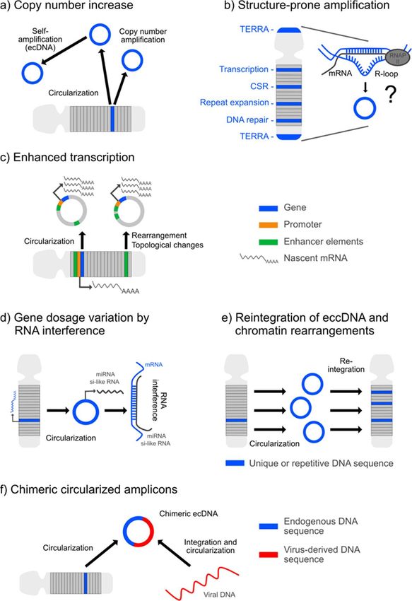
Figure 1.1.Mechanisms
Figure Mechanisms andand concepts
concepts of extrachromosomal
of extrachromosomal circularcircular DNA (ecc/ecDNA)-derived
DNA (ecc/ecDNA)-derived genomic
plasticity. (a) Biogenesis of ec/eccDNAs from linear chromosomes leads toleads
genomic plasticity. (a) Biogenesis of ec/eccDNAs from linear chromosomes to aingain
a gain the in the copy
gene gene
copy number
number that might
that might be potentiated
be potentiated by self-amplification
by self-amplification of the amplicon.
of the amplicon. (b) Several (b)lines
Several lines of
of evidence
evidencethe
suggest suggest the involvement
involvement of R-loopsofin R-loops in the generation
the generation of ecc/ecDNA.of ecc/ecDNA.
Associated Associated
are factorsaresuch
factors
as:
such as: enrichment of transcriptionally active genes on ecc/ecDNAs; transcription-coupled
enrichment of transcriptionally active genes on ecc/ecDNAs; transcription-coupled efficiency of class
efficiency
switch of class switch
recombination recombination
(CSR) that (CSR) involvement
generates eccDNA; that generates eccDNA;
in repeat involvement
transcription, in repeat
transcriptional
transcription,and
termination transcriptional
DNA instability, termination
as evidencedand inDNA instability,
several as evidenced
repeat expansion in several
disorders repeat
and, thus, in
expansion
genomic disorders and,
constellations thus,
prone to in genomic
form eccDNA; constellations
interaction withpronedifferent
to formDNA-repair
eccDNA; interaction
strategies with
that
different
can DNA-repair
give rise to eccDNA; strategies
and RNA that can give rise
transcribed fromtotelomere
eccDNA; and RNA
repeats transcribed
(TERRA) that servefromas atelomere
marker
repeats (TERRA) that serve as a marker of ALT, which is characterized by
of ALT, which is characterized by the generation of circular extrachromosomal telomere repeats the generation of circular
extrachromosomal
(ECTRs). (c) Enhanced telomere repeats
transcription (ECTRs).
from (c) Enhanced
a gene due transcription
to the circularization from mediated
process, a gene due to the
by altered
circularization
topological process,ormediated
influences by sequenceby altered topological
rearrangement influences
on the or Repressive
circle. (d) by sequenceeffects
rearrangement
on a gene by on
the generation
the circle. (d) Repressive
of miRNA and effects on RNA
si-like a gene by the generation
sequences derived from of eccDNA.
miRNA and si-like RNA
(e) Genetic sequences
rearrangements
derived
by from eccDNA.
the reinsertion (e) Genetic
of ecc/ecDNA rearrangements
into by the reinsertion
the linear chromatin. of ecc/ecDNA
(f) Generation of chimeric into the linear
circularized
amplicons consisting of endogenous and viral-derived DNA sequences.Int. J. Mol. Sci. 2020, 21, 2477 6 of 29
2.2. eccDNA Abundance—A Result of Genomic Instability and DNA Repair
Beyond ecDNA production in tumor tissue, the molecular and genetic processes of eccDNA
biogenesis are still not fully understood. As aforementioned, it is suggested that genomic instability,
apart from DNA replication and replication-related repair attempts, are primarily involved. Accordingly,
augmented eccDNA load is found as a result of double-strand break (DSB) induction [36], which thus
might operate as a trigger for eccDNA formation both in a physiological and pathological context.
Of note, several lines of evidence indicate that eccDNA biogenesis does not absolutely depend on
homologous recombination (HR) but can be produced by excision from chromosomal sequences, e.g.,
during the process of HR-independent DNA repair [36]. It has even been suggested that neither HR nor
non-homologous end joining (NHEJ; or classical NHEJ, c-NHEJ) are required for eccDNA production
and that the eccDNA amount is weakly correlated with the proliferation rate of an individual tissue
type [9]. Such discovery sets the basis for an exploration of eccDNA in the broadly post-mitotic tissue
context of the CNS (Table 1). Like other cellular entities, neural cell populations harbor individual
somatic break points and deletion events, and thus, it appears highly probable that eccDNA contributes
to intra-individual genetic mosaicisms also in the CNS [10].
Table 1. Processes related to extrachromosomal circular DNA (ecc/ecDNA) biogenesis with putative
operation in the central nervous system (CNS) environment.
Mechanisms Arguing for eccDNA Synthesis in the CNS
DSB
Oxidative stress
Genomic instability Repeat expansions
R-loops
TEs
Main DNA-repair mechanisms Replicative and non-replicative cells Replicative cells
related to eccDNA biogenesis
NHEJ HR
MM-EJ
NAHR
MMR
Telomere-maintenance mechanisms
related to ECTR
ALT-like processes? ALT
TERRA
Genomic hotspots for eccDNA formation (e.g., Agrin and Titin)
High transcriptional activity in neurons
Chromatin organization via NPC
NPC dysfunction in CNS aging and neurodegeneration
DSB: double-strand breaks, TEs: transposable elements, ECTR: extrachromosomal telomere repeats, NHEJ:
non-homologous end joining, MM-EJ: microhomology-mediated end joining, NAHR: non-allelic homologous
recombination, MMR: mismatch repair, ALT: alternative lengthening of telomeres, HR: homologous recombination,
and NPC: nuclear pore complexes.
This notion gains support from recent characterizations of the repertoire of DNA-repair
mechanisms engaged in eccDNA formation. As a basic proof of principle connecting eccDNA with an
HR-restricted neural milieu, Cohen and Lavi demonstrated that eccDNA release from high molecular
weight DNA can principally be independent of de novo DNA synthesis and HR but will alternatively
rely on chromatin excision [36]. This finding is emphasized by the requirement of active DNA ligase
IV, a major enzyme necessary to ligate DSB in NHEJ, in the formation of eccDNA in mammalian
cells [37]. Accordingly, the broader involvement of HR-independent repair and recombination paths in
eccDNA biogenesis has recently been illustrated with the aid of next-generation sequencing technologies
coupled to chimeric read mapping strategies in human cell-free plasma [38] and by defining the junction
sequences in both ecDNA and eccDNA in bloodstream and tissue [27,38]. According to the junction
analyses reported by Zhu and colleagues, eccDNA appears likely to arise from non-replicative NHEJ, asInt. J. Mol. Sci. 2020, 21, 2477 7 of 29
deduced from the frequent joining of blunt ends or very short (1–3bp) microhomology domains, from
the process of microhomology-mediated end joining (MM-EJ, also denoted as alternative end joining
(a-EJ)), as defined by 4–16bp of homology at the junction ends, as well as non-allelic homologous
recombination (NAHR) underlying repetitive elements of high homology (>90%), such as L1 repeats
and segmental duplications (SD) or low copy repeats (LCR) [38]. NAHR is assumed to account
for ~10–20% of all genomic rearrangements in the human genome [39,40]. Thus, considering that
LCR or SD constitute ~5% of the human genome and that their recombination via NAHR poses a
threat to genomic stability, this repair mechanism might represent a relevant source for eccDNA
generation [41,42]. Accordingly, in plasma, non-replicative repair mechanisms appeared to be strongly
involved in eccDNA formation [38], supporting the notion of a similar eccDNA release in the CNS
(Table 1).
To generate a comprehensive overview of the DSB-repair cascades engaged in eccDNA biogenesis,
chicken B-cell-derived DT40 cell lines with a targeted defect in key DNA-repair molecules operating
either in NHEJ, HR or mismatch repair (MMR) were analyzed with regard to their eccDNA abundance [9].
Strikingly, all these mutants retained their capability to produce eccDNA, except for the DT40 MSH3−/−
cell line, which displayed a drastic reduction of double-stranded eccDNA by 81%. These data strongly
suggest an MMR-mediated mode of eccDNA generation, since the MutS homolog 3 (MSH3)-encoded
MSH3 protein is essential in mismatch recognition and repair initiation. In support of these results, the
authors conclude that eccDNA generation by mechanisms regularly resulting in threatening mutations
might be unlikely but rather arise from repair strategies that reconstitute a normal genome [9]. Overall,
short microhomology domains of 4–8bp contributed by 75% to the ligation of microDNA ends [9].
Mechanistically, the authors propose that slippage of the DNA polymerase at microhomology domains
will generate single-stranded loops that are excised, circularized and converted into double-stranded
eccDNA, with loop excision requiring MMR proteins such as MSH3 [9]. As homozygous mutations in
MMR genes cause the mismatch repair cancer syndrome involving brain tumors, this process might
have importance for the maintenance of genomic stability also in the CNS (Table 1).
As HR-independent repair paths are of major importance for differentiated neurons to repair
DSB, such evidence sets the cornerstone for the generation of eccDNA in largely post-replicative
tissues such as the CNS. Neuron-intrinsic restriction of DNA-repair choices arises from the cell
cycle phase-dependence of DSB-repair pathways, whereby HR is limited to the availability of sister
chromatids in the S and G2 phases. NHEJ that requires no or minimal homology ( 20 bp) serve for the alignment of broken
DNA strands, operate with similar risk of incomplete DSB repair [43,44]. Although expected to be
low in G0 /G1 and favored in S/G2 , MM-EJ and SSA activity might complement NHEJ in DSB repair in
post-mitotic neurons, at least in those that can aberrantly reactivate an early S phase [21,45–48]. This is
similar to their up-regulation in HR-negative tumor cells, as these processes are operating not only
after but also prior to DNA replication [43,44]. A further, yet unproven, option for CNS neurons might
represent the MMR system, as delineated above [9], which purposes the correction of wrongly paired
bases either arising from errors in replication and recombination or during the DNA-damage response
(DDR) [49,50]. Indeed, adult brain cellular extracts enriched for neuronal nuclei have been shown to
express several MMR proteins [51], which are also assumed to play a crucial role in the propagation of
repeat expansions in non-replicative cells and, thus, in the generation of several neurodegenerative
disorders, including Huntington’s disease (HD) [52]. In summary, though limited in HR, the DSB
repair pathways prevailing in the CNS and, particularly, in post-mitotic neurons, all appear likely to
contribute to eccDNA biogenesis (Table 1).
Moreover, incomplete repair or non-repaired demise that will accumulate in non-replaceable
neurons over time might pose further predilections for the acquisition of somatic break points, evenInt. J. Mol. Sci. 2020, 21, 2477 8 of 29
without the presence of a heritable susceptibility factor, and thus, render the CNS a preconditioned
environment for tissue-specific eccDNA abundance. Such susceptibility might be potentiated by
a continuous exposure of the CNS environment to elevated oxidative stress levels due to the high
metabolic activity of neurons, which do not only cause base oxidations but also single-strand breaks
(SSB) and DSB [53] (Table 1). Apart from non-dividing neurons, the brain harbors glial populations
with reactive mitotic potential, which can additionally contribute to HR-related eccDNA formation and
account for events of eccDNA generation by break-independent replication errors. Moreover, it is now
well-established that, under certain stress conditions, post-mitotic neurons can reactivate an abortive
cell cycle at least up to an S phase that is entailed by partial or complete DNA replication [45–48].
Whether such events are also a source of eccDNA production, e.g., by the induction of DNA-repair
mechanisms reactive to the sensing of DNA-content alterations, is still not explored.
Concrete evidence for eccDNA production in the CNS from excised genomic loci was recently
provided by Shibata and colleagues [10]. When characterizing eccDNA during murine brain
development, they found eccDNA with a size of 200–400 bp at the highest frequency, with 98%
remaining below a size of 1kbp. Generally, the ratio of these microDNA circles to higher size circles was
estimated to be 50:1 [10]. Electron microscopy identified both single- and double-stranded eccDNA,
apparently with similar prevalence. Single-stranded eccDNA was proposed to originate from ligated
Okazaki fragments or from DNA produced in surplus along with replication slippage, or from the
digestion of double-stranded nicked DNA circles by nuclease [10]. Overall, microDNA structures were
found enriched in the 50 untranslated region (50 -UTR) of genes, in exons and in CpG islands. In 37% of
the eccDNA entities, the eccDNA junctional regions displayed short microhomology repeats of 2–15
bp in length, which were more than 10-fold above a random calculation model [10]. Thus, in general,
the finding of excised elements forming eccDNA in the developing brain are in high accordance with
recent data characterizing eccDNA in a broad panel of replicative and post-mitotic tissues, including
the adult brain, which is composed of occasionally replicative glia and post-mitotic neurons [9].
Moreover, the recent profiling of eccDNA in muscle supports a role of eccDNA in post-mitotic,
regeneration-competent tissues. Among a catalogue of ~100,000 different eccDNAs identified in muscle
tissue from 16 healthy male donors that apparently originated from all chromosomes, 8.1% of the
eccDNA reads mapped to rDNA, 3.5% to long interspersed nuclear elements (LINEs), 3.1% to short
interspersed nuclear elements (SINEs), 1.2% to satellites and 0.8% to telomeres [19]. Whether the
prevalence of repetitive elements such as telomeric and interspersed sequences arises either from HR
or, in light of the low proliferative activity of differentiated non-damaged muscle, from a continuous
excision of repetitive sequences from the genome, requires further investigation [19].
Considering such evidence, deletions of any chromosomal origin, including spontaneous deletions
or those arising from DNA-repair mechanisms, might give rise to eccDNA both in replicative and
post-mitotic tissues and organs.
3. Sequence Specificity and eccDNA Abundance
3.1. Structural Preponderance
As delineated above, eccDNA biogenesis is related to cancer and genomic instability.
Sequence-specific structural predispositions for eccDNA formation represent a further level of intensive
research. As an apparently conserved feature, virtually any part of the genome might possess the
potential to circularize and form eccDNA [19,54], implying that genic, intergenic and noncoding
regions contribute to eccDNA production, including unique and repetitive sites [9,10,55].
Segmented analyses of individual chromosomes, however, revealed a positive correlation between
microDNA release and the density of genes and guanine-cytosine (GC)-rich sequences, suggesting
that such loci might represent eccDNA hotspots and that genome-wide eccDNA production might
pursue a non-random pattern [9]. In support, both ecDNA from tumor cells and eccDNA that derived
from normal tissue were found to be substantially enriched in genic regions [9,19,27]. Thereby, largerInt. J. Mol. Sci. 2020, 21, 2477 9 of 29
ecDNAs harbor entire genes to a higher percentage than smaller eccDNAs, which predominantly
contain gene fragments [27]. Concrete estimates on the genome coverage of eccDNA reported from
recent whole-genome analyses in yeast, however, are commented as putative underestimation due
to the preponderance of unique eccDNAs that might have low inter-individual overlap [56]. This is
reflected by the reported eccDNA coverage of 23% of the total yeast genome, wherein the entire amount
of unique eccDNAs originating from individual out of several yeast populations amounted up to
72% [56]. Moreover, in a comprehensive approach screening eccDNA in a wide panel of tissues and cell
lines, shared characteristics included GC content above the genome average and an over-representation
of genic regions, particularly of 50 -UTRs of genes, exons and CpG islands [9,10]. Further enriched were
segments harboring full-length LINE-1 retrotransposons and those associated with RNA polymerase
II, suggesting that active transcription and associated DNA:RNA hybrids, or R-loops, might pose a
structural propensity for eccDNA biogenesis [9] (Figure 1b).
As aforementioned, repetitive DNA elements such as telomere repeats and satellite DNA, as well
as rDNA and transposon-related repetitive elements, including long terminal repeats (LTRs), are
frequent sources of eccDNA [9,19,37,57]. Overall, it was assumed that eccDNAs map to repetitive
elements in up to 40–50%, according to the representation of repetitive structures in the entire murine
genome [9]. Within the different categories of repetitive elements, however, some subclasses might
be over-represented. Likewise, in a recent study setting the focus on retrotransposons as a class of
repetitive elements, the Ty1 subfamily of LTR retrotransposons was found over-represented among
yeast eccDNA [58]. When assessed in mice, SINEs and LTRs were suggested to contribute equally to
the eccDNA load irrespective of the originating tissue (e.g., brain, heart and skeletal muscle, kidney,
spleen, lung and liver), whereas LINEs appeared to be enriched particularly in sperm [9].
In spite of repetitive elements apparently representing susceptibility loci for DNA break points
and looping-out events, it remained not fully clarified whether repeat elements were over-represented
relative to the global content of repeats within the individual genome. To conclusively resolve this
question, a genome-wide approach served to compare eccDNA abundances and read identities between
the evolutionarily compacted genome of pigeons (~1.11 Gb) in relation to the less-condensed and
larger human genome (~3.23 Gb), which contains ~10-times higher amounts of repetitive elements as
compared to the pigeon genome [54]. Considering such multipliers, the authors found that repetitive
elements as a putative source of eccDNAs are not per se over-represented but manifest in proportion to
the overall abundance of repetitive elements within the genome, which is dependent on the overall
genome size [54]. Sub-analyses revealed that, within the eccDNA reads that mapped to repetitive
sequences, the classes of LINEs and LTRs were slightly concentrated in pigeons, whereas, in humans,
the classes of simple repeats, SINEs and satellites were enriched relative to expected values. The
assumption of a linear representation of eccDNA containing repetitive elements in proportion to the
entire genome size is consistent with other studies [9].
Several aspects, particularly the densification of exons and gene-dense regions in eccDNA but also
the notion that R-loops might imply a preponderance for eccDNA formation and the aforementioned
association with MMR strategies, strongly suggest a role of transcription in the process of eccDNA
biogenesis (Figure 1b). In a recent study addressing the relation between eccDNA production and
transcription, eccDNA mapping to promoter regions was found to be more than 10-fold enriched
over random values. This observation coincided with an enrichment of intron-exon junctions and
chromatin marks of active transcription. The authors concluded that RNA transcription and consecutive
pre-mRNA splicing are amplifiers of eccDNA production [9]. Of note, R-loops are proposed to be a
risk factor for DNA damage due to the single-stranded nature of the displaced DNA strands or due to
interference with the DNA-replication process, followed by the induction of recombination-directed
repair pathways [59,60].Int. J. Mol. Sci. 2020, 21, 2477 10 of 29
3.2. eccDNA Structures Related to Function
In humans, eccDNA has been found to originate from some functional hotspots that structurally
can comprise either entire genes or solely gene fragments. One such hotspot originates from the
recombination of genes coding for the variable regions of the T-cell receptors and of immunoglobulin
light chains [61,62]. Additionally, eccDNA production arises from an isotype switch of activated
B cell-derived immunoglobulin heavy-chain classes, also called class switch recombination (CSR),
involving the induction of DSB and deletions within the loci encoding the constant regions of the
heavy-chain locus, which are unrelated to antigen specificity [63]. The strand ends are then ligated via
NHEJ. Programmed DSB induction in the switch regions involves active transcription and replication
origins that are determined by R-loop formation [64] (Figure 1b). To what extent R-loops, which are
essential for efficient CSR [65], contribute to eccDNA genesis in the process of CSR has to be explored.
Thus, eccDNA is a by-product of genetic rearrangements essential for the establishment of the defense
function of the adaptive immune response.
At this point, the question arises as to the extent that eccDNA can modulate self-tolerogenic
immune functions and autoimmunity, which are crucial in the immune evasion of tumor cells and
in the development of autoimmune spectrum disorders. Of note, CSR of antibodies directed against
myelin epitopes were shown to play a critical role in the induction and progression of nervous system
deficits in an autoimmune-mediated human myelin oligodendrocyte glycoprotein (hMOG)-based
experimental model of multiple sclerosis [66]. Moreover, though not derived from immune effectors
but from pathogens themselves, it has recently been demonstrated in insects that circular viral DNA
templated from the defective virus genome post-infection is a source of siRNAs that modulate antiviral
immunity towards a protective function [67] (Figure 1d). It will be interesting to explore whether the
profiling of eccDNA amplicons generated by a recombination in genes guiding adaptive immunity,
either acquired via B- or T-cell adaptions, might help identify antigens involved in autoimmune
spectrum disorders such as multiple sclerosis.
3.3. Telomere-Specific eccDNAs
Telomeres, the end caps of chromosomes, are nucleoprotein complexes comprising multiple copies
of tandem TTAGGG repeats and a hexameric protein complex, the shelterin or telosome complex.
Molecularly, they terminate in a single-stranded G-rich overhang of 50–400 nucleotides at the 30 terminal
(G-strand), whereas the complementary 50 end is C-rich (C-strand). Telomere repeat binding factor 2
(TRF2), a component of the shelterin complex, is auxiliary in the telomeric DNA to form a secondary
structure, called telomere loop or T-loop, by invasion of the single-stranded 30 overhang of the G-strand
into the double-stranded telomeric DNA [68]. TRF2 together with the DNA-binding protection of
telomere 1 (POT1), a further shelterin component that specifically interacts with the single-stranded
overhang, prevent the ataxia telangiectasia-mutated (ATM) signaling and ataxia telangiectasia and
Rad3-related (ATR) pathways to elicit a DDR at the chromosomal ends [69]. The special interrelations
of the DDR and telomere structures have been excellently reviewed [70].
In dividing cells, erosion of telomeres occurs gradually with each round of cell division, thereby
inducing senescence. Loss of telomeric DNA can be counteracted by the telomere replenishing
enzyme telomerase, implementing a human telomerase reverse transcriptase enzyme (hTERT) and a
telomerase RNA (hTR) component, the second of which serves as a template for telomere sequence
synthesis [71,72].
In telomerase-negative tumor cells, however, telomere attrition can be counterbalanced by a
telomerase-independent telomere restitution process, referred to as the alternative lengthening of
telomeres (ALT) [73], the principal mechanism of which is assumed to rely on a DNA-copying
step primarily dependent on HR, though it is likely to involve additional molecular mechanisms.
Accordingly, any telomere-length maintenance strategy that operates independently of the telomerase
enzyme was proposed to be categorized as ALT [74]. Although ALT is reported mainly as a
telomere-maintenance mechanism specifically in tumor cells lacking telomerase enzyme activity,Int. J. Mol. Sci. 2020, 21, 2477 11 of 29
it is also active in murine somatic cells [75]. One phenotypic identifier of ALT-positive cells represents
the occurrence of telomere-specific eccDNA, termed extrachromosomal telomeric repeats (ECTRs),
comprising, apart from linear double-stranded DNA, double-stranded T-circles and single-stranded 50
C-rich C-circles. Further distinct features include hypervariability of the telomere length, the presence
of ALT-associated promyelocytic leukemia bodies (APBs) harboring telomeric chromatin and elevated
levels of long noncoding TERRAs that also control telomere lengths [76,77] (Figure 1b). These TERRA
transcripts localize to critically short telomeres, where they form DNA:RNA hybrids, or R-loops, that
activate the DDR and promote telomere restitution via telomeric sister chromatid exchange (T-SCE) [78].
Thus, increased T-SCE is another signature of ALT. An interesting and still open question, in analogy to
the aforementioned, is whether TERRA-formed R-loops are a susceptibility region for ECTR secession
and how they influence ALT.
Accumulation of ECTRs occurs, as currently assumed, in 10–15% of the cancers that maintain
the telomere length by a telomerase-independent ALT pathway [79]. Molecularly, ECTR in the form
of double-stranded T-circles is generated by T-SCE via HR, thereby originating from the resolution
of T-loops. These circular ECTRs then provide a template for the expansion of telomere tracts by
a rolling circle amplification mechanism [79]. Moreover, T-circles can also originate as a result of
HR-mediated telomere rapid deletions (TRD) evoked by dysfunctional TRF2. Apart from repressing
DDR signaling at the telomere, TRF2 is essential for the formation of telomeric T-loops, and it prevents
their excision by blocking the DNA-repair proteins Nijmegen breakage syndrome 1 (NBS1) and X-ray
repair cross-complementing 3 (XRCC3) [80]. A further factor essential for telomere replication, which
operates in association with the MRE11-RAD50-NBS1 (MRN) complex, is the C-terminal binding protein
(CtBP)-interacting protein (CtIP). Absence of this DNA-repair protein leads to a drastic reduction of
telomere length, increased generation of T-circles, DNA damage and chromosomal aberrations [81].
A similar role in telomere replication, maintenance and repair applies to SAE2, the yeast homolog of
CtIP [82–84]. Interestingly, the inducible loss of CtIP, as investigated in hTERT-immortalized epithelial
RPE1 cells derived from the retina, leads to the accumulation of T-circles but not of C-circles [81], the
second of which has recently been established as a specific marker of ALT [85]. However, silencing of
CtIP in the ALT-positive U2OS cell line resulted in a two-fold increment in C-circles, inferring that CtIP
protects telomeres in both scenarios, i.e., a telomerase-positive as well as an ALT-positive environment,
and suppressed the release of ECTRs [81].
Previously, Hande and colleagues reported telomere shortening and an accumulation of ECTR
DNA in fibroblasts of ataxia-telangiectasia patients and Atm−/− mice [86]. Along with nuclear ECTR
DNA signals, cytoplasmic signs of telomeric DNA were detected, though the exact structure of these
ectopic ECTR entities was not defined [86]. A nuclear-to-cytoplasmic translocation of linear and circular
ECTRs has also been described in ALT-positive cancer cells [79]. Although the role of ATM and its yeast
homolog TEL1 in telomere maintenance is well-established [87], it is not yet clear if it directly contributes
to the generation and accumulation of ECTRs, e.g., by TRD processes. Such studies, however, provide
a link between extra-nuclear eccDNA and the DNA-sensing cyclic GMP-AMP synthase (cGAS),
together with the class of stimulator of interferon genes (STING) (cGAS-STING) pathway, which
elicits an interferon-mediated inflammatory reaction in response to highly immunogenic misplaced
DNA [88,89]. The functional interconnection between ECTR and cGAS-STING might have potential
in the surveillance of ALT-related tumor development, as the ectopic accumulation of ECTRs in
telomerase-positive immortalized human fibroblasts expressing a mutant TRF2 allele reduced cell
proliferation by the activation of interferon-responsive cytosolic DNA-sensing pathways [79]. Whether
an analogous mechanism of ectopic ECTR accumulation in combination with cytosolic DNA-sensing
signaling cascades will also operate in normal human somatic cells deserves further investigation.
Telomere lengths in human cancer cell lines, in human embryonic stem cells (hESCs) and
human induced pluripotent stem cells (hiPSCs), all of which overexpress hTR, are further controlled
by a process that trims over-elongated telomeres [76,90]. Thus, telomere-length homeostasis is
apparently maintained by a balance between telomerase-dependent telomere elongation and aInt. J. Mol. Sci. 2020, 21, 2477 12 of 29
telomeric cropping mechanism that will operate independently from replication-associated telomere
attrition. The telomere-trimming process activated as a response to over-extended telomeres in a
telomerase-positive cellular milieu results in circular ECTR formation and shares features with the
ALT process, such as telomere-length heterogeneity, though APBs remain an inconstant feature [76,90].
Moreover, these cells apparently do not exert T-SCE, which is, by contrast, upregulated under ALT.
Although telomere trimming in hESCs and hiPSCs involves the MRN complex member NBS1 and
XRCC3, which either participate in or prepare HR, the telomere-cropping mechanism in these cells is
described to occur independently of the activation of HR [76]. Thereby, XRCC3 and NBS1 mediate
telomere curtailment via the formation of single-stranded C-rich telomeric DNA and double-stranded
T-circles derived from T-loops, respectively. Apart from hESCs and hiPSCs, ECTRs were also suggested
to originate from telomere-trimming processes in normal neural cells and neuroblastoma cell lines [91].
As aforesaid, another important type of circular ECTR is represented by C-circles. Though specific
technical tools and detection assays are now available to investigate ALT activity using C-circles as
a specific marker for ALT activity [85], the molecular basis for their synthesis in ALT-positive cells
is poorly understood. More recently, a putative mechanism underlying the biogenesis of C-circles
was suggested in ALT-positive U2OS cells [92]. Hu and colleagues showed that the partial inhibition
of telomerase activity in telomerase-positive human fibrosarcoma HTC75 cells exhibiting telomeric
DNA damage leads to the generation of C-circles and APBs, which points to the possible switch from
telomerase-positive cancer cells to cells exerting ALT-like mechanisms [93]. Mechanistically, as a
result of endogenous telomere DNA damage, SSB and DSB induce replication fork collapse, which
triggers the NHEJ-dependent release of C-circles from the lagging strand and C-rich overhangs from
the leading strand. These observations imply that the production of C-circles and C-rich overhangs
are linked to DDR and replication stress at the telomeres. The HR-independent release of C-circles
via NHEJ might also suggest the involvement of a telomere-trimming process for the development of
C-circles, whereas telomere elongation via T-SCE characterizes active HR at telomeres [92].
Moreover, C-circles and C-strands were recently detected in the spinal cord and cerebellum,
suggesting that telomere-trimming mechanisms [91], or ALT-like processes, may exist in the CNS, which
comprises post-mitotic neurons and occasionally replicative glia cells. Whether these mechanisms
and ALT-like processes, which need HR-independent operations, will occur in neurons requires future
investigation. In general, the load of ECTR in individual tissues might depend, at least partially,
on its replicative index, since replication stress propagates the generation of telomere-related C-circles,
as shown for hESCs harboring extended telomeres due to the over-expressing hTR. However, the
compromised telomere stability of extra-long telomeres might equally add to this observation under
the conditions applied [76].
As aforementioned, apart from externalization, eccDNA/ecDNA can also cause genomic
rearrangements by insertional events. Analyses of reintegration sites revealed that circle reinsertions
can be placed proximal to the telomerase reverse transcriptase (TERT) gene and, thus, might enhance
TERT expression [27].
4. Replication, Segregation and Degradation of eccDNA
4.1. Self-Replication and Self-Limitation of eccDNA Production
The tissue-specific load in eccDNA might not only be a passive consequence of DNA replication,
DNA integrity and repair, as delineated before, but also implicate a self-determined regulation.
Accordingly, about 80% of all eccDNAs in yeast were found to possess either an origin of replication,
an autonomously replicating sequence or at least its related core consensus sequence, as shown by
several studies and approved for histone genes in Saccharomyces cerevisiae [56,94,95]. Likewise, in the case
of tumors, ecDNAs autonomously self-replicate and, thus, contribute to oncogene amplifications [96].
Such self-replication might account for a stable level of eccDNA in a given tissue and organ and
influence, if present, its regulatory role on gene copy numbers, e.g., in tumors, neurodegeneration andInt. J. Mol. Sci. 2020, 21, 2477 13 of 29
aging. Likewise, extrachromosomal rDNA circles (ERCs) that encode for ribosomal RNA accumulate
in mother cells by asymmetric segregation during mitosis and are associated with mechanisms that
propagate yeast aging and reduce lifespan [97]. Apart from self-amplification capacity, optimal eccDNA
amounts and related gene copy numbers will therefore also require a restriction control mechanism.
Addressing intrinsic control mechanisms of eccDNA abundance, it has been demonstrated
in Saccharomyces cerevisiae that the copy number of ERCs is under a tight self-limiting regulation,
implicating the potential to activate a need-adapted clonal expansion of ERCs, which will be reintegrated
in an amount required to keep cellular homeostasis. Accordingly, a direct correlation between the
production and deletion of ERCs was illustrated. Thus, ERCs are considered as a dynamic pool
exploited for the rapid adjustment of gene copy numbers to physiological needs [98]. Maintenance of
an optimal ERC steady state appeared unlikely to be an inherited feature established under selection
pressure, as ERCs are, in most cases, not transferred to daughter cells but almost entirely retained in the
mother cell during cell division. Such data strongly support the notion that eccDNA might not only
indicate genomic instability or an aberrant chromosomal state but also imply beneficial roles relevant
in health maintenance and pathology prevention. Thus, these findings appear of importance in light
of the presence of eccDNA also under healthy conditions. Further studies are necessary to explore
the existence of a relay mechanism that allows for steady-state levels of eccDNA in a tissue-specific
manner also in higher vertebrates.
4.2. Cellular Segregation
Due to their acentric structure, eccDNAs segregate unequally during cell division and, thus,
can contribute to somatic mosaicisms of copy number variations [33]. Currently, the mechanisms
that control eccDNA partitioning during cell division are not fully clarified. In a recent study in
yeast, Denoth-Lippuner and colleagues proposed a model of asymmetric circular DNA segregation
involving the operation of the multifunctional Spt-Ada-Gcn5 acetyltransferase (SAGA) complex that
has prominent roles in posttranslational histone modification, transcriptional elongation and mRNA
export [99]. In yeast, SAGA appears required to confine non-chromosomal circular DNAs to the mother
cell and prevent segregation into daughter cells during mitosis by mediating their tethering to nuclear
pore complexes (NPCs), the transport channels within the nuclear envelope. The authors show that
this process of eccDNA attachment to the NPC, exemplified for ERC, involves the transcription and
export complex (TREX)-2 of the pore, as well as the multicomponent SAGA complex in its role as
a recruitment factor of chromatin. As a result of SAGA-mediated chromatin targeting, the circular
DNA in conjunction with the ERC-loaded NPC will be retained in the mother cell. This asymmetric
segregation both of ERCs and NPCs will promote cellular aging and shorten the lifespan, with ERCs
being the drivers of NPC accumulation. By contrast, ERCs alone were insufficient to accumulate or
to promote aging and modulate lifespan. Of note, the authors stated that ERC require anchoring
to a moiety of nucleoporins (Nups) that are stably integrated at the NPC, such as those of the core
region [97]. It will be interesting to explore whether eccDNA tethering to NPC structures is also a
mechanism to modulate eccDNA load in mammalian cells (Table 1) and to define the impact of its
deregulation on the development of diseases, which implicate disturbed NPC-associated functions,
such as evidenced for C9ORF72-related amyotrophic lateral sclerosis (ALS) [100–102].
4.3. eccDNA Degradation
The turnover of eccDNA within cells or in cell-free DNA of the circulation has not yet been
investigated. Cellular DNA is partly degraded by TREX1 (DNA 30 repair exonuclease I) [103,104].
Generally, mutations in at least five gene loci are identified in humans to be associated with defective
nucleic acid metabolism, wherein particularly that coding for TREX1 is evidenced to be important for
the clearance of cytosolic DNA species derived from endogenous retro-elements [105,106]. Elimination
of extrachromosomal genetic elements has also been associated with structures termed micronuclei.
In particular, it was shown that micronuclei were enriched in ecDNA after low-dose exposure toInt. J. Mol. Sci. 2020, 21, 2477 14 of 29
the DNA replication inhibitor hydroxyurea, and this was also associated with a loss of amplified
genes on ecDNAs [107]. Furthermore, the entrapment of ecDNAs with amplified N-myc was found in
micronuclei from neuroblastoma tumor cells in vivo [108]. Whether this mechanism applies to other
cell types remains to be elucidated.
Generally, cell-free DNAs display short half-lives of several minutes to hours and are eliminated
from the blood system by the liver and kidneys [109]. The DNA of a circularized structure shows
resistance towards digestion by exonucleases and RNases and, therefore, is more persistent than linear
DNA or RNA after release into the blood [110]. Interestingly, most of the extracellular DNA is enclosed
in large extracellular vesicles, which contributes to both their dissemination and stability [111]. Hence,
results from ongoing and future studies will further shed light on the intra- and extracellular dynamics
and turnover rates of eccDNA and contribute to define their utility as “liquid biopsies”, as discussed
below [112].
5. eccDNA in CNS Aging and Neurodegeneration
5.1. Role in CNS Aging
EccDNAs have long been associated with aging in yeast and mammalian cells and tissues [20,29,95].
In particular, more than 20 years ago, Sinclair and Guarente provided evidence that ERCs accumulate
in old yeast and might be causal for fungal aging [95]. Moreover, eccDNA abundance has even been
proposed to serve as a putative index of cellular aging, as they were found to be amplified during aging
in senescence-resistant SAM-R mice, a process that was accelerated in the senescence-prone SAM-P
progeria model [113]. In support of this finding, mutations in the sgs1 gene, the yeast homolog of the
Werner’s syndrome gene, lead to a stronger ERC load, associated with characteristics of premature
aging and reduced lifespan [95]. In addition, the study by Sinclair and Guarente proposed that
the replication of eccDNA in the S phase of the cell cycle, where it was assumed to increase in an
exponential manner, might represent the clock that determines the lifespan in yeast. The rise in ERC
numbers also correlated with the exponential rise in the mortality rates in yeast [95].
Likewise, in a recent study, the effects of temporal ERC accumulation on the replicative lifespan
(RLS) were determined in yeast by using a microfluidics-dependent real-time approach [114]. At the
single cell level, the authors longitudinally assessed the yeast cellular vitality and RLS as a function
of ERC accumulation. Accordingly, they divided the entire lifespan of each single cell into three
major phases related to the senescence entry point (SEP), thereby defining pre-SEP, SEP and post-SEP.
Intriguingly, exponential ERC production eventuated before the onset of SEP in the life of mother
cells. In this pre-SEP phase, ERC accumulation in the nucleolus was accompanied by an increment in
rRNA levels, due to the transcription of ERC-related rDNA. However, the production of functional
ribosomes from these rRNAs remained defective. Thus, the miscoordination between abundant rRNA
and ribosome biogenesis led to nucleolar stress and impaired the nuclear homeostasis and cellular
growth in the post-SEP phase, which might ultimately lead to cell death. Moreover, post-SEP mother
cells underwent asymmetric segregation of ERC due to an unequal partitioning of the nucleolus and
nucleoplasm, which might serve as a mechanism for the rejuvenation of daughter cells [114]. Hence,
this study defined a series of events that might explain how ERC accumulation is associated with
cellular aging and death.
Another study highlighted the relationship between transcriptional activity and ecDNA formation
in the context of aging. In aging budding yeast, the generation of protein-coding eccDNAs was
illustrated to be triggered by the transcriptional stimulation of certain genes sensitive to environmental
stimuli [115]. Likewise, in yeast cells that aged under copper sulfate treatment, the CUP1 gene was
transcribed in a site-specific manner, exclusively leading to the accumulation of CUP1 eccDNA. Apart
from such transcriptional control, the CUP1 eccDNA load was amplified in the aging mother cell by
asymmetric segregation and the retention of eccDNAs [115]. Mechanistically, transcription of the CUP1
gene resulted in DSB and, consecutively, eccDNA formation via Sae2, Mre11- and Mus81-dependentYou can also read