The Regulation of Astrocytic Glutamate Transporters in Health and Neurodegenerative Diseases - MDPI
←
→
Page content transcription
If your browser does not render page correctly, please read the page content below
Review

The Regulation of Astrocytic Glutamate Transporters
in Health and Neurodegenerative Diseases
Alison C. Todd 1,2 and Giles E. Hardingham 1,2,*
 1 UK Dementia Research Institute at the University of Edinburgh, Chancellor’s Building,
   Edinburgh Medical School, Edinburgh EH16 4SB, UK; Alison.Todd@ed.ac.uk
 2 Centre for Discovery Brain Sciences, University of Edinburgh, Hugh Robson Building, George Square,

   Edinburgh EH8 9XD, UK
 * Correspondence: giles.hardingham@ed.ac.uk

 Received: 24 October 2020; Accepted: 11 December 2020; Published: 17 December 2020

 Abstract: The astrocytic glutamate transporters excitatory amino acid transporters 1 and 2 (EAAT1
 and EAAT2) play a key role in nervous system function to maintain extracellular glutamate levels
 at low levels. In physiology, this is essential for the rapid uptake of synaptically released glutamate,
 maintaining the temporal fidelity of synaptic transmission. However, EAAT1/2 hypo-expression or
 hypo-function are implicated in several disorders, including epilepsy and neurodegenerative
 diseases, as well as being observed naturally with aging. This not only disrupts synaptic information
 transmission, but in extremis leads to extracellular glutamate accumulation and excitotoxicity. A
 key facet of EAAT1/2 expression in astrocytes is a requirement for signals from other brain cell types
 in order to maintain their expression. Recent evidence has shown a prominent role for contact-
 dependent neuron-to-astrocyte and/or endothelial cell-to-astrocyte Notch signalling for inducing
 and maintaining the expression of these astrocytic glutamate transporters. The relevance of this non-
 cell-autonomous dependence to age- and neurodegenerative disease-associated decline in astrocytic
 EAAT expression is discussed, plus the implications for disease progression and putative
 therapeutic strategies.

 Keywords: Astrocyte; glutamate; excitotoxicity; neurodegenerative disease; EAAT1; EAAT2

1. Introduction
      Glutamate is the predominant excitatory neurotransmitter in the brain, activating post-synaptic
ionotropic N-methyl-D-aspartate (NMDA) and α-amino-3-hydroxy-5-methyl-4-isoxazolepropionic
acid (AMPA)/kainate receptors. Co-activation of these receptors allows the influx of Na+ ions, which
depolarises the cell’s membrane potential, triggering an action potential. Once released, it is
important that glutamate is rapidly cleared, since failure of this has two key consequences. Firstly, it
will continue to stimulate the post-synaptic receptors after the initial signal has been sent, impairing
the detection of the next signal that arrives—akin to a saturation phenomenon, and potentially
leading to cell swelling due to ion influx [1]. Secondly, if this glutamate escapes from the synaptic
zone it was released into it could activate unintended synapses, triggering activity where it should
not. Significantly, if glutamate escapes the synaptic region it can activate extrasynaptic NMDA
receptors: too much Ca2+ influx via these extrasynaptic NMDA receptors induces signalling cascades
that initiate cell death programs [2,3].
      Astrocytes have long been known to be important in promoting the survival of neurons and for
counteracting the toxic effects of glutamate [4–7]. This protection is largely due to their ability to take
glutamate up from the extracellular environment via transporters located on astrocytic membranes,
preventing excitotoxicity and associated oxidative stress [8,9]. Once inside the astrocytes, glutamate

Int. J. Mol. Sci. 2020, 21, 9607; doi:10.3390/ijms21249607                        www.mdpi.com/journal/ijms
Int. J. Mol. Sci. 2020, 21, 9607                                                                         2 of 32

is then either converted into α-ketoglutarate by glutamate dehydrogenase (GDH) or transaminases
and shunted into the astrocytic TCA cycle, or else converted into glutamine by the enzyme glutamine
synthetase (GS) [10,11]. Glutamine is not toxic to neurons, and is extruded by the SNAT3 glutamine
transporter into the extrasynaptic space, which can then be taken up by neurons and converted back
into glutamate via the neuronally expressed phosphate-activated glutaminase (PAG), thus
replenishing pre-synaptic glutamate stores [12–15]. This glutamate recycling pathway is referred to
as the glutamate-glutamine cycle (see Figure 1).
     Astrocytic glutamate uptake and recycling is a vital part of CNS function. Key within this
machinery are the two astrocytic glutamate transporters, excitatory amino acid transporters 1 and 2
(EAAT1 and EAAT2), that are responsible for the bulk of glutamate uptake. The astrocytic glutamate
transporters EAAT1 and EAAT2 belong to the solute carrier 1A family of transporters (SLC1A), which
includes two alanine serine cysteine transporters, ACST1 and ACST2, along with the five excitatory
amino acid transporters, EAAT1-5 [16]. The EAATs are electrogenic secondary-active transporters,
using the concentration gradients of their co- and counter-transported ions (Na+, H+, and K+) to drive
the transport of glutamate against its concentration gradient into the cell [17]. Astrocytes are able to
regulate any fluxes in their pH and membrane potential caused by this transport process through
their high expression of membrane K+ channels (especially KIR4.1), their Na+/H+ exchangers and
Na+/NCO3- cotransporters, as well as dissipating charge and ionic changes throughout the astrocytic
network via connexon coupling (see [18] for a recent review on astrocyte physiology).
     Unsurprisingly, reductions in the astrocytic glutamate transporters’ expression and function
have been implicated in a number of CNS diseases, particularly epilepsy, along with several
neurodegenerative diseases, as outlined below. It is therefore of interest to understand how these
transporters are functionally regulated, as boosting their expression and function may offer a novel
way to reduce severity and progression of neurodegenerative diseases.

      Figure 1. Glutamate-glutamine cycle. Glutamate (Glu-) released after excitatory transmission is
      collected by astrocytic EAAT transporters 1 and 2. The glutamate is then either converted into α-
      ketoglutarate (α-KG) via glutamate dehydrogenase (GDH) or transaminase reaction and enters the
      TCA cycle, or else is converted into glutamine (Gln) by glutamine synthetase (GS). Astrocytes excrete
      Gln back into the extracellular environment via the Na+ driven SNAT3 transporter, which is then
      taken up by an as yet unconfirmed neuronal Gln transporter. Neurons then convert Gln back to Glu-
      via a phosphate-activated glutaminase (PAG) reaction to replenish their vesicular Glu- stores.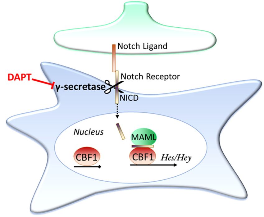
Int. J. Mol. Sci. 2020, 21, 9607                                                                  3 of 32

2. Glutamate Transporters in the Brain
     Glutamate is found at high concentrations in the brain, at a concentration of approximately 10–
14 mmol/L depending on region [19,20]. However, most of this glutamate is kept within intracellular
compartments, with very low levels maintained in the extracellular fluid (around 3-4 μmol/L in the
extracellular space of the hippocampus) [21,22]. Accordingly, there are numerous transporter
proteins in the brain that are capable of facilitating glutamate transport to ensure that the right
concentration of glutamate is maintained in the right compartment. These transporters fall into two
broad categories: those which are found in intracellular compartments, such as the three vesicular
glutamate transporters (vGLUT1-3) which package glutamate into synaptic vesicles, and those
located on the plasma membrane of cells that can transport glutamate into (or out of) the cell [1,23,24].
The glutamate transporters that are found in the plasma membranes of brain cells consist of five
sodium-dependent co-transporters, and one sodium-independent exchanger [17,25,26]. The sodium-
independent exchanger, xCT, is found almost exclusively on astrocytes, but preferentially transports
cysteine into the cell in exchange for extruding a glutamate molecule out of the astrocyte [27–29]. Due
to the need to transport glutamate into cells against its electrochemical gradient, it is therefore the
sodium-dependent class of transporters that are responsible for quickly sequestering extracellular
glutamate back into cells. There are five known members of this family of transporters, excitatory
amino acid transporters 1–5 (EAAT1-5).

2.1. Excitatory Amino Acid Transporters
     In the early 1970s, a high affinity sodium-dependent uptake system for the negatively charged
amino acids L-glutamate and L-aspartate was first described in synaptosomal preparations, which
was hypothesised to be responsible for the accumulation of the putative excitatory neurotransmitter
glutamate into cells [30,31]. A few years later Balcar and colleagues went on to show that this
glutamate uptake system was also present in glial cells, but not until 1992 were EAATs first purified,
with four independent groups cloning three distinct EAAT family members: Glt-1 (EAAT2), GLAST
(EAAT1), and EAAC (EAAT3) [32–36]. The final two members were cloned in 1995 (EAAT4) and 1997
(EAAT5) [37,38].
     All members of the EAAT family transport L-glutamate into cells under normal conditions using
the electrochemical gradients of Na+ and K+ [1,17]. The EAAT family of transporters and the sodium-
independent exchanger xCT both display high affinity for glutamate transport compared to the three
vesicular glutamate transporters (KM ≈ 2–100 μM for EAAT1-5 and KM ≈ 20–55 μM for xCT versus KM
≈ 1.5–3.5 mM for vGLUT1-3) [37–50]. Although the EAATs have relatively high affinity for glutamate,
enabling them to sequester low concentrations of extracellular glutamate to prevent excitotoxicity,
they interestingly have relatively slow transport cycle times (see Table 1). This problem may in part
be overcome by rapid surface diffusion and transporter trafficking of the EAATs upon glutamate
stimulation [51,52].
     The different EAAT subtypes are found throughout the body, and within the brain they are
found on different cell types and in different brain regions. Although they all transport glutamate
into cells, each subtype possesses a different degree of chloride permeability, and it appears the
function of each of these subtypes may vary. A summary of the five EAAT transporters is given in
Table 1. Note well, for clarity in this review the transporters encoded by SLC1A3/Slc1a3,
SLC1A2/Slc1a2, and SLC1A1/Slc1a1 will be consistently referred to as EAAT1, EAAT2, and EAAT3,
respectively, independent of species. Rodent EAAT1, EAAT2, and EAAT3 are typically referred to as
GLAST (Slc1a3), Glt-1 (Slc1a2), and EAAC (Slc1a1) in other studies. However, there is high amino
acid homology between human and rodent proteins, with 96% identity shared between human
EAAT1 and rat GLAST, 95% identity between EAAT2 and rat Glt-1, and 92% identity between
EAAT3 and rabbit EAAC1 [39].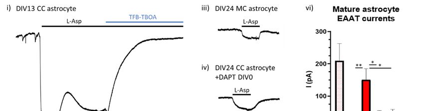
Int. J. Mol. Sci. 2020, 21, 9607                                                                               4 of 32

2.2. Location of Excitatory Amino Acid Transporters
      The two subtypes EAAT1 (i.e., GLAST) and EAAT2 (i.e., Glt-1) are referred to as the astrocytic
glutamate transporters as they are the only EAAT subtypes expressed on astrocytes, where they are
predominantly found on fine astrocytic processes opposed to glutamatergic synapses [53–55]. EAAT1
is found in astroglia (including the Bergmann and Müller glia) throughout the brain, on which they
are exclusively expressed, and are the primary collectors of glutamate in both the cerebellum and
retina (via Bergmann and Müller glia, respectively) [56–62]. EAAT2 is the primary glutamate
transporter in all other brain regions, most prominently in the hippocampus and cortex [54,57]. The
location of EAAT2 is less astrocyte-exclusive, with some evidence suggesting it is also found to a
small degree in neurons, particularly in the hippocampus and retina (see Zhou and Danbolt, 2013 for
discussion). Combined, EAAT1 and EAAT2 make up a significant proportion of the total protein in
the brain, representing ~2.1% of protein in the molecular layer of the cerebellum, 1.6% in the
hippocampal stratum radiatum, and 1% of protein in forebrain tissue, and are by far the most abundant
EAAT subtypes in the CNS [56].
      A more recent study using label-free quantitative (LFQ) tandem mass-spectrometry to survey
the regional protein expression of postnatal human brain tissue has confirmed that EAAT1 and
EAAT2 are abundantly expressed in all regions sampled: the cerebellar cortex (CBC), mediodorsal
thalamic nucleus (MD), striatum (STR), amygdala (AMY), hippocampus (HIP), primary visual cortex
(V1C), and the dorsal prefrontal cortex (DFC) [63]. Apart from the cerebellar cortex (where EAAT2
was within the 93rd percentile), EAAT2 was the most abundantly expressed of the transporters across
all regions, sitting in the 97th (MD), 98th (STR, AMY, HIP, and DFC), and 99th (V1C) percentile of
most abundant proteins within each region. EAAT1 on the other hand was more abundantly
expressed in the CBC, being in the 99th percentile of expressed proteins, with consistently high,
although lower than EAAT2, expression across other regions: 94th percentile in the AMY; 95th in
MD, STR, and HIP; and 97th percentile in the V1C and DFC (calculated from supplementary data
Table 10 of Carlyle et al., 2017) [63].

                                   Table 1. Excitatory amino acid transporter family overview.

                                 Cl-                                                                 Protein
  Protein         Gene                          Kinetics                 Location
                               Conduct.                                                             Abundance

                                                                                                  99th percentile of
                                                                                                  protein found in
                                               KM = 22–48     Astrocytes (incl. Bergmann &
                                                                                                    human CBC;
                                                  μM         Müller glia); Predominant EAAT
  EAAT1         SLC1A3              Mod                                                            ≥95th in V1C,
                                               Cycle time    subtype in cerebellum (1.8 mg/g
                                                                                                 DFC, MD, STR and
                                                = 62 ms           of protein) and retina
                                                                                                        HIP;
                                                                                                    94th in AMY

                                                                                                 99th percentile of
                                                               Astrocytes (and some sparse       protein found in
                                               KM = 25–97
                                                              neurons); Predominant EAAT           human V1C;
                                                  μM
  EAAT2         SLC1A2              Low                        subtype in hippocampus (1.3        ≥95th in DFC,
                                               Cycle time
                                                              mg/g of protein) and cortex (0.8    HIP, AMY, STR
                                                = 70 ms
                                                                          mg/g)                  and MD; 93rd in
                                                                                                       CBC

                                                                                                 26th percentile of
                                                                                                 protein found in
                                               KM = 42–62      Neurons (typically on spines);
                                                                                                   human MD;
                                                  μM             Highest concentration in
  EAAT3         SLC1A1              Mod                                                             21st in HIP;
                                               Cycle time      hippocampus (0.013 mg/g of
                                                                                                  ≤20th in AMY,
                                                = 10 ms                  protein)
                                                                                                  STR, CBC, DFC
                                                                                                     and V1C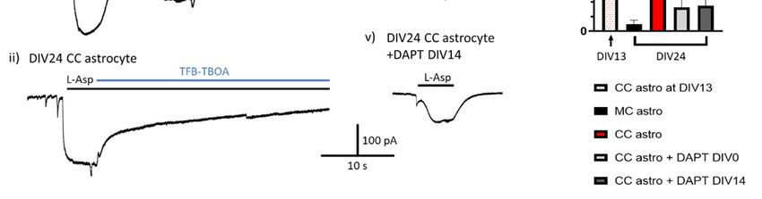
Int. J. Mol. Sci. 2020, 21, 9607                                                                          5 of 32

                                                                                             89th percentile of
                                                                                             protein in human
                                           KM = 2.5
                                                       Cerebellar Purkinje cells (0.2 mg/g          CBC;
                                             μM
  EAAT4         SLC1A6             High                of protein in cerebellar molecular      9th in MD and
                                          Cycle time
                                                                      layer)                        AMY;
                                           >166 ms
                                                                                             1000 ms
      Abbreviations: CBC = cerebellar cortex; V1C = primary visual cortex; DFC = dorsal prefrontal cortex;
      MD = mediodorsal thalamic nucleus; STR = striatum; HIP = hippocampus and AMY = amygdala. See
      ref [64] for references.

      The EAAT3 subtype is exclusively neuronal, and is found on neurons throughout the brain
[65,66]. It typically localises on dendritic spines and not axon terminals, with its highest expression
seen in the hippocampus, at a concentration of 0.013 mg/g [65]. This is 100 times lower than EAAT2
levels in the same region, so even in the hippocampus it may only contribute modestly to glutamate
clearance. Consistent with these earlier reports, the more recent LFQ mass spectrometry data showed
a relatively low protein abundance of EAAT3 ranging from the 13th percentile in the V1C to the 26th
percentile in the MD, and sitting at just the 21st percentile of abundance in the hippocampus (from
supplementary Table 10, Carlyle et al., 2017) [63]. This is compared to EAAT1 and EAAT2 which
were in the top percentiles of expressed proteins across all regions in these samples (>90th percentile
for both EAAT1/2 in all regions) [63].
      EAAT4 is another neuronally expressed glutamate transporter, however its expression profile is
more restricted than that of EAAT3, being found primarily on Purkinje cells of the cerebellum, with
some sparse expression in certain subregions of the forebrain and midbrain [67,68]. It represents
about 0.2% of protein in the molecular layer of the cerebellum, which is approximately 10 times less
than the predominant astroglial subtype in this region, EAAT1 (which represents 1.8% of total
cerebellar protein), and similar to EAAT2 levels (0.3% of total protein) [56,67]. Again, consistent with
these earlier findings, Carlyle and colleagues more recent mass spectrometry data revealed EAAT4
to be most abundantly expressed in the cerebellar cortex, in the 89th percentile of proteins. This is
less than both EAAT1 and EAAT2 in this region, but significantly higher than EAAT4′s expression
across all other regions (8th percentile in HIP, 10th in V1C, 14th in STR and DFC and 19th in MD and
AMY), confirming that EAAT4 is primarily restricted to the cerebellum [63]. The final member of the
family, EAAT5, has only been found in the eye, where it is located on synaptic terminals of retinal
rod bipolar cells as well as rod and cone photoreceptors [38,69]. Again, the astrocytic EAAT1 subtype
is expressed more strongly in the retina (located on Müller glia) than EAAT5, and there is evidence
that EAAT5 may physiologically act as a chloride channel rather than a glutamate transporter in these
retinal neurons [70,71].

2.3. Structure and Function of Excitatory Amino Acid Transporters—The Case for Astrocytic EAAT1 and
EAAT2
     Given that the glutamate concentration within cells is over 1000-fold higher than that in the
extracellular space (≈10 mmol/L compared to 4 μmol/L), combined with the fact that glutamate is an
anion carrying −1 charge, transporters responsible for sequestering glutamate must overcome both
concentration- and electrochemical gradients. All EAATs support the transport of L-glutamate as
well as D and L-Aspartate, displaying a relatively high affinity for inward L-glutamate transport,
with reported KM for glutamate ranging from 10–100 μM (see Table 1) [38,39,72]. This high affinity
allows the transporters to continue to work efficiently to uptake glutamate under the low
concentrations of the seen in the extracellular space. However, the times to complete one transport
Int. J. Mol. Sci. 2020, 21, 9607 6 of 32
cycle of a glutamate molecule for the EAATs are relatively low, from 10 ms for EAAT3, 60–70 ms for
EAAT1 and 2, >100 ms for EAAT4 and >1000 ms for EAAT5 [40,41,43–45]. Although this may seem a
limitation as glutamate is required to be quickly cleared, the transporters (especially EAAT1 and
EAAT2) are some of the most abundantly expressed proteins in the CNS (see Table 1), and
furthermore appear to undergo rapid surface diffusion and shuttling, helping to overcome
transporter saturation and slow transport cycle times [51].
The mechanism of EAAT transport was debated for some time, but it has now been established
that the EAATs combine the transport of 1 glutamate molecule with the co-transport of 3 Na+ and 1
H+, whilst counter-transporting 1 K+ [32,73–75]. This has been supported by more recent studies using
the homologous archaeal glutamate transporters GltPh and GltTk whose structures were crystallised
in 2004 and 2013 [76–79]. As a result of this stoichiometry, there is a net +2 charge per molecule of
glutamate transported, facilitating the inward movement of the otherwise negatively charged
glutamate by using the Na+ and K+ electrochemical gradients to drive transport into the cell. This
stoichiometry is estimated to allow the internal glutamate concentration to be in the order of 106 times
greater than the external concentration under physiological conditions, ensuring the transporters
work to take up rather than extrude glutamate under normal conditions where the concentration
differential is only in the order of 103 [1,19,20,22]. The electrogenic nature of EAAT function also
enables their function to be measured by both patch clamp and two-electrode voltage clamp
electrophysiology [80–82]. Although this transporter stoichiometry is believed to be common to all
members of the EAAT family, meaning all EAATs could help facilitate glutamate clearance, there is
a difference between the EAATs. As well as functioning as a transporter, EAATs can also act as
ligand-gated ion channels, with glutamate activation leading to an uncoupled conductance of Cl-
through the channel [37,40,41,83]. However, the level of anion conductance varies largely between
the different subtypes [84]. EAAT4 and EAAT5 display the largest ion conductance, with their Cl-
conductance being greater than that of their glutamate uptake, EAAT1 and EAAT3 have intermediate
ion conductance, while EAAT2 displays very little conductance at all [26,37,38,41]. Recent work has
suggested that EAAT5 uses this anionic conductance to act as an “inhibitory” glutamate receptor in
retinal cells, hyperpolarising these cells’ membrane potentials following glutamate activation
[71,85,86]. It is likely that both the EAAT4 and EAAT5 subtypes do not physiologically function as
glutamate uptake systems, but instead act as Cl− channels.
The first support for astrocytic glutamate transporters EAAT1 and EAAT2 being primarily
responsible for glutamate clearance rather than neuronal subtypes came from studies using
autoradiographic localization, which found the bulk of cleared glutamate was seen in glial cells
[87,88]. Electrophysiological recordings later went on to show that this astrocytic glutamate clearance
was mediated by EAAT1 and EAAT2 [89,90]. Final evidence that it is the astrocytic EAAT1 and
EAAT2 subtypes, and not the neuronal EAAT3, that are responsible for glutamate clearance comes
from knockout studies.
An EAAT2 knockout animal was generated in 1997, which developed lethal seizures resulting
in an 80% death rate by 13 weeks of age, compared to 100% survival in controls [91]. It was found
that there was a slower clearance of synaptically released glutamate in knockout animals, with
neuronal degeneration appearing specifically in the hippocampal CA1 region [91]. In 1998, the group
went on to generate an EAAT1 knockout animal [92]. Unlike the EAAT2 knockout, removal of EAAT1
did not appear to be lethal, and brain development appeared normal. The group focused on the
cerebellum, given that is EAAT1′s prominent region of expression, and found that in EAAT1
knockouts glutamate uptake in this region was nearly half that of wild-types. Although finding no
difference in basic motor tasks, they found a significant impairment in the knockouts’ ability to
complete a more challenging rotor-rod experiment. Furthermore, they found that mutant EAAT1
animals, but not wild-types, were susceptible to cerebellar edema following cold injury [92].
Inevitably, in 2006 the group reported on a double EAAT1/EAAT2 knockout animal. Unlike the single
mutants, the double knockout of EAAT1 and EAAT2 was embryonic lethal, with mice dying by E17-
18, and brain-wide abnormalities in structure observed [93]. These studies highlight the vital
importance of these transporters in the CNS.Int. J. Mol. Sci. 2020, 21, 9607 7 of 32
In 1997, a second group generated a mouse knockout for the neuronal glutamate uptake
transporter, EAAT3 [94]. Contrary to the neurological deficits seen in EAAT1 or EAAT2 knockout
animals, removal of EAAT3 had no negative effect on brain formation or function over a period of
>12 months. There was no impairment in motor skills, nor in memory, nor in susceptibility to induced
seizures [94]. A limitation of all these studies, particularly for the EAAT2 knockout, is that they
utilised global knockout models, and not astrocyte specific. As EAAT2 is reportedly expressed on
neurons, this does not rule out neuronal EAAT2 glutamate uptake as being an important source of
glutamate clearance to prevent excitotoxicity. Addressing this limitation, Rosenberg and colleagues
produced conditional neuronal and conditional astrocytic EAAT2 knockout lines [95]. Whilst
neuronal knockouts showed no difference in growth and lifespan, astrocytic EAAT2 mutants had
lower weight gain and significantly higher mortality rates compared to controls [95]. In proteo-
liposome preparations from forebrains they found glutamate uptake in astrocytic mutants was 25%
of that in controls, whereas there was no difference in glutamate uptake in these preparations
between neuronal knockouts and controls. EEG recordings further showed astrocytic EAAT2
knockouts to have significantly more seizure events than controls, with no difference between the
conditional neuronal knockouts and controls [95].
Altogether, the evidence shows that it is EAAT1 and EAAT2 expressed on astrocytes that are
primarily responsible for clearing extracellular glutamate to prevent excitotoxicity. The stoichiometry
of the EAAT transporters provides one explanation for why astrocytes and not neurons are primarily
responsible for glutamate homeostasis: uptake can result in significant depolarisation (up to 2+
charge per molecule). If neurons were required to take up the bulk of released glutamate, this process
in itself would cause significant neuronal depolarisation, potentially leading to a hyper-excitable
feedback loop. Additionally, uptake would result in a significant increase in internal Na+
concentration, which is counterproductive to the neuron’s need to remove internal Na+ following an
action potential and could represent a metabolic strain on the neurons and impede their ability to
sustain action potential firing.
3. Astrocytic EAAT Regulation
Before the different EAAT isoforms were isolated, it was observed that culturing cerebellar
astrocytes in the presence of cortical neuronal conditioned media increased glutamate uptake [96].
Over a decade later, astrocytic EAAT1 and EAAT2 were first found to be significantly downregulated
in the striatum following glutamatergic denervation, and the following year it was reported that
EAAT1 was upregulated in cortical astrocyte cultures following activation of astrocytic AMPA and
kainate receptors [97,98]. These reports implicated a role for neurons and neuronal activity in
regulating astrocytic glutamate transporters. Following on from this work, Swanson and colleagues
cultured cortical astrocytes alone or in the presence of cortical neurons, finding that isolated
astrocytes expressed very low levels of EAAT1 and EAAT2, that was robustly induced by neuronal
co-culture [99]. It was further documented by Gegelashvili and colleagues that physical culture of
neurons with cortical astrocytes increased astrocytic EAAT1 expression, as well as inducing EAAT2
expression [100]. Additionally, the group showed that feeding pure cortical astrocytes with neuronal
conditioned media was able to induce EAAT2 expression in astrocytes, although they did not find
EAAT1 to be affected by conditioned media. They concluded from this work that a neuronally
released soluble factor was responsible for the regulation of EAAT2, whereas contact mediated
interactions were predominant in the regulation of EAAT1 [100]. Interestingly, this group had earlier
reported that glutamate was able to increase cortical astrocytic EAAT1 expression, suggesting soluble
factors could play a role in regulating this transporter as well [98]. Work since then has focused on
discovering the signalling molecules and pathways behind this neuronal regulation (see Table 2 for
overview, references: Hardingham lab unpublished data, [97–109]).Int. J. Mol. Sci. 2020, 21, 9607 8 of 32
Table 2. Regulation of astrocytic glutamate transporters.
Treatment Slc1a3/EAAT1 Slc1a2/EAAT2 Species
Robust induction of Mouse and rat;
Neuronal coculture Increased expression
expression in vitro
Neuronal Conditioned
No Yes Mouse; in vitro
Media
Increases expression and Robust increases in
cAMP Mouse; in vitro
function expression and function
Downregulated by
glutamaterigic denervation; Downregulated by Mouse and Rat;
Glutamate
Upregulated by AMPA glutamaterigic denervation in situ
receptor activation
Increased expression.
Epidermal growth Mouse, rat and
No Overlap with NCM and
factor/NF-κB human; in vitro
cAMP pathway
Induces expression in pure
astrocytes; knockdown
Pax6 No Mouse; in vitro
represses neuron coculture
induction
Notch (Neuron/endothelial Increases expression; Increased expression; Mouse, rat and
cell to astrocyte contact inhibition decreases inhibition decreases drosophila; in
dependant) expression expression vitro and in vivo
3.1. Regulation of EAAT Expression by Soluble Factors
3.1.1. Cyclic AMP Signalling
One of the first chemicals shown to induce glutamate transporter function in astrocytes was the
cyclic AMP analogue, dibutyryl cyclic AMP (db-cAMP) [110]. Primary astrocytes grown alone
showed little response to glutamate and appeared flat, whilst astrocytes fed with db-cAMP became
more morphologically complex with significantly greater glutamate uptake [98,110,111]. As a result,
many researchers began to treat astrocyte cultures with db-cAMP as standard practice, to make them
more reminiscent of their in vivo counterparts. This suggests that one potential mechanism for the
neuronal regulation of astrocytic glutamate transporters is through an induction of the astrocytic
cAMP signalling pathway.
Further evidence for a role of cAMP signalling in astrocytic EAAT regulation comes from studies
showing that application of the adenyl cyclase activator forskolin, to stimulate cAMP production, to
both astrocyte cultures and striatal homogenates is able to increase glutamate uptake [102,112]. The
mechanism behind this cAMP induced upregulation is less clear, with one group finding inhibition
of cAMP’s downstream target of protein kinase A (PKA) to be sufficient to block the effects of
forskolin, whilst others found an effect of PKA inhibition in pure astrocyte cultures but no effect on
astrocyte glutamate transporter function when grown in the presence of neurons [102,112]. The latter
finding suggests that although PKA activation may upregulate EAAT1 and EAAT2 activity in
astrocytes in the absence of neurons, in the presence of neurons this pathway is occluded by other
mechanisms that are responsible for the observed neuronal regulation of astrocytic EAATs. Also
unclear is what neuronally derived factor could be responsible for astrocyte cAMP elevation, since
astrocytes express many adenylate cyclase-activating Gs-coupled receptors. Indeed, different ones
may play a role in different circumstances.
3.1.2. Glutamatergic Signalling and EAAT Trafficking
Despite early reporting that astrocytic AMPA and kainate receptor activation may upregulate
EAAT expression, and that denervation decreases EAAT2 expression, there still lacks a consensus as
to whether glutamatergic synaptic activity has a role in astrocytic EAAT expression. Both work from
our lab and others have found no effect of pharmacological blockade of neuronal activity onInt. J. Mol. Sci. 2020, 21, 9607 9 of 32
astrocytic EAAT expression [101,102]. Contrary to these findings, it has been reported that in
hippocampal astrocyte-neuron co-cultures pharmacological block of synaptic activity does reduce
protein levels of both EAAT1 and EAAT2 [113]. Furthermore, acute kainate injections to induce
seizure activity in rats were seen to initially cause a significant increase in cortical EAAT2 expression,
peaking after 4 h, before ultimately decreasing below baseline levels (following neuronal death) [114].
Although glutamatergic signalling’s role in regulating EAAT expression is not yet clear, it has
been observed that glutamate treatment can increase functional astrocytic glutamate clearance, and
that this increase in function is mediated directly by the activation of the astrocytic glutamate
transporters, rather than glutamate receptors [115]. The mechanism for this glutamate mediated
increase in EAAT function was found to be due to an induction of EAAT1 surface expression in
mouse astrocytic cultures, with no change in total transporter protein expression [115]. More recently,
it has been reported by two groups using quantum dot tracking that glutamate treatment increases
astrocytic glutamate clearance via robustly increasing the surface diffusion of EAAT2 in rodent
astrocytes from primary culture, organotypic hippocampal slice and acute slice preparations [51,52].
This increase in diffusion, especially in the synaptic region, leads to a faster turnover of unoccupied
transporters thereby enabling more efficient clearance of the perisynaptic glutamate. Indeed,
immobilising the diffusion of EAAT2 was found to alter synaptic kinetics, increasing both the rise
and decay time of spontaneous excitatory postsynaptic currents, although total glutamate clearance
was unaltered [51]. As well as surface diffusion, EAATs also undergo surface trafficking, with
glutamate triggering Ca+-dependent internalization of EAAT2 through endocytosis [116,117]. On the
other hand, surface expression of EAAT1 has been shown to be increased by glutamate application,
as well as insulin-like growth factor through phosphatidylinositol-3-kinase signalling, whereas the
ubiquitin ligase neuronal developmentally downregulated gene 4, isoform 2 (Nedd4-2) has been
shown to decrease EAAT1 surface expression [115,118,119]. These dynamic mechanisms for
regulating EAAT surface expression are likely to have a significant impact on the functional
glutamate clearance speed and capacity of astrocytes, especially given the EAATs rather slow
transport cycle times.
3.1.3. Other Secreted Signals
The EAAT2 transporter is able to be regulated by neuronal secreted factors, and much of the
work investigating EAAT regulation has been focused on EAAT2 in particular [100]. Epidermal
growth factor application has been shown to upregulate EAAT2 expression through activation of NF-
κB signalling, with neuron-dependent induction of astrocytic NF-κB having been shown to
upregulate astrocytic EAAT2 expression [103–105]. Additionally, enhanced expression of Pax6, a well
characterized transcription factor in astrocytes and the CNS [120,121], in pure astrocyte cultures has
been recently shown to induce EAAT2 expression, while knockdown of Pax6 in astrocytes grown
with neurons was seen to strongly repress neuron-induced EAAT2 expression [106]. However, the
authors do not speculate upon the neuronally released factor(s) that may modulate EAAT2
expression through astrocytic Pax6.
3.2. Contact Dependent Regulation: Notch Signalling
In contrast to neuronally released factors, relatively little work had investigated the role of
contact-dependent signalling pathways in neuronal control of astrocytic EAAT1 and/or EAAT2
expression. It has been reasonably well established that unlike EAAT2, neuronal upregulation of
EAAT1 expression is via a contact-dependent mechanism and not through a soluble factor , but the
mechanism remained unclear [122]. Furthermore, it had not been established if contact-dependent
signalling also has a role in EAAT2 regulation.
Notch is an important contact dependent signalling pathway present in astrocytes; in fact, it is
the interaction of Notch ligands expressed on neuronally committed precursor cells with
uncommitted precursors that first initiates the precursors’ development into astrocyte lineage cells
[123]. An overview of the Notch signalling pathway is shown in Figure 2. Briefly, when Notch ligands
(for example Delta and Jagged1 & 2) contact the receptors (Notch1-4) the receptors undergo cleavageInt. J. Mol. Sci. 2020, 21, 9607 10 of 32
by the enzyme γ-secretase, releasing the Notch intracellular domain (NICD) of the receptor. The
NICD then translocates into the cell nucleus, where it associates with the Notch effector (CBF1) and
Mastermind-like (MAML) to activate transcription, with the Hes and Hey family of genes being well-
established examples of NICD/CBF1 target genes [124,125].
Figure 2. The Notch signalling pathway. Notch is a contact dependent signalling pathway. When a
Notch ligand contacts a Notch receptor this initiates a cleavage event through the enzyme γ-secretase,
releasing the Notch intracellular domain (NICD). The NICD then translocates into the cell nucleus,
where it pulls down various proteins, such as MAML, and associates with the Notch effector, CBF1.
This association turns on transcription, with the Hes and Hey family of genes prominent examples of
genes transcribed by this cascade. The γ-secretase inhibitor DAPT is able to prevent activation of the
Notch signalling pathway as the NICD is unable to be cleaved.
In drosophila only the EAAT1 subtype of high affinity glutamate transporters are found, where
it is located on glia cells. Using this model system it was observed that Notch signalling mediated by
neuronally expressed Delta ligands induced the expression of EAAT1 in glia cells [107]. If this is a
conserved process, these results could suggest a role for Notch signalling not only in allowing
astrocyte cell type differentiation, but also in inducing astrocytic EAAT expression.
Strengthening the case for Notch, our lab recently demonstrated using a mixed-species culture
model that neuron-to-astrocyte Notch signalling was a major regulator of astrocytic Slc1a2 (EAAT2)
and Slc1a3 (EAAT1) expression, functionally boosting glutamate transporter activity in mouse
astrocytes [101]. We also found, by expressing a constitutively active form of CBF1, that driving
canonical Notch signaling is sufficient to induce glutamate uptake capacity in astrocytes [101].
Around the same time, another group found that endothelial cells were likewise able to induce
EAAT1 and EAAT2 expression in mouse astrocytes through contact dependent Notch signalling
[108]. The group has now gone on to show that the endothelial Notch ligands responsible for inducing
the astrocytic Notch signalling pathway, and downstream increases in astrocytic Slc1a2 and Slc1a3
expression, are the two Delta-like Notch ligands, Dll1 and Dll4 [109].
Interestingly, a link between cAMP signalling and Notch in astrocytes has been reported [126].
Application of db-cAMP was observed to increase the amount of NICD that translocated into the cell
nucleus, and that either application of the γ-secretase inhibitor DAPT to block NICD cleavage, or
application of the PKA inhibitor H89 to prevent cAMP mediated PKA signalling, was sufficient to
prevent this cAMP induced increase [126]. They confirmed that db-cAMP was able to induce Notch
transcription, first by showing increased CBF1 activity via a luciferase assay. They then demonstrated
that db-cAMP treatment increased both Hes5 gene and Hes5 protein expression, which was also
prevented by inhibition of either Notch (via DAPT application) or PKA (via H89) signalling [126].
This suggests the possibility that neuron contact-dependent and -independent signaling may
converge on the Notch pathway, with neuron secreted factors that boost cAMP signaling,
potentiating Notch signaling.Int. J. Mol. Sci. 2020, 21, 9607 11 of 32
Is Ongoing Notch Signalling Required to Maintain Glutamate Uptake Capacity?
Having found that Notch signalling was required to functionally increase the activity of the
astrocytic glutamate transporters, a key conceptual question remained for us: is this signalling
required to maintain expression throughout development, or is EAAT expression set after their initial
induction? If constant Notch signalling is required to maintain EAAT activity, then faulty neuronal
Notch signalling could be a cause of impaired astrocytic glutamate clearance and increased
excitotoxicity.
To investigate this possibility, we set up a mature coculture paradigm, growing established
mouse astrocytes in the absence (monoculture, MC) or presence (coculture, CC) of rat neurons for 24
days (to neuron DIV24). The γ-secretase inhibitor DAPT was then applied to some cocultured cells
from the point of neuronal plate down (DIV0) or else after two weeks of astrocyte-neuron coculture
(DIV14) to block Notch signalling either from the outset or after the establishment of functional
astrocytic glutamate transporters had occurred. Astrocyte glutamate transporter function was
assessed by electrophysiological recording of astrocytic EAAT currents both on the 13th day of
coculture with neurons in untreated astrocytes to confirm EAAT functional establishment, and then
again on DIV24 across all conditions.
We found that after two weeks exposure to neurons (DIV13) there was robust induction of
astrocytic glutamate transport activity in astrocytes (Figure 3i). As expected, by DIV24 there was
significantly greater EAAT activity in astrocytes cultured in the presence of neurons compared to
astrocytes cultured without neurons (Figure 3ii,iii). The transport activity of cocultured astrocytes
that had Notch inhibited from the outset was (as expected) lower than untreated cocultured cells,
although some residual transport activity was seen (Figure 3iv). Most significantly, the EAAT
transport activity in cocultured astrocytes that had Notch signalling inhibited on DIV14 of coculture
(i.e., after the induction of EAAT function) was indistinguishable from those cells who had Notch
signalling inhibited from the outset (Figure 3v,vi, Hardingham lab unpublished data). These data
support a model whereby continuous neuron-to-astrocyte Notch signalling is required in order to
maintain astrocytic glutamate transporter activity. This is consistent with the observation that in
astrocytes isolated ex vivo both EAAT1/2 expression and Notch target genes decline in culture
compared to their levels immediately post-isolation [101,127].
Figure 3. Notch signalling is needed to maintain astrocytic EAAT function. Example traces of EAAT
mediated currents in response to 200 μM L-Asp in (i) DIV13 neuronal cocultured (CC) astrocyte, (ii)
DIV24 CC astrocyte, (iii) DIV24 monocultured (MC) astrocyte, (iv) DIV24 CC astrocyte +DAPT from
DIV0, and (v) DIV24 astrocyte +DAPT from DIV14. Cells were voltage-clamped at −80 mV and all
recordings were done in the presence of 100 μM AP-5. (vi) At DIV24 there was a significantly larger
EAAT mediated response in control CC astrocytes compared to MC astrocytes (p = 0.009, LMEInt. J. Mol. Sci. 2020, 21, 9607 12 of 32
ANOVE, df = 29), as well as CC astrocytes treated with DAPT from either DIV0 or DIV14 (p = 0.028 &
0.027, for DIV0 and DIV14 respectively, LME ANOVA, df = 29). There was no difference in EAAT
response in CC astrocytes treated with DAPT from DIV0 or from DIV14 after currents had been
established. Recordings were taken from cells across at least three independent culture batches. * p <
0.05, ** p < 0.001.
3.3. Epigenetic Regulation of EAATs
There is a growing interest into the role of epigenetic regulation of glutamate transporter
expression in astrocytes, in particular the regulation of EAAT2. Astrocytes express both a number of
epigenetic ‘writers’—enzymes capable of DNA and histone protein modification usually through the
processes of methylation or acetylation, and ‘erasers’—enzymes capable of removing the epigenetic
marks—i.e., demethylation and deacetylation—allowing for the epigenetic control of astrocytic gene
transcription [128–133]. With respect to the glutamate transporters, it has been shown that
overexpression of the epigenetic ‘erasers’ histone deacetylase (HDAC) 1, 3, 6, and 7 all reduce EAAT2
promotor activity, which can be reversed by treatment with HDAC inhibitors [134]. There are
suggestions that altered epigenetic regulation of EAAT expression may be a factor in various
diseases. For example it has been observed that in tissue samples from malignant glioma tumors there
is a near total absence of astrocytic EAAT2 expression, which was explained by the pronounced
hypermethylation of the EAAT2 promotor, resulting in the abolishment of EAAT2 transcription
[135,136]. Treatments to inhibit both DNA methyltransferase (an epigenetic ‘writer’ that causes
epigenetic transcriptional repression) and HDAC (which likewise leads to transcriptional repression)
were successfully able to increase astrocytic EAAT2 expression in these glioma cell lines [135]. These
findings raise the possibility of targeting epigenetic mechanisms as a way of controlling astrocytic
EAAT expression in the context of disease [137].
4. Astrocytic EAAT in Ageing and Neurodegenerative Disease
As noted above, disruption to glutamate homeostasis has the capacity to disturb synaptic
transmission, and by extension, synaptic connectivity and synaptic plasticity of both the classical and
homeostatic type. [138–141], although direct evidence so far extends to Hebbian, spike timing-
dependent plasticity [142]. Given the importance of plasticity in shaping circuits in development, one
can envisage that even mild disruption to astrocyte-mediated glutamate homeostasis may contribute
to deficits in early life brain disorders. Indeed, EAAT2 variants have been associated with cerebral
palsy in pre-term infants [143], and with grey matter deficits and working memory in schizophrenia,
along with altered EAAT1 and EAAT2 mRNA expression seen in some CNS regions of schizophrenic
patients [144–146]. Alterations in astrocytic EAAT expression have further been associated with
attention deficit hyperactivity disorder [147,148], autism [147,149], and depressive illnesses [150–152].
Additionally, given the protective effects of transient, phasic synaptic NMDAR activation, [153,154],
perturbations which interfere with this are likely to be deleterious. Moreover, aberrant glutamate
homeostasis leads to toxic extrasynaptic NMDAR activation, implicated in the pathophysiology of a
number of disorders [155], including stroke [156], epilepsy [157,158], Huntington’s disease [159,160],
and Alzheimer’s disease [161–164]. Though pharmacological inhibition [165], altering
synaptic/extrasynaptic signaling balance [166,167], or uncoupling extrasynaptic NMDARs from
downstream cascades [168,169] can mitigate these effects, it is important to understand the situations
that lead to glutamate dyshomeostasis in the first place. A number of accounts have linked astrocytic
glutamate transporter dysfunction both to epilepsy and the neurodegenerative diseases, such as
amyotrophic lateral sclerosis (ALS), multiple sclerosis, Alzheimer’s disease, and Parkinson’s disease
(see [170] for a recent review of EAATs in diseases of the CNS). An overview of diseases and disorders
that aberrant expression and function of the two astrocytic glutamate transporters have been
associated with is provided in Table 3. Interestingly, there have been recent suggestions that decreases
in astrocytic EAAT expression may also be a natural feature of ageing in humans.Int. J. Mol. Sci. 2020, 21, 9607 13 of 32
Table 3. CNS diseases and disorders associated with astrocytic glutamate transporter dysfunction.
Disease Transporter Observed Association Refs
EAAT1/2 function and expression
reduced by amyloid β;
Aberrant EAAT1 expression in AD
Alzheimer’s Disease EAAT1,
patient neurons; [171–179]
(AD) EAAT2
Reduced function and expression of
EAAT1 and 2 in hippocampal and
cortical AD tissue
Impaired Glu uptake in patients with
sporadic ALS;
Reduced EAAT2 protein in tissue
from motor regions;
One reported case of a patient with a
mutation in SLC1A2 causing reduced
Amyotrophic lateral
EAAT2 activity;
sclerosis (ALS)/motor EAAT2 [101,180–187]
Familial ALS with SOD1 mutations
neuron disease
expected to reduce functional EAAT2
protein;
Deletion of slc1a2 in mice spinal cord
leads to motor neuron degeneration;
Reduction in slc1a2 in P301S
tauopathy mouse model
Reduced EAAT2 in TLE patients with
hippocampal sclerosis;
Epilepsy/temporal EAAT1,
Reduced EAAT1 & 2 in treatment [91,188–190]
lobe epilepsy (TLE) EAAT2
resistant TLE patients;
Mouse EAAT2 KO → lethal epilepsy
Increased EAAT1 and EAAT2 mRNA
and protein in MS optic nerve, with
increased glutamate uptake;
Loss of EAAT1 and EAAT2 in areas
surrounding cortical lesions of MS
patients;
Multiple sclerosis EAAT1,
In rat EAE model cortex, increased [191–193]
(MS) EAAT2
EAAT2 mRNA and protein,
increased EAAT1 mRNA but
decreased protein. In rat EAE model
cerebellum, increased EAAT1 and
EAAT2 mRNA, but decreased
EAAT1 and EAAT2 protein.
Increased EAAT1 and EAAT2
expression following injection of
α-synuclein oligomers in mouse
striatum;
Synucleinop-athies
EAAT1, Decreased EAAT1 and EAAT2 in rat
(including Parkinson’s [194–198]
EAAT2 striatum following dopaminergic
disease -PD)
denervation via MPTP treatment or 6-
ODHA induced lesion rat models;
Reduced glutamate uptake in
platelets from PD patients;Int. J. Mol. Sci. 2020, 21, 9607 14 of 32
PD-related mutation DJ-1 mouse
model showed reduced EAAT2
function
Decrease in EAAT2 mRNA
expression in neostriatum of HD
patients, decrease corresponding to
Huntington’s disease
EAAT2 disease severity; [199,200]
(HD)
Decease in EAAT2 mRNA and
protein in mice expressing mutant
huntingtin;
Increased mRNA expression of
EAAT1 in Brodmann’s area (BA)9;
Increased mRNA expression of
EAAT1 and EAAT2 in BA10;
EAAT1,
Schizophrenia (SCZ) Increased EAAT1 mRNA and [145,146,201,202]
EAAT2
decreased protein in post mortem
SCZ CNS tissue;
Clozapine (used to treat SCZ)
decreased EAAT2 expression;
Reduced mRNA expression of
EAAT1 and EAAT2 in anterior
cingulate, dorsolateral prefrontal
Major depressive EAAT1, cortex, locus coeruleus and
[150–152,203]
disorder (MDD) EAAT2 hippocampus of human MDD
patients;
Decreased protein in orbitofrontal
cortex of MDD patients;
EAAT1 mRNA expression
upregulated;
EAAT1,
Autism Decreased functional EAAT2 in [149,204]
EAAT2
conditional Fmr1 KO mouse
astrocytes (mouse model of fragile-X)
Duplication of SLC1A3 gene observed
in clinical case of ADHD;
Attention deficit
SLC1A3 rs1049522 allele significantly
hyperactivity disorder EAAT1 [147,148]
associated with ADHD;
(ADHD)
Increased EAAT1 mRNA expression
in cerebellar cortex
Decreased EAAT2 mRNA in rostral
ventromedial medulla and spinal
cord in rodent chronic pain models;
Administration of EAAT2 antagonist
Chronic pain EAAT2 [205–209]
alleviates hyperalgesia in rats;
Analgesic effects of valproic acid
suggested to be due to increasing
EAAT1 expression.
Caused by mutations in SLC1A3
Episodic ataxia type 6 EAAT1 [210]
altering properties of EAAT1
4.1. Epilepsy and EAATs
The first demonstration that increased glutamate concentrations are a feature of epilepsy came
from the results of an in vivo microdialysis investigation into the concentrations of GABA andInt. J. Mol. Sci. 2020, 21, 9607 15 of 32
glutamate in the hippocampi of epilepsy patients from 1989 to 1992 [211]. The investigators found
that an increase in glutamate concentration appeared in the epileptogenic hippocampus
approximately 1.5 min prior to seizure onset, but not in the contralateral hippocampus. At the onset
of seizure glutamate levels became further elevated, with concentrations in glutamate also beginning
to increase in the opposing hippocampus. Ten minutes post seizure, the non-epileptic hippocampus
glutamate concentrations had returned to baseline, whereas the epileptic side had persistently
elevated glutamate levels >15 min post seizure [211]. The patients went on to receive surgical
resection of the epileptic hippocampus, with microscopy of the removed tissue revealing moderate
to severe pyramidal neuron loss throughout the hippocampal tissue along with reactive gliosis [211].
Given the deleterious effects of seizure-induced glutamate elevation in epilepsy, later work
investigated the potential role of the glutamate transporters. A reduction in astrocytic EAAT2
expression was found in patients with temporal lobe epilepsy (TLE) that went on to develop
hippocampal sclerosis, but no change was found in EAAT2 expression in patients without neuronal
loss [188,189]. More recently, it was reported that there was a decrease in both EAAT1 and EAAT2 in
epileptic hippocampi of patients with intractable treatment-resistant TLE [190]. It is not known if
reduced astrocytic glutamate transporter function initiates some epileptic disorders or exacerbates it,
or even if it is simply an outcome of prolonged disease in humans. However, animal models have
shown that removal of functional astrocytic EAAT2, and not neuronal EAAT3, is sufficient to cause
lethal epilepsy, demonstrating the possible contribution of astrocytic glutamate transporter
dysfunction or hypo-expression in the development of certain epileptic disorders [91,212].
4.2. Neurodegenerative Diseases and EAATs
An increasing amount of research has gone into investigating the role of astrocytes and their
glutamate clearance in different neurodegenerative diseases, with some links to disease emerging.
One example is in amyotrophic lateral sclerosis (ALS), a motor neuron disease characterised by
progressive loss of motor neurons in the motor cortex, somatosensory cortex and spinal cord, with
around 90% of cases occurring sporadically and 10% with familial linkage. From 1992, it was
discovered that impaired glutamate uptake in motor regions was a feature of tissue samples from
patients with sporadic ALS, and in 1995 that there was a pronounced reduction specifically in EAAT2
protein levels in these tissue samples [180,181]. Additionally, one patient with sporadic ALS was
found to have a mutation in the SLC1A2 gene that resulted in an EAAT2 protein with reduced
glutamate transporter activity, suggesting EAAT2 dysfunction may cause some cases of disease [182].
Familial forms of ALS on the other hand were found to be associated with mutations in the
superoxide dismutase gene (SOD1), with SOD1 mutant protein being shown to reduce functional
EAAT2 protein levels in animal models by initiating the cleavage of EAAT2 by Caspase-3 [183–186].
Specific deletion of Slc1a2 in the spinal cord of mice was recently shown to be sufficient to lead to
motor neuron degeneration by the fifth month of the mice’s lives [187]. Also of note, motor neurons
carrying the C9ORF72 expansion are also vulnerable to glutamate excitotoxicity [213], underlying the
importance of glutamate homeostasis in this disorder. Finally, in work with our collaborators we
have found that in the P301S tauopathy model mouse [214] which results in motor neuron loss of the
spinal cord, there is an approximate 35% decrease in Slc1a2 expression in disease mice [101]. It is
unclear if it is reduced EAAT function that leads to initial motor neuron death, or if it is the death of
neurons that leads to the decrease in functional EAAT (perhaps due to reduced contact-dependent
Notch signalling), although a combination of both may be responsible.
Glutamatergic excitotoxicity has been suggested to play a role in PD and other
synucleinopathies, with elevated glutamate levels hypothesized as a potential trigger for
dopaminergic cell death. Most studies into EAAT dysfunction in PD have focused on animal models,
however one human study found that there was reduced glutamate uptake in platelets derived from
PD patients [197]. Many rodent models of PD have shown decreases in both EAAT1 and EAAT2
function and expression, including in the PD mutation TJ-1 mouse model, MPTP injection and 6-
ODHA lesion models [195,196,198]. However, some animal models have seemingly found the
opposite, including one recent study which found that application of pathological α-synucleinInt. J. Mol. Sci. 2020, 21, 9607 16 of 32
oligomers to mice in vivo caused an increase in EAAT1 and EAAT2 expression that persisted into the
timepoint when Parkinsonian phenotypes occurred [194]. No functional data for glutamate uptake
in this in vivo setting was provided, and it would be interesting for future studies to investigate
glutamate transporter function in this scenario, as the increase in expression may not necessarily be
accompanied by an increase in functional uptake. One other possibility for these incongruous results
is that astrocytes initially upregulate EAATs in response to α-synuclein oligomers (or their effect on
neuronal activity) to try and reduce the excitotoxic effects, but over time with disease this
upregulation is not sufficient, leading to neuronal loss and eventual reductions in EAAT expression.
As with motor neuron disease, there is interest in the role that glutamate dysregulation may play
in the progression of neurodegeneration in Alzheimer’s disease (AD) and other dementias. From the
1990s, it was observed that amyloid β, the main protein found in AD-associated plaques, reduced the
function and expression of EAAT1 and EAAT2 in rat hippocampal and cortical astrocytes [171–173].
Studies of human tissue samples have found the aberrant expression of both the normally astrocyte-
specific EAAT1 transporter and the enzyme glutamine synthetase in subsets of cortical pyramidal
neurons of AD patients, suggesting a marked dysfunction in astrocyte glutamate metabolism
[174,175]. Reduced expression of both EAAT1 and EAAT2 have further been observed in the
hippocampi of patients with AD, alongside a significant decrease in glutamate transporter function
in human AD cortices [176,177]. Altogether, accumulated evidence suggests that impaired astrocytic
glutamate recycling in the hippocampus and cortex is a feature of dementias and may play a role in
the pathological progression of these disorders.
4.3. Ageing and EAATs
It appears that there may be a natural decline in the expression of astrocytic EAAT transporters
with age. From a dataset produced by Barres and colleagues in 2016, where they reported the gene
expression in astrocytes purified from healthy human CNS tissue (subjects ranging from 8 to 63 years
old), the mean expression of the astrocytic glutamate transporters SLC1A2 and SLC1A3 was
approximately 35% lower in the six samples from subjects >40 years old compared to the six samples
from subjects < 40 years old (see Table 4; data from Zhang et al., 2016, Supplementary Table S6 [129]).
A decrease in astrocytic (EAAT1 and EAAT2), but not neuronal (EAAT3) protein has further been
observed as a feature in aged rats compared to younger animals, along with a decrease in Slc1a2
expression seen in aged animals [215–217]. Thus, reduction in glutamate uptake capacity in ageing
may contribute to it being by far the most important risk factor in neurodegenerative diseases.
Table 4. Notch pathway and astrocytic EAAT expression in human astrocytes with age. There is a
reduction in gene expression for both astrocytic EAAT transporters and Notch pathway associated
genes with age in astrocytes isolated from human tissue.
Mean Human Expression Mean Human Expression Relative Expression
Gene
in 40 y.o. (FPKM) with Older Age
Notch genes
HES1 9.31 6.57 0.71
HES6 2.91 1.28 0.44
HES5 3.47 0.92 0.26
HEY2 1.40 1.57 1.12
HEY1 10.43 9.76 0.94
BCL2 5.41 4.75 0.88
Total FPKM 32.93 24.84 0.75
Notch receptorsYou can also read