The taxonomy, host range and pathogenicity of coronaviruses and other viruses in the Nidovirales order
←
→
Page content transcription
If your browser does not render page correctly, please read the page content below
Zhou et al. Animal Diseases      (2021) 1:5
https://doi.org/10.1186/s44149-021-00005-9

 REVIEW                                                                                                                                             Open Access

The taxonomy, host range and
pathogenicity of coronaviruses and other
viruses in the Nidovirales order
Zhijian Zhou, Ye Qiu* and Xingyi Ge*

  Abstract
  The frequent emergence of coronavirus (CoV) epidemics has seriously threatened public health and stock farming.
  The major hosts for CoVs are birds and mammals. Although most CoVs inhabit their specific natural hosts, some
  may occasionally cross the host barrier to infect livestock and even people, causing a variety of diseases. Since the
  beginning of the new century, increasing attention has been given to research on CoVs due to the emergence of
  highly pathogenic and genetically diverse CoVs that have caused several epidemics, including the recent COVID-19
  pandemic. CoVs belong to the Coronaviridae family of the Nidovirales order. Recently, advanced techniques for viral
  detection and viral genome analyses have enabled characterization of many new nidoviruses than ever and have
  greatly expanded the Nidovirales order with new classification and nomenclature. Here, we first provide an overview
  of the latest research progress in the classification of the Nidovirales order and then introduce the host range,
  genetic variation, genomic pattern and pathogenic features of epidemic CoVs and other epidemic viruses. This
  information will promote understanding of the phylogenetic relationship and infectious transmission of various
  pathogenic nidoviruses, including epidemic CoVs, which will benefit virological research and viral disease control.
  Keywords: Coronavirus, Nidovirales, Hosts, S protein, Genetics, Evolution

Introduction                                                                           Middle East Respiratory Syndrome Coronavirus (MERS-
The ongoing coronavirus disease 2019 (COVID-19) pan-                                   CoV, 2012) (Zaki et al. 2012), and SARS-CoV-2 (2019)
demic caused by severe acute respiratory syndrome cor-                                 (Zhu et al. 2020), among which SARS-CoV, MERS-CoV,
onavirus 2 (SARS-CoV-2) has been prevalent in almost                                   and SARS-CoV-2 are highly pathogenic CoVs. Together
all regions of the world, resulting in 129,471,273 con-                                with 2 human CoVs (HCoV-229E and HCoV-OC43)
firmed cases and 2,825,407 deaths by April 1,                                          discovered in the 1960s, 7 types of CoVs have been
2021. COVID-19 was declared as a worldwide pandemic                                    found to infect humans to date (Vetterlein and Hesse
on February 20, 2020 (WHO n.d.). During the last two                                   1965; McIntosh et al. 1967). The frequent global CoV
decades, 5 types of coronaviruses (CoV) have been found                                pandemics in the new century have alarmed experts and
to infect humans, including SARS-CoV (2003) (Falsey                                    indicated the great threat of pathogenic CoVs to public
and Walsh 2003), human coronavirus NL63 (HCoV-                                         health worldwide.
NL63, 2004) (van der Hoek et al. 2004), human corona-                                     In addition to human CoVs, many animal CoVs also
virus HKU1 (HCoV-HKU1, 2005) (Woo et al. 2005),                                        cause diseases in domestic animals and serious harm to
                                                                                       livestock farming. For example, porcine CoVs are com-
* Correspondence: qiuye@hnu.edu.cn; xyge@hnu.edu.cn
                                                                                       mon porcine pathogens, including transmissible gastro-
Hunan Provincial Key Laboratory of Medical Virology, Institute of Pathogen             enteritis virus (TGEV) (Doyle and Hutchings 1946),
Biology and Immunology, College of Biology, Hunan University, 27 Tianma                porcine respiratory coronavirus (PRCV, Chen et al.
Rd., Changsha, Hunan, China

                                        © The Author(s). 2021 Open Access This article is licensed under a Creative Commons Attribution 4.0 International License,
                                        which permits use, sharing, adaptation, distribution and reproduction in any medium or format, as long as you give
                                        appropriate credit to the original author(s) and the source, provide a link to the Creative Commons licence, and indicate if
                                        changes were made. The images or other third party material in this article are included in the article's Creative Commons
                                        licence, unless indicated otherwise in a credit line to the material. If material is not included in the article's Creative Commons
                                        licence and your intended use is not permitted by statutory regulation or exceeds the permitted use, you will need to obtain
                                        permission directly from the copyright holder. To view a copy of this licence, visit http://creativecommons.org/licenses/by/4.0/.
                                        The Creative Commons Public Domain Dedication waiver (http://creativecommons.org/publicdomain/zero/1.0/) applies to the
                                        data made available in this article, unless otherwise stated in a credit line to the data.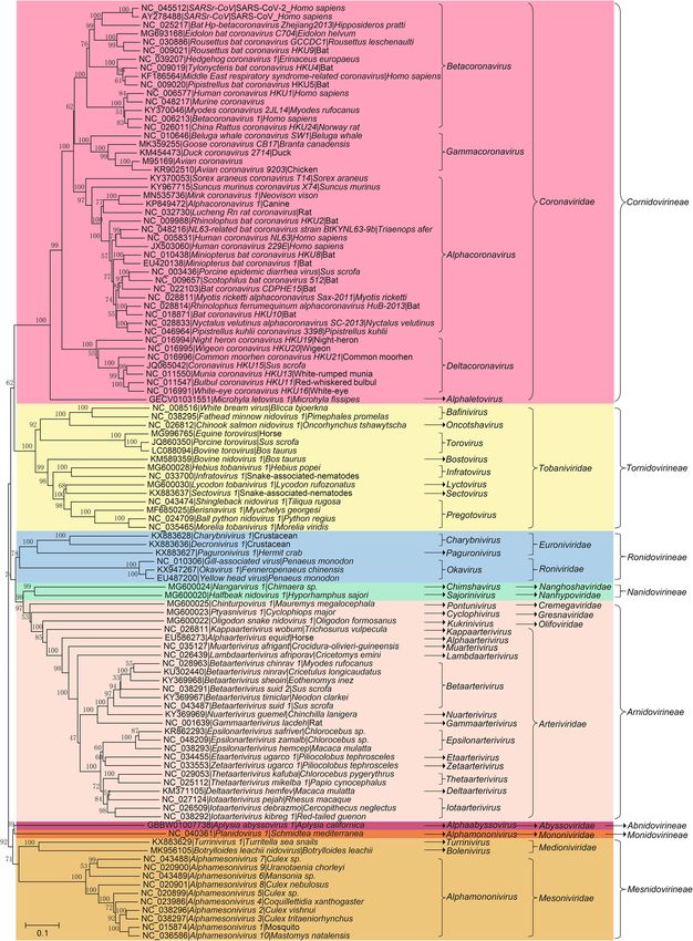
Zhou et al. Animal Diseases   (2021) 1:5                                                                      Page 2 of 28

2019), porcine epidemic diarrhea virus (PEDV, Pensaert       identified in various natural hosts of birds and mammals
and Bouck 1978), porcine deltacoronavirus (PDCoV,            that could not be classified using the former taxonomic
Woo et al. 2012), porcine hemagglutinating encephalo-        system. Thus, the system for CoV classification has been
myelitis virus (PHEV), and porcine enteric alphacorona-      adjusted several times, and the latest version was pro-
virus (PEAV), also known as swine acute diarrhea             posed (International Committee on Taxonomy of Vi-
syndrome coronavirus (SADS-CoV, Gong et al. 2017;            ruses Executive 2020). In addition, the widespread
Pan et al. 2017; Zhou et al. 2018). In addition to porcine   nature of CoVs raises concerns about the spread, spill-
CoVs, there are CoVs that infect other livestock and ani-    over and prevalence of epidemic viruses in the future,
mals, such as bovine coronavirus (BCoV, Castells et al.      which may impose significant respiratory and/or gastro-
2017), equine coronavirus (ECoV, Pusterla et al. 2016),      intestinal diseases on humans and economic losses to
canine coronavirus (CCoV, Decaro et al. 2010), and fe-       livestock farming. Therefore, it is critical to investigate
line coronavirus (FCoV, Li et al. 2019). Furthermore,        the characteristics of transmission and infection of
some avian CoVs, such as infectious bronchitis virus         CoVs. In this review, we provide an overview of the lat-
(IBV) and the closely related turkey coronavirus (TCoV)      est classification of viruses in the Nidovirales order and
(Jackwood et al. 2012; Brown et al. 2016) cause diseases     then focus on the genetic evolution, pathogenesis, and
in poultry.                                                  cross-species transmission of epidemic CoVs and other
   After the SARS-CoV epidemic in 2003, studies tra-         epidemic nidoviruses, aiming to provide a comprehen-
cing epidemic CoVs revealed the extensive presence           sive understanding of the current progress of studies
of nonpathogenic CoVs with remarkable genetic di-            concerning CoVs and nidoviruses.
versity in many species of wild animals. The natural            To avoid any divergence, information about the viruses
hosts of these CoVs include bats, rodents, cats, wild        listed in this review is preferentially in accordance with
birds, marine mammals and so on (Woo et al. 2012;            the latest update in the taxonomy system of the Inter-
Ge et al. 2013; Ge et al. 2017; Hu et al. 2017). Ac-         national Committee on Taxonomy of Viruses Executive
cording to genomic sequencing and phylogenetic ana-          on Taxonomy of Viruses (ICTV) (https://talk.ictvonline.
lyses, most human CoVs have been shown to                    org/taxonomy/), if available, which may not be com-
originate from wild animals at different time points         pletely consistent with the description of the original lit-
and through different paths (Cui et al. 2019; Zhou           erature. At the same time, only the classified virus
et al. 2020b). It’s particularly noteworthy that the 3       strains listed on the ICTV website are involved, while
highly pathogenic CoVs, including SARS-CoV, MERS-            some virus strains reported elsewhere may not be men-
CoV and SARS-CoV-2, may originate from bats (Ge              tioned. In addition, at present, the origin and spread of
et al. 2013; Azhar et al. 2014; Wang et al. 2014b;           nidoviruses are still controversial topics, and some de-
Zhou et al. 2020b). The HKU-2-related bat corona-            scriptions in this review may represent the authors’ per-
virus, named PEAV/SADS-CoV, was recently trans-              sonal views only.
mitted from bats to swine and caused an epidemic in
2016-17 (Zhou et al. 2018). On the other hand, the           Summary of the Nidovirales order
long-term persistence and rapid mutagenesis of native        Taxonomy of the Nidovirales order
CoVs may lead to the emergence of new types of epi-          The Nidovirales order was first proposed by ICTV in
demic CoVs, which has brought many challenges to             1996 and was named after the Latin term nido, which
the prevention and control of these diseases. For ex-        means “nest” (Pringle 1996). Initially, the order con-
ample, epidemiological studies have shown an increas-        tained only 2 viral families, Coronaviridae and Arteriviri-
ing genotype diversity of PEDV in pigs and IBV in            dae (Pringle 1996). However, with improvements in
poultry (Zhou et al. 2014; Li et al. 2017; Zeng et al.       techniques of virus detection and viral metagenomics,
2017). At the same time, the diversity and evolution         additional viral genomes have been detected (Ge et al.
of CoVs in their natural reservoirs, such as bats and        2012; Shi et al. 2018). Characterization of these novel
birds, have also caused increasing concerns about            viral genomes has profoundly changed the classification
their potential for interspecific transmission and to        of viruses. At present, 8 suborders have been established
cause epidemics (Hu et al. 2017).                            under the Nidovirales order: Abnidovirineae, Arnidoviri-
   CoVs are membrane-enveloped viruses with a linear,        neae, Cornidovirineae, Mesnidovirineae, Monidovirineae,
single-stranded and positive RNA genome. CoVs belong         Nanidovirineae, Ronidovirineae, and Tornidovirineae
to the Orthocoronavirinae subfamily of the Coronaviri-       (Walker et al. 2019). These 8 suborders contain 14 viral
dae family in the Cornidovirineae suborder of the Nido-      families, 25 subfamilies, 39 genera, 65 subgenera, and a
virales order (https://talk.ictvonline.org/taxonomy/)        total of 109 viral species (Tables 1, 2 and 3). Genetic re-
(Walker et al. 2019). After the SARS epidemic in 2013, a     lationship among the 109 representative virus species is
large number of CoVs with high genetic diversity were        shown in Fig. 1.
Zhou et al. Animal Diseases            (2021) 1:5                                                                                                 Page 3 of 28

Table 1 Hosts and sampled countries for Cornidovirineae viruses
Subfamily               Genus            Subgenus             Host                                        Country
Letovirinae             Alphaletovirus Milecovirus            Microhyla fissipes                          Unknown
Orthocoronavirinae Alpha-                Colacovirus          Bat                                         USA, Canada
                   coronavirus
                                         Decacovirus          Bat                                         China
                                         Duvinacovirus Homo sapiens, Camel, Bat                           USA, Netherlands, Italy, Sweden, Ghana, Saudi
                                                                                                          Arabia, Germany, Kenya, Haiti, United Arab Emirates
                                         Luchacovirus         Rat                                         China, United Kingdom
                                         Minacovirus          Mink, Neovison vison                        USA, Netherlands, Japan, China, Denmark
                                         Minunacovirus Bat                                                China
                                         Myotacovirus         Myotis ricketti                             China
                                         Nyctacovirus         Bat                                         China, Italy
                                         Pedacovirus          Sus scrofa                                  China, South Korea, USA, Mexico, Viet Nam, Canada,
                                                                                                          Ukraine, Belgium, France, Italy, Thailand, Viet Nam,
                                                                                                          Slovenia, Colombia, United Kingdom, Hungary,
                                                                                                          Japan, Germany, Russia, Philippines, Spain
                                         Rhinacovirus         Bat, Sus scrofa                             China
                                         Setracovirus         Homo sapiens                                Netherlands, USA, China, Haiti, Kenya, South Korea
                                         Soracovirus          Sorex araneus                               China
                                         Sunacovirus          Suncus murinus                              China
                                         Tegacovirus          Sus scrofa, Dog, Cat                        USA, China, Italy, Netherlands, Germany, United
                                                                                                          Kingdom, Belgium, Denmark, Mexico, Brazil, Spain
                        Beta-            Embecovirus          Homo sapiens, Bovine, Alpaca, Antelope,     Japan, Australia, USA, France, Belgium, China,
                        coronavirus                           Deer, Rat, Oryctolagus cuniculus, Dog,      Germany, South Korea, United Arab Emirates, Saudi
                                                              Camel, Sus scrofa, Horse, Pan troglodytes   Arabia, United Kingdom, Bangladesh, Mexico,
                                                              verus                                       Malaysia, Cote d'Ivoire, Uganda, Thailand, Kenya,
                                                                                                          Ethiopia, Morocco, Nigeria
                                         Hibecovirus          Bat                                         Nigeria, China
                                         Merbecovirus         Bat, Homo sapiens, Camel, Hedgehog,         China, United Kingdom, Germany, Jordan, South
                                                              Lama glama                                  Africa, Saudi Arabia, United Arab Emirates, France,
                                                                                                          Qatar, Egypt, USA, South Korea, Oman, Thailand,
                                                                                                          Italy, Ethiopia, Morocco, Burkina Faso, Nigeria,
                                                                                                          Kenya
                                         Nobecovirus          Bat                                         China, India, Singapore, Cameroon
                                         Sarbecovirus         Bat, Civet, Homo sapiens, Malayan           Global
                                                              pangolin, Felis catus, Mink, Dog, Tiger
                        Delta-           Andecovirus          Wigeon                                      China
                        coronavirus
                                         Buldecovirus         Sus scrofa, Birds, Leopard cat              USA, South Korea, China, Thailand, Laos, Viet Nam,
                                                                                                          Japan, United Arab Emirates, Poland, Australia
                                         Herdecovirus         Night-heron                                 China
                        Gamma-           Brangacovirus Branta canadensis                                  Canada
                        coronavirus
                                         Cegacovirus          Delphinapterus leucas, Bottlenose dolphin   USA
                                         Igacovirus           Chicken, Turkey, Duck, Pheasant, Goose,     China, USA, Canada, Netherlands, Nigeria, Sweden,
                                                              Partridge, Pigeon, Peafowl                  South Korea, Australia, Ukraine, South Africa, Italy,
                                                                                                          Belgium, France, India, Poland, Brazil, Sudan, Jordan,
                                                                                                          Pakistan, Egypt, United Kingdom, Uruguay, Iran,
                                                                                                          Malaysia, Peru, Australia, Norway
Notes: Some related but unclassified viruses are not listed

Common features of viruses in the Nidovirales order                                    nidoviruses share some significant common features.
Members of the Nidovirales order are membrane-                                         The most typical feature is transcription of multiple
enveloped viruses with a single strand positive RNA gen-                               3'-nested subgenomic RNAs from the 5’ terminus to the
ome. Although the viral particle size, morphology, and                                 3’ terminus along the genome during viral gene expres-
genome size of viruses in these 8 suborders vary greatly,                              sion, which endows the order name of nido-“nest”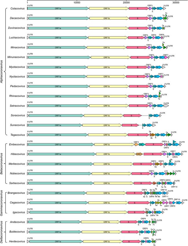
Zhou et al. Animal Diseases             (2021) 1:5                                                                                                 Page 4 of 28

Table 2 Hosts and sampled countries of Arnidovirineae viruses
Family              Subfamily           Genus                 Subgenus                 Host                            Country
Arteriviridae       Crocarterivirinae Muarterivirus           Muarterivirus afrigant   Crocidura olivieri guineensis   Guinea
                    Equarterivirinae    Alphaarterivirus      Alphaarterivirus equid   Horse, Donkey                   USA, France, United Kingdom, Chile,
                                                                                                                       Serbia, Poland
                    Heroarterivirinae Lambdaarterivirus Lambdaarterivirus              Cricetomys emini                Cameroon
                                                        afriporav
                    Simarterivirinae    Deltaarterivirus      Hedartevirus             Monkey                          USA
                                        Epsilonarterivirus    Sheartevirus             Monkey                          USSR, South Africa
                                        Etaarterivirus        Etaarterivirus ugarco 1 Monkey                           Uganda
                                        Iotaarterivirus       Debiartevirus            Monkey                          Cameroon
                                                              Kigiartevirus            Monkey                          Uganda
                                                              Pedartevirus             Monkey                          USA
                                        Thetaarterivirus      Kaftartevirus            Monkey, Papio                   Zambia
                                                              Mitartevirus             Papio                           Tanzania
                                        Zetaarterivirus       Zeta-arterivirus ugarco Monkey                           Uganda
                                                              1
                    Variarterivirinae   Betaarterivirus       Ampobartevirus           Sus scrofa                      USA, South Korea, India, China
                                                              Chibartevirus            Myodes rufocanus,               China
                                                                                       Eothenomys inez
                                                              Eurpobartevirus          Sus scrofa                      Netherlands, France, Hungary, China,
                                                                                                                       Russia
                                                              Micartevirus             Neodon clarkei                  China
                                        Gammaarterivirus Gammaarterivirus              Rat                             Unknown
                                                         lacdeh
                                        Nuarterivirus         Nuarterivirus guemel     Chinchilla lanigera             China
                    Zealarterivirinae Kappaarterivirus        Kappaarterivirus         Trichosurus vulpecula           Australia, New Zealand
                                                              wobum
Cremegaviridae Rodepovirinae            Pontunivirus          Chinturpovirus 1         Mauremys megalocephala          China
Gresnaviridae       Reternivirinae      Cyclophivirus         Ptyasnivirus 1           Cyclophiops major               China
Olifoviridae        Gofosavirinae       Kukrinivirus          Oligodon snake           Oligodon formosanus             China
                                                              nidovirus 1
Notes: Some related but unclassified viruses are not listed

(Posthuma et al. 2006). The other common features in-                                  3CLpro, NiRAN, RdRp, ZBD, and HEL1 located in
clude similar genomic organization (encoding the non-                                  the genome and proteins are illustrated in Fig. 2
structural polyprotein upstream and the structural                                     using the SARS-CoV-2 model.
protein downstream of the genome), expression of
the polyprotein by ribosomal frame shifting mode,                                      The taxonomy, host range and pathogenicity of
and some nonstructural proteins (NSPs) with unique                                     Cornidovirineae
protease activities (Fig. 2) (Posthuma et al. 2008).                                   Suborder Cornidovirineae
Due to the complexity of the genome, the new classi-                                   Among all 8 suborders of the Nidovirales order,
fication standard of order Nidovirales is based on                                     Cornidovirineae harbors the most epidemic viruses.
amino acid (aa) sequences of several hallmark genes                                    Currently, the Cornidovirineae suborder contains 1
only, which include 3CLpro (3C-like protease),                                         viral family, Coronaviridae, which is further divided
NiRAN (nidovirus RdRp-associated nucleotidyltrans-                                     into 2 subfamilies, Letovirinae and Orthocoronavirinae
ferase), RdRp (RNA-directed RNA polymerase), ZBD                                       (Bukhari et al. 2018). The subfamily Letovirinae con-
(Zn-binding domain covalently linked to HEL1), and                                     tains 1 genus Alphaletovirus, 1 subgenus Milecovirus,
HEL1 (helicase of superfamily 1) domains of the                                        and 1 species Microhyla letovirus 1 (MLeV), which is
replicase protein (International Committee on                                          represented by a 22.3 kb potentially partial genome
Taxonomy of Viruses Executive 2020). Domains of                                        found in an ornamented pygmy frog (Microhyla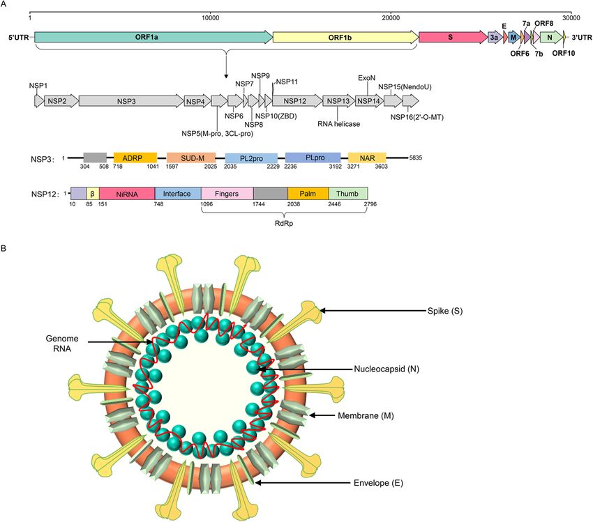
Zhou et al. Animal Diseases            (2021) 1:5                                                                                              Page 5 of 28

Table 3 Hosts and sampled countries of Ab-, Mes-, Mo-, Na-, Ro- and Tor-Nidovirineae viruses
Suborders           Family                Subfamily           Genus              Subgenus        Host                    Country
Abnidovirineae Abyssoviridae              Tiamatvirinae       Alphaabyssovirus   Aplyccavirus    Aplysia californica     Unknown
Mesnidovirineae Medioniviridae            Medionivirinae      Turrinivirus       Beturrivirus    Turritella sea snails   China
                                          Tunicanivirinae     Bolenivirus        Balbicanovirus Botrylloides leachii     New Zealand
                    Mesoniviridae         Hexponivirinae      Alphamesonivirus Casualivirus      Coquillettidia          Australia
                                                                                                 xanthogaster
                                                                                 Enselivirus     Culex                   Cote d'Ivoire
                                                                                 Hanalivirus     Culex                   Cote d'Ivoire
                                                                                 Kadilivirus     Culex                   Brazil
                                                                                 Karsalivirus    Culex                   Viet Nam, Indonesia
                                                                                 Menolivirus     Uranotaenia             Cote d'Ivoire
                                                                                 Namcalivirus    Aedes, Rat, Culex,      Viet Nam, Cote d'Ivoire, Indonesia,
                                                                                                 Uranotaenia             Thailand, China, South Korea,
                                                                                                                         Senegal, Ghana, Australia, Italy,
                                                                                                                         USA, Austria, Mexico, Sweden,
                                                                                                                         Brazil, Spain
                                                                                 Ofalivirus      Mansonia                Brazil
Monidovirineae      Mononiviridae         Mononivirinae       Alphamononivirus Dumedivirus       Schmidtea               USA
                                                                                                 mediterranea
Nanidovirineae      Nanghoshaviridae Chimanivirinae           Chimshavirus       Nangarvirus 1   Chimaera sp.            China
                    Nanhypoviridae        Hyporhamsavirinae Sajorinivirus        Halfbeak        Hyporhamphus            China
                                                                                 nidovirus 1     sajori
Ronidovirineae      Euroniviridae         Ceronivirinae       Charybnivirus      Cradenivirus    Charybdis crab          China
                                                                                 Wenilivirus     Crustacean              China
                                          Crustonivirinae     Paguronivirus      Behecravirus    Hermit crab             China
                    Roniviridae           Okanivirinae        Okavirus           Tipravirus      Penaeus                 Australia, Thailand, Egypt, China
Tornidovirineae     Tobaniviridae         Piscanivirinae      Bafinivirus        Blicbavirus     Blicca bjoerkna         Germany
                                                                                 Pimfabavirus    Pimephales promelas USA
                                                                                 unclassified    Salmo salar, Yellow     Canada, China
                                                                                 Bafinivirus     catfish
                                                              Oncotshavirus      Salnivirus      Oncorhynchus            Canada, China, United Kingdom
                                                                                                 tshawytscha,
                                                                                                 Macrognathus
                                                                                                 aculeatus, Crucian
                                                                                                 carp, Carassius
                                                                                                 auratus
                                          Remotovirinae       Bostovirus         Bosnitovirus    Cattle                  USA
                                          Serpentovirinae     Infratovirus       Hepoptovirus    Hebius popei            China
                                                                                 Xintolivirus    Snake-associated        China
                                                                                                 nematodes
                                                              Lyctovirus         Rebatovirus     Lycodon rufozonatus China
                                                              Pregotovirus       Roypretovirus   Python regius,          USA, Switzerland
                                                                                                 Morelia viridis
                                                                                 Snaturtovirus   Myuchelys georgesi      Australia
                                                                                 Tilitovirus     Tiliqua rugosa          Australia
                                                              Sectovirus         Sanematovirus Snake-associated          China
                                                                                               nematodes
                                          Torovirinae         Torovirus          Renitovirus     Sus scrofa, Cattle,     China, USA, Japan, Switzerland
                                                                                                 Horse
Notes: Some related but unclassified viruses are not listed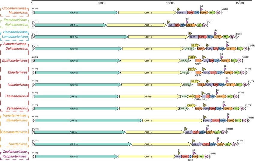
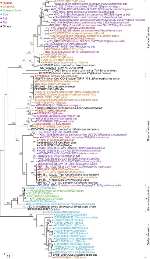
Zhou et al. Animal Diseases      (2021) 1:5                                                                                         Page 6 of 28

 Fig. 1 Phylogenetic tree of RNA-dependent RNA polymerase (RdRp) amino acid sequences of 109 viral species in the Nidovirales order. The tree
 was constructed using the neighbor-joining method with a p-distance model and 1000 bootstraps in the MEGA V. 7.0.14

fissipes) (Bukhari et al. 2018). In contrast, the subfam-                 (Coronaviridae Study Group of the International
ily Orthocoronavirinae contains 4 genera, Alphacoro-                      Committee on Taxonomy of Viruses 2020). In this re-
navirus      (14    subgenera      and     19     species),               view, “CoVs” refers to viruses of the subfamily Ortho-
Betacoronavirus (5 subgenera and 14 species), Delta-                      coronavirinae (Figs. 2, 3, 4 and 5).
coronavirus (3 subgenera and 7 species), and Gamma-
coronavirus (3 subgenera and 5 species), which is the                     Subfamily Orthocoronavirinae
largest subfamily in the Nidovirales order, including                     CoVs are spherical, enveloped viral particles (approxi-
some highly pathogenic viruses, such as SARS-CoV,                         mately 100 nm in diameter) with S proteins on the sur-
MERS-CoV,         and      SARS-CoV-2        (Table     1)                face. The subfamily Orthocoronavirinae is divided into 4
Zhou et al. Animal Diseases       (2021) 1:5                                                                                             Page 7 of 28

 Fig. 2 Genome structure, coding proteins, and viral particle structure diagram of coronavirus represented by SARS-CoV-2. a Genome structure,
 functional domains and locations of SARS-CoV-2. NSP, nonstructural proteins; 3Clpro, 3C-like protease; NiRAN, nidovirus RdRp-associated
 nucleotidyltransferase; RdRp, RNA-directed RNA polymerase; ZBD, Zn-binding domain covalently linked to HEL1; HEL1, helicase of superfamily 1;
 NendoU, endonuclease; 2′-O-MT, 2′-O-methyltransferase; ExoN, 3′-5′ exonuclease; SUD-M, SARS-unique domains; ADRP, ADP-ribose-1″-
 phosphatase; PLpro, papain-like protease; NAR, nucleic acid-binding domain. b Viral particle diagram of SARS-CoV-2. Structural proteins of S, N, M,
 and E are labeled, and the red line indicates the RNA genome

genera: Alphacoronavirus (Alpha-CoV), Betacoronavirus                        subgenera Duvinacovirus and Setracovirus, respectively,
(Beta-CoV), Deltacoronavirus (Delta-CoV), and Gamma-                         have been identified (McIntosh et al. 1967; van der Hoek
coronavirus (Gamma-CoV) (Wong et al. 2019). Alpha-                           et al. 2004). These CoVs are prevalent worldwide in chil-
CoVs and beta-CoVs infect mammals, while delta-CoVs                          dren and adults and cause respiratory infections, usually
and gamma-CoVs mostly infect birds, but a few of them                        leading to mild cold-like symptoms and acute respiratory
do infect mammals (Woo et al. 2012). Currently, there                        disease (El-Sahly et al. 2000; van der Hoek et al. 2004).
are 14 subgenera (from Colacovirus to Tegacovirus), in-                      Genetic evidence has shown that HCoV-229E and NL63
cluding 19 species in Alphacoronavirus with 5 subgenera                      share common ancestors with bat CoVs, indicating the
(Embecovirus, Hibecovirus, Mergecovirus, Nobecovirus                         possibility of viral origin from bats (Tao et al. 2017).
and Sarbecovirus), 14 species in Betacoronavirus with 3                      229E-like bat CoVs, such as strains BtKY229E-1,
subgenera (Andecovirus, Buldecovirus and Herdecovirus),                      BtKY229E-8 and BtCoV/FO1A-F2, have been detected
7 species in Deltacoronavirus with 3 subgenera (Branga-                      in Hipposideros bats in Africa (Pfefferle et al. 2009;
covirus, Cegacovirus, and Igacovirus), and 5 species in                      Crossley et al. 2012). Moreover, CoVs highly similar to
Gammacoronavirus (Table 1, Fig. 3).                                          229E were also detected in camelids, including alpacas
                                                                             and dromedary camels (Crossley et al. 2012; Corman
Alpha-CoVs                                                                   et al. 2016). The 229E-related CoVs isolated from drom-
Human related alpha-CoVs                                                     edary camels can use human aminopeptidase N (APN)
Alpha-CoVs contain 14 viral genera and 19 species and                        as a receptor and efficiently replicate in human hepa-
are the largest genus in the subfamily Orthocoronaviri-                      toma cells, which are neutralized by antibodies in human
nae (Fig. 3). Two human CoVs (HCoVs) in alpha-CoVs,                          sera positive for 229E, suggesting a close genetic rela-
HCoV-229E and HCoV-NL63, which belong to                                     tionship between 229E and 229E-related CoVs (Corman
Zhou et al. Animal Diseases (2021) 1:5 Page 8 of 28 Fig. 3 The maximum likelihood tree of coronaviruses based on amino acid sequences of RdRp gene. The tree was constructed using an IQ-tree with 10,000 ultrafast bootstraps and the most appropriate substitution model of LG+F+I+G4, which was calculated by ModelFinder
Zhou et al. Animal Diseases      (2021) 1:5                                                                                         Page 9 of 28

 Fig. 4 The neighbor-joining tree of spike gene nucleotide sequences in Sarbecovirus. The tree was constructed using the p-distance model and
 1000 bootstraps in the MEGA V. 7.0.14. Viral strains derived from humans, bats, and other animals are indicated by red, purple, and
 cyan, respectively

et al. 2016). Due to the long-term contact and habitat                    Executive 2020). NL63-like CoVs were also found in
overlap between humans and camels, one of the con-                        Triaenops bats, such as strains BtKYNL63-9a,
jectures about the transmission path of 229E is that                      BtKYNL63-9b, and BtKYNL63-15 (Tao et al. 2017).
the ancestor bat CoV was transmitted to camels first                      These NL63-like bat CoVs exhibited less than 90%
and then from camels to humans (Corman et al.                             similarity in their aa sequences compared to NL63 in
2016). Bat and camel 229E-related CoVs have been                          the concatenated protein domains of 3CLpro, NiRAN,
designated into the same species represented by 229E                      RdRp, ZBD and HEL1, and thus, they were classified
because all of these CoVs share more than 95%                             into NL63-related bat coronavirus strain BtKYNL63-
amino acid (aa) sequence identity (higher than 92.4%                      9b species, parallel to the human coronavirus NL63
according to the species demarcation criteria), with                      species in the subgenus Setracovirus. However, NL63-
229E in the concatenated protein domains of 3CLpro,                       related CoVs have not yet been detected in livestock
NiRAN, RdRp, ZBD and HEL1 (Sabir et al. 2016;                             or other animals, so the origin and intermediate
International Committee on Taxonomy of Viruses                            host(s) of NL63 are still unclear. Notably, NL63-
Zhou et al. Animal Diseases      (2021) 1:5                                                                                      Page 10 of 28

 Fig. 5 Gene composition of 25 subgenera in 4 viral genera of Orthocoronavirinae. The reference strains used for mapping in each subgenus are
 as follows: Colacovirus (Bat-CoV CDPHE15/USA/2006, NC_022103.1), Decacovirus (Bat-CoV HKU10, NC_018871.1), Duvinacovirus (229E-related bat-
 CoV BtKY229E-1, KY073747.1), Luchacovirus (AcCoV-JC34, NC_034972.1), Minacovirus (FRCoV-NL-2010, NC_030292.1), Minunacovirus (Bat-CoV HKU8,
 NC_010438.1), Myotacovirus (BtMr-CoV/SAX2011, NC_028811.1), Nyctacovirus (Bat-CoV HKU33, MK720944.1), Pedacovirus (PEDV, NC_003436.1),
 Rhinacovirus (SADS-CoV, MT199592.1), Setracovirus (NL63-related bat-CoV BtKYNL63-9b, NC_048216.1), Soracovirus (Shrew-CoV/Tibet2014,
 KY370053.1), Sunacovirus (Wencheng Sm shrew CoV Xingguo-74, KY967715.1), Tegacovirus (CCoV/NTU336/F/2008, GQ477367.1), Embecovirus
 (Murine CoV, JX169867.1), Hibecovirus (Bat Hp-betacoronavirus/Zhejiang2013, NC_025217.1), Merbecovirus (Human MERS-CoV, NC_019843.3),
 Nobecovirus (Rousettus bat-CoV GCCDC1 356, NC_030886.1), Sarbecovirus (SARS-CoV-2, NC_045512.2), Brangacovirus (Canada goose CoV
 Cambridge_Bay_2017, MK359255.1), Cegacovirus (Beluga Whale CoV SW1, NC_010646.1), Igacovirus (Turkey CoV, NC_010800.1), Andecovirus
 (Wigeon CoV HKU20, NC_016995.1), Buldecovirus (Common-moorhen CoV HKU21, NC_016996.1), Herdecovirus (Night-heron CoV
 HKU19, NC_016994.1)

related and 229E-related bat CoVs exhibit high gen-                      Bat alpha-CoVs and derived CoVs infecting other animals
etic diversity in bats, and recombination of bat CoVs                    Bats carry many different CoVs and have been recog-
may accelerate the occurrence of new viruses and the                     nized as important natural reservoirs for CoVs. Bat
possibility of cross-species transmission (Tao et al.                    CoVs occupy 11 of 19 viral species in alpha-CoVs. The
2017).                                                                   representative species of subgenus Colacovirus is bat
Zhou et al. Animal Diseases   (2021) 1:5                                                                     Page 11 of 28

coronavirus CDPHE15, which was identified in Myotis           agent of porcine epidemic diarrhea (PED), which infects
lucifugus (little brown bat) (Subudhi et al. 2017). The       pigs of all ages and causes significantly high mortality in
subgenus Decacovirus contains 2 species, bat corona-          piglets under 7 days old with symptoms including diar-
virus HKU10 and Rhinolophus ferrumequinum alphacor-           rhea, vomiting, anorexia and dehydration (Chen et al.
onavirus HuB-2013, both of which were isolated from           2013). Since the recognition of PEDV in 1978 in
bats. HKU10- and HKU10-related CoVs were identified           Belgium, this virus has caused epidemics worldwide and
in 2 species of bats, Rousettus leschenaultii (Lesche-        has continuously impaired pig farming industry (Pen-
nault’s Rousettes) and Hipposideros pomona (Pomona            saert and Bouck 1978). During long-term epidemics,
leaf-nosed bat), which were named Ro-BatCoV HKU10             novel PEDV variants continuously emerge, deriving the
and Hi-BatCoV HKU10, respectively (Lau et al. 2012).          virulent variants of SINDEL groups and different geno-
Ro-BatCoV HKU10 and Hi-BatCoV HKU10 share high                type groups including GI and GII (even more groups or
aa similarity (>99%) among 3CLpro, NiRAN, RdRp, ZBD           subgenotypes) (Zeng et al. 2017). In addition, the coex-
and HEL1 but only 60.5% aa identity in the S proteins.        istence or coinfection of multiple genotypes or variants
Interspecies transmission of HKU10 from Leschenault’s         of PEDV has been reported, which makes the prevention
Rousettes to Pomona leaf-nosed bats, 2 different bat          and control of PEDV more difficult (Zeng et al. 2017).
suborders, has been reported in a previous study (Lau            The subgenus Rhinacovirus has 1 species, Rhinolophus
et al. 2012). The HKU10-related bat CoV was also iden-        bat coronavirus HKU2 (Bat-CoV HKU2). Bat-CoV
tified in Rhinolophus sinicus (Rs-BatCoV HKU32), which        HKU2 was first reported in 2006 and was detected in
may be a potential novel species (Lau et al. 2019b). The      Rhinolophus sinicus (Chinese horseshoe bat) (Woo et al.
species of Rhinolophus ferrumequinum alphacoronavirus         2006a). Subsequently, HKU2-like viruses were identified
HuB-2013 has several strains, among which the strain          in other bat species, such as Rhinolophus ferrumequinum
BtRf-Alpha-CoV/HuB2013 was detected in Rhinolophus            and Rhinolophus affinis (Lau et al. 2007; Wu et al. 2016).
ferrumequinum, and the strain BtMs- alpha-CoV/                HKU2 is genetically distinct from other known bat
GS2013 was detected in Myotis bats (Wu et al. 2016).          alpha-CoVs and forms a unique phylogenetic lineage
The subgenus Minunacovirus includes 2 species, Mini-          (Fig. 3) (Wu et al. 2016). Another remarkable feature of
opterus bat coronavirus 1 and Miniopterus bat corona-         HKU2 is the shortest aa sequence of its S protein, which
virus HKU8, which are common CoVs in Miniopterus              has deletions in the N-terminal region and extremely
bats, such as Miniopterus schreibersii, Miniopterus fuligi-   low (< 30%) aa identities with the other S proteins of
nosus and Miniopterus pusillus (Chu et al. 2008; Wu           CoVs (Lau et al. 2007). These significant differences in-
et al. 2016).                                                 dicate that HKU2 has a special evolutionary history. Be-
   The subgenus Myotacovirus contains 1 species, Myotis       cause HKU2 has a long phylogenetic relationship with
ricketti alphacoronavirus Sax-2011, which was found in        common alpha-CoV pathogens, it was not predicted to
Myotis ricketti in China in 2011 (Wu et al. 2016). The        exhibit cross species transmission or lead to diseases
subgenus Nyctacovirus contains 2 species, Nyctalus velu-      until a similar virus was detected in piglets with diarrhea
tinus alphacoronavirus SC-2013 (BtNv-Alpha-CoV/               symptoms in China from 2016 to 2017 (Gong et al.
SC2013) and Pipistrellus kuhlii coronavirus 3398 (Bat-        2017; Pan et al. 2017). In October 2016 and February
CoV/P.kuhlii/Italy/3398), which were detected in Nycta-       2017, outbreaks of newborn piglet diarrhea occurred in
lus and Pipistrellus bats, respectively (Wu et al. 2016; De   commercial pig farms in Guangdong Province, charac-
Sabato et al. 2019). Recently, bat CoV HKU33 (Tr-Bat-         terized by acute vomiting and watery diarrhea with a
CoV HKU33) was discovered in Tylonycteris robustula           mortality rate over 35% in piglets (Pan et al. 2017). The
(greater bamboo bats) in Guizhou Province in China            etiology was identified as a Bat-HKU2 alpha-CoV called
(Lau et al. 2019b). Tr-BatCoV HKU33 is most closely re-       swine enteric alphacoronavirus (SeACoV), porcine en-
lated to BtNv-alpha-CoV/SC2013. However, in the               teric alphacoronavirus (PEAV), or SADS-CoV (Zhou
concatenated protein domains, aa sequence identity is         et al. 2018). The genome sequence of SADS-CoV
76.3% between Tr-BatCoV HKU33 and BtNv-Alpha-                 showed approximately 95% nucleotide (nt) identity with
CoV/SC2013, indicating that Tr-BatCoV HKU33 repre-            the Bat HKU2 strains (Pan et al. 2017). However, at the
sents a novel CoV species and a potential novel sub-          nt and aa levels, the S of SADS-CoV only exhibited ap-
genus (less than 14.7% homology with the 3CLpro,              proximately 80% and 86% sequence identities with that
NiRAN, RdRp, ZBD and HEL1 domains) in the alpha-              of bat HKU2 (Pan et al. 2017; Zhou et al. 2018). Recent
CoV genus (Lau et al. 2019b).                                 studies showed that bat HKU2 strains derived from bats
   The subgenus Pedacovirus contains 2 species, PEDV          in Guangdong Province were genetically diverse in dif-
and Scotophilus bat coronavirus 512 (BtCoV/512).              ferent Rhinolophus bats and exhibited different homolo-
BtCoV/512 was discovered in Scotophilus kuhlii bats in        gies to SADS-CoV, from 95.09% to 98.48% (Zhou et al.
Hainan, China (Tang et al. 2006). PEDV is the etiologic       2018). Virological, epidemiological, and evolutionary
Zhou et al. Animal Diseases   (2021) 1:5                                                                    Page 12 of 28

evidence suggested that SADS-CoV might have recently          to Alphacoronavirus 1 have not been detected in wild
spread from bats to pigs and cause an outbreak of dis-        animals, such as bats, and thus, the exact origin of these
ease (Zhou et al. 2018). One study reported that SADS-        viruses is unclear.
CoV caused an epidemic across 4 farms, leading to the
death of 24,693 piglets from October 2016 to January          Other alpha-CoVs
2017 (Zhou et al. 2018). After that, new SADS cases           The subgenus Minacovirus has 1 species, mink corona-
were reported in Fujian Province of China in 2018 until       virus 1 (MCoV). MCoV causes epizootic catarrhal
January 2019 (Yang et al. 2020b) and in Southern China        gastroenteritis (ECG) in mink with high morbidity
in February 2019 (Zhou et al. 2019). Moreover, experi-        (approximately 100 %) and low mortality (
Zhou et al. Animal Diseases   (2021) 1:5                                                                    Page 13 of 28

clustered together with rodent alpha-CoVs in the ORF1b      subgenus Merbecovirus. Eidolon bat coronavirus C704,
tree but showed a certain genetic distance from other       Rousettus bat coronavirus GCCDC1and Rousettus bat
rodent alpha-CoVs (Tsoleridis et al. 2019). Surprisingly,   coronavirus HKU9 in subgenus Nobecovirus, and se-
rodent alpha-CoVs cluster together with HKU2, includ-       vere acute respiratory syndrome-related coronavirus in
ing SADS-CoV and other related bat CoVs, in the beta-       subgenus Sarbecovirus.
CoV group based on the spike tree, which indicates an
ancient recombination event between them (Pan et al.        Human beta-CoVs
2017; Tsoleridis et al. 2019). Rodentia is the largest
mammalian order, harboring ~2,200 species. Rodents          HCoV-OC43 and HKU1 Five types of beta-CoVs clas-
carry diverse pathogens and are an important source of      sified into 4 representative species have been reported
emerging viral infections. Recently, an increasing num-     to infect humans: HCoV-OC43 of Betacoronavirus 1,
ber of CoVs have been isolated from rodents, and more       HCoV-HKU1, MERS-CoV of Middle East respiratory
are expected in the future. Since rodents and many kinds    syndrome-related coronavirus (MERSr-CoV), SARS-
of animals share habitats, monitoring rodent CoVs could     CoV and SARS-CoV-2 of severe acute respiratory
be beneficial for the prevention and control of CoV-        syndrome-related coronavirus (SARSr-CoV) (Hu et al.
related diseases.                                           2021). OC43 and HKU1 were discovered in patients
  The subgenera Soracovirus and Sunacovirus each            with respiratory diseases in 1967 and 2005, respect-
contain 1 viral species, Sorex araneus coronavirus          ively (Hamre and Procknow 1966; Woo et al. 2005).
T14 (Shrew-CoV/Tibet2014) and Suncus murinus                OC43 and HKU1, together with the other 2 alpha-
coronavirus X74 (Xīngguō-74 or known as WESV),              CoVs, 299E and NL63, are associated with a wide
respectively. Shrew-CoV/Tibet2014 and Xīngguō-74            range of respiratory outcomes, including bronchiolitis
have been identified in different shrews, Sorex ara-        and pneumonia and cause nonrespiratory organ
neus and Suncus murinus, respectively, in China             system diseases, such as enteric and nervous systems
(Wang et al. 2017; Wu et al. 2018). Both of them            (Zeng et al. 2018). Although these CoVs are consid-
belong to the Soricidae family and Soricomorpha             ered one of the most common respiratory pathogens
order. In addition to Xīngguō-74, other strains were        associated with respiratory tract infections in children
also identified in shrews in China, including Wénch-        and elderly individuals, particularly severe infections
éng shrew virus (WESV) and Yúdū shrew virus                 in infants, clinical epidemiological investigations have
(Yúdū-76), which showed moderate genetic diversity          revealed a higher positive rate of these CoVs in
(Wang et al. 2017). CoVs detected in shrews in              adults, ranging from 50 to 59 years old (Cabeça et al.
China, which are represented by Shrew-CoV/                  2013). In some seasons, OC43 could be the most
Tibet2014 and Xīngguō-74, are clustered together,           frequent virus detected in acute respiratory tract in-
forming a distinct group. However, shrew CoVs               fection cases and had a more frequently abnormal
show distinct phylogenetic relationships to other           pulmonary rate than the other 3 CoVs (Zeng et al.
known alpha-CoVs, indicating that these viruses are         2018).
from different origins and have distinct evolutionary         OC43 has a global distribution, and circulating OC43
characteristics (Wang et al. 2017; Wu et al. 2018).         viruses have high genetic diversity with at least 5 distinct
                                                            genotypes (A to E) (Kin et al. 2015). Recombination of
Beta-CoVs                                                   different types promotes the generation of new viral
The genus Betacoronavirus currently contains 5 subgen-      types. For example, OC43-D emerged via recombination
era (Embecovirus, Hibecovirus, Mergecovirus, Nobecov-       between genotypes B and C, and OC43-E emerged via
irus, and Sarbecovirus) and 14 species. Beta-CoV is well    recombination among genotypes B, C, and D (Zhang
known because it includes the most pathogenic CoVs to       et al. 2015). In viral classification, OC43 belongs to the
human beings, such as SARS-CoV, MERS-CoV and                species Betacoronavirus 1, together with various closely
SARS-CoV-2, as well as a large number of related bat        related CoVs detected in other animals (Fig. 3). In
CoVs (Hu et al. 2015; Wong et al. 2019; Hu et al.           addition to OC43, the other related CoVs in species
2021). The Beta-CoV species include Betacoronavirus         Betacoronavirus 1 include HKU23 detected in dromed-
1, China rattus coronavirus HKU24, human corona-            ary camels, bovine CoV (BCoV) in bovine and alpaca,
virus HKU1, murine coronavirus and Myodes corona-           SACoV in Sable antelope, GiCoV in giraffe, canine re-
virus 2JL14 in subgenus Embecovirus, bat Hp-                spiratory CoV (CRCoV) in dog, porcine hemagglutinat-
betacoronavirus Zhejiang2013 in subgenus hibecovirus,       ing encephalomyelitis virus (PHEV) and equine CoV
hedgehog coronavirus 1, Middle East respiratory             (ECoV) (Fig. 3) (Woo et al. 2014b). Phylogenetically,
syndrome-related coronavirus, pipistrellus bat corona-      CoVs of betacoronavirus 1 are most closely related to
virus HKU5 and tylonycteris bat coronavirus HKU4 in         another species, China Rattus coronavirus HKU24
Zhou et al. Animal Diseases (2021) 1:5 Page 14 of 28
(HKU24), in the subgenus Embecovirus (Fig. 3). HKU24 days in 95% of symptomatic cases and even longer in
and similar viruses have been found in rats, such as Rat- some asymptomatic cases (Yang et al. 2020a). The
tus norvegicus, Apodemus agrarius, and Apodemus chev- COVID-19 pandemic has become a serious global public
rieri (Lau et al. 2015; Wang et al. 2015; Ge et al. 2017). health problem that has challenged our knowledge of
The RdRp and Hel proteins of OC43 and HKU24 exhibit virology and systems of viral disease control.
91.8% and 93.5% aa sequence identity, respectively (Lau The overall genomic sequence identity between SARS-
et al. 2015). Current evidence indicates that OC43 likely CoV and SARS-CoV-2 is 79.6%; however, aa identities of
originated from rodents and may be transmitted to ORF1a and ORF1b between them are 80.9% and 95.7%,
humans via livestock, such as bovines (Wang et al. 2015; respectively (Zhou et al. 2020b). Based on the new
Forni et al. 2017; Ge et al. 2017). demarcation criteria, they belong to the same species, se-
HKU1 was first reported in 2005 in a 71-year-old pa- vere acute respiratory syndrome-related coronavirus
tient with pneumonia and bronchiolitis in Hong Kong (SARSr-CoV) Coronaviridae Study Group of the Inter-
(Woo et al. 2005). Since then, HKU1 has been detected national Committee on Taxonomy of Viruses 2020). In
in France, USA, Brazil, Australia, etc., indicating its glo- CoV taxonomy, SARS-CoV, SARS-CoV-2, most bat
bal distribution (Siu et al. 2014; Zeng et al. 2018). Strains CoVs and a few CoVs detected in civets, pangolins and
of HKU1 are divided into 3 genotypes, A, B and C, based other animals are classified in the SARSr-CoV subgenus
on their phylogenetic relationships (Woo et al. 2006b). Sarbecovirus (Fig. 4) (Coronaviridae Study Group of the
Recombination between different genotypes may lead to International Committee on Taxonomy of Viruses
the emergence of novel genotypes. For instance, the re- 2020). Since the appearance of SARS, investigations on
combination of hemagglutinin esterase (HE) coding re- the origin of SARS-CoV have led to the discovery of a
gions between A and B generated genotype C (Woo large number of novel CoVs in various animals, particu-
et al. 2006b; Dominguez et al. 2014). HKU1 is closest to larly in bats, and greatly promoted the understanding of
murine coronavirus (known as mouse hepatitis virus, the existence and spread of CoVs in nature (Hu et al.
MHV) according to phylogenetic analysis, and different 2015; Wong et al. 2019). In 2005, CoV strains highly
strains of murine coronavirus have been detected in rats homologous to SARS-CoV were identified in the
(rat CoV Parker) and mice (MHV, Fig. 3) (Das Sarma Chinese horseshoe bat (Rhinolophus sp. ), including
et al. 2001). One hypothesis is that HKU1 originates Rp3, HKU3, etc. (Li et al. 2005; Woo et al. 2005).
from rodents, but its intermediate host is still unclear SARS-CoV uses angiotensin-converting enzyme 2
(Forni et al. 2017; Cui et al. 2019). (ACE2) as its receptor to enter cells (Li et al. 2003).
In 2013, WIV1, a living bat CoV using the same
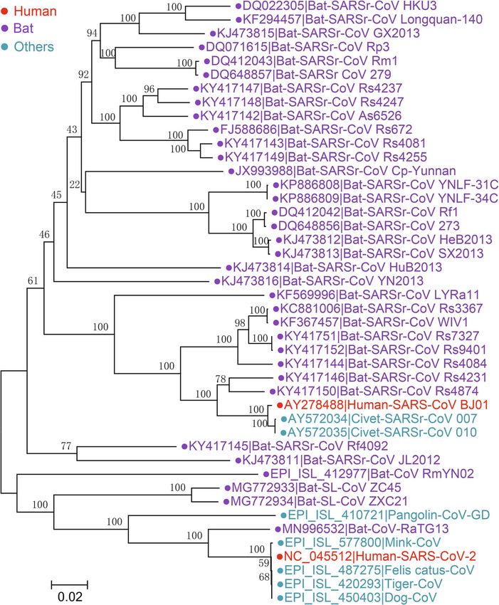
SARS-CoV and SARS-CoV-2 Severe acute respiratory ACE2 receptor as SARS-CoV, was isolated from a
syndrome coronavirus (SARS-CoV) and severe acute re- Rhinolophus sinicus sample (Li et al. 2003; Ge
spiratory syndrome coronavirus 2 (SARS-CoV-2) are et al. 2013). The overall nt identity of WIV1 genome
highly pathogenic human CoVs (Falsey and Walsh 2003; with human SARS-CoV is 95.4%. In 2015, another bat
Zhu et al. 2020). SARS-CoV caused a relatively short CoV strain, WIV16, which shows higher genomic se-
epidemic of SARS in 2002–2003, resulting in more than quence identity (96% nt identities) to SARS-CoV, was
8,000 clinical cases with a mortality of 10% (Parry 2003; isolated (Yang et al. 2015). A 5-year surveillance of
Stadler et al. 2003). However, ongoing SARS-CoV-2 has bat SARSr-CoVs in Yunnan Province, China, revealed
caused an extensive pandemic, termed coronavirus dis- that the SARSr-CoVs circulating in bats are highly di-
ease 2019 (COVID-19), beginning the end of 2019, lead- verse and have high genetic similarity to SARS-CoV
ing to more than a hundred million cases and millions in the hypervariable N-terminal domain (NTD) and
of deaths worldwide (Hu et al. 2021). Both SARS-CoV receptor-binding domain (RBD) of S1 gene, ORF3 and
and SARS-CoV-2 are primarily transmitted and enter ORF8, respectively (Hu et al. 2017). During the SARS
through the respiratory tract, and the primary symptom epidemic, SARS-CoV was detected in some animal
is acute severe pneumonia (Huang et al. 2020a; Zhu species, such as civets in southern China, which
et al. 2020). However, compared to SARS-CoV, SARS- showed genomic nt identities of 99.8% to human
CoV-2 reportedly has a wider histotropic range of infec- SARS-CoV, indicating interspecies transmission of
tion, including the intestinal tract, kidney, nervous sys- SARS-CoV between animals and humans (Guan et al.
tem and so on (Chen et al. 2020a; Hu et al. 2021; Huang 2003).
et al. 2020a). In addition, SARS-CoV-2 exhibits high Benefitting from previous SARS-CoV and SARSr-CoV
transmissibility, with an R0 value estimated as 2.3 but research, within a short time after the outbreak of
that could be as high as 5.7, and a long incubation SARS-CoV-2, a bat CoV strain (RaTG13) showing high
period of 5.4 median incubation days is estimated for similarity to SARS-CoV-2 was identified (Zhou
SARS-CoV-2 infection, which can be as long as 13.7 et al. 2020b). The overall nt identity of genomes betweenZhou et al. Animal Diseases (2021) 1:5 Page 15 of 28
SARS-CoV-2 and RaTG13 is 96.2%, and the aa identities MERS-CoV MERS-CoV is detected in both humans and
of ORF1a, ORF1b, and S protein between SARS-CoV-2 dromedaries. Several closely related bat CoVs are classi-
and RaTG13 are 98%, 99.3%, and 97.7%, respectively, fied into the species Middle East respiratory syndrome-
suggesting that SARS-CoV-2 and RaTG13 are highly related coronavirus in the subgenus Merbecovirus (Wong
homologous (Zhou et al. 2020b). Although the aa iden- et al. 2019). Symptoms caused by MERS-CoV infection
tity of S proteins between SARS-CoV and SARS-CoV-2 range from none to mild or severe respiratory ailments,
is only approximately 77%, SARS-CoV-2 also uses ACE2 including fever, cough, shortness of breath, and, on oc-
as its entry receptor (Zhou et al. 2020b). Other CoV casion, pneumonia and gastrointestinal symptoms (de
strains close to SARS-CoV-2 include bat SARSr-CoV Wit et al. 2016). Since the first report of MERS in Saudi
ZC45, ZXC21 and RmYN02 (Fig. 4) (Hu et al. 2018; Arabia in 2012, 2519 laboratory-confirmed cases of
Zhou et al. 2020a, b), but their genomes exhibit lower MERS-CoV have been reported, causing 866 deaths
homology to that of SARS-CoV-2 than RaTG13. It is (34.3% mortality) in 27 countries globally (Zaki et al.
worth noting that RaTG13, ZC45/ZXC21 and RmYN02 2012; http://www.emro.who.int/pandemic-epidemic-
were found in different Rhinolophus bat species: Rhinolo- diseases/mers-cov/mers-situation-update-january-2020.
phus affinis, Rhinolophus pusillus, and Rhinolophus html). To date, MERS cases have been reported in 12
malayanus, respectively. In addition to bats, CoV strains countries in the Middle East and are prevalent primarily
(pangolin-CoV) close to SARS-CoV-2 were also detected in Saudi Arabia, where 80% of all MERS cases have been
in pangolins (Lam et al. 2020; Xiao et al. 2020). Pango- reported (Chafekar and Fielding 2018). MERS cases have
lins infected with pangolin-CoV showed clinical signs also been reported in countries outside the Middle East,
and histological changes, indicating that pangolins are likely due to international travel (Fanoy et al. 2014). Un-
probably not a natural reservoir but an intermediate host like SARS-CoV and SARS-CoV-2, MERS-CoV exhibits
of SARS-CoV-2 (Xiao et al. 2020). However, all SARSr- limited human-to-human transmission and does not
CoVs detected in bats (RaTG13, ZC45/ZXC21, and usually cause sustainable epidemics (Mailles et al. 2013).
RmYN02) and pangolins (pangolin-CoV) are different However, an explosive MERS epidemic was reported in
from human SARS-CoV-2 in their genomic sequences Korea from 20 May to 13 June 2015, in which an
and certain special genes. In particular, S protein of imported MERS case led to 185 infections and 36 deaths
SARS-CoV-2 harbors a unique furin cleavage site that is (Korean Society of Infectious Diseases and Korean Soci-
lacking in animal SARSr-CoVs, suggesting that SARSr- ety for Healthcare-associated Infection Control and Pre-
CoVs are not the origin of SARS-CoV-2 (Wang et al. vention 2015).
2020b). Due to the long-term pandemic of SARS-CoV-2, MERS-CoV is a zoonotic virus, and transmission of
it has been detected in a wide variety of animals, includ- this virus from dromedaries to humans through direct
ing cats, tigers, minks and dogs, but they are likely to be or indirect contact with infected animals has been docu-
transmitted from COVID-19 patients (Oude Munnink mented (The Health Protection Agency Uk Novel Cor-
et al. 2021). Both SARS-CoV and SARS-CoV-2 exhibited onavirus Investigation Team 2013; Memish et al. 2014).
broad ACE2 usage, indicating their wide range of hosts CoVs isolated from dromedaries showed >99% genomic
(Wang et al. 2020a). nt identity with human MERS-CoV, further demonstrat-
In the phylogenetic tree based on S proteins, ing transmission of the virus between camels and
SARSr-CoV formed 3 clusters. Human and civet humans (Chu et al. 2014; Haagmans et al. 2014). Nucleic
SARS-CoV strains are clustered with various bat acid detection, sequencing and phylogenetic evidence in-
SARSr-CoVs derived from Rhinolophus sinicus, Rhino- dicates that MERS-CoV exists in dromedaries in Africa
lophus pusillus, Rhinolophus ferrumequinum, Rhinolo- and the Middle East, confirming that the virus is preva-
phus macrotis, etc. However, the most homologous lent in dromedaries (Reusken et al. 2014; Sabir et al.
strains were all detected from Rhinolophus sinicus, 2016). In addition, serological evidence on MERS-CoV
such as WIV1, indicating the host specificity of these revealed that antibodies in dromedaries in Africa and
CoVs. The SARS-CoV-2 strains cluster with RaTG13, the Middle East exhibit high neutralizing titers against
pangolin-CoV, ZC45/ZXC21 and RmYN02. Moreover, MERS-CoV (Reusken et al. 2013; Ommeh et al. 2018).
the other 2 bat SARSr-CoVs, JL2012 and Rf4092, Antibodies against MERS-CoV in dromedaries were de-
cluster together. These results suggest that Rhinolo- tected as early as the 1980s, indicating the long-term ex-
phus bats are a natural reservoir of SARSr-CoVs and istence and infection of MERS-CoV or MERSr-CoV in
bat SARSr-CoVs occasionally spill over across species dromedaries (Müller et al. 2014). As a result, the exact
to infect humans or other animals. However, the time in history when MERS-CoV spread between camels
exact timing of the spillover of SARSr-CoVs remains and humans and among humans remains uncertain.
obscure. In bats, a few strains of MERSr-CoV have been de-
tected. In 2014, a bat CoV detected in the South AfricanZhou et al. Animal Diseases (2021) 1:5 Page 16 of 28
Neoromicia capensis bat (NeoCoV) was clustered as a been observed in Hipposideros pratti bats in China (Wu
basal sister clade of MERS-CoV. NeoCoV had 92.7%, et al. 2016). BtHp-BetaCoV/ZJ2013 showed 78% RdRp
96.4%, 98.6% and 98.5% aa sequence identities with aa identity with beta-CoVs and represents a separate
ORF1ab polyprotein, 3CLpro, RdRp and Hel proteins of clade in the tree of beta-CoVs (Fig. 3). BtHp-betaCoV/
MERS-CoV, respectively (Corman et al. 2014a). In ZJ2013 also displays a unique genomic structure con-
addition to NeoCoV, bat MERSr-CoVs were also discov- taining an ORF2 between ORF1ab and S gene (Fig. 5)
ered in Pipistrellus bats in Romania and Ukraine, Eptesi- (Wu et al. 2016). CMR704 was detected in Eidolon hel-
cus bats in Italy, Nyctinomops bats in Mexico, Vespertilio vum bats in Cameroon (Yinda et al. 2018). Ro-BatCoV
superans bats in China (BtVs-BetaCoV/SC2013), Pipi- GCCDC1 was first identified in Rousettus leschenaulti
strelle bats in China (HKU25), etc., and these bat bats in Yunnan, China in 2016. The most remarkable
MERSr-CoVs share more than 96% aa identity with feature of the Ro-BatCoV GCCDC1 genome is a 3′-end
MERS-CoV in the partial RdRp protein (Yang et al. derived from reovirus, indicating a cross-family recom-
2014a; Hu et al. 2015; Lau et al. 2018b). Therefore, these bination event between CoV and reovirus (Huang et al.
bat CoVs and MERS-CoV originated form the MERSr- 2016). Similar recombination was also reported in a CoV
CoV species. Nevertheless, bat MERSr-CoVs are unlikely detected in lesser dawn bats (Eonycteris spelaea) in
to be the direct ancestor of MERS-CoV due to the Singapore (Paskey et al. 2020). HKU9 was first identified
extremely low similarity in some genes between bat in Leschenault’s rousette bats (Rousettus lechenaulti) in
MERSr-CoVs and MERS-CoV. For example, NeoCoV China and has been commonly observed in Rousettus
and MERS-CoV share 64.3-64.6% aa identity only in S bats (Woo et al. 2007). It is worth mentioning that
proteins (Corman et al. 2014a). The transmission of BtHp-BetaCoV/ZJ2013, Ro-BatCoV GCCDC1, and
MERSr-CoVs among bats, dromedaries and humans re- HKU9 in the subgenus Nobecovirus all form a distinct
mains to be further revealed. branch in the tree and are detected in fruit bats only, in-
In the subgenus Merbecovirus, there are 3 other viral dicating the host specificity of these viruses (Fig. 3).
species, Hedgehog coronavirus 1 (HedCoV1), Tylonycteris
bat coronavirus HKU4 (HKU4), and Pipistrellus bat cor- Rodent beta-CoVs
onavirus HKU5 (HKU5) (Lau et al. 2019a). After the All rodent beta-CoVs are classified in the subgenus
emergence of MERS-CoV, HedCoV1 was identified in Embecovirus. In addition to HKU24 and murine corona-
hedgehogs in Europe and China and named EriCoV and virus (MHV) mentioned above, another rodent viral spe-
Ea-HedCoV HKU31 (HKU31), respectively (Corman cies, Myodes coronavirus 2JL14 (RtMruf-CoV-2/JL2014),
et al. 2014b; Lau et al. 2019a). EriCoV and HKU31 are is in the subgenus Embecovirus (Wu et al. 2018). 2JL14
classified in HedCoV1 species, which is the species clos- was detected in gray-sided voles (Myodes rufocanus) in
est to MERSr-CoV according to their genomic similarity China in 2018 (Wu et al. 2018). Similar to other rodent
and phylogenetic relationship (Lau et al. 2019a). Before beta-CoVs, it’s also close to CoVs in the species Betacor-
the emergence of MERS-CoV in 2007, HKU4 and HKU5 onavirus 1 in the subgenus Embecovirus (Fig. 3).
were detected in lesser bamboo bats (Tylonycteris pachy-
pus) and Japanese pipistrelle bats (Pipistrellus abramus), Gamma-CoVs
respectively (Woo et al. 2007). HKU4 and HKU5 came The genus Gammacoronavirus consists of 3 subgenera:
form 2 independent sister species, which are close to the Igacovirus, Brangacovirus and Cegacovirus (Lefkowitz
other two species, HedCoV1 and MERSr-CoV (Fig. 3) et al. 2018). Igacovirus subgenus contains 3 species:
(Lau et al. 2018b). Evidence that bat CoVs, such as avian coronavirus, avian coronavirus 9203, and duck cor-
HKU4 and HKU25, use human dipeptidyl peptidase 4 onavirus 2714. Viruses of Igacovirus subgenus primarily
(hDPP4), the MERS-CoV receptor, for cell entry sug- infect birds, such as chickens (Gallus gallus), turkeys,
gests a bat origin for MERS-CoV (Yang et al. 2014b; Lu ducks, geese, pheasants, partridges, pigeons and peafowl
et al. 2015; Lau et al. 2021). (Cavanagh et al. 2002; Jonassen et al. 2005; Cavanagh
2007; Sun et al. 2007; Domanska-Blicharz et al. 2020).
Other bat beta-CoVs Infectious bronchitis virus (IBV) is the prototype virus of
In addition to bat CoVs HKU4, HKU5, MERSr-CoV, Igacovirus subgenus, and various serotypes of IBV have
and SARSr-CoV mentioned above, there are 4 other spe- been identified. IBV is the causal agent of infectious
cies of bat beta-CoVs, including bat Hp-betacoronavirus bronchitis that is prevalent worldwide and primarily in-
Zhejiang2013 (BtHp-BetaCoV/ZJ2013) in the subgenus fects the upper respiratory tract, but several IBV sero-
Hibecovirus, Eidolon bat coronavirus C704 (CMR704), types also infect kidney, digestive tract and reproductive
Rousettus bat coronavirus GCCDC1 (Ro-BatCoV GCCD system (Reddy et al. 2015; Hou et al. 2020). The genome
C1), and Rousettus bat coronavirus HKU9 (HKU9) in of IBV contains at least 10 ORFs characterized by the
the subgenus Nobecovirus. BtHp-betaCoV/ZJ2013 has following organization: 5’UTR-ORF1a/1b-S-3a-3b-E-M-Zhou et al. Animal Diseases (2021) 1:5 Page 17 of 28 5a-5b-N-3’UTR. High rates of mutation and recombin- Brangacovirus and Cegacovirus lack the homologs of ation have been observed in IBV genome and are con- accessory proteins 3a and 3b, which are expressed by Iga- sidered to be the key factor driving adaptive evolution covirus (Papineau et al. 2019), indicating high genomic and host shift in this virus (Hewson et al. 2011; Latinne similarity between Brangacovirus and Cegacovirus but not et al. 2020; Marandino et al. 2017). Genomic mutations Igacovirus (Fig. 5). A recent study indicated that Branga- of IBV are primarily located in the coding region of S covirus is likely a transition stage in the evolution from protein, among which some mutations are reported to Igacovirus to Cegacovirus (Papineau et al. 2019). reduce the virulence of IBV, and most adaptive marker mutations occur in S1 subunit (Cavanagh et al. 1988). Delta-CoVs Nevertheless, mutations of S2 subunit have been re- There are 3 subgenera (Andecovirus, Buldecovirus, and ported to alter the membrane fusion ability of viral parti- Herdecovirus) in the genus Deltacoronavirus, harboring cles, leading to potential infectivity in nonbird species, CoVs infecting mammals (Asiana leopard cat, Chinese such as mammals (Fang et al. 2005). In addition to S ferret badger, and porcine) and birds (bulbul, thrush, protein, coding regions of ORF1ab, especially those of munia, white eye, sparrow, magpie robin, night heron, NSP2, NSP3 and NSP16, also harbor many hotspots for wigeon, common moorhen, falcon, houbara, pigeon, and mutation and recombination (Thor et al. 2011). Similar quail) (Dong et al. 2007; Woo et al. 2009; Woo et al. to mutations, most recombination events of IBV genome 2012; Lau et al. 2018a; Wang et al. 2021). Among mam- also tend to reduce the virulence of IBV (Huang et al. malian delta-CoVs, porcine deltacoronavirus (PDCoV) 2020b). has become one of the primary pathogens causing pig Subgenus Brangacovirus contains only 1 species, goose diarrhea since 2014 (Li et al. 2014; Wang et al. 2014a). coronavirus CB17, which is named after the newly identi- PDCoV was first discovered in a surveillance study in fied gammacoronavirus Canada goose coronavirus Hong Kong in 2012, and 2 strains with completed ge- (CGCoV) that infects the goose species Branta canaden- nomes were named HKU15-44 and HKU15-155 (Woo sis (Papineau et al. 2019). Genome of CGCoV aligns with et al. 2012). Subsequently, PDCoVs were reported in the following structure: 5’UTR-ORF1a/1b-S-ORF3-4a-E- other countries, including the United States, South M-5b-ORF6-7a-7b-8a-8b-N-ORF10-ORF11-3’UTR. Simi- Korea, Japan, mainland China, Thailand, Laos, Vietnam lar to avian coronaviruses (ACoVs), several transcription and other regions (Lee and Lee 2014; Li et al. 2014; Song regulatory sequences (TRSs) are located at the end of et al. 2015; Janetanakit et al. 2016; Lorsirigool et al. each leader sequence of CGCoV, but the number of TRSs 2016; Le et al. 2018; Suzuki et al. 2018). in CGCoV is twice as many as that of ACoVs (Papineau PDCoV infects the digestive tracts of pigs, resulting in et al. 2019). acute diffuse, severe atrophic enteritis with vomiting, Subgenus Cegacovirus is represented by 2 viral strains, watery diarrhea and severe dehydration with similar clin- beluga whale coronavirus SW1 (BWCoV SW1) and ical symptoms to PED or TGE (Zhang 2016). The mor- bottlenose dolphin coronavirus HKU22 (BdCoV HKU22), tality of PDCoV-infected pigs ranges from 30 to 50% in which infect marine mammals (Mihindukulasuriya et al. piglets, which has seriously threatened pig breeding in- 2008; Woo et al. 2014a). BWCoV SW1 belongs to the spe- dustry (Zhang 2016). Porcine aminopeptidase N (pAPN) cies of beluga whale coronavirus SW1 characterized by the has been demonstrated to be a functional receptor suffi- genomic structure 5’UTR-ORF1a/1b-S-E-M-5a-5b-5c- ciently mediating PDCoV entry (Wang et al. 2018). ORF6-ORF7-ORF8-ORF9-ORF10-N-3’UTR. Comparative However, either knockout of pAPN or pAPN-specific genomic analyses demonstrated that BdCoV HKU22 antibody treatment cannot block the entry of PDCoV, harbors the same genomic structure as BWCOV SW1, indicating the existence of unknown redundant recep- particularly with respect to the putative TRSs, indicating tor(s) for PDCoV (Zhu et al. 2018). In addition, PDCoV that these 2 viruses can be classified into 1 species; how- may induce cellular autophagy (Qin et al. 2019). ever, this classification is still controversial since they The typical genomic organization of PDCoV is 5’UTR- share only 74.3-74.7% aa identity on their spike genes ORF1a/1b-S-E-M-ORF6-N-7a/b/c-3’UTR, and the (Woo et al. 2014a). Currently, it is widely believed that complete genome nucleotide identity of PDCoV strains BWCoV SW1 and BdCoV HKU22 both evolved from in different regions ranges from 97.1% to 99.9%, indicat- ACoVs (Woo et al. 2014a). ing a single genotype of PDCoV around the world Gamma-CoVs do not express active NSP1, such as (Zhang 2016). PDCoV can be divided into 4 genetic line- alpha-CoVs and beta-CoVs, to inhibit host protein pro- ages: the United States-Japan-South Korea lineage, the duction due to a lack of cleavage sites between NSP1 and Chinese lineage, the Thailand-Laos-Vietnam lineage and NSP2. Alternatively, IBV expresses 5b protein, performing the early Chinese lineage (He et al. 2020b). Each lineage similar functions to NSP1 of other coronaviruses (Kint is relatively evolutionarily independent, while a recent et al. 2016). Among the subgenera of gamma-CoVs, study found that PDCoV strains SD2018/10 and
You can also read