Tick Immune System: What Is Known, the Interconnections, the Gaps, and the Challenges - Frontiers
←
→
Page content transcription
If your browser does not render page correctly, please read the page content below
REVIEW
                                                                                                                                                published: 02 March 2021
                                                                                                                                        doi: 10.3389/fimmu.2021.628054

                                          Tick Immune System: What Is
                                          Known, the Interconnections,
                                          the Gaps, and the Challenges
                                          Andréa C. Fogaça 1*, Géssica Sousa 1, Daniel B. Pavanelo 1, Eliane Esteves 1,
                                          Larissa A. Martins 2,3, Veronika Urbanová 2, Petr Kopáček 2 and Sirlei Daffre 1*
                                          1 Department of Parasitology, Institute of Biomedical Sciences, University of São Paulo, São Paulo, Brazil, 2 Institute of

                                          Parasitology, Biology Centre, Czech Academy of Sciences, Ceske Budejovice, Czechia, 3 Laboratory of Bacteriology,
                                          Tick-Pathogen Transmission Unit, National Institute of Allergy and Infectious Diseases, Hamilton, MT, United States

                                          Ticks are ectoparasitic arthropods that necessarily feed on the blood of their vertebrate
                                          hosts. The success of blood acquisition depends on the pharmacological properties of
                                          tick saliva, which is injected into the host during tick feeding. Saliva is also used as a
                                          vehicle by several types of pathogens to be transmitted to the host, making ticks versatile
                            Edited by:    vectors of several diseases for humans and other animals. When a tick feeds on an
                   Nathalie Boulanger,    infected host, the pathogen reaches the gut of the tick and must migrate to its salivary
    Université de Strasbourg, France
                                          glands via hemolymph to be successfully transmitted to a subsequent host during the next
                      Reviewed by:
               Dana Kathleen Shaw,
                                          stage of feeding. In addition, some pathogens can colonize the ovaries of the tick and be
        Washington State University,      transovarially transmitted to progeny. The tick immune system, as well as the immune
                       United States
                                          system of other invertebrates, is more rudimentary than the immune system of
                          Zhen Zou,
         Institute of Zoology (CAS),      vertebrates, presenting only innate immune responses. Although simpler, the large
                               China      number of tick species evidences the efficiency of their immune system. The factors of
                 *Correspondence:         their immune system act in each tick organ that interacts with pathogens; therefore, these
                   Andréa C. Fogaça
                      deafog@usp.br
                                          factors are potential targets for the development of new strategies for the control of ticks
                          Sirlei Daffre   and tick-borne diseases. The objective of this review is to present the prevailing
                  sidaffre@icb.usp.br
                                          knowledge on the tick immune system and to discuss the challenges of studying tick
                  Specialty section:
                                          immunity, especially regarding the gaps and interconnections. To this end, we use a
        This article was submitted to     comparative approach of the tick immune system with the immune system of other
              Microbial Immunology,       invertebrates, focusing on various components of humoral and cellular immunity, such as
              a section of the journal
             Frontiers in Immunology      signaling pathways, antimicrobial peptides, redox metabolism, complement-like
      Received: 10 November 2020          molecules and regulated cell death. In addition, the role of tick microbiota in vector
       Accepted: 11 January 2021          competence is also discussed.
        Published: 02 March 2021
                                          Keywords: cell-mediated immunity, immune signaling pathway, immune system, microbiota, tick-borne pathogen
                            Citation:
 Fogaça AC, Sousa G, Pavanelo DB,
 Esteves E, Martins LA, Urbanová V,
 Kopáček P and Daffre S (2021) Tick
                                          INTRODUCTION
   Immune System: What Is Known,
                                          Ticks (Acari: Ixodida) are ectoparasitic arthropods that obligatorily feed on the blood of a diverse
            the Interconnections, the
          Gaps, and the Challenges.
                                          list of vertebrate hosts, including mammals, birds, reptiles, and even amphibians. More than 950
         Front. Immunol. 12:628054.       tick species have been described to date, which, according to morphological and physiological
   doi: 10.3389/fimmu.2021.628054         characteristics, are divided into two main families, Ixodidae (hard ticks), comprising more than

Frontiers in Immunology | www.frontiersin.org                                      1                                           March 2021 | Volume 12 | Article 628054
Fogaça et al.                                                                                                                  Tick Immune System

75% of tick species, and Argasidae (soft ticks); a third family,             A BRIEF HISTORY OF STUDIES ON THE
known as Nuttalliellidae, is monospecific (1, 2). As a result of              IMMUNE SYSTEM OF ARTHROPODS
blood spoliation [a single ixodid adult female can ingest more
than ~1 mL of blood (3)], the host can suffer from anemia,                   The first records of studies on arthropod disease date to the 19th
which negatively impacts the productivity of livestock and                   century, when Louis Pasteur investigated the cause of brown dots
causes a huge economic burden worldwide. For example, the                    on the cuticle of larvae of Bombyx mori that predestined larvae to
estimated annual losses due to reductions in weight gain and                 death and affected silk production in France (12). In the 1980s,
milk production caused by the cattle tick Rhipicephalus                      the isolation of several immune factors from the hemolymph of
microplus are approximately 3.24 billion dollars in Brazil                   arthropods that have a large volume of hemolymph, such as
alone (4).                                                                   larvae of dipteran and lepidopteran insects, horseshoe crabs and
    In addition to ingesting blood, ticks also secrete saliva into the       crayfish, was achieved. Indeed, the first animal AMP to be
host during feeding. Tick saliva, produced by their salivary                 characterized was cecropin, isolated from the hemolymph of
glands, returns excess water and ions to the host, thereby                   the moth Hyalophora cecropia (13). After that, AMPs were
concentrating the blood meal (5). Tick saliva contains an                    identified as important effectors of mammalian immunity (14,
arsenal of bioactive molecules that modulate host hemostasis                 15). In addition to AMPs, components of the PPO cascade from
and immune reactions, thus enabling blood acquisition (6, 7).                the hemolymph of H. cecropia (16), B. mori (17), and the crayfish
The antihemostatic and immunomodulatory properties of saliva                 Pacifastacus leniusculus (18) were also elucidated. Some years
can also facilitate the infection of pathogens that use saliva as a          later, the components of the coagulation cascade, another
vehicle to be transmitted to the host during tick blood feeding (6,          important arthropod immune reaction, were characterized in
8). Indeed, ticks are versatile vectors of viruses, bacteria,                P. leniusculus (19) and horseshoe crabs (20).
protozoans and nematodes, which cause life-threatening                           In the 1990s, relevant studies on the immune pathways that
diseases to humans as well as to other animals, including                    regulate AMP production were conducted using the fruit fly
livestock, pets, and wildlife (9). Among human diseases, we                  Drosophila melanogaster (hereafter referred to as Drosophila) as
highlight Lyme disease, the most common tick-borne zoonosis,                 a model (21–24). Among them, we highlight the identification of
which is caused by spirochetes from the Borrelia burgdorferi                 a kappa B (kB)-binding region in the promotor region of certain
sensu lato complex. After transmission by the bite of an infected            insect AMP genes (25) and the identification of Toll receptors,
tick, the typical clinical sign of Lyme disease is erythema migrans,         posteriorly identified to be homologous to interleukin-1 receptor
but infection can spread and affect joints, heart, and the nervous           of mammals (26). Some years later, with the improvement of
system (10).                                                                 molecular techniques and funding by major support agencies,
    The first organ that a pathogen acquired within the blood                 such as the MacArthur Foundation, the World Health
meal interacts with is the tick gut (Figure 1). Then, the                    Organization, and the National Institutes of Health (USA),
pathogen must colonize the gut epithelial cells and/or cross                 studies on the arthropod immune system were redirected to
the gut epithelium to enter the hemocoel, an open body cavity                vectors of human diseases, principally mosquitoes (27). In this
filled with hemolymph, the fluid that irrigates all the tissues                period, Sanger-based technology was largely used to elucidate
and organs in the tick. The pathogen must then reach the                     genomes and generate datasets of expressed sequence tags
salivary glands. In each of these organs, the pathogen must                  (ESTs). After the development of next-generation sequencing
counteract tick immune factors to be successfully transmitted                (NGS) technologies, additional information on arthropod
through saliva to the vertebrate host in a subsequent blood-                 genomes and transcriptomes was added to public databases
feeding (11). Some pathogens also have the ability to invade                 (28). Indeed, currently, more than 40 arthropod genomes are
tick ovaries and can therefore be transovarially transmitted to              available in the VectorBase database (https://www.vectorbase.
progeny (Figure 1). Thus, elucidation of the immune factors                  org/organisms).
involved in the interactions between ticks and tick-borne                        Knowledge of vector genomes and ESTs allowed in silico
pathogens (TBPs) in each of these steps is essential to                      comparisons of immune factors among species [for example, see
understand the biology of tick-transmitted diseases and may                  (29–33)]. Moreover, studies with diverse approaches, such as
help to identify targets for the development of new strategies to            transcriptomics, proteomics, and metabolomics analyses, to
block pathogen transmission. In this review, we present an                   assess the arthropod response to different microbial stimuli
update on humoral and cellular tick immunity components                      were significantly expanded in the postgenomic era (28). The
(Figure 1), including signaling pathways, antimicrobial                      development and application of RNA interference (RNAi) and
peptides (AMPs), redox metabolism, complement-like                           CRISPR-Cas9 technologies to arthropods [(34, 35), respectively]
proteins, and regulated cell death. Using a comparative                      were also important to determine the role played by immune
approach with the immune system of other invertebrates, we                   factors in the interaction between vectors and vector-
highlight the challenges of studying tick immunity, the gaps,                borne pathogens.
such as prophenoloxidase (PPO) and coagulation cascades, and                     Despite the importance of ticks as disease vectors, studies on
the interconnections, such as immune system signaling                        their genomes and the molecular factors involved in their
pathway crosstalk. In addition, the role of tick microbiota in               interactions with pathogens are scarce compared to studies on
vector competence is also discussed.                                         other arthropod vectors. The large size of tick genomes and the

Frontiers in Immunology | www.frontiersin.org                            2                                  March 2021 | Volume 12 | Article 628054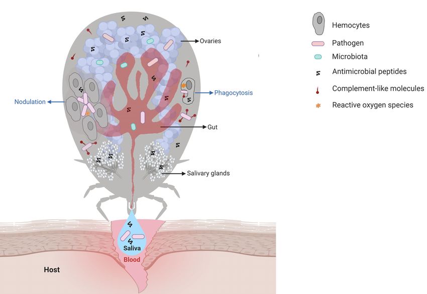
Fogaça et al.                                                                                                                                    Tick Immune System

 FIGURE 1 | Main interactions among tick immune system components, microbiota, and pathogens. Pathogens ingested within the blood meal initially reach the tick
 gut, where they interact with components of the gut microbiota and with cytotoxic molecules, such as AMPs (hemocidins and endogenous AMPs) and possibly with
 factors of redox metabolism, despite not being fully comprised. Pathogens must colonize and/or cross the gut epithelium to reach the hemocoel, which is filled with
 hemolymph. In hemolymph, complement-like molecules attach to pathogens that can be engulfed or trapped by hemocyte-mediated processes named
 phagocytosis and nodulation, respectively. Invaders can also be killed by several types of effector molecules, including AMPs, complement-like molecules, and
 factors of redox metabolism. The tick salivary glands return excess water and ions from the blood meal to the host through saliva, which also contains
 antihemostatic and immunomodulatory molecules. Pathogens use tick saliva as a vehicle to be transmitted to the host, in which infection can be facilitated by saliva
 properties. Some pathogens can also colonize the tick ovaries and are transmitted to progeny. In the tick salivary glands and ovaries, as in the gut, pathogens must
 deal with the members of resident microbiota as well as tick immune reactions. Additional studies are required to elucidate the molecules responsible for hemolymph
 clotting and melanization in ticks.

high contents of repetitive regions make genome assembly                              types, has also contributed considerably to studies on tick biology
difficult. Indeed, the size of tick genomes is approximately 1.3                       and their interactions with TBPs (41).
Gbp in argasids and 2.6 Gbp in ixodids (36). However, the
genome of the cattle tick R. microplus is even larger and has been
estimated to be approximately 7.1 Gpb, which is more than twice                       TICK IMMUNE SIGNALING PATHWAYS
the size of the human genome (37). In addition, approximately
70% of the tick genome includes repetitive regions (37, 38). For                      Blood feeding represents a challenge for hematophagous
this reason, until very recently, only the genome of the tick Ixodes                  arthropods due to the large diversity of pathogens to which
scapularis had been annotated (38). Additional genomes were                           these animals are exposed. In contrast to other arthropods, hard
recently assembled by the use of NGS (37, 39). The scarcity of                        ticks are strictly hematophagous, feeding on the blood of their
studies on the molecular factors involved in ticks and TBPs is in                     host for several days. In addition, some species feed on a different
part due to the need for sophisticated structures to raise                            host in each developmental stage (larvae, nymphs, and adults),
vertebrate animals to feed ticks, which is laborious and                              thereby increasing the chance of either acquiring or transmitting
involves ethical concerns. In the last few years, artificial feeding                   pathogens. Therefore, ticks are important vectors of a large list of
systems have been successfully used to maintain laboratory tick                       disease-causing pathogens (42). In addition to host pathogens,
colonies; however, an animal blood source is still required (40).                     ticks are in close contact with the microbiota of the host skin,
Finally, the development of continuous cell lines derived from                        which may also be acquired within the blood meal (43). Ticks are
tick embryos, despite representing a mixture of different cell                        also exposed to microorganisms in the environment during the

Frontiers in Immunology | www.frontiersin.org                                     3                                         March 2021 | Volume 12 | Article 628054
Fogaça et al.                                                                                                                                      Tick Immune System

nonparasitic phases of their life cycle. Hence, the immune system                      Arthropoda phylum (30). Tick immunity is, however, still greatly
of ticks must be activated continuously to protect them from                           neglected and unexplored (45). Hence, we review the prevailing
harmful infections.                                                                    knowledge on tick immune signaling pathways alongside the
    Most of our knowledge on arthropod immune responses has                            connections between them and other equally important factors,
come from studies on dipteran insects, especially Drosophila and                       such as AMPs, redox metabolism, complement-like proteins, and
the mosquitoes Aedes spp. and Anopheles spp. In Drosophila,                            regulated cell death.
invading microorganisms are mainly recognized by the Toll,
immune deficiency (IMD), Jun-N-terminal kinase (JNK), Janus                             Nuclear Factor-Kappa B Signaling
kinase/signal transducer and activator of transcription (JAK/                          Pathways: Molecular Regulators for
STAT) and/or RNAi pathways (44). Nonetheless, the hypothesis                           Pathogen Recognition
that the level of conservation of arthropod immune responses                           The Unexplored Toll Pathway
might be high has been rejected by several studies on ticks, mites,                    The Toll signaling pathway is well studied in Drosophila, in
lice, hemipterans, and others, and it is now recognized that the                       which it is preferentially activated in the presence of bacterial [by
immune system displays remarkable diversification across the                            recognition of lysine-type peptidoglycan (PGN) from the cell

                               A                                          B                                        C

 FIGURE 2 | Tick signaling-related genes in the three main immune signaling pathways of arthropods: (A) Toll, (B) IMD, and (C) JAK/STAT. (A) A previous in silico
 study (31) showed that components of the Toll signaling pathway of arthropods are conserved in ticks: extracellular cytokine Spatzle (Spz), transmembrane cytokine
 receptor Toll, Toll-interacting protein (TOLLIP), adaptor protein MyD88, kinases Tube (interleukin-1 receptor-associated kinase 4 or IRAK4), Pelle (interleukin-1
 receptor-associated kinase 1 or IRAK1), Pelle-interacting protein Pellino, TNF receptor associated factors (TRAFs), evolutionarily conserved signaling intermediate in
 toll pathway (ECSIT), sterile alpha- and armadillo-motif-containing protein (SARM), Rel/NF-kappa B transcription factor Dorsal, Dorsal inhibitor protein IkappaB
 Cactus (IkB), and interacting protein Cactin of the IkB. (B) Regarding the IMD pathway, genes encoding downstream members of both the NF-kB/Relish and Jun
 N-terminal kinase (JNK) branches were identified: peptidoglycan recognition proteins (PGRPs), enzymes involved in ubiquitination (UEV1a, Effete/Ubc13 and
 Bendless/Ubc5), X-linked inhibitor apoptosis protein (XIAP), negative regulators Caspar (Fas-associating factor 1) and POSH (E3 ligase Plenty of SH3), transforming
 growth factor-beta activated kinase 1 (TAK1), TAK1-binding protein 2 (TAB2), IRD5 and Kenny/NEMO (IKKg), and Relish-like Rel/NF-kB transcription factor. The
 adaptor protein IMD (immune deficiency), its associated molecule FAAD (Fas associated protein with death domain), the caspase DREDD (death related ced-3/
 Nedd2-like) and Dnr1 (defense repressor 1) have not yet been described in ticks. Components of the JNK branch of the tick IMD pathway include mitogen-activated
 protein (MAP) kinase hemipterous (HEP), Jun-kinase basket (BSK), activator protein 1 (AP-1) transcription factors JRA (Jun-related antigen) and KAY (Fos-related
 antigen, Kayak). Some IMD pathway components were functionally characterized by (48) (Insert). The authors showed that the IMD pathway is activated by PODAG
 (1-palmitoyl-2-oleoyl diacylglycerol) or POPG (1-palmitoyl-2-oleoyl-sn-glycero-3-phosphoglycerol). Once activated, XIAP interacts with the heterodimer Bendless :
 UEV1a, leading to the ubiquitination of p47 in a K63-dependent manner. Ubiquitylated p47 connects to Kenny (also named NEMO) and induces the phosphorylation of
 IRD5 and Relish. (C) Components of the Janus kinase/signal transducer and activator of transcription (JAK/STAT) signaling pathway are also conserved in ticks: the
 transmembrane cytokine receptor Domeless, tyrosine kinase JAK (Hopscotch), transcription factor STAT, signal transducing adaptor molecule (STAM) and the inhibitor
 proteins PIAS (protein inhibitor of activated STAT) and SOCS (suppressor of cytokine signaling). The ligand of the Domeless receptor (UPD gene) was not identified in
 ticks (C). Activated transcription factors are represented in dark blue; the immune signaling pathway components not yet described in ticks are represented in green.

Frontiers in Immunology | www.frontiersin.org                                      4                                          March 2021 | Volume 12 | Article 628054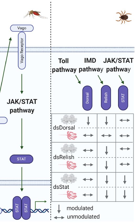
Fogaça et al.                                                                                                                                       Tick Immune System

wall of Gram-positive bacteria) and fungal (by recognition of                           components. These results suggest that A. marginale may
(1,3)-glucan polymers of D-glucose from the cell wall] pathogen-                        downregulate Toll pathway components in an attempt to favor
associated molecular patterns (PAMPs) (44, 46). In silico and                           vector colonization, which might correspond to coevolutionary
genomic analyses have shown that ticks encode most Toll                                 adaptation. Of note, similar results were found for the IMD, JNK,
pathway components (31, 33, 38, 47) (Figure 2A), including                              and JAK/STAT signaling pathways (31). However, studies on the
the NF-kB Dorsal, indicating that conserved mechanisms of Toll                          mechanisms used by this pathogen to overcome tick immune
pathway activation may exist. Indeed, the NF-kB transcription                           responses are warranted to confirm the authors’ hypothesis. In
factor dorsal-related immunity factor (DIF) is the only                                 adult R. microplus, only Dorsal was downregulated in both the gut
component of the Toll pathway not yet reported in any                                   and salivary glands of A. marginale-infected ticks, while Relish and
tick species.                                                                           STAT remained unmodulated (49). Moreover, Dorsal silencing
    How the tick Toll pathway operates is largely unclear. Rosa and                     promoted an increase in the A. marginale burden as well as
collaborators showed that the Toll pathway components are                               knockdown of Relish and STAT. However, while Relish dsRNA
differentially expressed in the tick cell line BME26, which is                          (dsRelish) specifically silenced Relish, this transcription factor was
derived from the tick R. microplus, in response to live                                 also downregulated in both the dsDorsal and dsSTAT groups,
Anaplasma marginale and Rickettsia rickettsii (two obligate                             which might explain the increase in the A. marginale load in these
intracellular bacteria) and heat-killed Saccharomyces cerevisiae                        two groups as well. To determine the pathway responsible for
(yeast), Enterobacter cloacae (Gram-negative bacterium) and                             infection control, the gene expression of specific effectors of each
Micrococcus luteus (Gram-positive bacterium) (31). Interestingly,                       immune signaling pathway, which are currently unknown, is
heat-killed microorganisms upregulated the gene expression of the                       warranted. As Dorsal-, Relish-, and STAT-encoding genes do
majority of the Toll pathway components, R. rickettsii upregulated                      not exhibit significant sequence similarity, the authors suggested
some Toll pathway components and downregulated others, and                              the existence of putative crosstalk among the Toll, IMD, and JAK/
infection with A. marginale (a pathogen naturally transmitted by                        STAT signaling pathways (49) (Figure 3C). Nonetheless, an off-
R. microplus) downregulated most of the Toll pathway                                    target effect cannot be ruled out. It is also possible that the

                       A                                               B                                         C

 FIGURE 3 | Immune pathway crosstalk. (A) In Drosophila, the DIF-Relish heterodimer activates the expression of both Toll and IMD pathway effectors, resulting in a
 stronger response against infection (50). (B) In Culex, after recognition of West Nile virus (WNV) dsRNA by Dcr-2, TNF receptor-associated factor (TRAF) stimulates
 Relish, upregulating Vago expression (51, 52). Vago is then secreted by the infected cell and activates the JAK/STAT pathway in adjacent cells, upregulating the
 expression of antiviral genes. (C) In R. microplus, knockdown of Dorsal downregulates both Dorsal and Relish expression in salivary glands, while the levels of all the
 transcription factors remain unaltered in the gut. Relish is also downregulated in the gut and salivary glands of STAT-deficient ticks. Conversely, knockdown of Relish
 results in the specific silencing of its target gene in both the gut and salivary glands (49).

Frontiers in Immunology | www.frontiersin.org                                       5                                         March 2021 | Volume 12 | Article 628054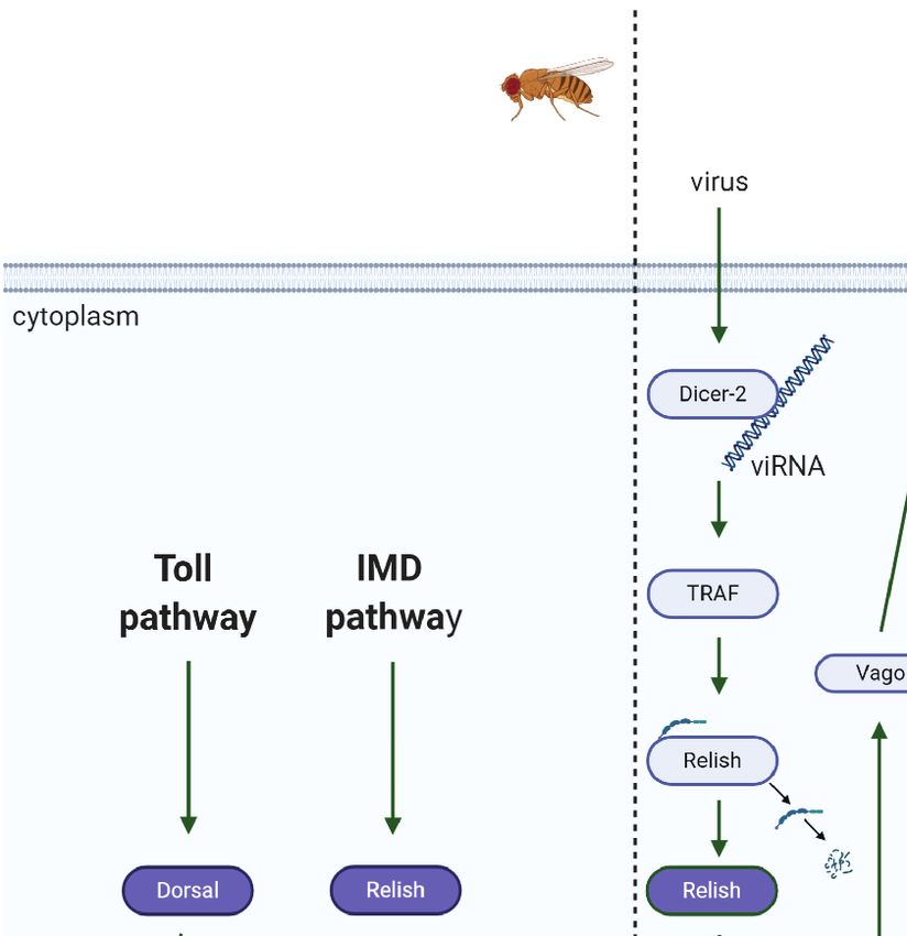
Fogaça et al.                                                                                                              Tick Immune System

knockdown of immune signaling transcription factors exerts an            RNAi silencing of several IMD pathway components, including
effect on the gut microbiota, which, in turn, may modulate their         Bendless, ubiquitin E2 variant 1A (UEV1a), Relish, and Caspar
gene expression. In contrast to the results obtained with R.             (Figure 2B, insert), showed that this cascade controls A.
microplus, gene silencing of Toll (ISCW018193) did not exhibit           phagocytophilum and B. burgdorferi burden in I. scapularis
any effect on the Anaplasma phagocytophilum burden in the                nymphs (48). In contrast to the classical Drosophila model of
salivary glands of I. scapularis nymphs (53). However, gut               DAP-PGN recognition by PGRPs (44, 55), glycerophospholipids
colonization was not evaluated; therefore, it is not possible to         from bacterial membranes, including 1-palmitoyl-2-oleoyl-sn-
guarantee that the Toll pathway is not involved in controlling A.        glycerol-3-phosphoglycerol (POPG) and 1-palmitoyl-2-oleoyl
phagocytophilum infection in this tick species. In a study carried       diacylglycerol (PODAG), were reported to act as PAMPs for
out with I. ricinus cells (IRE/CTVM20), it was shown that the            IMD pathway activation in ticks (48) (Figure 2B, insert).
expression of a Toll gene (homologous to the Toll ISCW022740 of          However, the mechanisms of POPG and PODAG recognition
I. scapularis) is upregulated after 72 and 120 h of infection with       remain unclear, but it is hypothesized that they are sensed by a
flaviviruses [tick-borne encephalitis virus (TBEV) and louping ill        yet uncharacterized pattern-recognition receptor. X-linked
virus (LIV)] but remained unmodulated in response to A.                  inhibitor of apoptosis protein (XIAP) is an upstream signaling
phagocytophilum (54). Conversely, infection with these                   component of the IMD pathway and, when activated, specifically
flaviviruses downregulated the expression of three other Toll             and directly interacts with the heterodimer E2 conjugating
transcripts (homologous to ISCW017724; ISCW007727;                       enzyme complex Bendless : UEV1a (48). Upon microbial
ISCW007724 of I. scapularis), while Toll ISCW00727 expression            activation, XIAP, together with Bendless : UEV1a, binds and
was downregulated by A. phagocytophilum (54). The expression of          ubiquitylates its p47 substrate in a K63-dependent manner.
another component of the Toll pathway, MyD88, was also                   Ubiquitylated p47 connects to Kenny (also named NEMO)
downregulated by infection with these three pathogens,                   and induces, by a yet unknown mechanism, phosphorylation
suggesting that they might suppress this pathway to promote              of the inhibitor of NF-kB kinase (IKK) b (also known as IRD5)
vector colonization. To confirm this hypothesis, it is necessary to       and Relish, the IMD transcription factor. Consequently, Relish is
functionally characterize the role played by Toll components in          cleaved and translocated to the nucleus (58) (Figure 2B, insert).
pathogen proliferation.                                                  On the other hand, RNAi knockdown of two other components
                                                                         of the IMD pathway, transforming growth factor-b activated
The Unconventional Immune Deficiency Pathway                              kinase 1 (TAK1) and TAK1 adaptor protein 1 (TAB1) (Figure
In Drosophila, bacterial infections caused by Gram-negative              2B), presented no effect on the A. phagocytophilum burden in the
bacteria and certain Gram-positive bacteria, such as Bacillus            salivary glands of I. scapularis nymphs (53). Therefore, studies
and Listeria species, are mainly controlled by the IMD pathway           carried out by Dr. Pedra’s group (48, 58) showed how the IMD
through the recognition of diaminopimelic acid (DAP)-type                pathway is activated in ticks, which is highly relevant since there
PGN, which is present in the bacterial cell wall, by PGN-                is a lack of components in this pathway, different from the classic
recognition proteins (PGRPs) (44, 55). Genomic and in silico             Drosophila model (44). However, the effector molecule(s)
studies have shown that ticks lack orthologs of many key                 regulated by the IMD pathway that control(s) infections by
elements of the IMD pathway, including the transmembrane                 pathogens such as A. phagocytophilum and B. burgdorferi still
PGRP, the Fas-associated protein with death domain (FADD),               need to be identified.
the adaptor molecule IMD, and the death-related ced-3/Nedd2-                 In adult R. microplus, RNAi silencing of immune signaling of
like protein (DREDD) (31, 33, 38, 48) (Figure 2B). Losses of             the Toll, IMD, and JAK/STAT pathway transcription factors
IMD pathway components are not exclusive to ticks since they             identified the IMD pathway as the main controller of A.
have also been described in other arachnids and hemipterans (30,         marginale infection in the tick gut and salivary glands (49).
56). Nevertheless, it is important to highlight that some                The expression of the genes encoding the AMPs microplusin,
arthropods have unusual gene architectures, resulting in                 defensin, ixodidin, and lysozyme was analyzed in the gut and
inaccurate annotation due to the use of software based on                salivary glands of R. microplus after knockdown of Relish and
standard gene structures, as reported for the kissing bug                infection with A. marginale. Interestingly, only the microplusin
Rhodnius prolixus (57). Gathering data from the genome and               transcript levels were downregulated in dsRelish ticks,
transcriptome associated with reciprocal BLAST (Basic Local              implicating this AMP as an effector of the IMD signaling
Alignment Search Tool) and hidden Markov model profile                    pathway, which may act against A. marginale (49). However,
searches, the authors showed that most of the missing IMD                although microplusin appears to be under IMD pathway
pathway components are present in this hemipteran. Therefore,            regulation, possible coregulation by the JAK/STAT pathway
it is possible that the missing IMD pathway components might             cannot be discarded (49).
be a consequence of incorrect annotations due to structural                  The other branch that constitutes the IMD pathway is JNK
divergences. Indeed, assays showed that the IMD cascade is               signaling (Figure 2B). In Drosophila, JNK has been shown to be
functional in the insect fat body and is predominantly responsive        involved in a wide range of biological processes, including
against Gram-negative bacterial infection (57).                          cellular immune and stress responses, but it seems to not be
    Despite missing several elements, the tick IMD pathway is            required to induce AMP gene expression (59). Although
functional and responsive to distinct pathogens (48, 49, 58).            activation of both the JNK and Relish branches of the IMD

Frontiers in Immunology | www.frontiersin.org                        6                                  March 2021 | Volume 12 | Article 628054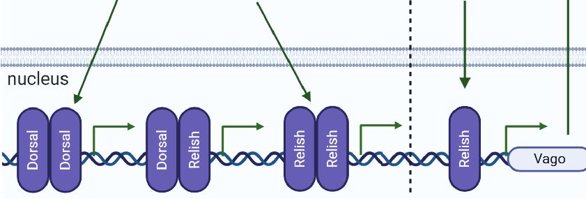
Fogaça et al.                                                                                                                  Tick Immune System

pathway occurs via TAK1 in Drosophila (59), additional studies                another pathogen naturally transmitted by I. scapularis (64).
are warranted to determine the activation of JNK pathways in                  Infection with A. phagocytophilum upregulates a tick antifreeze
ticks (48, 53, 58).                                                           glycoprotein, which, in turn, alters bacterial biofilm formation
                                                                              and, consequently, disturbs the natural gut microbiota. This
JAK/STAT Pathway: Just a Support                                              microbiota alteration affects the integrity of the peritrophic
Molecular Circuit?                                                            matrix, favoring pathogen colonization (64). Knockdown of
In Drosophila, the JAK/STAT signaling pathway only plays an                   peritrophin-1 and, therefore, the reduction in the thickness of
indirect role in controlling bacterial and fungal infection.                  the peritrophic matrix increases the A. phagocytophilum load in
Therefore, this pathway is considered a support circuit to the                the tick gut (64).
Toll and IMD pathways; however, it is especially sensitive to
viral infections (60). Beyond its effects on the immune response,             RNAi as a Tick Innate Immunity
the JAK/STAT signaling pathway also regulates multiple                        Component
biological processes, including repair and renewal of the                     RNAi is a biological process that plays an important role in the
gut epithelial layer (61), a function that was also reported to               defense of arthropods against viruses and transposable elements.
occur in ticks (62).                                                          Four main RNAi-related pathways have been described based on
    Although still poorly understood, the tick JAK/STAT                       the origin of the activating small RNAs. The origin of three of
pathway (Figure 2C) was reported to be functional, playing an                 these small RNAs is endogenous [microRNA (miRNA), small
important role in the control of pathogens (53, 62, 63). However,             interfering RNA (endo-siRNA), and piwi-interacting RNA
it is not clear how ticks activate the JAK/STAT signaling                     (piRNA)], while the origin of the fourth is exogenous (siRNA)
pathway, as the unpaired (Upd) encoding gene, a cytokine-like                 (65). The exogenous siRNA pathway is especially important and
signaling molecule ligand of the transmembrane receptor                       has been proposed to be the main antiviral response in
Domeless, is missing. In I. scapularis, knockdown of the                      Drosophila and mosquitoes (66). In general, after infection,
transcription factor STAT and JAK yielded evidence that this                  long viral dsRNA is recognized and cleaved by Dicer-2 (Dcr-2)
pathway is key to the control of A. phagocytophilum infection                 into 21 nucleotide (nt) siRNAs, known as viRNAs (65, 66). These
(53). The results also showed that the 5.3-kDa AMP is an effector             viRNAs are then transferred to Argonaute-2 (Ago2), which
regulated by the JAK/STAT pathway, which is essential to restrict             couples to other members of the RNA-induced silencing
A. phagocytophilum proliferation in tick salivary glands and                  complex (RISC). Only one strand of the viRNA remains
hemolymph but not in the gut, indicating that additional                      coupled to RISC and guides the degradation of complementary
effectors under JAK/STAT pathway regulation are required in                   viral RNA (65, 66). miRNAs use a similar mechanism, although
this organ (53). Interestingly, it was reported that I. scapularis            involving Dcr-1 and Ago-1 (67).
employs a sophisticated immune strategy that uses a vertebrate                   The genome of I. scapularis exhibits significant gene
host-derived cytokine to stimulate its own JAK/STAT immune                    expansion in RNAi elements, including five Ago homologous
pathway (63). During feeding, the interferon-gamma (INFg)                     genes: Ago-78, homologous to insect Ago-1, and Ago-96, -68, -16,
acquired within the infected bloodmeal activates STAT by a                    and-30, homologous to insect Ago-2 (68). Additionally, two Dcr
yet unknown receptor and, through mediation of a Rho-like                     genes, Dcr-89 and -90, were clustered with Drosophila Dcr-2 and
GTPase, leads to the synthesis of the AMP domesticated amidase                -1, respectively. Similar gene expansion was identified in
effector 2 (Dae2), limiting the level of B. burgdorferi. Other                Hyalomma asiaticum RNAi components (viz., two copies of
evidence that indicates that the JAK/STAT pathway is associated               Dcr-2 and five copies of Ago-2) (69). Infection of I. scapularis
with the regulation of AMPs was reported by Capelli-Peixoto                   IDE8 cells with Langat virus (LGTV) showed that Ago-16 and
and collaborators in adult R. microplus (49). The authors                     Ago-30 neutralized both LGTV and its replicon, as well as Dcr-
observed the downregulation of the AMPs ixodidin and                          90, despite the clustering of the last element with insect Dcr-1,
lysozyme in the salivary glands and defensin in the gut and                   which is involved in miRNA processing but not in siRNA (68).
salivary glands of STAT-deficient ticks.                                       Shortly thereafter, knockdown of Ago-30 and Dcr-90 confirmed
    Effectors from signaling pathways, such as JAK/STAT, can act              their antiviral role upon LGTV infection in I. scapularis IDE8
as either positive or negative regulators of infection. As presented          and I. ricinus IRE/CTVM19 cell lines (70).
above, Dae2 (63) and the 5.3-kDa AMP (53) are negative                           Interestingly, viral or endogenous siRNAs were shown to be
regulators, as they control pathogen proliferation. In contrast,              mostly 22 nt in length depending on the tick (68), in contrast
peritrophin-1, another effector from the tick JAK/STAT pathway,               with Drosophila and mosquito viRNAs and endo-siRNAs, which
was reported to increase B. burgdorferi survival in the gut of                contain 21 nt (66). Moreover, these viRNAs mapped at the
I. scapularis nymphs (62). Knockdown of STAT had a direct                     highest frequency around the 5’ and 3’ UTRs of the viral
impact on the gut epithelium, affecting its mitotic activity as well as       genome and antigenome (68). The 3’ UTRs of LGTV and TBEV
decreasing peritrophin-1 expression, which consequently                       express subgenomic flavivirus RNAs (sfRNAs), which are a
disrupted the structural integrity of the peritrophic matrix (62).            counterdefense against the tick RNAi system, assuring vectorial
Therefore, peritrophin-1, which is a component of the peritrophic             competence (68). Of note, sfRNAs are expressed by almost all
matrix, favors B. burgdorferi establishment (62). Interestingly,              Flaviviridae members as an evolved balance between arthropods
peritrophin-1 exhibits the opposite effect on A. phagocytophilum,             and viruses (67).

Frontiers in Immunology | www.frontiersin.org                             7                                 March 2021 | Volume 12 | Article 628054
Fogaça et al. Tick Immune System
Grubaugh and collaborators (71) validated the in vitro data activation of effectors from both pathways and, consequently,
previously obtained by Schnettler et al. (68), showing that most targeting a broader spectrum of infectious microorganisms (50).
viRNAs are, indeed, 22 nt in length and originate from the UTR Another example of a certain effector being regulated by more
of the viral genome and antigenome of I. scapularis in its life than one immune pathway occurs in the hemipteran stinkbug
stages (larvae, nymphs, and adults) naturally infected with Plautia stali (56). As shown by Nishide and collaborators,
Powassan virus (POWV) (71). Moreover, the viral genetic knockdown of IMD, as well as Toll pathway components,
diversity in ticks is lower than that in mice, suggesting that modulates effectors of both pathways. Interruption of both
ticks exert stronger viral control than their vertebrate hosts. pathways at the same time had a more conspicuous effect on
Therefore, POWV evolution seems to depend on RNAi- AMP production, strengthening crosstalk (56). The authors
mediated diversification and selective constraints (71). proposed an intriguing hypothesis that the redundancy
Regarding endogenous miRNAs, recent studies have shown between these two immune signaling pathways may have
that pathogens, such as viruses (72) and bacteria (73), modulate predisposed them to and facilitated the loss of some IMD-
tick miRNA profiles, with a potential role in controlling related genes in P. stali.
pathogen replication within the vector (72, 73). On the other The crosstalk between RNAi and immune signaling pathways
hand, the piRNA response to infection is still unknown in ticks. has been shown in recent publications (51, 52, 78, 79). In Culex
Nonetheless, the piRNA response has been implicated in the mosquitoes, Dcr-2, a central component of the siRNA pathway,
response of mosquitoes to viral infections (74, 75). Moreover, recognizes West-Nile virus (WNV) dsRNA and activates a
Hess and colleagues (76) suggested that the mosquito piRNA signaling cascade to stimulate Relish via tumor necrosis factor
response precedes the RNAi-Dcr-2-dependent (siRNA) response (TNF) receptor-associated factor (TRAF) to increase Vago
during viral infection. In contrast with siRNAs, piRNA activation expression (Figure 3B) (51, 52). Following this transcriptional
seems to be mediated by single-stranded RNAs that are Dcr1- upregulation, Vago is secreted from infected cells and acts as a
and Dcr2-independent and possibly mediated by the vertebrate cytokine functional homolog, binding to a still
endonuclease activity of Piwi proteins, resulting in 24–30 nt unknown cellular receptor in surrounding cells and triggering
small RNAs, as found in Drosophila. In addition to antiviral the JAK/STAT pathway. Activation of the JAK/STAT pathway
activity, piRNAs seem to have important roles in controlling the ultimately results in an appropriate antiviral response in
activity of transposable elements in the genome and in uninfected cells, such as upregulation of vir-1 and other
the development of reproductive tissues (65). Considering the antiviral genes. These studies thereby revealed a paracrine
knowledge of the role played by RNAi in the defense of insects signaling response mediated by a complex network of
against infections, the tick RNAi system represents a wide and crosstalk, opening up several intriguing lines of investigation
still unexplored field awaiting investigation. for future studies on arthropod immunity. Other studies have
shown crosstalk between RNAi and the Toll pathway in Ae
Independent Immune Pathways or aegypti Aag2 cells (78) and Drosophila (79). In the first study, the
Dynamic and Indispensable Crosstalk? miRNA aae-miR-375 upregulated Cactus, inhibiting the
Although the term crosstalk is commonly applied to the arthropod activation of the NF-kB transcription factor and reducing
immunity literature, its definition remains conflicting, and in AMP synthesis, consequently enhancing dengue virus (DENV)
many cases, the mechanism by which it occurs remains infection (78). In Drosophila, on the other hand, four distinct
unknown. Here, we consider crosstalk to occur when (i) the members of the miR-310 family directly regulate drosomycin
same effector is regulated by more than one immune signaling expression, a Toll-derived AMP (79). In addition to the
pathway (50, 56, 77) and (ii) the components of a specific immune connection between RNAi and signaling pathways, the
signaling pathway modulate the components of other pathways redundancy of distinct miRNAs cotargeting the same
(49, 51, 52, 78–80). transcript highlights the tight regulation imposed by miRNAs
The regulation of AMP expression by Toll and IMD pathways on the innate response.
was initially established in Drosophila, as well documented in the It was also shown that the transcription factors activator
historical review by Imler (24). Originally, it was accepted that protein 1 (AP-1; from the JNK pathway) and STAT neutralize
AMPs were regulated by a specific immune pathway; however, Relish-mediated activation during the innate immune response
subsequent studies carried out by different research groups in Drosophila, which is necessary for a proper and balanced
showed that this regulation was more complex than initially immune response. The mechanism for controlling Relish-
known, and crosstalk among immune pathways could occur, as mediated transcriptional activation is through the formation of
described in the examples below. Although AMPs are mostly a complex composed of AP-1 and STAT with the dorsal switch
regulated by either Toll or IMD pathways in Drosophila, it has protein (Dsp1), which recruits a histone deacetylase to prevent
been reported that some AMP-encoding genes can be activated Relish transcription (80).
synergistically by both immune pathways (50) (Figure 3A). It In ticks, to the best of our knowledge, there is only one study
was shown that the NF-kB transcription factors Dorsal, DIF, and reporting putative crosstalk among the immune signaling
Relish can dimerize as homo- or heterodimers with varying pathways, which was reported by Capelli-Peixoto and
degrees of efficiency. The DIF-Relish heterodimer mediates the collaborators (49). The authors showed that knockdown of the
crosstalk between the Toll and IMD pathways, resulting in the transcription factors Dorsal, Relish, or STAT downregulates
Frontiers in Immunology | www.frontiersin.org 8 March 2021 | Volume 12 | Article 628054Fogaça et al. Tick Immune System
Relish expression (Figure 3C), with a consequent increase in the embryos before and after oviposition. Microplusin exhibits an
A. marginale load in R. microplus salivary glands. In contrast, a-helical globular domain and chelates metal ions (95). The
Dorsal-deficient ticks presented no effects on Relish expression in bacteriostatic activity of microplusin against the Gram-
the gut, where, intriguingly, ticks exhibited only modest silencing positive bacterium M. luteus was reversed by the addition of
of Dorsal itself. Relish levels were also diminished in STAT- copper II but not iron II. Indeed, microplusin interferes with
deficient guts. Only treatment with dsRelish resulted in specific the respiration (a copper-dependent process) of both M. luteus
silencing of its target gene in both the gut and salivary glands (95) and the fungus Cryptococcus neoformans (96). Microplusin
(Figure 3C). Nonetheless, the A. marginale burden was higher in was also reported to affect melanization and capsule formation,
the gut of ticks from all groups (dsDorsal, dsRelish, and dsSTAT) which are important virulence factors of C. neoformans
than in the control (49). As similarities among Dorsal, Relish, and (96). Interestingly, knockdown of microplusin increased the
STAT gene sequences were insignificant, the authors hypothesized load of R. rickettsii in Amblyomma aureolatum (97). On the
that crosstalk of the immune pathways in ticks might occur to other hand, this AMP had no effect on either rickettsial
enhance the immune response. However, an off-target effect transmission or tick fitness. Defensins compose another class of
cannot be completely disregarded. Although the regulation of AMPs that have been described in several tick species, displaying
AMPs by the IMD and JAK/STAT pathways has been established, activity against different types of microorganisms [for review,
as already described above, it is still necessary to silence AMP- see (11, 83)]. For example, defensin-2 of Dermacentor variabilis
encoding genes to assign their role in A. marginale control. was shown to protect against another bacterium of the genus
Therefore, the tick immune system, as shown in some insects, is Rickettsia, R. montanensis, as its neutralization with antidefensin-
also integrated, versatile, and possibly capable of making a 2 IgG increased the rickettisal load in the tick gut (98). Defensin-2
network of connections among innate signaling pathways, giving causes permeabilization of the bacterial membrane with
rise to effective antimicrobial responses. consequent leakage of cytoplasmic proteins (98).
Dae2 is an AMP of I. scapularis that was acquired by
horizontal bacterial gene transfer and has become an important
ANTIMICROBIAL PEPTIDES: MAY THE effector to control B. burgdorferi infection (99), although it does
“SOURCE” BE WITH YOU! not exhibit direct action on this pathogen (63). Indeed, it was
recently shown that Dae2 is physically unable to overcome the
AMPs are important effectors of the immune systems of both outer membrane structure of the outer membrane of Gram-
invertebrates and vertebrates, having a broad spectrum of negative bacteria; thus, it does not present lytic activity against
activity against microorganisms (81). In ticks, the main sites of B. burgdorferi, suggesting the need for other factors, such as
AMP expression are hemocytes, fat body, gut, ovaries, and membrane-permeabilizing agents (100). As Dae2 is delivered to
salivary glands, where they can be modulated in response to the vertebrate host bite site via saliva and exhibits strong activity
either blood feeding or microbial challenge (82). Several reviews against bacteria usually encountered in the host skin, this AMP
of tick AMPs addressing their characterization, as well as their may protect ticks from the acquisition and proliferation of host
interaction with microorganisms, have been published in the last skin microbes (100).
decade (11, 33, 83, 84). Serine proteinase inhibitors have also been reported to play a
Interestingly, ticks use host hemoglobin, one of the most role in the arthropod immune system. For instance, serine
abundant proteins within the blood meal, as a source for the proteinase inhibitors mediate both coagulation and melanization
production of antimicrobial-derived fragments (85–88). processes of hemolymph and the production of AMPs (101). In
Hemoglobin-derived AMPs, referred to as hemocidins (89), are addition, serine proteinases may also exert antimicrobial activity,
produced by the proteolytic activity of aspartic and cysteine possibly inhibiting proteinases that microorganisms use to
(catepsin-L like) proteinases from the tick gut (90). Structural colonize host tissues and evade the immune system (102). The
studies with the synthetic amidated hemocidin Hb33-61a of R. first report of a tick serine proteinase inhibitor with antimicrobial
microplus showed that its a-helical C-terminus is responsible for properties was the ixodidin of R. microplus (103), which presents
the permeabilization of the microbial membrane (91). However, the key features of trypsin inhibitor-like domain proteins (104).
it is still unknown whether hemocidins act intracellularly or if Interestingly, one Kunitz inhibitor was reported to control
they are released to the tick gut lumen, where they can fight R. montanensis infection in the gut of D. variabilis (105). In
against microorganisms. contrast to defensin, D. variabilis Kunitz-type inhibitors present a
In addition to hemocidins, ticks also produce endogenous bacteriostatic effect on R. montanensis (106). Therefore, serine
(ribosomally synthesized) AMPs (11, 83). Among the several tick proteinase inhibitors are also used by ticks as powerful
AMPs identified to date, microplusins (also known as hebraeins) antimicrobial molecules.
are among the most well characterized. Microplusin is a cysteine- Despite the diverse nature of molecules used by ticks as
and histidine-rich AMP that was first isolated from the antimicrobials, little information on their synthesis regulation
hemolymph of adult R. microplus (92) and Amblyomma is available, as discussed above. Therefore, additional studies on
hebraeum (93). Microplusin was also identified in the ovaries the regulation of tick AMPs by immune signaling pathways are
and eggs of R. microplus (94), suggesting that in addition to required to better understand their role in the control of
protecting adults, it may also play a role in the protection of distinct pathogens.
Frontiers in Immunology | www.frontiersin.org 9 March 2021 | Volume 12 | Article 628054Fogaça et al. Tick Immune System
REDOX METABOLISM AS AN IMPORTANT survival and indirectly prevents the induction of borreliacidal
PLAYER IN THE INFECTION CONTROL agents in the tick gut (116).
Intriguingly, A. marginale upregulated the genes encoding
ORCHESTRA antioxidant enzymes, including superoxide dismutase, catalase,
In addition to AMPs, triggering of the production of reactive glutathione peroxidase, glutathione S-transferase, thioredoxin,
oxygen species (ROS) and reactive nitrogen species (RNS) in thioredoxin reductase, and peroxiredoxin, whereas genes
response to infection has been described in several arthropods, encoding ROS-generating enzymes, such as DUOX and
such as Drosophila (107) and mosquitoes (108). ROS have an endoplasmic reticulum oxidase, were downregulated in R.
essential role in infection-related physiological as well as microplus-derived BME26 cells (117). Conversely, R. rickettsii
pathophysiological processes, such as signaling, regulation of and heat-killed S. cerevisiae, E. cloacae or M. luteus triggered
tissue injury and inflammation, cell survival, proliferation, the opposite gene expression pattern (117). Furthermore,
differentiation, and apoptosis (109, 110). simultaneous RNAi knockdown of catalase, thioredoxin, and
In ticks, there is still little available information on ROS glutathione peroxidase, three representative members of the tick
metabolism and their impact on pathogen control. Nonetheless, antioxidant enzymatic system, as well as the oxidation resistance 1
it is recognized that hemocytes produce ROS under stimulation. (OXR1), which regulates the expression of ROS detoxification
Gram-positive bacteria, zymosan, and phorbol 12-myristate 13- enzymes, decreased A. marginale infection (117). Therefore, while
acetate elicit the production of hydrogen peroxide (H2O2) and BME26 cells respond to infection, producing an oxidant
superoxide (O−2 ) by hemocytes of R. microplus (111). In contrast, environment, A. marginale seems to subvert this response to
stimulation with lipopolysaccharide (LPS), the major create an antioxidant environment, which is required for its
component of the Gram-negative bacterial outer membrane, survival (117). It is possible that A. marginale manipulates R.
failed to induce ROS generation, indicating that different microplus redox metabolism (and production of immune signaling
mechanisms or roles for ROS upon infection with either pathway effectors, as aforementioned) to favor its proliferation.
Gram-positive or Gram-negative bacteria may exist (111). Additional studies are required to elucidate the mechanisms that
Further studies with R. microplus showed that cytochrome c this bacterium uses to subvert tick immune responses.
oxidase subunit III (COXIII), an enzyme of mitochondrial
electron transport complex IV involved in mitochondrial ATP
and ROS generation, is important for the transmission of A. CELL-MEDIATED IMMUNITY IN TICKS
marginale to calves (112). It is possible that COXIII knockdown
imbalances tick redox metabolism, affecting its ability to Hemocytes, which are sessile or circulating cells from arthropod
transmit this pathogen (112). The peroxiredoxin Salp25D hemolymph, are responsible for several immune responses.
from I. scapularis had no effect on the transmission of B. The nomenclature of hemocytes varies considerably depending
burgdorferi but instead played a role in spirochete acquisition on the arthropod species and/or the approaches of the study (118).
by the tick (113). RNAi-mediated silencing of Salp25D affects Earlier morphological, ultrastructural, and physiological studies of
bacterial acquisition by ticks fed on B. burgdorferi-infected mice. the hemocyte repertoire in different tick species consistently
The same effect was obtained when ticks were fed on Salp25- reported the presence of three basic types of hemocytes, namely,
immunized mice (113). It is possible that Salp25 may detoxify phagocytic plasmatocytes and granulocytes and nonphagocytic
ROS at the tick feeding site and gut, thus affording a survival granulocytes (119–121). These cells apparently differentiate from
advantage to B. burgdorferi. rarely occurring prohemocytes (120, 122). More recent studies
In the mosquito An. gambiae, an extracellular matrix have described additional types of tick hemocytes, namely,
crosslinked by dityrosine covalent bonds catalyzed by dual adipohemocytes in Rhipicephalus sanguineus (123) and
oxidase (DUOX) and heme peroxidase is located in the gut spherulocytes and oenocytoids in R. microplus (122). The most
ectoperitrophic space (between the epithelial cell layer and the important immune responses of arthropod hemocytes are
peritrophic matrix). This extracellular matrix acts as an additional phagocytosis, encapsulation, nodulation (which involves
physical barrier to decrease gut permeability to bacterial PAMPs, melanization by the PPO cascade), coagulation, and production
impairing immune response activation by the resident microbiota of immune-related molecules.
(114). Importantly, the dityrosine network also provides a The role of tick hemocytes in the phagocytosis of a variety of
favorable environment for Plasmodium development, as it microbes, including bacteria, yeast, spirochetes, and foreign
prevents the activation of nitric oxidase synthase (iNOS), a particles, has been investigated by several studies [for example,
nitric oxide-generator enzyme (114). iNOS is responsible see (111, 124–127)]. By contrast, very little is known about the
for parasite nitration, a key step in the action of the encapsulation and nodulation mechanisms. Indeed, there is only
antiplasmodium complement-like molecule TEP1. Later, it was one report on encapsulation (128) and one on nodulation (129),
shown that the heme peroxidase 2/NADPH oxidase 5 system both in D. variabilis. After the inoculation of ticks with
plays a central role in epithelium nitration, therefore potentiating Escherichia coli, hemocytes did not form circular layers but
the antiparasitic effect of nitric oxide (115). Similar to An. gambiae aggregated around the bacteria, which is a characteristic
(114), an extracellular matrix was described in the tick feature of nodule formation (129). As the encapsulation study
I. scapularis, which acts as a shield that favors B. burgdorferi was performed using an implant of Epon−Araldite under the tick
Frontiers in Immunology | www.frontiersin.org 10 March 2021 | Volume 12 | Article 628054Fogaça et al. Tick Immune System
cuticle, it is still unknown whether it also occurs against in vitro assays are needed to ultimately resolve the question of the
microorganisms (128). existence of hemolymph clotting in ticks.
In invertebrates such as insects and crustaceans, hemocytes Tick hemocytes (83), as well as the hemocytes of other arthropods,
produce components of the melanization response, which such as mosquitoes (142), also produce a series of immune-
involves an enzymatic cascade referred to as the PPO related molecules. Intriguingly, the hemocytome of I. ricinus
activating system, ultimately resulting in the production of showed that only 1.48% of the 15,716 coding sequences (CDSs)
melanin (130, 131). This process can be locally activated by identified were related to immune factors (143). Of the identified
cuticle injury or systemically triggered by microbial invasion of CDSs, 327 were five times more highly expressed in hemocytes
the hemocoel. Interestingly, in the cotton bollworm Helicoverpa than in salivary glands and the gut, among which 11 encode
armigera, infection with the baculovirus HearNPV decreased immune factors, including AMPs and proteins involved in
the levels of the majority of PPO cascade components, while pathogen recognition. As presented in this section, hemocytes
serpin-9 and serpin-5 (which were also shown to regulate the are versatile components of the arthropod immune system that
proteases cSP4 and cSP6, respectively) were increased (132). In play diverse and key roles. The principal insect tissue that
addition, in vitro assays showed that hemolymph melanization produces the majority of soluble immune molecules in
can kill baculovirus, an effect abolished by the specific PO hemolymph is the fat body (44). The role of tick fat body in
inhibitor phenylthiourea. Together, the results suggest that the tick immune system requires further investigation.
baculovirus inhibits the melanization response to ensure its
survival in H. armigera (132). There is no evidence of the
existence of the PPO cascade in ticks based on available THE PRIMORDIAL COMPLEMENT
genomic and transcriptomic data. In line with this, no PPO SYSTEM OF TICKS
activity has been reported to be present in the hemolymph of
the hard ticks Amblyomma americanum, D. variabilis, and One important branch of both cellular and humoral innate
I. scapularis (133). In contrast, two studies reported PPO-like immunity in vertebrate and invertebrate metazoan organisms
activity using L-DOPA as a substrate in the hard tick R. is carried out by the complement system. In higher vertebrates,
sanguineus (134) and in the soft tick Ornithodoros moubata the complement system is composed of approximately thirty
(135). However, the enzymes responsible for such activity have components arranged in classical, lectin, and alternative
not yet been identified, and enzymatic assays did not employ pathways, which recognize foreign cells (microbes), specifically
phenylthiourea as a control. tag them via opsonization, and ultimately, eliminate them via
Coagulation is another important immune response of phagocytosis or cell lysis (144). The common denominator of all
arthropods. The final product of coagulation is a protein clot, three pathways is the proteolytic activation of the central C3
which is essential to avoid the loss of hemolymph in cases of an complement component. The occurrence of this molecule can be
injury and the spread of an invader microorganism throughout traced back in most ancient invertebrates, such as horseshoe
the hemocoel (136). In horseshoe crabs, the clotting process crabs (subphylum Chelicerata, class Merostomata), implying
involves a serine-protease cascade that leads to the activation of that an ancestor of the complement system existed on Earth
the clotting enzyme that converts the coagulogen into the for more than 500 mil. years (145, 146). For ticks, which are also
insoluble clot (137), while in crayfish, the process depends on chelicerates, advanced knowledge of the primitive complement
direct transglutaminase (TG)-mediated cross-linking of a system of horseshoe crabs gathered during the past two decades
specific plasma protein homologous to vitellogenins (19, 136). presents the best matching comparative model (137, 145, 147).
TG is also involved in the final step of coagulation in horseshoe Microbial pattern recognition by the vertebrate lectin
crabs, cross-linking coagulin with hemocyte surface proteins pathway is mediated by multimeric mannose-binding lectins
named proxins (138). Interestingly, factors of the coagulation (MBLs) or ficolins. The horseshoe crab counterparts of
cascade interact with hemocyanin, causing it to present PO mammalian ficolins are lectins named tachylectin-5 or
activity in the horseshoe crab Tachypleus tridentatus, carcinolectin-5 (148–150). These lectins share a fibrinogen-
demonstrating crosstalk between melanization and coagulation related protein (FRED) with ficolins but lack the N-terminal
cascades (139). In Drosophila, coagulation and PO activity were collagen-like domain responsible for forming complexes
also described to be tightly associated (140). Wound sealing in with MBL-associated serine proteases (MASPs) (151), which
flies involves two steps: in the first step, TG-mediated are absent in arthropods (146). The lectin Dorin M, purified
crosslinking of hemolymph proteins occurs, and in the second from the plasma of the soft tick O. moubata (152), was shown to
step, PO-dependent crosslinking takes place, hardening the clot be a clear ortholog of the horseshoe crab tachylectins-5 (153),
and producing melanin. In ticks, putative coagulation was and similarly to ficolins and tachylectins, it forms high
uniquely reported for D. variabilis, where a fibrous matrix was molecular weight multimers in the native state (152). The
observed around an inert implant (128). TGs and proclotting search for homologous lectins in I. ricinus (154) and in the
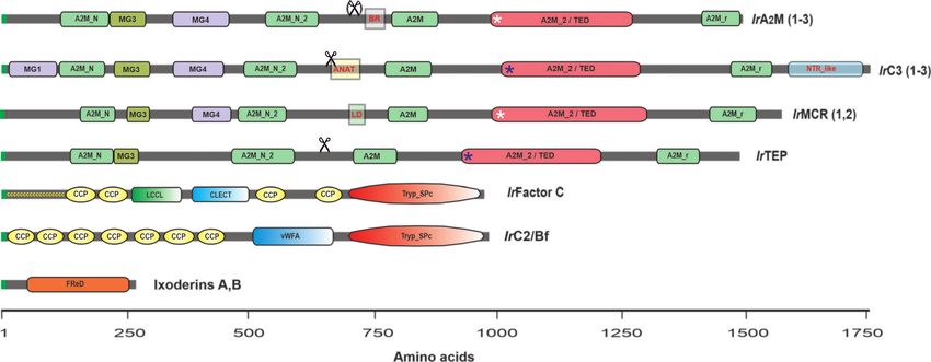
enzyme precursors have been detected in tick genomes (33). genome of I. scapularis (155) revealed the existence of two
Moreover, an injury-responsive multidomain serine protease phylogenetically distinct families, further referred to as ixoderin
homologous to Limulus Factor C has been characterized in I. A and ixoderin B (Figure 4). Ixoderin A is mainly present in
ricinus (141). Therefore, additional studies based on appropriate plasma and is responsible for the hemagglutination of mouse
Frontiers in Immunology | www.frontiersin.org 11 March 2021 | Volume 12 | Article 628054You can also read