Treating Bladder Cancer: Engineering of Current and Next Generation Antibody-, Fusion Protein-, mRNA-, Cell- and Viral-Based Therapeutics - TUprints
←
→
Page content transcription
If your browser does not render page correctly, please read the page content below
REVIEW
                                                                                                                                                  published: 27 May 2021
                                                                                                                                          doi: 10.3389/fonc.2021.672262

                                              Treating Bladder Cancer:
                                              Engineering of Current and Next
                                              Generation Antibody-, Fusion
                                              Protein-, mRNA-, Cell- and
                                              Viral-Based Therapeutics
                                              Jan P. Bogen 1,2*, Julius Grzeschik 2, Joern Jakobsen 3, Alexandra Bähre 3, Björn Hock 4
                                              and Harald Kolmar 1*
                                              1 Institute for Organic Chemistry and Biochemistry, Technical University of Darmstadt, Darmstadt, Germany, 2 Ferring

                                              Darmstadt Laboratory, Biologics Technology and Development, Darmstadt, Germany, 3 Ferring Pharmaceuticals,
                                              International PharmaScience Center, Copenhagen, Denmark, 4 Global Pharmaceutical Research and Development,
                          Edited by:
                                              Ferring International Center S.A., Saint-Prex, Switzerland
                       Ja Hyeon Ku,
            Seoul National University,
                        South Korea
                                              Bladder cancer is a frequent malignancy and has a clinical need for new therapeutic
                        Reviewed by:
                                              approaches. Antibody and protein technologies came a long way in recent years and new
                      Massimo Fantini,
             Precision Biologics, Inc.,       engineering approaches were applied to generate innovative therapeutic entities with
                         United States        novel mechanisms of action. Furthermore, mRNA-based pharmaceuticals recently
                          Anja Rabien,
   Charité Universitätsmedizin Berlin,
                                              reached the market and CAR-T cells and viral-based gene therapy remain a major
                             Germany          focus of biomedical research. This review focuses on the engineering of biologics,
                           Kisung Ko,
                                              particularly therapeutic antibodies and their application in preclinical development and
  Chung-Ang University, South Korea
                                              clinical trials, as well as approved monoclonal antibodies for the treatment of bladder
                *Correspondence:
                     Harald Kolmar            cancer. Besides, newly emerging entities in the realm of bladder cancer like mRNA, gene
    Harald.Kolmar@TU-Darmstadt.de             therapy or cell-based therapeutics are discussed and evaluated. As many discussed
                     Jan P. Bogen
          Bogen@Biochemie-TUD.de
                                              molecules exhibit unique mechanisms of action based on innovative protein engineering,
                                              they reflect the next generation of cancer drugs. This review will shed light on the
                   Specialty section:         engineering strategies applied to develop these next generation treatments and
         This article was submitted to
              Genitourinary Oncology,
                                              provides deeper insights into their preclinical profiles, clinical stages, and ongoing trials.
               a section of the journal       Furthermore, the distribution and expression of the targeted antigens and the intended
                 Frontiers in Oncology        mechanisms of action are elucidated.
        Received: 25 February 2021
           Accepted: 11 May 2021              Keywords: antibody engineering, protein engineering, bladder cancer, urothelial carcinoma, ADC, immunotherapy,
           Published: 27 May 2021             immuno-oncology, recombinant viral vector vaccines

                            Citation:
              Bogen JP, Grzeschik J,
   Jakobsen J, Bähre A, Hock B and
                                              INTRODUCTION
   Kolmar H (2021) Treating Bladder
      Cancer: Engineering of Current
                                              The genitourinary system encompasses reproductive organs and the urinary system. The latter
     and Next Generation Antibody-,
       Fusion Protein-, mRNA-, Cell-
                                              comprises the kidneys, which are being connected to the bladder in the lower pelvis via the ureters.
       and Viral-Based Therapeutics.          The bladder is a hollow organ, which releases upon muscle contraction urine via the urethra.
            Front. Oncol. 11:672262.          Bladder cancer (BC) is the second most common genitourinary malignant disease and the 10th most
     doi: 10.3389/fonc.2021.672262            common cancer, causing nearly 570.000 new cases and 210.000 deaths worldwide each year (1, 2). It

Frontiers in Oncology | www.frontiersin.org                                          1                                           May 2021 | Volume 11 | Article 672262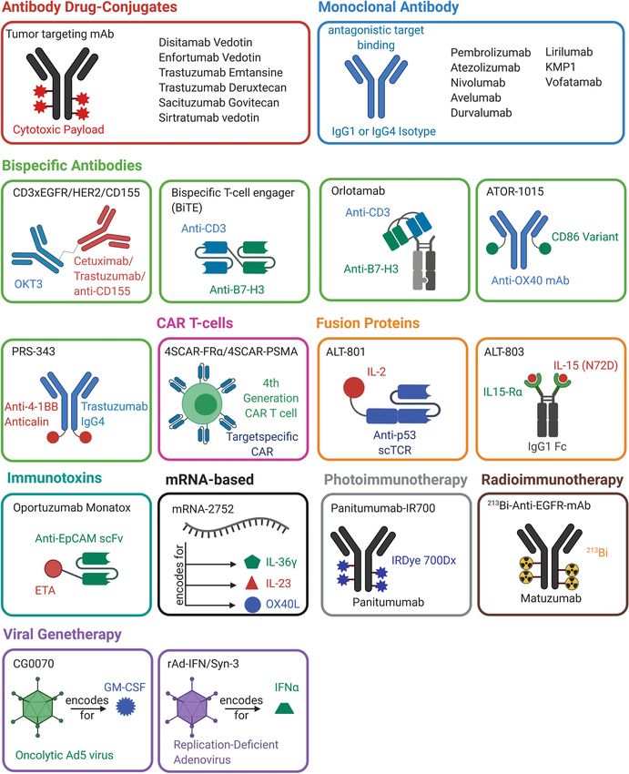
Bogen et al.                                                                                                                  Engineering Therapeutics for Bladder Cancer

typically affects older adults, and peak incidence occurs in the                         invaded the inner or the outer detrusor muscle, and at T3, the
seventh and eighth decades of life. Males are four times more                            tumor has invaded beyond the muscle layer and the surrounding
likely to develop bladder cancers. Hence BC is the sixth most                            fat tissue. At T4, the tumor has begun to grow through the
common cancer in males and the ninth leading cause of cancer-                            bladder wall into the pelvic or abdominal wall, invading adjacent
related deaths (2).                                                                      organs and/or started to spread to nearby lymph nodes or even to
    Bladder cancer is categorized by cell type and by stage of                           more distant organs (metastasis, M1) (6).
tissue invasion. Besides urothelial carcinoma/transitional cell                              This staging system was created by the American Joint
carcinoma (UC/TCC), which is by far the most common type                                 Committee on Cancer (AJCC) and the International Union
of bladder cancer (90-95%), other malignancies as squamous cell                          Against Cancer (UICC). Histological grading follows the 2004
carcinoma (2-5%), adenocarcinoma (0.5–2%), and small-cell                                World Health Organization (WHO) grading system for flat and
carcinoma (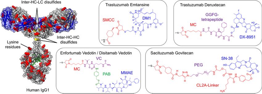
Bogen et al.                                                                                          Engineering Therapeutics for Bladder Cancer

To evaluate the cell type and stage of tissue invasion, a                    Immunotherapy approaches revolutionized cancer therapy in
Transurethral Resection of the Bladder (TURB) is performed to            the last decades. It is based on the concept of activating the
confirm the diagnosis and stage the patient correctly.                    patients’ immune system to eradicate the malignant cells. Even
    All papillary tumors are resected completely during the TURB,        though a successful biotherapy applied for approximately 40
which in some cases can be considered a cure. CIS cannot be              years, the anti-tumor mechanisms associated with BCG are not
resected completely during the TURB and thus need additional             fully understood (29). Nevertheless, BCG-mediated effects are
instillation therapy to reduce tumor burden, recurrence, and/or          presumably based on the involvement of CD4+ and CD8+
progression (4). For high and intermediate risk NMIBC patients,          lymphocytes, natural killer (NK) cells, and granulocytes, as
there is evidence of significantly reducing recurrence rate by            well as a variety of cytokines (29). As BCG-failure is observed
additional instillation therapy to the bladder of either a               in 30% to 40% of NMIBC patients (18, 19), there is a need for
chemotherapeutic agent, like mitomycin c, or Bacillus Calmette           new immunotherapy-based drugs. Different engineering
Gué r in (BCG). Single instillation (SI) of mitomycin c                 strategies and antibody formats were investigated in (pre-)
(postoperative), a potent chemotherapeutic agent which                   clinical settings over the last decades to employ new
crosslinks DNA, has been shown to reduce the 5-year recurrence           mechanism of actions. On the one hand, novel approaches
rate by 14% (59%-45%) (13). For most NMIBC patients,                     based on antibody engineering like monoclonal antibodies
intravesical immunotherapy utilizing BCG is the standard of              (mAbs), antibody-drug-conjugates (ADCs), and bispecific
care (14, 15). BCG is a non-pathogenic bacillus derived from             molecules have been explored. On the other hand, gene
Mycobacterium bovis and represents the only available vaccine for        therapeutics, mRNA- and cell-based therapies are emerging as
tuberculosis (16). Most of the time, BCG treatment starts after          promising tools to treat urologic cancers. Here we review those
TURB, aiming at preventing tumor recurrence (17).                        engineering strategies, illuminate the mechanism of action, and
    Although failure after BCG instillation therapy is observed in       discuss clinical trials (Table 1).
30% to 40% of cases (18, 19), patients who experience early
failure are defined as having BCG unresponsive NMIBC disease
(20, 21). These patients are very unlikely to benefit from further        MONOCLONAL ANTIBODIES
BCG therapy and are recommended radical cystectomy, which
describes the complete removal of the bladder. The primary               With over 90 mAbs approved for a diverse set of diseases like
treatment for patients with MIBC depends on the M (metastasis)           cancer, infectious and autoimmune diseases, monoclonal
status. For localized MIBC (M0), the primary and standard                antibodies are essential in modern medical therapy options
treatment is the complete removal of the bladder and creation            (30). While some rely on Fc-mediated effector functions like
of a urinary diversion (radical cystectomy) +/- neoadjuvant              antibody-dependent cellular cytotoxicity (ADCC) and
therapy. In very select cases, a bladder-sparing approach can            complement-dependent cytotoxicity (CDC), others achieve
be adopted. Controversies exist regarding age and type of                clinical benefit through blocking the interaction between the
diversion (5). Non-eligible patients can be offered radiotherapy.        tumor cell and the immune system, like checkpoint inhibitors.
10-15% of patients with muscle-invasive disease are already
metastatic at diagnosis (22). As first-line treatment in advanced         Checkpoint Inhibitors
stages, platinum-based chemotherapeutics, like cisplatin, are            Over the last years, immune checkpoint inhibition showed a
utilized, which interfere with DNA replication in fast-growing           massive impact on cancer therapies. Particularly antibodies
cells (23). Furthermore, intravenous application of the                  blocking the interaction of the programmed cell death protein
nucleoside-analog gemcitabine, structurally related to cytidine,         1 (PD-1) and its ligand (PD-L1) showed impressive outcomes in
and instillation of mitomycin c are chemotherapeutic treatment           clinical applications (31), resulting in the approval of six anti-
options (24). Even though bladder cancer is relatively                   PD-(L)1 antibodies (Pembrolizumab, Nivolumab, Cemiplimab,
chemosensitive, reoccurrence after first-line treatment with the          Durvalumab, Avelumab, Atezolizumab). Today, except
chemotherapeutics gemcitabine plus cisplatin results in poor             Cemiplimab, all these anti-PD-(L)1 antibodies are in clinical
prognosis (25). Resistance mechanisms include the elevated               use for UCs (32, 33).
drug efflux as well as reduced influx. Furthermore, increased                  Studies investigating the expression of PD-1 and PD-L1 in
repair of the DNA leads to drug resistance. Multiple enzymes and         UC samples showed an elevated expression level of PD-(L)1 in
repair mechanisms, including ERCC1, PARP, Nrf2, CTR1, are                high-risk tumors compared to low-risk tumors (34).
involved in the development of drug resistance (26). A detailed          Immunohistochemistry (IHC) studies on surgically resected
review on resistance of bladder cancer against conventional              urothelial cancer specimens found PD-L1 expression in 21.1%
therapies can be found elsewhere (26, 27). If treatment fails, it        of cases (35). In patients failing BCG-treatment, PD-L1
might become necessary to perform cystectomy, where the                  expression correlated with higher tumor grades. While 7% of
bladder is partly or completely removed (28). As this is a               pTa tumors expressed PD-L1, 30% of pT3/4 and 45% of CIS
radical measure with a significant impact on the quality of life          tumors exhibited PD-L1 expression. The frequent observation
for respective patients, new treatment options are necessary,            that BCG treatment strongly induces PD-L1 expression might be
allowing for a more precise treatment and circumventing side             a reason for the lost effectiveness of BCG treatment over time
effects that are observed in classical chemotherapies.                   (36). Furthermore, PD-L1 expression was associated with tumor

Frontiers in Oncology | www.frontiersin.org                          3                                   May 2021 | Volume 11 | Article 672262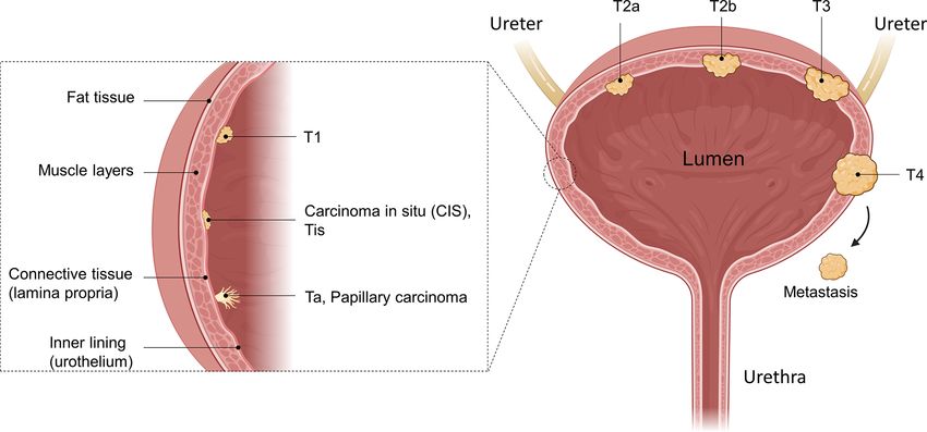
Bogen et al. Engineering Therapeutics for Bladder Cancer
TABLE 1 | Overview of clinical trials targeting bladder cancer with engineered biologics.
Drug (Target) NCT Number Combined with (or Disease Number of Phase Status
compared to) patients
(estimated
or actual
enrolled)
Monoclonal Lirilumab NCT03532451 Nivolumab Bladder Cancer 43 I Active, not
Antibody (KIR2DL) recruiting
Vofatamab NCT02401542 Docetaxel Locally Advanced or Metastatic Urothelial Cell Carcinoma, 71 I/II Terminated
(FGFR3) Urinary Bladder Disease, Urological Diseases (program
has been
put on hold
by the
sponsor)
NCT02925533 Pembrolizumab Bladder Cancer 1 I Terminated
(Terminated
due to
safety
concerns.)
NCT03123055 Pembrolizumab Locally Advanced or Metastatic Urothelial Cell Carcinoma, 28 I/II Terminated
Urinary Bladder Disease, Urological Diseases (program
has been
put on hold
by the
sponsor)
Bispecifc Orlotamab NCT02628535 / Mesothelioma, Bladder Cancer, Melanoma, Squamous 67 I Terminated
Antibody (CD3×B7-H3) Cell Carcinoma of the Head and Neck, Non-Small Cell (Business
Lung Cancer, Clear Cell Renal Cell Carcinoma, Ovarian decision
Cancer, Thyroid Cancer, Breast Cancer, Pancreatic (not for
Cancer, Prostate Cancer, Colon Cancer, Soft Tissue safety
Sarcoma reasons))
NCT03406949 Retifanlimab Advanced Solid Tumors 25 I Active, not
(formerly MGA012) recruiting
ATOR-1015 NCT03782467 / Solid Tumor Neoplasms 53 I Recruiting
(CTLA-4 ×
OX40)
PRS-343 NCT03330561 / HER2-positive Breast Cancer, HER2-positive Gastric 110 I Suspended
(HER2 × 4- Cancer, HER2-positive Bladder Cancer, HER2-positive (Partial
1BB) Solid Tumor clinical hold)
NCT03650348 Atezolizumab HER2-positive Breast Cancer, HER2-positive Gastric 45 I Suspended
Cancer, HER2-positive Bladder Cancer, HER2-positive (Partial
Solid Tumor clinical hold)
Fusion ALT-801 (anti- NCT01326871 Cisplatin/ Transitional Cell Carcinoma of Bladder, Urethra Cancer, 90 I/II Unknown
Protein p53- Gemcitabine Ureter Cancer, Malignant Tumor of Renal Pelvis
scTCR×IL2) NCT01625260 Gemcitabine non-muscle-invasive Bladder Cancer 52 I/II Unknown
ALT-803 NCT02138734 BCG non-muscle-invasive Bladder Cancer 596 I/II Recruiting
(IL15-N72D: NCT03022825 BCG Bladder Cancer 183 II Recruiting
IL15Ra-Fc) NCT03228667 ALT-803 Non-Small Cell Lung Cancer, Small Cell Lung Cancer, 636 II Recruiting
+Pembrolizumab, Urothelial Carcinoma, Head and Neck Squamous Cell
ALT-803 + Carcinoma, Merkel Cell Carcinoma, Melanoma, Renal Cell
Nivolumab, Carcinoma, Gastric Cancer, Cervical Cancer,
ALT-803 + Hepatocellular Carcinoma, Microsatellite Instability,
Atezolizumab, Mismatch Repair Deficiency, Colorectal Cancer
ALT-803 +
Avelumab,
ALT-803 +
Durvalumab,
ALT-803 +
Pembrolizumab +
PD-L1 t-haNK,
ALT-803 +
Nivolumab + PD-L1
t-haNK,
(Continued)
Frontiers in Oncology | www.frontiersin.org 4 May 2021 | Volume 11 | Article 672262Bogen et al. Engineering Therapeutics for Bladder Cancer
TABLE 1 | Continued
Drug (Target) NCT Number Combined with (or Disease Number of Phase Status
compared to) patients
(estimated
or actual
enrolled)
ALT-803 +
Atezolizumab + PD-
L1 t-haNK,
ALT-803 +
Avelumab + PD-L1 t-
haNK,
ALT-803 +
Durvalumab + PD-L1
t-haNK
ADC Disitamab NCT03809013 / Urothelial Carcinoma 60 II Recruiting
Vedotin NCT04073602 / Urothelial Carcinoma 18 II Recruiting
(HER2)
Enfortumab NCT03219333 / Carcinoma, Transitional Cell, Urinary Bladder Neoplasms, 219 II Active, not
Vedotin Urologic Neoplasms, Renal Pelvis Neoplasms, Urothelial recruiting
(Nectin-4) Cancer, Ureteral Neoplasms, Urethral Neoplasms
NCT01409135 / Tumors, Medical Oncology, Neoplasms 34 I Completed
NCT03474107 docetaxel, vinflunine, Ureteral Cancer, Urothelial Cancer, Bladder Cancer 608 III Active, not
paclitaxel recruiting
NCT03288545 Pembrolizumab, Carcinoma, Transitional Cell, Urinary Bladder Neoplasms, 407 I/II Recruiting
cisplatin, carboplatin, Urologic Neoplasms, Renal Pelvis Neoplasms, Urothelial
gemcitabine Cancer, Ureteral Neoplasms, Urethral Neoplasms
NCT03869190 Atezolizumab, Urothelial Carcinoma 385 I/II Recruiting
Niraparib, Hu5F9-
G4, Tiragolumab,
Sacituzumab
Govitecan,
Tocilizumab,
RO7122290,
RO7121661
ado- NCT02999672 / Bladder Cancer, Pancreas Cancer, Cholangiocellular 20 II Completed
Trastuzumab Carcinoma
Emtansine NCT02675829 / Solid Tumor Cancers, Lung Cancer, Bladder Cancer, 100 II Active, not
(HER2) Urinary Tract Cancers recruiting
Trastuzumab NCT03523572 Nivolumab Breast Cancer, Urothelial Carcinoma 99 I Recruiting
Deruxtecan
(HER2)
Sacituzumab NCT03547973 Pembrolizumab Urothelial Carcinoma 201 II Recruiting
Govitecan NCT03992131 Rucaparib, Lucitanib Ovarian Cancer, Triple-negative Breast Cancer, Urothelial 329 I/II Recruiting
(Trop2) Carcinoma, Solid Tumor
Sirtratumab NCT01963052 / Metastatic Urothelial Cancer 93 I Completed
Vedotin
(SLITRK6)
Immunotoxin Oportuzumab NCT02449239 / Bladder Cancer 134 III Active, not
Monatox (anti- recruiting
EpCAM- NCT00462488 / Urinary Bladder Cancer, Bladder Cancer, Bladder 46 II Completed
scFv×ETA) Neoplasms, Bladder Tumors
NCT03258593 Durvalumab Urinary Bladder Neoplasms 40 I Recruiting
mRNA mRNA-2752 NCT03739931 Durvalumab Dose Escalation: Relapsed/Refractory Solid Tumor 126 I Recruiting
(encodes Malignancies or Lymphoma
IL23, OX40L, Dose Expansion: Triple Negative Breast Cancer, Head
IL-36g) and Neck Squamous Cell Carcinoma, Non-Hodgkin
Lymphoma, and Urothelial Cancer
CAR-T cell 4SCAR-FRa/ NCT03185468 / Bladder Cancer, Urothelial Carcinoma Bladder 20 I/II Recruiting
therapy 4SCAR-
PSMA
Gene CG0070 NCT00109655 / Carcinoma, Transitional Cell, Bladder Neoplasms 75 I Unknown
therapy NCT02365818 / Bladder Cancer 66 II Completed
NCT04387461 Pembrolizumab, n- non-muscle-invasive Bladder Cancer 37 II Not yet
dodecyl-B-D-maltoside recruiting
(Continued)
Frontiers in Oncology | www.frontiersin.org 5 May 2021 | Volume 11 | Article 672262Bogen et al. Engineering Therapeutics for Bladder Cancer
TABLE 1 | Continued
Drug (Target) NCT Number Combined with (or Disease Number of Phase Status
compared to) patients
(estimated
or actual
enrolled)
NCT04452591 n-dodecyl-B-D- Non Muscular Invasive Bladder Cancer 110 III Not yet
maltoside recruiting
rAd-IFN/Syn-3 NCT01162785 / Bladder Cancer 7 I Completed
NCT01687244 / Superficial Bladder Cancer 40 II Completed
NCT02773849 / Superficial Bladder Cancer 157 III Active, not
recruiting
infiltration of mononuclear cells (36). CD4+ and CD8+ tumor- (45). Screening of the resulting antibodies resulted in the
infiltrating lymphocytes (TILs) were analyzed by flow cytometry isolation of the IgG4 antibody IPH2101 (1-7F9), which
for the expression of PD-1. In most patients, PD-1 expression on effectively antagonized KIR signaling by blocking the
TILs was highly upregulated compared to peripheral blood interaction of HLA class I to inhibitory KIR2DLs. IPH2101 (1-
lymphocytes (35). While in one study no correlation in PD-(L) 7F9) showed enhanced NK-cell mediated cytotoxicity in vitro
1 expression on overall survival was observed (34), another study and in vivo (45). An antibody variant with stabilized hinge region
found that PD-L1 is a significant prognostic factor for termed IPH2102 (Lirilumab) is currently in a phase I study
postoperative recurrence and survival (35). aimed at evaluating its effect in combination with Nivolumab in
After receiving breakthrough therapy designation, priority bladder cancer patients (NCT03532451).
review status, and accelerated approval, the anti-PD-L1 mAb
Atezolizumab (Tecentriq) was the first approved checkpoint KMP1
inhibitor for MIBC in 2016 (37). In the following year, the Food In 2018, Chen and coworkers generated murine monoclonal
and Drug Administration (FDA) granted approval for the PD-1 antibodies by immunizing mice with the bladder cancer cell line
specific mAbs Nivolumab (Opdivo) (37) and Pembrolizumab EJ. Subsequently, hybridomas were generated, and resulting
(Keytruda) (38) as well as for the PD-L1 targeting antibodies antibodies were verified for binding to the tumor cells,
Avelumab (Bavencio) (37, 38) and Durvalumab (Imfinzi) (38, 39) resulting in the isolation of KMP1. By affinity chromatography
for locally advanced or metastatic UC showing tumor progression and mass spectrometry, CD44 was identified as the antigen of
during or after adjuvant therapy utilizing platinum-containing KMP1. IHC studies with patient-derived biopsies showed that
chemotherapy or neoadjuvant treatment. Today, PD-1/PD-L1 patients exhibiting the epitope recognized by KMP1 had a worse
specific antibodies became the standard of care as a second-line prognosis compared to those with a weak KMP1-staining.
treatment for bladder cancer patients. Furthermore, KMP1 mediated inhibition of proliferation,
At the beginning of 2020, Pembrolizumab received approval migration, and adhesion in EJ cells in vitro and inhibited
for the treatment of BCG-unresponsive NMIBC patients tumor growth of EJ-derived tumors in xenografts (46).
unwilling or unfit for cystectomy with CIS with or without CD44 emerged as a promising bladder cancer target as its
papillary tumors after being granted priority review. Today, expression correlates with a higher aggressiveness of the tumor
Pembrolizumab is the only antibody approved for the defined by a higher invasion ability compared to CD44 negative
treatment of NMIBC. cells in vitro. Interestingly, it was shown that Interleukin-6 (IL-6)
While Atezolizumab, Avelumab, and Durvalumab are IgG1 signaling facilitates a favorable microenvironment for CD44
antibodies, Nivolumab and Pembrolizumab belong to the IgG4 expression (47). Furthermore, CD44 overexpressing tumors
isotype (37, 38). Given the number of approved PD-1/PD-L1 exhibited a lower complete response rate and lower survival.
specific mAbs for UCs and their impact in other oncologic areas, Interestingly, in vivo experiments showed that CD44+ tumors were
checkpoint inhibitors have become an essential tool for the treatment more resistant to irradiation, both for immunocompromised and
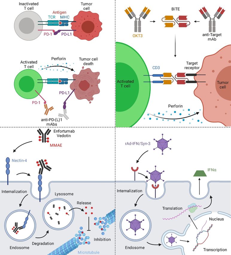
of bladder cancer. Their mechanism of action is depicted in Figure 2A. immunocompetent hosts (48).
Lirilumab Vofatamab
Human natural killer (NK) cell activity is modulated by killer cell Vofatamab is a clinical-stage mAb targeting the fibroblast growth
immunoglobulin-like receptors (KIR) (40). KIRs are classified as factor receptor 3 (FGFR3). FGFR3 signaling has been linked to
activating and inhibitory (41) and recognize predominantly HLA development, differentiation, growth, and survival (49). In
class I as their ligand (42). Blockage of the inhibitory KIR2DL1 patients suffering from bladder cancer, 13 different activating
has been shown to promote ADCC in tumor models (43). missense mutations have been identified, with three alternations,
In 2009, Romagné et al. immunized transgenic mice (44) with R248C, S249C, and Y375C, accounting for over 85% of all
BW5417 thymoma cells stably transfected with KIR2DL1, with observed mutations (50). The cysteine residues generated by
subsequent booster immunizations utilizing soluble KIR2DL3 the mutations presumably lead to ligand-independent receptor
Frontiers in Oncology | www.frontiersin.org 6 May 2021 | Volume 11 | Article 672262Bogen et al. Engineering Therapeutics for Bladder Cancer
A B
C D
FIGURE 2 | Mechanisms of action of immune and viral therapies for bladder cancer. (A) T cells can recognize cancer cells via the interaction of their T cell receptor
(TCR) with the major histocompatibility complex (MHC) on the target cell, resulting in cytotoxic activity eventually leading to cell death of the malignant cells. However,
a prominent escape mechanism of cancer is the upregulation of checkpoint inhibitors like PD-L1, inhibiting cancer-specific T cells from attacking the malignant cells.
By blocking the interaction of PD-1 on T cells with its ligand PD-L1 on the tumor cell, T cells are reactivated and can effectively target the tumor. (B) Bispecific T cell
engagers (BiTEs) consist of two binding arms, simultaneously binding to a tumor-associated antigen on the tumor cell and CD3, a part of the TCR complex, on T
cells, generating a synthetic immunological synapse. This results in T cell activation, eventually leading to the induction of a cytotoxic effect against the targeted tumor
cell. (C) Upon binding of Enfortumab Vedotin to nectin-4, the antibody-drug-conjugate (ADC) gets subsequently internalized into the endosome. Next, the complex is
transported to the lysosome, where the ADC is degraded. The valine-citrulline linker is cleaved by cathepsin B resulting in the release of monomethyl auristatin E
(MMAE) and the subsequent inhibition of the microtubule, eventually leading to cell death. (D) The rAd-IFN/Syn-3 adenovirus is internalized into cells of the bladder
after prior instillation. The genetic information of the virus is translocated into the nucleus and upon transduction, these cells produce and secrete IFNa, resulting in
an anti-tumor response by activating nearby immune cells. Created with BioRender.com.
dimerization and subsequent activation (51). Furthermore, One study on FGFR3 in the context of NMIBC found that
FGFR3-fusion proteins were observed, where the complete 32% of T1 tumors exhibit FGFR3 mutation variants, and 96% of
FGFR3 sequence except the final exon was C-terminally fused those are overexpressed. In tumors exhibiting a wild-type FGFR3
with the sequences of TACC3 (52, 53) or BAIAP2L1 (54), gene (68%), overexpression was found in 47% of cases. Overall,
resulting in a phenotype associated with higher grades (50, 55). 63% of the study population exhibited overexpression of FGFR3
Frontiers in Oncology | www.frontiersin.org 7 May 2021 | Volume 11 | Article 672262Bogen et al. Engineering Therapeutics for Bladder Cancer
(56). Consistently, another study showed that 85% of tumors action of T cell engagers is depicted in Figure 2B. The molecular
exhibiting mutations within FGFR3 showed overexpression structure T cell engagers, as well as of all other discussed
while being low-grade. Furthermore, it seems that higher molecules, is illustrated in Figure 3.
FGFR3 expression is elevated in more differentiated tumors In 2018 Juan Ma and coworkers generated CD3×EGFR and
(57). This is in line with the finding that higher FGFR3 CD3×HER2 bispecific antibodies by reacting the anti-epidermal
expression was found in pTa stage tumors compared to pT1 growth factor receptor (EGFR) mAb Cetuximab or the anti-
tumors and is therefore associated with lower-grade (57, 58) as HER2 mAb Trastuzumab, respectively, with sulfo-succinimidyl
well as a favorable prognosis. In contrast, wtFGFR3 expression 4-(N-maleimidomethyl)cyclohexane-1-carboxylate (sulfo-
did not show any influence on clinical parameters (56). Only SMCC), followed by conjugation with OKT3 after activation
42% of tumors exhibiting wtFGFR3 showed overexpression, with Traut’s reagent (62). The mouse-derived OKT3 mAb
where 66% were high-grade, and 68% invasive (58). recognizes the CD3ϵ domain in the T cell receptor (TCR)
In 2009, Qing and coworkers isolated an anti-FGFR3 complex and is commonly used for the construction of T cell
monoclonal antibody, termed R3mab via phage display, which engagers, as e.g., Blinatumomab (63). By analyzing human-
blocked the ligand binding as well as receptor dimerization (59). derived active T cells, it could be demonstrated that the
Furthermore, R3mab was able to bind not only wtFGFR3 but also bispecific constructs exhibited a stronger cytotoxic activity
the most common FGFR3 mutants and inhibited proliferation as against bladder cancer cells and mediated secretion of the
well as FGFR3 signaling in bladder cancer cells, exhibiting wild activation markers Interferon gamma (IFN-g), tumor necrosis
type and mutated variants of FGFR3. In subsequent in vivo factor alpha (TNF-a), and IL-2 (62). The following year, the
studies utilizing xenografts of either WT or mutated FGFR3 same group utilized this system to generate a CD3×CD155
cells, R3mab revealed remarkable anti-tumor effects. In 2015 bispecific antibody (64).
R3mab, now termed as Vofatamab, was tested in a phase I/II CD155, also known as nectin-like protein 5, belongs to the Ig
dose-escalation study alone or in combination with docetaxel for superfamily and is overexpressed in multiple malignancies,
metastatic urothelial cell carcinoma (NCT02401542). 20% of including lung adenocarcinoma, pancreatic, colon cancer as
enrolled patients experienced serious adverse events when well as in muscle-invasive bladder cancer (65–70).
treated with Vofatamab as a monotherapy and over 50% when Peripheral blood mononuclear cells (PBMC), isolated from
treated in combination with docetaxel, resulting in the healthy donors and bladder cancer patients, were activated and
termination of the study. Furthermore, a phase I study in 2016 subsequently treated with the CD3×CD155 bsAb, resulting in
combined with Pembrolizumab (NCT02925533) was conducted, cytotoxic effects against bladder cancer cell lines (64). A major
but terminated as well due to safety concerns. A phase I/II study drawback in chemical antibody conjugation strategy lies in the
in 2017 for locally advanced or metastatic urothelial cell fact that it is not site-specific, and therefore it yields a
carcinoma (NCT03123055) was terminated since over 46% of heterogenous population of bispecific molecules.
patients experienced serious adverse events. Several alternative routes exist to overcome this problem. A
frequently used format is based on the genetic fusion of two
single chain Fv (scFv) fragments that consist of an antibody
BISPECIFIC ANTIBODIES variable domain of the heavy and the light chain connected by a
linker sequence, each addressing different targets resulting in a
Over recent years, bispecific antibodies (bsAb) have gained bispecific T cell engager (BiTE)-like architecture (Figure 3) (71).
interest of pharmaceutical and academic research. The dual In frame of development of bispecific T cell engagers for the
targeting binding properties can mediate mechanisms of action, treatment of bladder cancer, Li and coworkers generated a
being impossible to accomplish with their monospecific tandem BiTE-based CD3×B7-H3 bispecific antibody (72, 73).
counterparts or a combination of multiple standard IgGs (60). B7-H3 is naturally expressed by antigen-presenting cells
To this day, only three bispecific antibodies were approved. One is (APC) and is involved in inhibition of T cells (74–76).
the by now withdrawn Catumaxomab for the treatment of ascites, However, overexpression can also be found on several cancer
the second one is Blinatumomab, which is approved for the cells (77–83), where B7-H3 plays a role in cell migration and
treatment of acute lymphatic leukemia (ALL), and Emicizumab, invasion (84). Interestingly, B7-H3 exhibits a high expression
approved for hemophilia A treatment, is the third. However, a level in bladder cancer cells. In vitro cytotoxicity studies of this
variety of bsAbs is in (pre-)clinical development for the treatment CD3×B7-H3 BiTE, as well as in vivo experiments, revealed a
of different indications, including bladder cancer, exhibiting a notable cell-killing activity of bladder cancer cells, which was
diverse spectrum of antibody formats. further improved by combination with trametinib, a small
molecule MEK inhibitor (72).
Anti-CD3 Bispecifics The only T cell engaging molecule that was in clinical testing for
T-cell engagers are bispecific molecules that bridge cytotoxic T bladder cancer was Orlotamab/MGD009 (NCT02628535), a
cells to tumor cells leading to the generation of a synthetic humanized Dual Affinity Re-Targeting (DART) protein which
immunologic synapse. This results in cytotoxic activity on tumor comprises a CD3 and a B7-H3 binding moiety (85). DART
cells mediated by T-cells. Commonly, CD3-specific antibodies molecules exhibit a diabody-like architecture with the VH
are utilized as T cell binding moieties (61). The mechanism of domain of one binder linked to the VL of the second binder and
Frontiers in Oncology | www.frontiersin.org 8 May 2021 | Volume 11 | Article 672262Bogen et al. Engineering Therapeutics for Bladder Cancer FIGURE 3 | Overview of antibody-, protein-, mRNA-, cell- and viral-based drugs that are approved or are in (pre-)clinical development for the treatment of bladder cancer. The molecules belonging to one drug class are marked in the same color. A schematic representation of the molecular architecture is given. Created with BioRender.com. Frontiers in Oncology | www.frontiersin.org 9 May 2021 | Volume 11 | Article 672262
Bogen et al.                                                                                                  Engineering Therapeutics for Bladder Cancer

the VH of the second binding moiety attached to the VL of the first.              HER2, 4-1BB is clustered on nearby T cells, resulting in T cell
Furthermore, disulfides are utilized to stabilize the construct (60,              activation and a cytotoxic response against the tumor.
86). The DART of MGD009 is linked to an Fc fragment, resulting                       The PRS-343 architecture is based on a Trastuzumab variant
in a monovalent antibody-like molecule (Figure 3). The Fc part is                and a 4-1BB specific anticalin (98) (Figure 3). Anticalins are
mutated to reduce effector functions while allowing recyclization                engineered variants of tear lipocalin and neutrophil -gelatinase–
via the neonatal Fc receptor to mediate extended in vivo half-life               associated lipocalin (NGAL), where multiple loops are
(85). The first clinical testing of Orlotamab/MGD009 against                      randomized by concerted mutagenesis (98–100). Via phage
bladder cancer, among others, was terminated due to business                     display, a 4-1BB-binding anticalin, termed J10, was isolated
decisions and not for safety issues (85) (NCT02628535). However,                 from an anticalin library. This molecule exhibits a 2 nM
a phase I study is currently ongoing for the treatment of advanced               affinity, shows cross-specific binding to 4-1BB from
solid tumors (87) (NCT03406949).                                                 cynomolgus monkeys, and does not block ligand binding (98).
                                                                                 Trastuzumab VH and VL domains were subcloned into an IgG4
ATOR-1015                                                                        format to silence Fc-mediated effector functions. Furthermore,
Immune checkpoint inhibitors such as the anti-CTLA-4 mAb                         the S228P mutation was implemented, suppressing the naturally
Ipilimumab can have significant effects in cancer patients.                       occurring Fab-arm exchange found in IgG4 molecules (101). Fab-
Kvarnhammar and coworkers developed a bispecific CTLA-                            arm exchange describes the naturally occurring and dynamic
4×OX40 bsAb (ATOR-1015) (88). To this end, OX40 binding                          dissociation of an IgG4 homo-tetramer into two half-molecules
Fab domains were isolated from an scFv phage display library.                    consisting of one heavy and one light chain. This is followed by
For CTLA-4 binding, the V-like domain of the CTLA-4 ligand                       the reassociation with a second half antibody, leading to an
CD86 was engineered in five residues to achieve a 100-fold                        antibody with two different Fab arms (102). This mechanism
increased affinity towards CTLA-4. This engineered CD86                           would lead to antibodies with reduced specificity and therapeutic
domain was C-terminally fused to the kappa light chain of the                    efficacy and is therefore avoided in PRS-343 (103). Additionally,
anti-OX40 IgG1 molecule (88) (Figure 3). The resulting                           the F234A and the L235A mutation, which reduce the binding to
bispecific antibody bound CTLA-4 with 3.0 nM affinity and                          Fcg-receptors and further silence effector function, were
blocked the interaction with CD80 and CD86.                                      implemented in PRS-343 (104, 105). Since the distance between
    The co-stimulatory molecule OX40 (CD134) belongs to the                      effector and target cells is crucial for T cell activation and
next generation of immune therapeutic targets. CTLA-4 and                        cytotoxicity (105), the anticalin moiety was N- and C-
OX40 are upregulated in tumor-infiltrating, but not peripheral,                   terminally fused to the heavy and light chains of the engineered
regulatory T cells (Tregs). ATOR-1015 binds domain 2 on OX40                     Trastuzumab variant utilizing a (G4S)3 linker. While all variants
with an affinity of 1.6 nM and blocks interaction with OX40L and                  exhibited similar biophysical properties, the C-terminal heavy
furthermore induced T cell activation in vitro by blocking CTLA-                 chain fusion showed the strongest T cell activation.
4. By crosslinking OX40 upon bind to either CTLA-4 or to                             In vivo experiments demonstrated recruitment of TILs, while
FcgRIIIa (CD16a), ATOR-1015 leads to NK-cell or T cell-                          lymphocytes in peripheral blood remained unaffected (98). Due
mediated depletion of OX40-overexpressing Tregs. Those                           to its favorable properties, phase I dose-escalation studies were
Tregs can negatively regulate the immune response (89–92),                       conducted in patients with HER2-positive cancers, including
and their depletion can enhance anti-tumor activity (93–95). By                  bladder cancer, either as a monotherapy (NCT03330561) or in
targeting both receptors in a bispecific manner, the activity of the              combination with Atezolizumab (NCT03650348). Both studies
bsAb is not only directed to the tumor side, eventually                          were suspended, but very recently, the clinical hold was lifted by
introducing anti-tumor responses, but also may result in a                       the FDA.
more favorable safety profile and better efficacy compared to
standard anti-CTLA4 mAbs (88, 96).
    In vivo experiments utilizing the MB49 bladder cancer model                  FUSION PROTEINS
showed a reduction in tumor growth and prolonged survival.
Furthermore, ATOR-1015 enhanced the effect of anti-PD1                           Like bsAbs, fusion proteins can mediate mechanisms of action that
treatment in in vivo models. As ATOR-1015 shows promising                        are impossible to achieve with a mixture of the parental,
results in preclinical cancer models, including colon, pancreas, and             monofunctional fusion partners. For example, Etanercept or
bladder cancer, it is currently in phase I clinical trials (NCT03782467).        Belatacept are antibody-derived Fc fusion proteins where the Fab
                                                                                 fragment of the antibody is replaced by either the extracellular
PRS-343                                                                          domain of tumor necrosis factor receptor or CTLA4, respectively.
PRS-343 is a bispecific fusion protein targeting HER2 and 4-1BB                   While those molecules are approved for the treatment of
(CD137), which is a costimulatory receptor on T cells. Despite 4-                rheumatoid arthritis or organ transplant rejection for years
1BB being a therapeutic target of major interest, prior clinical                 (106–108), no fusion proteins are approved for bladder cancer
studies utilizing the monospecific 4-1BB-binding IgG4 antibody                    yet, but some have entered clinical development.
Urelumab were stopped due to hepatoxicity (97). The bispecific
antibody PRS-343 has the potential to overcome these side-                       ALT-801
effects by its tumor-specific mechanism of action. Upon                           In 1995, Theobald and coworkers used transgenic mice to
accumulation in HER2-positive tumors and clustering of                           generate cytotoxic T lymphocytes (CTL) targeting the human

Frontiers in Oncology | www.frontiersin.org                                 10                                   May 2021 | Volume 11 | Article 672262
Bogen et al. Engineering Therapeutics for Bladder Cancer
p53 protein (109). From the population of CTLs the clone no. 5, Pharmacokinetic experiments demonstrated that the complex
specific to the p53 peptide 264–272, was isolated by limiting exhibited an in vivo half-life of 18 to 25 h, which is significantly
dilution (110). To generate a soluble T-cell receptor, Belmont enhanced compared to wtIL-15 (0.64 h). In vivo, the complex
et al. amplified the Va and Vb/Cb regions by RT-PCR. mediated proliferation of NK cells and CD8+ T cells in mice
Subsequently, the C-terminal end of the Va was fused to the (114). As this super agonistic construct can activate NK cells, it
N-terminal end of the Vb utilizing a (G4S)4 linker. The Cb region was in vitro demonstrated that the ALT-803 is able to modulate
directly fused to Vb was truncated to lack the final cysteine as well ADCC activity of a cancer-specific antibody. Furthermore, it led
as the transmembrane and cytoplasmic regions. Via a peptide to the upregulation of activating NK cell receptors, antiapoptotic
linker (encoding for VNAKTTAPSVYPLAPV), human IL-2 was factors, and factors involved in the NK cytotoxicity while
linked to the single-chain TCR (scTCR) (Figure 3). The construct simultaneously downregulating NK cell inhibiting factors
was produced in mammalian cell culture and the resulting fusion (116). Furuya and coworkers investigated the effect of ALT-803
protein bound to p53-peptide-loaded MHC on T2 cells, as well as in an orthotopic mouse model, demonstrating that subcutaneous
to the IL-2 receptor. Furthermore, the scTCR-IL-2 construct administration of ALT-803 is non-inferior to intravesical
exhibited similar biologic effects in vitro as recombinant IL-2. It treatment of BCG. While subcutaneous administered ALT-803
was shown that this construct could bring p53-peptide-loaded activated CTLs, NK cells and NKT cells, in combination with
MHC-bearing cells in proximity with cells exhibiting the IL-2R. intravesical BCG additional immunomodulating cytokines in the
Pharmacokinetic studies showed a half-life in mice of 1.6 to 3h, serum were detected (117). Additional mouse models
which is elevated compared to free IL-2, as it has a half-life of demonstrated that ALT-803 increased PD-L1 expression in
5 min. In vivo experiments using xenografts showed beneficial tumors in vivo and elucidated the safety and efficacy of
outcomes of the scTCR-IL-2 construct in comparison to IL-2 combination treatment with PD-L1-specific checkpoint
alone (111). To reduce the immunogenicity of the construct, blockage, paving the way for clinical combination treatment
the murine Cb was exchanged to its human counterpart while (118). Currently, ALT-803 is in phase I/II and phase II studies
retaining a promising profile in in vivo experiments (112). In investigating its effect on patients suffering from NMIBC or
2011 and 2012, phase I/II studies of NMIBC and bladder bladder cancer, respectively, as monotherapy or in combination
cancer were initiated, but the current status is unknown with BCG (NCT02138734, NCT03022825). Furthermore, a
(NCT01326871, NCT01625260). currently recruiting phase II study will investigate the efficacy
However, this engineering approach’s versatility was of ALT-803 in combination with the checkpoint inhibitors
demonstrated as Zhu and coworkers substituted the IL-2 Pembrolizumab, Nivolumab, Atezolizumab, Avelumab or
moiety with an IL-15 variant (N72D), which exhibits an Durvalumab. One cohort, in addition to the combination of
improved binding towards the IL-15Rb, resulting in an scTCR ALT-803 with one of the checkpoint inhibitors, will be weekly
fusion construct exhibiting a super agonistic mechanism of dosed with PD-L1 t-haNK cells (NCT03228667). These cells are
action for the IL-15 pathway (113). derived from NK-92 cells, exhibit the high-affinity CD16
receptor, produce an IL-2 variant, which is retained in the
ALT-803 endoplasmic reticulum, and carry an additional chimeric
Based on the IL-15 N72D mutein, Han and coworkers antigen receptor addressing PD-L1 (119).
constructed in 2011 a fusion protein of the sushi domain of
IL-15Ra (aa 1-66) and a C-terminal human IgG1 Fc part.
Subsequently, plasmids encoding for IL-15 N72D and IL- ANTIBODY-DRUG-CONJUGATES
15Ra-hFc were co-transfected in Chinese hamster ovary
(CHO) cells. IL-15 is poorly produced in mammalian cell Antibody-drug-conjugates (ADC) comprise a cancer-targeting
culture due to its fast degradation. However, in complex with monoclonal antibody, which is conjugated to a cytotoxic agent
IL-15Ra, the cytokine is shielded from degradation in the (Figure 3). The specificity of the mAb ensures a selective
endoplasmic reticulum, resulting in high expression levels of targeting of tumor cells. Upon antigen-binding, ADCs are
the IL-15 N72D:IL-15Ra-hFc complex. Utilizing ion-exchange internalized by receptor-mediated endocytosis and trafficked to
chromatography methods allowed for the specific purification of the endosome and ultimately to the lysosome. Depending on the
IL-15Ra-hFc molecules fully occupied by two IL-15 N72D linker utilized, acidic pH or lysosomal proteases mediate linker
variants (114) (Figure 3). cleavage, resulting in the release of the toxin and eventually cell
IL-15 stimulates the proliferation of NK cells and T cells and death (120, 121). The linkers, the toxins, and the conjugation
induces the generation of CD8+ cytotoxic lymphocytes. sites of all clinical-stage ADCs for bladder cancer are illustrated
Naturally, both IL-15 and IL-15Ra are expressed on dendritic in Figure 4. While coupling efficiency and heterogeneity of the
cells and interact with NK cells and T cells in trans, which conjugate are common hurdles (122), nine ADCs are approved
express cell-bound IL-2/IL-15Rb and the common gamma chain for several malignancies, including solid tumors such as breast
(gc), resulting in heterotrimeric receptor formed inside the cancer and hematologic malignancies (123–127). Currently, 16
immunologic synapse (115). ALT-803 can bind to IL-2/-15Rb ADCs are in phase II and III studies, with many more in phase I
and gc on T cells and NK cells and induce the IL-15 pathway studies and preclinical development (121). With the recent
leading to stimulation of cell proliferation. approval of Enfortumab Vedotin (Padcev) and the clinical
Frontiers in Oncology | www.frontiersin.org 11 May 2021 | Volume 11 | Article 672262Bogen et al. Engineering Therapeutics for Bladder Cancer
evaluation of multiple other ADCs, the focus is shifting to intermolecular disulfide bonds and a subsequent conjugation
urothelial malignancies (128). utilizing a thiol-reactive maleimido-valine-citrulline-dipeptide-
Two marketed ADCs are based on the monoclonal antibody linker, monomethyl auristatin E (MMAE) was conjugated,
Trastuzumab targeting the human epidermal growth factor resulting in the ADC RC48 (135). MMAE inhibits tubulin
receptor 2 (HER2), a well-studied tumor antigen overexpressed polymerization, resulting in phase arrest and apoptosis (136). The
in a variety of malignant diseases, including breast cancer (129, linker is effectively cleaved by human cathepsin B upon
130) and lung cancer (131, 132). Furthermore, HER2 emerged as internalization in HER2 positive cells but stable in human plasma
a promising therapeutic target for bladder cancer: (137). In vivo studies using xenografts demonstrated a significant and
Fluorescence in situ hybridization (FISH) and IHC studies of durable regression, underlining the high potency of this ADC (135).
tissue arrays derived from high- and low-grade UCs, as well as In 2015, a dose-escalating phase I study was conducted in
papillary urothelial neoplasms, revealed that up to 9% of high- HER2-overexpressing cancer patients, showing a manageable
grade UCs exhibit a HER2 gene amplification, which can be safety profile while exhibiting low liver toxicity (137, 138)
associated with higher recurrence and a worse prognosis. (NCT02881190). Currently, RC48, renamed as Disitamab
Additionally, a subset of high-grade NMIBC patients exhibits a Vedotin, is in phase II studies for locally advanced or
HER2 gene amplification, which can be correlated with metastatic urothelial cancer in HER2 overexpressing
aggressive tumor growth (133). Consistently, a silver-enhanced (NCT03809013) or HER2-negative patients (NCT04073602)
in situ hybridization (SISH) analysis of NMIBC patients showed and recently gained breakthrough therapy status.
that 4.2% of the tumors exhibited stronger HER2 staining. All
HER2-positive NMIBC samples belonged to the high-grade Enfortumab Vedotin (Padcev)
subset, and HER2-expression is an independent predictor of For the treatment of PD-1/PD-L1 unresponsive patients
tumor prognosis (134). Today, multiple anti-HER2-ADCs are in suffering from locally advanced or metastatic UC, the FDA
clinical development for the treatment of bladder cancer (128). approved Enfortumab Vedotin in 2018 (139). This was based
on a clinical trial including 125 participants (NCT03219333),
Disitamab Vedotin where 44% of enrolled patients had tumor shrinkage or growth
In order to generate a novel HER2-specific antibody, Yao and arrest, including 12% showing a complete response, meaning the
coworkers immunized mice with the extracellular domain (ECD) tumor disappeared entirely (140). This ADC consists of a nectin-
of HER2, extracted the spleen, and generated hybridomas (135). The 4 targeting antibody, conjugated to a microtubule inhibitor and
clone exhibiting the best binding activity was sequenced and was granted breakthrough therapy designation, priority review
subsequently humanized by CDR grafting. The resulting mAb status, and accelerated approval.
variant was termed Hertuzumab and showed a higher affinity The cell adhesion molecule (CAM) nectin-4 belongs to the family
compared to gold-standard Trastuzumab. By partial reduction of of nectin-like proteins (Necl), consisting of nectin-1, -2, -3, -4,
FIGURE 4 | Cytotoxic payloads and conjugation strategies of approved and clinical-stage antibody-drug-conjugates (ADCs) for the treatment of bladder cancer. On
the left, a human IgG1 antibody is shown, where cysteine (yellow) and lysine residues (red) are highlighted, the glycosylation is marked in green, while the light chains
are depicted in blue. The heavy chain is illustrated in grey. Depending on the conjugation strategy, either cysteine residues or lysine residues are modified with the
toxins illustrated on the right side. Except for ado-Trastuzumab Emtansine, all clinical-stage ADCs for bladder cancer are based on the partial reduction of inter-chain
disulfide bonds (yellow), resulting in a maximal drug to antibody ratio (DAR) of 8, while simultaneously reducing the number of potential ADC variants. In ado-
Trastuzumab Emtansine conjugation is achieved by coupling to lysine residues (red). The respective linkers and toxins of the payloads are color-coded and named
separately. SMCC, succinimidyl 4-(N-maleimidomethyl)cyclohexane-1-carboxylate; DM1, maytansine; MC, maleimidocaproyl; PAB, p-aminobenzyl; MMAE,
monomethyl auristatin E; PEG, polyethylene glycol.
Frontiers in Oncology | www.frontiersin.org 12 May 2021 | Volume 11 | Article 672262Bogen et al. Engineering Therapeutics for Bladder Cancer
and -5, which trans-interact with one another (141–143). Besides urothelial bladder carcinoma were dosed with 3.6 mg/kg qw,
breast (144), lung (145), and ovarian cancer (146), it is expressed in after the safety of Trastuzumab Emtansine was assessed by an
malignancies of the bladder (147), where it is involved in metastasis independent data management committee after dosing the first
and proliferation (145, 148–150). six patients with 2.4 mg/kg qw. Partial response (defined as a
The parental human IgG1 antibody (AGS-22M6) was 30% decrease in the sum of the diameters of the target lesions
generated by utilizing the XenoMouse technology, where taking as a reference the baseline sum diameter according to the
transgenic mice are genetically engineered with a humanized RECIST 1.1 criteria) was observed in 38.5% of patients. No
humoral immune system (151). Immunization was performed patients achieved complete response. Median OS (95%CI) was
with the extracellular domain (ECD) of human nectin-4, 7.03 months (3.75 to NE). A second study on HER2 amplified or
followed by the generation of over 50 hybridomas (152). mutated cancers, including bladder cancer, is currently
Besides binding to human nectin-4, AGS-22M6 showed cross- ongoing (NCT02675829).
specificity to its monkey and rat orthologues. Using flow
cytometry, it was demonstrated that AGS-22M6 bound its Trastuzumab Deruxtecan (Enhertu)
human target with a KD value of 10 pM, which was also DS-8201a is another HER2-targeting ADC. It is based on
confirmed for its MMAE-conjugated counterpart. By deletion Trastuzumab, which was conjugated with DX-8951 (DXd), a
mutagenesis of nectin-4, it was shown that the epitope of AGS- topoisomerase I inhibitor via a GGFG peptide linker, using
22M6 is located on the V-domain, enabling inhibition of the maleimide chemistry, after partial reduction of interchain
heterodimerization with nectin-1 (152). For recombinant disulfides. This tetrapeptide linker is cleaved by lysosomal
expression, VH and VL genes were extracted from the proteases cathepsins B and L in HER2-positive tumor cells
hybridoma and subcloned into expression vectors for upon receptor-mediated endocytosis (161). The coupling
production in CHO cells. For ADC generation, the results in a high DAR of 7.7, enabling anti-tumor effects even
microtubule-disrupting toxin MMAE was conjugated utilizing in HER2 low-expressing models not being feasible with ADCs
the cleavable linker maleimidocaproylvaline-citrulline-p- exhibiting lower DAR (including T-DM1/ado-Trastuzumab
aminobenzyloxycarbonyl (136), which was conjugated by Emtansine) (161, 162).
partial reduction of interchain disulfide bond (153, 154), This higher DAR might help to overcome drug resistance,
resulting in a drug to antibody ratio (DAR) of approximately 4 which is observed in T-DM1 treated patients. Even though the
(152). In preclinical xenograft models, the ADC showed drug resistance in the context of T-DM1 is not fully understood,
significant growth inhibition in breast, pancreatic, lung, and one of the main reasons might be the downregulation of HER2,
bladder cancer models (152), paving the way for its phase I impaired internalization, enhanced recycling of HER2, and
study, which started in 2011 (NCT01409135). lysosomal degradation (155, 163). However, due to its different
Currently, Enfortumab Vedotin is in further clinical trials mechanism of action and efficacy in low HER2-expressing
investigating its effect on bladder cancer in comparison to cancers, it is hypothesized that DS-8201a might be effective
chemotherapy (NCT03474107), in combination with against T-DM1 resistant tumors (161). Currently, DS8201a,
checkpoint inhibitors and chemotherapy (NCT03288545), or renamed as Trastuzumab Deruxtecan, is under clinical
combined with other immunotherapies after failure of investigation for advanced breast and urothelial cancer in
platinum-containing chemotherapy (NCT03869190). combination with Nivolumab (NCT03523572) (164).
Trastuzumab Deruxtecan is approved for treatment of patients
ado-Trastuzumab Emtansine (Kadcyla) suffering from unresectable or metastatic HER2-positive
In 2008, Phillips and coworkers conjugated the HER2-specific breast cancer.
antibody Trastuzumab to a maytansine derivative (DM1) via the
non-reducible thioether linker SMCC (155, 156). Trastuzumab Sacituzumab Govitecan (Trodelvy)
exhibits 88 lysine residues that could potentially be utilized for In 1990, Stein and coworkers utilized the hybridoma technology
conjugation. While the DAR is approximately 3.5, at least 70 to generate the antibody RS7-3G11 by immunizing mice with
lysine residues are partially conjugated, resulting in a crude membrane preparations of squamous cell carcinoma of the
heterogeneous product (157). The resulting ADC, termed T- lung (165). It was observed that the resulting mAb RS7-3G11 is
DM1, was effective in Trastuzumab-sensitive and -insensitive specific to multiple malignancies, including breast, colon, renal,
cancer models in vitro as well as in vivo. T-DM1 is internalized and prostate cancers (166). In 1995, Basu and coworkers
upon binding to HER2, resulting in accumulation in the identified Trop2 as the target of RS7-3G11 (167).
endosome (155, 158). As the linker utilized is non-reducible, Trop2, also known as human trophoblastic cell surface
DM1 release occurs by antibody degradation in the lysosome antigen 2, is an oncogene involved in tumorigenesis and tumor
(156, 159). After lysosomal escape, DM1 inhibits microtubule invasion (168–170). With a low expression in healthy tissue and
assembly, resulting in cell death (155, 160). T-DM1, further being upregulated in several cancers, it emerged as a promising
termed as ado-Trastuzumab Emtansine, was approved in 2013 target (171, 172).
for HER2 positive breast cancer. Recently, a phase II study It was observed that RS7 facilitates fast internalization and
investigating the tumor response in HER2 expressing tumors, was evaluated for in vivo efficacy (173). Upon humanization by
including bladder cancer, was completed (NCT02999672). In CDR-grafting, the now termed hRS7 was subjected to radio-
cohort 1, 13 patients with locally advanced or metastatic labeled in vivo studies (174). Additionally, ADCC effects in
Frontiers in Oncology | www.frontiersin.org 13 May 2021 | Volume 11 | Article 672262Bogen et al. Engineering Therapeutics for Bladder Cancer
cervical cancers were observed utilizing hRS7 (174). Later on, low-risk patients and up to 3-fold higher in high-risk patients
hRS7 was conjugated with the topoisomerase inhibitor SN-38, (180). Other studies found by IHC that 15.1% of UC patients had
resulting in the ADC IMMU-132 or Sacituzumab Govitecan low and 26.2% had high EGFR expression (181), where one study
(175, 176). Conjugation was performed by mild reduction of found EGFR overexpression in 74% of UC patients (182). No
interchain disulfides followed by formation of a thioether bond, association of EGFR expressing UCs and mutations in the tyrosine
resulting in DARs of less than 4 (176). Subsequent phase I studies kinase domain are known to this date (182). However, EGFR
showed acceptable toxicity and promising results (177). expression was correlated with prognostic factors like grade, deep
Currently, Sacituzumab Govitecan is in clinical phase II studies muscle invasion, and recurrence (180, 181). Other IHC studies of
for metastatic UC (NCT03547973) and phase I/II studies in tissue microarrays revealed that 70% of samples exhibited a
combination with chemotherapy for multiple carcinomas, relative staining intensity of 1 or higher staining. Of those
including UC (NCT03992131). In early 2020, Sacituzumab samples, 16% had staining intensities of 3, and 31% had staining
Govitecan was approved under the name Trodelvy for the intensities of 4 stainings. A higher rate of positive stainings was
treatment of triple-negative breast cancer. observed in squamous tumors (94%) in comparison with
nonsquamous tumors (69%). Interestingly, no correlation
Sirtratumab Vedotin between EGFR staining intensity and T stage was observed
Utilizing tumor microarrays, SLIT and NTRK-like protein 6 (183). However, Kassouf et al. found that tumors simultaneously
(SLITRK6) were found to be expressed in 88% of bladder cancer showing a high EGFR and low HER4 expression display a more
specimens, of which 67% showed strong to moderate expression. invasive phenotype in conjunction with a short recurrence time
Furthermore, transitional cell carcinoma exhibited SLITRK6 (184). In consistence, a study investigating the expression of all
expression in 90% of cases and metastatic bladder cancer in HER-family members in bladder cancer found that while EGFR
100% of cases (178). and HER2 expression rates alone do not correlate with survival,
In order to generate an anti-SLITRK6 ADC, the Xenomouse higher EGFR or HER2 expression combined with low HER3 and
technology was employed by performing immunization with HER4 expression result in a worse prognosis (185).
SLITRK6 expressing cells or recombinant SLITRK6 protein, In 2017 Railkar et al. conjugated the EGFR-specific and FDA-
resulting in hybridomas clones. The clone AGS-15C was approved mAb Panitumumab via NHS ester chemistry with IRDye
identified for its binding to SLITRK6 and subcloned for 700Dx, generating Panitumumab-IR700, intending to create a
recombinant expression (178). Subsequently, ASG-15C was targeted phototherapy, termed photoimmunotherapy (PIT) for
conjugated to MMAE via a valine-citrulline dipeptide linker, bladder cancer (183). IR700 is a hydrophilic agent with low
resulting in the ADC ASG-15ME. The ADC caused complete cytotoxicity. Its cytotoxic nature is activated by the association to
growth inhibition in xenograft lung cancer models. Interestingly, the cell membrane and subsequent activation using near-infrared
approximately 5% of tumor cells were negative for SLITRK6 radiation (NIR), resulting in the generation of cytotoxic oxygen
expression, underlining the effect this ADC has on heterogenic species (186). Since the EGFR-specific antibody mediates
cancers, which are typically found in patients (178). AGS-15ME, membrane proximity, only EGFR-positive cells are targeted by
also termed Sirtratumab Vedotin, was tested in a phase I clinical this approach. In vitro, Panitumumab-IR700 showed cytotoxic
study on metastatic urothelial cancer (NCT01963052). effects on EGFR-positive bladder cancer cells in a dose-
With the recent approval of Enfortumab Vedotin and the dependent manner (183). Additionally, the effects are dependent
large number of additional toxin-coupled mAbs in clinical on the expression level of EGFR and the applied NIR. As EGFR
testing for bladder cancer, ADCs have the chance to gain a expression increases, lower NIR is needed to achieve similar IC50
profound impact in the treatment of bladder cancer. The values. In xenograft models, the bladder cancer cell line UMUC-5
mechanism of action of the approved Enfortumab Vedotin is was utilized to induce tumors that were subsequently treated with
depicted in Figure 2C. Panitumumab-IR700. NIR treated tumors showed regression,
which was not observed in non-NIR-treated control groups.
Furthermore, xenografts generated with cells exhibiting a lower
PHOTOIMMUNOTHERAPY EGFR-expression profile, showed no significant difference between
NIR-treated and NIR-untreated animals, underlining the
The photoimmunotherapeutic approach is based on an ADC-like importance of antigen expression level on the tumor (183).
molecule that carries an inactive cytotoxic agent to the tumor cell Since anti-tumor effects were limited by the surface
by conjugation to a monoclonal antibody (Figure 3). By irradiation expression of the antigen, Siddiqui and coworkers generated a
with light of the appropriate wavelength, the cytotoxic agent is combinatorial approach, consisting of Panitumumab-IR700 and
activated and is able to mediate effective tumor-killing (179). Trastuzumab-IR700 (187). In vitro and in vivo experiments
utilizing the EGFR/HER2 double-positive bladder cancer cell
Panitumumab-IR700 line SW-780 showed more potent effects upon combinatorial
The photoimmuno-conjugate Panitumumab-IR700 is based on treatment in comparison to mono treatments (187). Even though
the EGFR-specific mAb Panitumumab. RT-PCR analysis of there is no clinical study ongoing utilizing Panitumumab-IR700,
TURB bladder washings of stage Ta, T1, and Tis NMIBC the IR700 conjugated anti-EGFR PIT Cetuximab Salotarocan is
patients revealed that EGFR expression is 1.7-fold elevated in currently under investigation in phase I/II studies for EGFR
Frontiers in Oncology | www.frontiersin.org 14 May 2021 | Volume 11 | Article 672262You can also read