SPECIAL EDITION - The Mastocytosis Chronicles - HEALTH CARE PROFESSIONALS EDITION
←
→
Page content transcription
If your browser does not render page correctly, please read the page content below
The Mastocytosis Chronicles
              The Mastocytosis Society, Inc. | 2020-2021, Volume 2

              SPECIAL EDITION
              HEALTH CARE PROFESSIONALS EDITION

                                                                     Mast Cell Diseases
© 2020-2021 The Mastocytosis Society, Inc. All rights reserved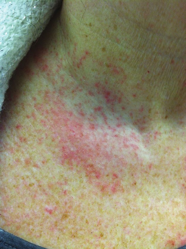
Our History

                                                                          presumed that Mastocytosis was one of the causes
                                                                          of death, when in fact the patient had often died of
                                                                          other causes, and the Mastocytosis was an incidental
               The Mastocytosis Society, Inc. (TMS) was founded           finding. On the other hand, more advanced cases
               in 1995 by Bill Abbottsmith, Linda Buchheit, Olive         of aggressive Mastocytosis were also recognized
               Clayson, Iris Dissinger, Bill Hingst, and Joe Palk. At     during post-mortem exams, leading pathologists to
               that time very little was known about Mastocytosis,        identify all forms of Mastocytosis as having a high
               so these pioneering individuals sought to fill a massive   associated mortality rate. Fortunately, that prognosis
               void with some answers to their multitude of questions     has improved as more patients are diagnosed and
               about this rare disease. They found one another            treated sooner, and more physicians research and treat
               through NORD, with sheer determination and                 this disease. Today, we know that pediatric patients
               extensive research.                                        have a 75% chance of outgrowing their disease at
                                                                          or before puberty, and adults with Indolent Systemic
               The first support group meeting was held in Baltimore      Mastocytosis can have a near normal life expectancy
               at the Inner Harbor in 1994 and was attended by Linda      if they avoid triggers and take their medication.
               Buchheit and Bill Hingst. The second meeting was held
               the following year at Linda Buchheit’s home in Ohio.       Founding Members: Today’s accomplishments are built
               Fourteen members attended that year. Little did they       on the foundations laid by the early volunteers, and we
               know how fruitful their efforts would be and what a        are grateful for their efforts. TMS is where it is today
               lifeline they would become as more and more patients       because of the seeds that they planted in 1994 and
               joined each year.                                          in the early years. Since then there have been many
                                                                          more champions who have served their fellow patients
               Until 1990 many patients diagnosed with Mastocytosis       and families affected by Mastocytosis and Mast Cell
               were given a very grim prognosis. Up until that            Activation Diseases by volunteering for TMS. We
               time, Mastocytosis was not often considered when           salute you!
               physicians were making a differential diagnosis,
               and many cases were completely missed, resulting           Past Board Members: THANK YOU to all of our past
               in patient death. At that point, signs of the disease      board members as they are our strong foundation for
               were then discovered on autopsy; however, because          all the wonderful and exciting things happening now
               so little was known about Mastocytosis, it was             and in the future for TMS!

2   tmsforacure.org | Special Edition 2020-2021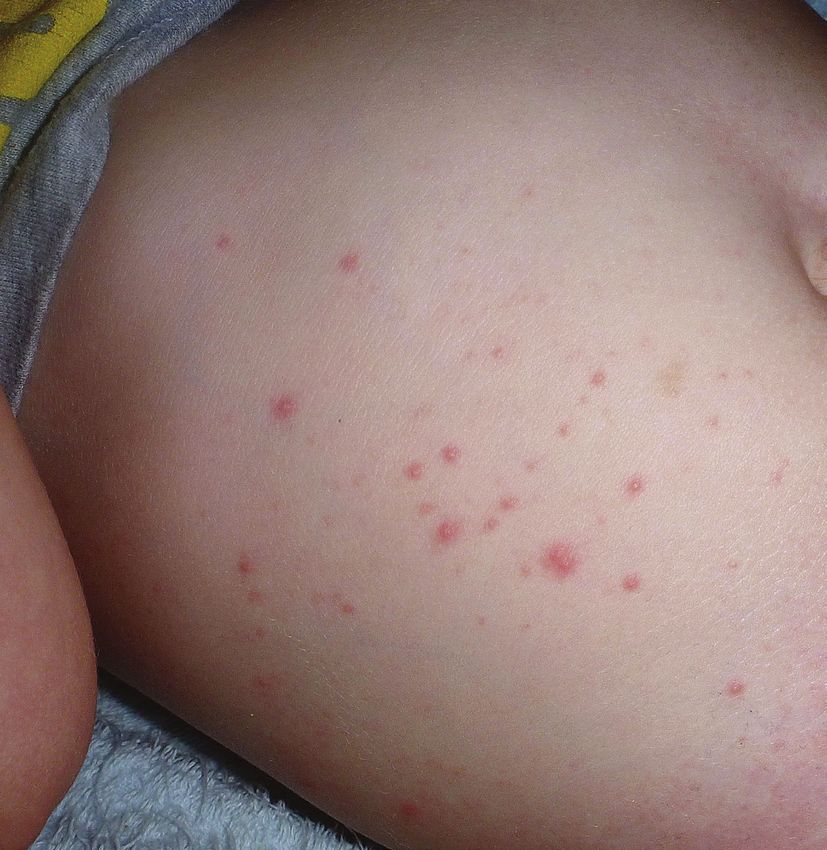
The Mastocytosis Chronicles                              American Initiative in Mast
                                                         Cell Diseases (AIM)
The Mastocytosis Society, Inc. | 2020-2021 - Volume 24
                                                         By Susan Jennings, PhD, and Valerie Slee, RN,
                                                         BSN – September 2019

                                                         Since 2014, The Mastocytosis Society, Inc. (TMS) has
In this issue
                                                         hosted small ancillary Mast Cell Disorder Challenges
                                                         meetings during specialty medical conferences. The
5	Overview, Definitions, Diagnosis
                                                         objectives of these meetings have been to bring together
   and Classification
                                                         specialist physicians, drug company representatives and
9 Cytology of Mast Cells                                 members of the TMS Research Committee to identify
                                                         primary challenges facing the mast cell disease community
10 Cutaneous Mastocytosis Variants                       in the United States and to explore possible actions to
                                                         address those challenges. A key conclusion from our initial
12 Systemic Mastocytosis Variants                        meetings was that the establishment of a US network
                                                         for mast cell diseases would be extremely helpful in
16 Mast Cell Activation Syndrome Variants
                                                         overcoming many of the challenges faced by our disease
17 Hereditary Alpha Tryptasemia                          community. During these meetings, our US physicians
                                                         have received significant support from many international
18 Signs, Symptoms And Triggers                          mast cell disease specialists, who have shared their
                                                         experiences of forming networks in their own countries
21 Tests                                                 and more broadly in Europe. TMS has collaborated with a
                                                         committee established for the formation of an American
25 Treatments For Mast Cell Diseases
                                                         network, under the direction of Jason Gotlib, MD, MS,
27 Medications To Treat Mast Cell Diseases               and Cem Akin, MD, PhD, as Co-Chairs. In May 2019, the
                                                         TMS Patient/Caregiver Conference was paired with the
29 Pediatric Mast Cell Diseases: Facts in Brief          inaugural investigator meeting of the American Initiative in
                                                         Mast Cell Diseases (AIM). AIM will be a network of centers
34 Visual Guide to Diagnosing Mastocytosis               established across North, Central and South America, with
                                                         a goal of excellence in patient diagnosis and treatment, and
38 	Medical & Research Centers that Treat Patients      collaboration on research initiatives. AIM will collaborate
    with Mast Cell Diseases                              closely with the European Competence Network on
41 Medical Advisory Board                                Mastocytosis (ECNM), which has centers established
                                                         throughout Europe.
43 Support Group Contacts
                                                         Please see www.aimcd.net for more information on
44 Mast Cell Connect Patient Registry Brochure           the American Initiative in Mast Cell Diseases.

                                                                          tmsforacure.org | Special Edition 2020-2021   3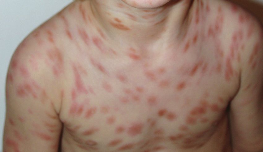
Committees
                                                        Board of Directors
    Advanced Systemic Mastocytosis Variants
    (advancedvariants@tmsforacure.org)                  Executive Board/Officers                         Rosemary Schultz: Interim Treasurer
    Valerie M. Slee, RN, BSN, Chair                     Valerie M. Slee RN, BSN: Chair                   treasurer@tmsforacure.org
    Michele Q. Kress, Smoldering SM Liaison
                                                        Medical Advisory Board Liaison
    Drug Shortage                                                                                        Other Board Members/Directors
    (drugshortage@tmsforacure.org)                      chairman@tmsforacure.org
    Valerie M. Slee, RN, BSN, Co-Chair                                                                   Courtney Rabb: Fundraising Chair
    Emily A. Menard, Co-Chair                           Jan Hempstead, RN: Interim Vice Chair
    Carlene Bartolotta RN, BSN                                                                           fundraising@tmsforacure.org
                                                        Patient Care Coordination Chair
    Education
    (education@tmsforacure.org)                         nurses@tmsforacure.org                           Jennifer Lockhart: Advocacy Chair
    Gail Barbera, Chair                                                                                  advocacy@tmsforacure.org
                                                        Gail Barbera: Secretary
    Fundraising
    (fundraising@tmsforacure.org)                       Education Chair                                  Rita Barlow: Retired, Director Emeritus
    Counrtney Rabb, Chair                               secretary@tmsforacure.org
    International Mastocytosis &
    Mast Cell Diseases Committee
                                                        education@tmsforacure.org
    Gail Barbera, USA Representative
    Jodylynn Bachiman, Website Design Committee
    Media Relations                                     Special Edition For Health Care Professionals
    (mediarelations@tmsforacure.org)
    Ariella Cohen, JD, Chair                            The special edition of The Mastocytosis Chronicles has been published specifically for physicians
    Medical Conference Planning                         and health care professionals since 2007. This edtion contains diagnostic and treatment
    (medicalconference@tmsforacure.org)
    Kathy Tomasic
                                                        information for mastocytosis and mast cell activation diseases, locations of mast cell disease
    Chantelle Bosco, Materials Management               treatment centers, physician contact information, documentation of research articles, and other
    Patient Care Coordination                           pertinent information. For additional information visit www.tmsforacure.org.
    (nurses@tmsforacure.org)
    Jan Hempstead, RN, Chair
    Pediatric
    (pediatrics@tmsforacure.org)                        TMS Medical Advisory Board
    Wendy Garr Ringer, Chair
    Political and Patient Advocacy                      Ivan Alvarez-Twose, MD           Tracy I. George, MD             Anne Maitland, MD, PhD
    (advocacy@tmsforacure.org)
    Jennifer Lockhart, Chair
                                                        K. Frank Austen, MD (Honorary)   Jason Gotlib, MD, MS            Larry Schwartz, MD, PhD
    Research
                                                        Patrizia Bonadonna, MD           Norton J. Greenberger, MD       Theoharis Theoharides, MD, PhD
    (research@tmsforacure.org)                          Joseph Butterfield, MD           (Honorary)                      Megha Tollefson, MD
    Susan Jennings, PhD, Chair
                                                        Mariana Castells, MD, PhD        Matthew J. Hamilton, MD         Celalettin Ustun, M.D.
    Support Groups
    (supportgroups@tmsforacure.org)                     Madeleine Duvic, MD              Olivier Hermine, MD, PhD        Peter Valent, MD
    Gail Barbera, Interim Support Group Chair           Luis Escribano, MD, PhD,         Nicholas Kounis, MD, PhD        Srdan Verstovsek, MD, PhD
    Jan Hempstead, RN Patient Care
    Coordination Chair                                         We thank each of these doctors for their time, caring, and expertise.
    Emily Bolden, Interim Support
    Group Coordinator
    Website Content
    (education@tmsforacure.org)                         TMS is a long-standing member
    Gail Barbera, Co-Chair                              of the National Organization for
    Susan Jennings, PhD, Co-Chair                       Rare Disorders (NORD)
    SUPPORTING CONTRACTORS
                                                        TMS is proud to be a Lay Organization member of The American
    Graphic Designers
    Rachael Zinman                                      Academy of Allergy Asthma and Immunology (AAAAI)
    John Gilligan
    Webmaster
    (webmaster@tmsforacure.org)                         Our Mission
    Ben Rabb                                            The Mastocytosis Society, Inc. is dedicated to providing multi-faceted support to patients,
                                                        families and medical professionals in our community and to leading the advancement of
                                                        knowledge and research in mast cell diseases through education, advocacy and collaboration.

4         tmsforacure.org | Special Edition 2020-2021
MAST CELLS AND MAST CELL DISEASES

Overview, Definitions, Diagnosis
and Classification

What are Mast Cells?                                           Mast cells have within them small sacs, or granules,
                                                               surrounded by membranes (Figure 1). The sacs contain
Mast cells (MC) are immune system cells that live in           many different kinds of substances called mediators,
the bone marrow and in body tissues, internal and              which participate in all of the roles above, including
external, such as the gastrointestinal tract, the lining       allergic response and anaphylaxis. The mediators are
of the airway, and the skin. Everyone has mast cells in        selectively released when there is an allergic or mast cell
their body, and they play many complex and critical roles      based reaction.1
in keeping us healthy. The positive roles that they play
include protecting us from infection, and helping our body     There is a difference between someone who is healthy,
by participating in the inflammatory process. However,         with mast cells that are functioning normally, and
mast cells are also involved in allergic reactions, from the   someone with a mast cell disease, whose mast cells may
tiny swelling that appears after a mosquito bite to a life     be activating inappropriately, sometimes in response to
threatening, full-blown anaphylaxis.                           triggers, or may also be proliferating and accumulating in
                                                               organ tissues.

Figure 1. Mast cell (electron micrograph)                      What are Mast Cell Diseases?

Mast cell granule (sac) which contains mediators               Mast cell diseases are caused by the proliferation and
                                                               accumulation of genetically altered mast cells and/or the
                                                               inappropriate release of mast cell mediators, creating
                                                                 symptoms in multiple organ systems.2 The three major
                                                                     forms of mast cell diseases are mastocytosis,
                                                                         mast cell activation syndromes (MCAS), and
                                                                            hereditary alpha tryptasemia (HaT). HaT
                                                                              is caused by a duplication or triplication
                                                                                of the alpha-tryptase gene.2a Mast
                                                                                  cell diseases can cause tremendous
                                                                                   suffering and disability due to
                                                                                    symptomatology from recurrent
                                                                                    mast cell mediator release, and/or
                                                                                    symptoms arising from infiltration
                                                                                    and accumulation of mast cells in
                                                                                   major organ systems. In addition,
                                                                                  those suffering from HaT may
                                                                                experience additional symptoms from

                                                                                                        Continued on page 6

                                                                                               tmsforacure.org | Special Edition 2020-2021   5
Overview, Definitions, Diagnosis and Classification
               Continued from page 5

               dysautonomia and connective tissue disease. Although          typically maculopapular cutaneous mastocytosis/urticaria
               systemic mastocytosis is a rare disease,3 those suffering     pigmentosa, is common in adult patients and can provide
               with MCAS and/or HaT have recently been increasingly          an important clue to accurate diagnosis.11, 12
               recognized and diagnosed. As a result, patients with
               MCAS and/or HaT appear to represent a growing                 Diagnosis and Classification13-17
               proportion of the mast cell disease patient population.4, 5
               It is important to note that the process of mast cell         CM is diagnosed by the presence of typical skin lesions and
               activation can occur in anyone, even without a mast           a positive skin biopsy demonstrating characteristic clusters
               cell disease, as well as in patients with mastocytosis,       of mast cells. The preferred method of diagnosing SM is via
               MCAS, and HaT.6                                               bone marrow (BM) biopsy. The WHO has established criteria
                                                                             for diagnosing SM, summarized18 as follows:
               MASTOCYTOSIS
                                                                                Major ª: Multifocal dense infiltrates of mast cells
               Definition                                                       (MCs) (> 15 MCs in aggregate) in tryptase stained
                                                                                biopsy sections of the bone marrow or other
                          Mastocytosis has been defined in the                  extracutaneous organ
                             literature as an abnormal accumulation
                                                                                Minorª:
                                of mast cells in one or more organ
                                                                                • More than 25% of MCs in bone marrow or
                                   systems. Previously classified by the
                                                                                  other extracutaneous organ(s) show abnormal
                                     World Health Organization (WHO)
                                                                                  morphology (i.e. are atypical MC type 1 or are
                                      as a myeloproliferative neoplasm,
                                                                                  spindle–shaped MCs) in multifocal lesions in
                                        mastocytosis is now classified in
                                                                                  histologic examination
                                        its own category under myeloid
                                         neoplasms.7 Broadly separated          • KIT mutation at codon 816V in extracutaneous
                                         into three categories – cutaneous        organ(s) (in most cases bone marrow cells are
                                         mastocytosis (CM), systemic              examined)
                                         mastocytosis (SM) and mast cell
                                        sarcoma – these diseases occur          • KIT+MCs in bone marrow show aberrant expression
                                      in both children and adults. CM is          of CD2 and/or CD25
                                    considered a benign skin disease
                                                                                • Serum total tryptase > 20 ng/mL (does not count in
                                  representing the majority of pediatric
                                                                                  patients who have SM-AHN-type disease.)
                               cases. In 67-80% of pediatric cases
                            seen, resolution will occur before or in            Abbreviation Key:
                         early adulthood.8-10 In pediatric mastocytosis,        KIT: Mast cell growth receptor/tyrosine kinase receptor
                     symptoms of mast cell mediator release may                 MC(s): Mast cells;
                 occur systemically as a result of mast cell mediators          SM-AHN: Systemic mastocytosis with associatiated
               released from skin lesions.10 This, however, does not            hematologic neoplasm.
               necessarily indicate systemic disease. The incidence
                                                                                ª If at least one major criterion and one minor criterion
               of systemic pediatric disease was previously unknown,
                                                                                OR at least three minor criteria are fulfilled, the
               but systemic forms have now been proven to exist in
                                                                                diagnosis of systemic mastocytosis can be established.
               some children.8-10 The majority of adult patients are            b
                                                                                  Activating mutations at codon 816, in most cases,
               diagnosed with systemic disease. Skin involvement,
                                                                                KIT D816V.
6   tmsforacure.org | Special Edition 2020-2021
MAST CELL ACTIVATION SYNDROMES

Definition

Existence of a subset of mast cell disease patients who
experience episodes of mast cell activation without
detectable evidence of a proliferative mast cell disease
was postulated over 20 years ago.19, 20 Over the last two
decades, with development of improved methodology
for identification of abnormal mast cells,21-24 it became
apparent that there were patients who exhibited                  The second required co-criterion for systemic mast cell
symptoms of mast cell mediator release who did not               activation depends on documentation that mast cells are
fulfill the criteria for SM.25, 26 Thus began the evolution of   directly involved in the symptomatology. An increase in the
discussions about other forms of mast cell diseases, both        serum level of tryptase, above baseline and within a narrow
clonal and nonclonal, which became known as Mast Cell            (generally accepted as one to two hour) window of time after
Activation Syndromes (MCAS).6, 27, 28                            a symptomatic episode, is proposed as the preferred method
                                                                 for providing evidence of mast cell involvement according
Diagnosis and Proposed Classification                            to these criteria.6, 28-30 The consensus article provides a
                                                                 method for calculating the required minimum rise in serum
Recognition by specialist physicians of the importance           tryptase.6 After a reaction, a level of serum tryptase that
of mast cell activation in disease led to an international       is a minimum of 20% above the basal serum tryptase level,
Mast Cell Disorders Working Conference emphasizing               plus 2 ng/ml, will meet the second criterion listed above
this topic in September of 2010. Consensus statements            for a mast cell activation event. Consensus members also
were published regarding classification of and diagnostic        agreed that when serum tryptase evaluation is not available
criteria for mast cell diseases,6 where mast cell activation     or when the tryptase level does not rise sufficiently to meet
plays a prominent role.                                          the required increase for the co-criterion, other mediator
                                                                 tests could suffice. A rise in urinary n-methyl histamine,
Mediators produced by mast cells have a considerable             prostaglandin-D2, or its metabolite, 11β-prostaglandin-F2α
effect on specific symptomatology. Symptoms, including,          (24-hour or spot urine test for any of the three), is considered
but not limited to flushing, pruritis (itching), urticaria       an alternative for the co-criterion related to a requirement
(hives), headache, gastrointestinal symptoms (including          for a mast cell mediator level rise during a systemic mast cell
diarrhea, nausea, vomiting, abdominal pain, bloating,            activation event.6
gastroesophageal reflux), and hypotension (low blood
pressure), allow a patient to meet the first of three            Finally, the third co-criterion requires a response (based
required co-criterion for systemic mast cell activation          on response criteria15) to medications that inhibit the
when the patient exhibits symptoms involving two                 action of histamine.6 In addition, in those with typical
or more organ systems in parallel, which recur, or are           mast cell activation symptoms, a “complete or major”
chronic, are found not to be caused by any other condition       response to drugs that inhibit other mediators produced
or disorder other than mast cell activation, and require         by mast cells or block mast cell mediator release can be
treatment or therapy.6, 28                                       regarded as fulfillment of the third co-criterion for MCAS.6, 28

                                                                                                             Continued on page 8

                                                                                                    tmsforacure.org | Special Edition 2020-2021   7
Overview, Definitions, Diagnosis and Classification
               Continued from page 7

               References                                                                     statements on diagnostics, treatment recommendations and
                                                                                              response criteria. Eur J Clin Invest. 2007 Jun;37(6):435-53.
               1.	Gilfillan AM, Austin SJ, Metcalfe DD. Mast cell biology:               16.	Horny HP, Akin C, Metcalfe DD, Escribano L, Bennett JM, Valent
                   introduction and overview. Adv Exp Med Biol. 2011;716:2-12.                 P, et al. Mastocytosis (mast cell disease) Swerdlow SH, Campo
                                                                                               E, Harris NL, Jaffe ES, Pileri SA, Stein H, et al., editors. World
               2.	Theoharides TC, Valent P, Akin C. Mast Cells, Mastocytosis, and             Health Organization (WHO) Classification of Tumours. Pathology
                   Related Disorders. N Engl J Med. 2015 Jul 9;373(2):163-72.                  and Genetics. Tumours of Haematopoietic and Lymphoid Tissues.
               2a. Lyons JJ, Yu X, Hughes JD, Le QT, Jamil A, Bai Y, et al. Elevated          Lyon: IARC Press; 2008.
                   basal serum tryptase identifies a multisystem disorder associated      17.	Valent P, Escribano L, Broesby-Olsen S, Hartmann K, Grattan C,
                   with increased TPSAB1 copy number. Nat Genet. 2016                          Brockow K, et al. Proposed diagnostic algorithm for patients with
                   Dec;48(12):1564-9.                                                          suspected mastocytosis: a proposal of the European Competence
               3.	Horny HP, Sotlar K, Valent P, Hartmann K. Mastocytosis: a                   Network on Mastocytosis. Allergy. 2014 Oct;69(10):1267-74.
                   disease of the hematopoietic stem cell. Dtsch Arztebl Int. 2008        18.	Valent P. Diagnostic evaluation and classification of mastocytosis.
                   Oct;105(40):686-92.                                                         Immunol Allergy Clin North Am. 2006 Aug;26(3):515-34.
               4.	Akin C, Valent P, Metcalfe DD. Mast cell activation syndrome:          19. 	Roberts LJ, 2nd, Oates JA. Biochemical diagnosis of systemic
                   proposed diagnostic criteria. J Allergy Clin Immunol. 2010                  mast cell disorders. J Invest Dermatol. 1991 Mar;96(3):19S-24S;
                   Dec;126(6):1099-104 e4.                                                     discussion S-5S.
               5.	Afrin LB. Presentation, diagnosis and management of mast cell          20.	Metcalfe DD. Classification and diagnosis of mastocytosis:
                   activation syndrome. In: Murray DB, editor. Mast cells: phenotypic          current status. J Invest Dermatol. 1991 Mar;96(3):2S-4S.
                   features, biological functions and role in immunity. Hauppauge:
                   Nova Science Publishers, Inc.; 2013. p. 155-232.                       21.	Nagata H, Worobec AS, Oh CK, Chowdhury BA, Tannenbaum
                                                                                               S, Suzuki Y, et al. Identification of a point mutation in the
               6.	Valent P, Akin C, Arock M, Brockow K, Butterfield JH, Carter                catalytic domain of the protooncogene c-kit in peripheral blood
                   MC, et al. Definitions, criteria and global classification of mast          mononuclear cells of patients who have mastocytosis with an
                   cell disorders with special reference to mast cell activation               associated hematologic disorder. Proc Natl Acad Sci U S A. 1995
                   syndromes: a consensus proposal. Int Arch Allergy Immunol.                  Nov 7;92(23):10560-4.
                   2012;157(3):215-25.
                                                                                          22.	Longley BJ, Tyrrell L, Lu SZ, Ma YS, Langley K, Ding TG, et al.
               7.	Arber DA, Orazi A, Hasserjian R, Thiele J, Borowitz MJ, Le Beau             Somatic c-KIT activating mutation in urticaria pigmentosa and
                   MM, et al. The 2016 revision to the World Health Organization               aggressive mastocytosis: establishment of clonality in a human
                   classification of myeloid neoplasms and acute leukemia. Blood.              mast cell neoplasm. Nat Genet. 1996 Mar;12(3):312-4.
                   2016 May 19;127(20):2391-405.
                                                                                          23.	Escribano L, Orfao A, Diaz-Agustin B, Villarrubia J, Cervero C,
               8.	Torrelo A, Alvarez-Twose I, Escribano L. Childhood mastocytosis.            Lopez A, et al. Indolent systemic mast cell disease in adults:
                   Curr Opin Pediatr. 2012 Aug;24(4):480-6.                                    immunophenotypic characterization of bone marrow mast cells
               9.	Fried AJ, Akin C. Primary mast cell disorders in children. Curr             and its diagnostic implications. Blood. 1998 Apr 15;91(8):2731-6.
                   Allergy Asthma Rep. 2013 Dec;13(6):693-701.                            24.	Horny HP. Mastocytosis: an unusual clonal disorder of bone
               10.	Meni C, Bruneau J, Georgin-Lavialle S, Le Sache de Peufeilhoux             marrow-derived hematopoietic progenitor cells. Am J Clin Pathol.
                    L, Damaj G, Hadj-Rabia S, et al. Paediatric mastocytosis:                  2009 Sep;132(3):438-47.
                    a systematic review of 1747 cases. Br J Dermatol. 2015                25.	Sonneck K, Florian S, Mullauer L, Wimazal F, Fodinger M, Sperr
                    Mar;172(3):642-51.                                                         WR, et al. Diagnostic and subdiagnostic accumulation of mast
               11.	Berezowska S, Flaig MJ, Rueff F, Walz C, Haferlach T, Krokowski            cells in the bone marrow of patients with anaphylaxis: monoclonal
                    M, et al. Adult-onset mastocytosis in the skin is highly suggestive        mast cell activation syndrome. Int Arch Allergy Immunol.
                    of systemic mastocytosis. Mod Pathol. 2014 Jan;27(1):19-29.                2007;142(2):158-64.

               12.	Hartmann K, Escribano L, Grattan C, Brockow K, Carter MC,             26.	Akin C, Scott LM, Kocabas CN, Kushnir-Sukhov N, Brittain E,
                    Alvarez-Twose I, et al. Cutaneous manifestations in patients with          Noel P, et al. Demonstration of an aberrant mast-cell population
                    mastocytosis: Consensus report of the European Competence                  with clonal markers in a subset of patients with “idiopathic”
                    Network on Mastocytosis; the American Academy of Allergy,                  anaphylaxis. Blood. 2007 Oct 1;110(7):2331-3.
                    Asthma & Immunology; and the European Academy of                      27.	Horny HP, Sotlar K, Valent P. Evaluation of mast cell activation
                    Allergology and Clinical Immunology. J Allergy Clin Immunol. 2016          syndromes: impact of pathology and immunohistology. Int Arch
                    Jan;137(1):35-45.                                                          Allergy Immunol. 2012;159(1):1-5.
               13.	Valent P, Horny HP, Escribano L, Longley BJ, Li CY, Schwartz LB,      28.	Valent P. Mast cell activation syndromes: definition and
                    et al. Diagnostic criteria and classification of mastocytosis: a           classification. Allergy. 2013 Apr;68(4):417-24.
                    consensus proposal. Leuk Res. 2001 Jul;25(7):603-25.
                                                                                          29.	Schwartz LB, Sakai K, Bradford TR, Ren S, Zweiman B, Worobec
               14.	Valent P, Horny H-P, Li CY, Longley JB, Metcalfe DD, Parwaresch            AS, et al. The alpha form of human tryptase is the predominant
                    RM, et al. Mastocytosis. Jaffe ES, Harris NL, Stein H, Vardiman            type present in blood at baseline in normal subjects and is
                    JW, editors. World Health Organization (WHO) Classification of             elevated in those with systemic mastocytosis. J Clin Invest. 1995
                    Tumours. Pathology and Genetics. Tumours of Haematopoietic and             Dec;96(6):2702-10.
                    Lymphoid Tissues. Lyon: IARC Press; 2001.
                                                                                          30.	Schwartz LB, Irani AM. Serum tryptase and the laboratory
               15.	Valent P, Akin C, Escribano L, Fodinger M, Hartmann K, Brockow K,          diagnosis of systemic mastocytosis. Hematol Oncol Clin North
                    et al. Standards and standardization in mastocytosis: consensus            Am. 2000 Jun;14(3):641-57.

8   tmsforacure.org | Special Edition 2020-2021
Cytology of Mast Cells1
By Tracy I. George, MD

Mast cell types Morphology                                                                               Types of disease
Normal/reactive      Round, well-granulated, with                                                        Normal marrow, mast
                     granules that fill the cytoplasm                                                    cell hyperplasia, well
                     and obscure the nucleus; round                                                      differentiated SM
                     to oval nucleus

Atypical type I      Hypogranular, enlarged, with                                                        Indolent SM, ASM,
                     cytoplasmic projections                                                             SM-AHN
(spindle shaped)

Atypical type II     Enlarged and round, hypogranular;                                                   Mast cell leukemia,
                     indented bilobed nuclei                                                             myelomastocytic
(promastocyte)                                                                                           leukemia

Metachromatic        Hypogranular with a few large                                                       Mast cell leukemia,
blast                metachromatic granules; high                                                        myelomastocytic
                     nuclear-to-cytoplasm ratio;                                                         leukemia
(immature)           smooth chromatin in nuclei

SM: Systemic mastocytosis                                                                                Reference
ASM: Aggressive systemic mastocytosis                                                                    1. G
                                                                                                             eorge TI, Horny HP. Systemic
                                                                                                            mastocytosis. Hematol
SM-AHN: Systemic mastocytosis with an associated hematologic neoplasm [previously referred to               Oncol Clin North Am. 2011
as SM-AHNMD (systemic mastocytosis with an associated (clonal) hematologic non-mast cell                    Oct;25(5):1067-83, vii.
lineage disease]

                                                                                        tmsforacure.org | Special Edition 2019-2020          9
Cutaneous an existing CM lesion with a wooden tongue depressor,
Mastocytosis approximately 5 times with moderate pressure. Within a few
minutes, a wheal and flare reaction of the lesion will be seen.
Variants A positive Darier’s sign is usually seen in pediatric patients,
but not always in adults. It may be decreased by treatment
with antihistamines. If the testing procedure for Darier’s sign
An international consensus task force of mast cell disease is not done properly, false positives or false negatives may
specialists has recently proposed updates to the diagnostic result. Darier’s sign is to be applied to the evaluation of fixed
criteria and classification for cutaneous disease.1 Typical skin cutaneous lesions except in the case of a pediatric patient
lesions found in mastocytosis, along with a positive Darier’s with cutaneous mastocytoma or nodular lesions. Testing for
sign (see below), is the major criterion for diagnosing skin Darier’s sign may provoke a systemic reaction and should
involvement in patients with mastocytosis. The two minor either be performed with the greatest of caution or avoided.
criteria are identified via skin lesion biopsy: increased mast
cell numbers and the presence of an (activating) KIT mutation.1, Dermatographism is a skin reaction characterized by a
2
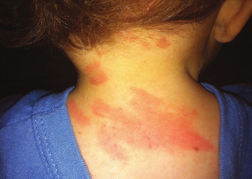
Cutaneous mastocytosis (CM) includes three variants: wheal and flare response when normal skin, not affected by
maculopapular cutaneous mastocytosis (MPCM), skin lesions, is stroked with a tongue depressor, finger nails
which includes urticaria pigmentosa (UP) and telangiectasia or other instrument. The nick-name for dermatographism is
macularis eruptiva perstans (TMEP), diffuse cutaneous skin writing disease.
mastocytosis (DCM), and cutaneous mastocytoma.1 The
taskforce recommends that telangiectasia macularis eruptiva A macule is a lesion that is flat and even with the
perstans (TMEP) be removed as a separate category because, surrounding skin, identified by a change in color compared
although some adult patients may have telangiectatic lesions to the surrounding skin.
on their chest, shoulders, neck and back, they may also A papule is a small bump or elevated lesion, up to 1 cm in
demonstrate maculopapular lesions in other places, therefore diameter, containing no visible fluid.
fulfilling criteria for MPCM. A nodule is a growth of abnormal tissue just below the skin.
A bulla is a large blister filled with fluid.
Most cases of pediatric mastocytosis fall into one of the
Telangiectasia is a vascular lesion formed by dilatation
above categories and may or may not include symptoms
of a group of small blood vessels.
of systemic mast cell activation, including anaphylaxis, as
a result of mediators released from the skin.3, 4 Pediatric
CM encompasses a variety of clinical manifestations. In VARIANTS OF CUTANEOUS MASTOCYTOSIS
children, some forms of CM will spontaneously resolve,
some will go on to be diagnosed as indolent systemic Maculopapular Cutaneous Mastocytosis (MPCM)/
mastocytosis (ISM), with a smaller percentage identified Urticaria Pigmentosa (UP)1
as well-differentiated systemic mastocytosis (WDSM).5
• May be seen in infants, children or adults
In most adults with skin lesions typical for mastocytosis • Adults presenting with maculopapular lesions have
(in particular, the maculopapular type), systemic disease a very high likelihood of systemic disease, most
will ultimately be found, leading to a diagnosis of systemic frequently indolent systemic mastocytosis (ISM)
mastocytosis, usually in an indolent form (indolent • Rarely, an adult presents with maculopapular lesions
systemic mastocytosis).1, 6 who does not have systemic disease, and has a
diagnosis of MPCM
Definitions1, 7 • Red maculopapular lesions tend to wheal when
scratched (positive Darier’s sign)
Darier’s sign is an important diagnostic finding of • Blister formation can occur with rubbing or stroking
patients with mastocytosis. It can be elicited by stroking of lesion and is associated with pruritis8
10 tmsforacure.org | Special Edition 2020-2021• Encompasses several clinical entities with different • Can appear at birth or early infancy; may persist into
outcomes, including: pitted melanotic macules, reddish adulthood, possibly as well differentiated systemic
brown telangiectatic macules, lightly pigmented mastocytosis (WDSM)5
papules, brownish papules, and small nodules • Blisters, some of which are hemorrhagic, and bullae
• This group is divided into two sub-variants may be present and dermatographism may be
° Monomorphic variant prominent
-M ostly seen in adults and in a small subgroup • Flushing is a common symptom
of children • Tryptase may be elevated due to increased mast cell
- S mall maculopapular lesions, similar in shape, burden in the skin and can be indicative of WDSM5
size and color
-A dults most typically express the KIT D816V Cutaneous Mastocytoma1
mutation in exon 17 of the KIT gene
- In adults, thigh, axilla, trunk, extremities and • Usually present at birth
neck may be involved • Elevated lesion(s) (up to a total of three lesions)
- 9 5% of adults diagnosed with ISM, 50% with which usually resolves during childhood
advanced systemic mastocytosis [systemic • Four cutaneous mastocytomas or more become a
mastocytosis with an associated hematologic diagnosis of MPCM
neoplasm (SM-AHN, formerly SM-AHMND) or • Multiple mastocytomas may evolve into adult WDSM5
aggressive systemic mastocytosis (ASM)] and
less than 50 % of mast cell leukemia patients References
exhibit this variant
1. Hartmann K, Escribano L, Grattan C, Brockow K, Carter MC,
- Children presenting with this form may have Alvarez-Twose I, et al. Cutaneous manifestations in patients with
increased serum tryptase and a tendency toward mastocytosis: Consensus report of the European Competence Network
on Mastocytosis; the American Academy of Allergy, Asthma &
systemic disease that persists into adulthood Immunology; and the European Academy of Allergology and Clinical
- T he type of lesions can vary during the course Immunology. J Allergy Clin Immunol. 2016 Jan;137(1): 35-45.
of the disease, i.e., nodules during infancy may 2. V alent P, Akin C, Escribano L, Fodinger M, Hartmann
K, Brockow K, et al. Standards and standardization in
turn into plaques at age 6 mastocytosis: consensus statements on diagnostics, treatment
° Polymorphic variant recommendations and response criteria. Eur J Clin Invest. 2007
Jun;37(6):435-53.
-M ostly seen in children
3. M atito A, Carter M. Cutaneous and systemic mastocytosis in
- C an be macular, plaque or nodular, with lesions children: a risk factor for anaphylaxis? Curr Allergy Asthma Rep.
of variable shape, color and size 2015 May;15(5):22.
-A lthough children typically express mutations 4. M eni C, Bruneau J, Georgin-Lavialle S, Le Sache de Peufeilhoux
L, Damaj G, Hadj-Rabia S, et al. Paediatric mastocytosis:
in exon 8, 9, 11 or 17 of the KIT gene, KIT a systematic review of 1747 cases. Br J Dermatol. 2015
mutations may be negative Mar;172(3):642-51.
-U sually involving head, neck and extremities 5. T orrelo A, Alvarez-Twose I, Escribano L. Childhood
mastocytosis. Curr Opin Pediatr. 2012 Aug;24(4):480-6.
-M ay involve blistering upon irritation until 3
years of age 6. B erezowska S, Flaig MJ, Rueff F, Walz C, Haferlach T,
Krokowski M, et al. Adult-onset mastocytosis in the skin is
- P rognosis is favorable with regression of highly suggestive of systemic mastocytosis. Mod Pathol. 2014
Jan;27(1):19-29.
disease in adolescence or young adulthood
7. Venes D, Thomas CL. Taber’s cyclopedic medical dictionary. 19
ed. Philadelphia: F.A. Davis Co.; 2001.
Diffuse Cutaneous Mastocytosis (DCM)1
8. C astells M, Metcalfe DD, Escribano L. Diagnosis and
treatment of cutaneous mastocytosis in children: practical
• Skin thickened, hyperpigmented and diffusely infiltrated recommendations. Am J Clin Dermatol. 2011 Aug 1;12(4):259-70.
• Can involve up to 100% of the skin with the trunk,
head and scalp heavily affected
tmsforacure.org | Special Edition 2020-2021 11Systemic Mastocytosis Variants
Systemic mastocytosis (SM) consists of a group of tryptase, and CD25 should be performed on sections
rare, heterogeneous diseases involving growth and of the biopsy.1-5
accumulation of abnormal mast cells (MC) in one or
multiple extracutaneous (non-skin) organ systems Recent Updates in Diagnosis
(Table 1). Standard technique can be used to obtain
an iliac crest bone marrow (BM) biopsy and aspirate A new diagnostic algorithm has been proposed by the
smear for diagnosis. Aspirated BM should be allocated European Competence Network on Mastocytosis for
for flow cytometry to assess for the presence of mast evaluating patients with suspected mastocytosis.6
cells with aberrant phenotype (i.e., co-expression Recommendations for KIT mutation analysis, including in
of CD25). Immunohistochemistry for KIT, mast cell peripheral blood, have also been recently published.7
Table 1. Major Variants of Systemic Mastocytosis8
ISM (Indolent systemic mastocytosis)
WHO criteria for SM met, MC burden low, +/- skin lesions, no C findings, no evidence of AHN
• Bone marrow mastocytosis: ISM variant with BM involvement, but no skin lesions
SSM (Smoldering systemic mastocytosis)
WHO criteria for SM met, typically with skin lesions, with 2 or more B findings, but no C findings.
Advanced Disease Variants
SM-AHN (SM with an associated hematologic neoplasm, formerly SM-AHNMD)
Meets criteria for SM and also criteria for an AHN (MDS, MPN, MDS/MPN, AML), or other WHO-defined myeloid
hematologic neoplasm, +/- skin lesions.
ASM (Aggressive systemic mastocytosis)
Meets criteria for SM with one or more C findings. No evidence of MCL, +/- skin lesions.
MCL (Mast cell leukemia)
Meets criteria for SM. BM biopsy shows a diffuse infiltration, usually compact, by atypical, immature MCs. BM
aspirate smears show 20% or more MCs.
Typical MCL: MCs comprise 10% or more of peripheral blood white cells. Aleukemic MCL: < 10% of peripheral blood
white cells are MCs. Usually without skin lesions.
*SM-AHN is the recently updated term from the 2016 WHO classification of mastocytosis;9 a lymphoproliferative disorder or plasma cell
dyscrasia may rarely be diagnosed with SM.
WHO: World Health Organization; BM: bone marrow; MC: mast cell; MDS: myelodysplastic syndrome; MPN: myeloproliferative neoplasm; MDS/
MPN: myelodysplastic syndrome/ myeloproliferative neoplasm overlap disorders; AML: acute myeloid leukemia.
12 tmsforacure.org | Special Edition 2020-2021Table 2. B and C Findings8
B Findings
BM biopsy showing > 30% infiltration by MCs (focal, dense aggregates) and serum total tryptase level > 200 ng/mL
Myeloproliferation or signs of dysplasia in non–MC lineage(s), no prominent cytopenias; criteria for AHN not met
Hepatomegaly and/or splenomegaly on palpation without impairment of organ function and/or lymphadenopathy on
palpation/imaging (> 2 cm)
C Findings*
Cytopenia(s): ANC < 1 x 10⁹/L, Hb < 10 g/dL, or platelets < 100 x 109/L
Hepatomegaly on palpation with impairment of liver function, ascites, and/or portal hypertension
Skeletal lesions: osteolyses and/or pathologic fractures
Palpable splenomegaly with hypersplenism
Malabsorption with weight loss from gastrointestinal tract MC infiltrates
*Must be attributable to the MC infiltrate.
INDOLENT SYSTEMIC MASTOCYTOSIS the systemic category, despite that 91% of patients
with WDSM have childhood onset of disease, with
The majority of adult patients fit into this category, fulfilling familial involvement in 39%. There is a heterogeneous
the criteria for indolent systemic mastocytosis (ISM).2, 10-12 presentation of lesions, maculopapular, nodular and
The bone marrow, gastrointestinal tract, skeletal system, diffuse cutaneous, that may involve a large percentage of
nervous system and skin may be affected. Some patients the skin.17 Severe mast cell symptoms can occur and the
may have enlarged livers and spleens and lymphadenopathy. variant may persist into adulthood in a low percentage
Mediator-related symptoms are common, but the grade of of cases. The mast cells often do not express CD25 or
bone marrow infiltration is low (usually less than 5 percent) CD2 that are part of the minor World Health Organization
with the bone marrow fulfilling the criteria for SM and (WHO) criterion for SM, but may have CD30. Also, roughly
80-90% of the patients exhibiting a positive D816V KIT 90% of WDSM patients don’t have the KIT D816V or
mutation. In most patients the serum tryptase concentration other exon 17 KIT mutations.17 Bone marrow analysis
exceeds 20 ng/mL, but a normal level of tryptase does identifies mast cells in WDSM patients as notably large,
not rule out either mastocytosis or another mast cell round, mature-appearing mast cells with the absence
activation disease. Treatment usually includes mediator- of the spindle-shaped mast cells typically seen in SM.15
targeting drugs, including antihistamines, but does not usually Baseline serum tryptase levels
require cytoreductive agents, although there are exceptions. Continued on page 14
Isolated bone marrow mastocytosis (BMM) is a variant of
indolent SM.12 BMM is characterized by the absence of
skin lesions, lack of multi-organ involvement, and an
increased incidence of anaphylaxis.13
91% of patients
with WDSM have
Well differentiated SM (WDSM) first described
in 200414, is reported in the literature as a rare childhood onset of
variant that fulfills the major criterion for SM
and continues to be studied by researchers.15-17 disease, with familial
WDSM is distinguished from pediatric
cutaneous mastocytosis by its inclusion in
involvement in 39%
tmsforacure.org | Special Edition 2020-2021 13Systemic Mastocytosis Variants
Continued from page 13
in these patients are usually lower than what is frequently MAST CELL LEUKEMIA21
detected in SM, except in a variable percentage of
children at onset. Imatinib mesylate has been used in In this extremely rare variant, mast cell leukemia (MCL)
some patients with severe cases of WDSM, since these patients fit the criteria for SM, and a bone marrow
patients do not usually carry the KIT D816V mutation, aspirate smear shows that 20% or more of the cells
which causes resistance to imatinib.18 are mast cells, or 10% or more mast cells are seen in
circulating blood.8, 21, 22 The mast cells have malignant
SMOLDERING SYSTEMIC MASTOCYTOSIS features. A 2014 international consensus proposal
recommends that MCL be separated into acute and
Smoldering systemic mastocytosis (SSM) was recently chronic23 subvariants based on whether or not C findings
moved out of the WHO ISM category and into its own (Table 2) are present.21 In addition, it recommends
category under SM.9 In SSM, two or more B findings, but a distinction between a primary form of MCL and a
no C findings (Table 2) are found and there is a greater secondary form that evolves from an existing mast cell
possibility that the disease will progress to a more neoplasm, such as ASM or mast cell sarcoma. There is a
aggressive variant. prognostic pre-phase identified in patients with ASM with
5-19% mast cells in bone marrow smears, associated with
Advanced Systemic rapid progression. It has been proposed that this condition
be called “ASM in transformation to MCL” (ASM-t).
Mastocytosis Variants8 Prognosis can be variable based on the form of disease;
life expectancy has been extended, in some cases, due
SM WITH AN ASSOCIATED HEMATOLOGIC to advances in cytoreductive therapy.24 It is important to
NEOPLASM (SM-AHN) note that myelomastocytic leukemia (MML), which is a
differential diagnosis, is not regarded by mast cell disease
SM-AHN is the recently updated term for SM-AHNMD specialists as a subvariant of MCL or SM and should be
from the 2016 WHO classification of mastocytosis.9 These considered a secondary condition.21
patients fit the criteria for SM and they fit the WHO criteria
for myelodysplastic syndrome (MDS), myeloproliferative References
neoplasm (MPN), MDS/MPN overlap disorder, or acute
1. Valent P, Akin C, Escribano L, Fodinger M, Hartmann K, Brockow K,
myeloid leukemia (AML), with or without skin lesions.8, 19, 20 et al. Standards and standardization in mastocytosis: consensus
Patients are treated for both the SM component and for the statements on diagnostics, treatment recommendations and
response criteria. Eur J Clin Invest. 2007 Jun;37(6):435-53.
associated hematologic neoplasm.
2. Alvarez-Twose I, Morgado JM, Sanchez-Munoz L, Garcia-
Montero A, Mollejo M, Orfao A, et al. Current state of biology and
AGGRESSIVE SYSTEMIC MASTOCYTOSIS diagnosis of clonal mast cell diseases in adults. Int J Lab Hematol.
2012 Oct;34(5):445-60.
In this rare variant, aggressive systemic mastocytosis 3. Horny HP, Sotlar K, Valent P. Mastocytosis: state of the art.
Pathobiology. 2007;74(2):121-32.
(ASM) patients fit the criteria for SM, with or without skin
4. Horny HP, Valent P. Diagnosis of mastocytosis: general
lesions, and also meet criteria for one or more C findings histopathological aspects, morphological criteria, and
(Table 2).8 Patients with ASM often require chemotherapy. immunohistochemical findings. Leuk Res. 2001 Jul;25(7):543-51.
5. Escribano L, Garcia Montero AC, Nunez R, Orfao A. Flow
cytometric analysis of normal and neoplastic mast cells: role in
diagnosis and follow-up of mast cell disease. Immunol Allergy Clin
North Am. 2006 Aug;26(3):535-47.
14 tmsforacure.org | Special Edition 2020-20216. Valent P, Escribano L, Broesby-Olsen S, Hartmann K, Grattan C, 16. Fried AJ, Akin C. Primary mast cell disorders in children. Curr
Brockow K, et al. Proposed diagnostic algorithm for patients with Allergy Asthma Rep. 2013 Dec;13(6):693-701.
suspected mastocytosis: a proposal of the European Competence
Network on Mastocytosis. Allergy. 2014 Oct;69(10):1267-74. 17. Alvarez-Twose I, Jara-Acevedo M, Morgado JM, Garcia-
Montero A, Sanchez-Munoz L, Teodosio C, et al. Clinical,
7. Arock M, Sotlar K, Akin C, Broesby-Olsen S, Hoermann G, immunophenotypic, and molecular characteristics of well-
Escribano L, et al. KIT mutation analysis in mast cell neoplasms: differentiated systemic mastocytosis. J Allergy Clin Immunol.
recommendations of the European Competence Network on 2016 Jan;137(1):168-78 e1.
Mastocytosis. Leukemia. 2015 Jun;29(6):1223-32.
18. Alvarez-Twose I, Gonzalez P, Morgado JM, Jara-Acevedo M,
8. Gotlib J, Pardanani A, Akin C, Reiter A, George T, Hermine O, et Sanchez-Munoz L, Matito A, et al. Complete response after
al. International Working Group-Myeloproliferative Neoplasms imatinib mesylate therapy in a patient with well-differentiated
Research and Treatment (IWG-MRT) & European Competence systemic mastocytosis. J Clin Oncol. 2012 Apr 20;30(12):e126-9.
Network on Mastocytosis (ECNM) consensus response
criteria in advanced systemic mastocytosis. Blood. 2013 Mar 19. Stoecker MM, Wang E. Systemic mastocytosis with
28;121(13):2393-401. associated clonal hematologic nonmast cell lineage disease:
a clinicopathologic review. Arch Pathol Lab Med. 2012
9. Arber DA, Orazi A, Hasserjian R, Thiele J, Borowitz MJ, Le Beau Jul;136(7):832-8.
MM, et al. The 2016 revision to the World Health Organization
classification of myeloid neoplasms and acute leukemia. Blood. 20. Wang SA, Hutchinson L, Tang G, Chen SS, Miron PM, Huh
2016 May 19;127(20):2391-405. YO, et al. Systemic mastocytosis with associated clonal
hematological non-mast cell lineage disease: clinical
10. Carter MC, Metcalfe DD, Komarow HD. Mastocytosis. Immunol significance and comparison of chomosomal abnormalities
Allergy Clin North Am. 2014 Feb;34(1):181-96. in SM and AHNMD components. Am J Hematol. 2013
Mar;88(3):219-24.
11. Valent P. Mastocytosis: a paradigmatic example of a rare disease with
complex biology and pathology. Am J Cancer Res. 2013;3(2):159-72. 21. Valent P, Sotlar K, Sperr WR, Escribano L, Yavuz S, Reiter A, et al.
Refined diagnostic criteria and classification of mast cell leukemia
12. Pardanani A. Systemic mastocytosis in adults: 2013 update on (MCL) and myelomastocytic leukemia (MML): a consensus
diagnosis, risk stratification, and management. Am J Hematol. proposal. Ann Oncol. 2014 Sep;25(9):1691-700.
2013 May 30.
22. Georgin-Lavialle S, Lhermitte L, Dubreuil P, Chandesris MO,
13. Z anotti R, Bonadonna P, Bonifacio M, Artuso A, Schena D, Rossini Hermine O, Damaj G. Mast cell leukemia. Blood. 2013 Feb
M, et al. Isolated bone marrow mastocytosis: an underestimated 21;121(8):1285-95.
subvariant of indolent systemic mastocytosis. Haematologica.
2011 Mar;96(3):482-4. 23. Valent P, Sotlar K, Sperr WR, Reiter A, Arock M, Horny HP.
Chronic mast cell leukemia: a novel leukemia-variant with distinct
14. Akin C, Fumo G, Yavuz AS, Lipsky PE, Neckers L, Metcalfe DD. morphological and clinical features. Leuk Res. 2015 Jan;39(1):1-5.
A novel form of mastocytosis associated with a transmembrane
c-kit mutation and response to imatinib. Blood. 2004 Apr 24. Gotlib J, Kluin-Nelemans HC, George TI, Akin C, Sotlar K,
15;103(8):3222-5. Hermine O, et al. Efficacy and Safety of Midostaurin in
Advanced Systemic Mastocytosis. N Engl J Med. 2016 Jun
15. Torrelo A, Alvarez-Twose I, Escribano L. Childhood mastocytosis. 30;374(26):2530-41.
Curr Opin Pediatr. 2012 Aug;24(4):480-6.
Mast Cell Sarcoma1, 2
Mast cell sarcoma is a rare tumor that may present in References
many different anatomic locations and age groups, and
prognosis is generally poor. Mast cell sarcoma is often 1. V alent P, Horny HP, Escribano L, Longley BJ, Li CY, Schwartz LB,
et al. Diagnostic criteria and classification of mastocytosis: a
misdiagnosed because the presenting cells bear little consensus proposal. Leuk Res. 2001 Jul;25(7):603-25.
resemblance to normal mast cells and spindle-shaped 2. H
orny HP, Akin C, Metcalfe DD, Escribano L, Bennett JM, Valent
mast cells frequently seen in systemic mastocytosis.3 The P, et al. Mastocytosis (mast cell disease) Swerdlow SH, Campo E,
Harris NL, Jaffe ES, Pileri SA, Stein H, et al., editors. World Health
cells of mast cell sarcoma more closely resemble “atypical Organization (WHO) Classification of Tumours. Pathology and
type II mast cells” or “promastocytes” that are associated Genetics. Tumours of Haematopoietic and Lymphoid Tissues. Lyon:
IARC Press; 2008.
with some cases of aggressive systemic mastocytosis.1, 3
3. R yan RJ, Akin C, Castells M, Wills M, Selig MK, Nielsen GP, et
Pathological examination of the tumor has shown it to be al. Mast cell sarcoma: a rare and potentially under-recognized
highly malignant with an aggressive growth pattern.3, 4 diagnostic entity with specific therapeutic implications. Mod
Pathol. 2013 Apr;26(4):533-43.
Patients with this tumor do not fulfill the criteria for SM.1
4. Georgin-Lavialle S, Aguilar C, Guieze R, Lhermitte L, Bruneau J,
The imatinib mesylate-resistant KIT D816V mutation has Fraitag S, et al. Mast cell sarcoma: a rare and aggressive entity-
not been found in reported mast cell sarcomas, such that -report of two cases and review of the literature. J Clin Oncol.
2013 Feb 20;31(6):e90-7.
use of imatinib has been attempted in some patients.3
tmsforacure.org | Special Edition 2020-2021 15Mast Cell Activation Syndrome Variants1-3
PRIMARY MCAS present with typical and recurrent signs and symptoms of
mast cell activation do not present with elevated levels of
Primary MCAS results from a clonal population of mast mediators for which we are currently able to test. Non-
cells, where a genetic alteration in the cells exists, and specialist physicians may most commonly use serum tryptase
may be due to mastocytosis or to monoclonal Mast levels to exclude a mast cell disease. However, some MCAS
Cell Activation Syndrome (MMAS). Primary MCAS specialists have indicated that tryptase rises are not seen
with mastocytosis can be diagnosed if the patient as often in patients with certain forms of MCAS, and that
fulfils criteria for MCAS and fulfills the WHO criteria for other changes in bloodwork and urine tests can sometimes
mastocytosis. MMAS is a distinct disease characterized be more reliable.13, 14 Additionally, there is a very narrow
by the presence of abnormal mast cells and fulfillment window of time (1-2 hours after symptoms begin) during
of criteria for MCAS, but where sufficient criteria for a which to obtain a serum tryptase test to indicate mast cell
diagnosis of mastocytosis are not identified.1-10 activation, 2 such that obtaining laboratory evidence of the
event can prove difficult in many circumstances. Some
SECONDARY MCAS specialists suggest that despite lack of proof of elevated
mast cell mediators, a response to mast cell or mast cell
Secondary MCAS is diagnosed when mast cell activation mediator blockers should be determined in such patients.12
occurs as an indirect result of another disease or If a patient responds well to anti-mediator treatment and
condition.1-3, 9, 11 Physician awareness of the presence of fulfills the other proposed criteria, 2 with the exception of
secondary MCAS will allow for more appropriate mast displaying a rise in mediators, then a diagnosis of idiopathic
cell activation-targeted treatments, in addition to primary MCAS remains open for consideration, as long as other
disease-related medications, to be provided. In addition diagnoses continue to be considered (please see Valent
to the widespread example of IgE-dependent allergy as a article noted below for more information on differential
cause of secondary MCAS, other diseases that can cause diagnoses). The patient should be periodically monitored
secondary MCAS have been reviewed in the literature.1-3, 11 to try to capture a rise in any of the mediators for which
commercial testing is both available and recognized as a
IDIOPATHIC MCAS widely accepted diagnostic standard.12
Idiopathic MCAS is proposed as a final diagnosis Even the co-criterion requiring a response to mast cell targeted
after proposed MCAS criteria have been fulfilled and therapy can be difficult to obtain in some patients. Sometimes
a thorough evaluation has excluded the possibility of multiple mast cell (or mast cell mediator) blocking therapies
another known underlying cause for this activation.2, must be tried before successful symptom resolution is
12
Idiopathic MCAS is therefore nonclonal, with regard attained.3, 16 Also, it is reported in another study, that only one
to current diagnostic capabilities related to mast cell third of MCAS patients experience a complete resolution with
analyses, and has been presented and discussed in the treatment; one third have a major response and another third
literature by a variety of mast cell disease specialists.1-3, have a minor response, and a combination of drugs is usually
9-13
Review of other causes of MCAS to aid physicians in
required to achieve control of symptoms.10
evaluation for the exclusionary diagnosis of idiopathic
MCAS have also been provided.1-3, 10
Please see the following article for more
Additional Considerations for MCAS information on mast cell activation syndromes,
including potential causes, symptoms, variants,
It is recognized by researchers that current diagnostic effects of comorbidities and other possible
methods for capturing a rise in mast cell mediators after a
diagnoses to exclude:
symptomatic episode are not ideal.12, 14, 15 Some patients who
16 tmsforacure.org | Special Edition 2020-2021Valent P. Mast cell activation syndromes: definition and 14. Molderings GJ, Brettner S, Homann J, Afrin LB. Mast cell
activation disease: a concise practical guide for diagnostic
classification. Allergy. 2013 Apr;68(4):417-24. workup and therapeutic options. J Hematol Oncol. 2011;4:10.
15. Afrin LB. Polycythemia from mast cell activation syndrome:
lessons learned. Am J Med Sci. 2011 Jul;342(1):44-9.
References
16. Afrin LB. Presentation, diagnosis and management of mast
cell activation syndrome. In: Murray DB, editor. Mast cells:
1. Akin C, Valent P, Metcalfe DD. Mast cell activation syndrome: phenotypic features, biological functions and role in immunity.
proposed diagnostic criteria. J Allergy Clin Immunol. 2010 Hauppauge: Nova Science Publishers, Inc.; 2013. p. 155-232.
Dec;126(6):1099-104 e4.
2. Valent P, Akin C, Arock M, Brockow K, Butterfield JH, Carter
MC, et al. Definitions, criteria and global classification of mast
cell disorders with special reference to mast cell activation
Hereditary Alpha
syndromes: a consensus proposal. Int Arch Allergy Immunol.
2012;157(3):215-25. Tryptasemia (HaT)
3. Valent P. Mast cell activation syndromes: definition and
classification. Allergy. 2013 Apr;68(4):417-24. Tryptase is a protein made primarily by mast cells and can
4. Akin C, Scott LM, Kocabas CN, Kushnir-Sukhov N, Brittain E, be used as a marker for mast cell activation. Hereditary
Noel P, et al. Demonstration of an aberrant mast-cell population alpha tryptasemia is an inherited genetic mutation causing
with clonal markers in a subset of patients with “idiopathic”
anaphylaxis. Blood. 2007 Oct 1;110(7):2331-3. extra copies of the alpha tryptase gene (TPSAB1), leading
to increased levels of tryptase in the blood. There is a
5. Valent P, Akin C, Escribano L, Fodinger M, Hartmann
K, Brockow K, et al. Standards and standardization in great variability from person to person with duplications
mastocytosis: consensus statements on diagnostics, treatment or triplications in terms of symptoms. If a patient’s
recommendations and response criteria. Eur J Clin Invest. 2007
blood tryptase level is above 10 ng/ml and he or she has
Jun;37(6):435-53.
another relative who has an elevated level, the patient is
6. Sonneck K, Florian S, Mullauer L, Wimazal F, Fodinger M,
Sperr WR, et al. Diagnostic and subdiagnostic accumulation of more likely to have hereditary alpha tryptasemia. Some
mast cells in the bone marrow of patients with anaphylaxis: patients with hereditary alpha tryptasemia may manifest
monoclonal mast cell activation syndrome. Int Arch Allergy the following symptoms: allergic-like symptoms such
Immunol. 2007;142(2):158-64.
as skin itching, flushing, hives, and even anaphylaxis;
7. Bonadonna P, Perbellini O, Passalacqua G, Caruso B, Colarossi
S, Dal Fior D, et al. Clonal mast cell disorders in patients with
gastrointestinal (GI) symptoms such as bloating, abdominal
systemic reactions to hymenoptera stings and increased pain, diarrhea and/or constipation (frequently diagnosed
serum tryptase levels. J Allergy Clin Immunol Pract. 2009 as irritable bowel syndrome or IBS), heartburn, reflux, and
Mar;123(3):680-6.
difficulty swallowing; connective tissue symptoms such as
8. Alvarez-Twose I, Gonzalez de Olano D, Sanchez-Munoz L, Matito
A, Esteban-Lopez MI, Vega A, et al. Clinical, biological, and
hypermobile joints and scoliosis; cardiac symptoms such as
molecular characteristics of clonal mast cell disorders presenting a racing or pounding heartbeat or blood pressure swings,
with systemic mast cell activation symptoms. J Allergy Clin
Immunol. 2010 Jun;125(6):1269-78 e2. sometimes with fainting; as well as anxiety, depression,
9. Akin C, Metcalfe DD. Mastocytosis and mast cell activation
chronic pain or panic attacks. It is not clear the extent to
syndromes presenting as anaphylaxis. In: Castells MC, editor. which activated mast cells contribute to this disease, nor
Anaphylaxis and hypersensitivity reactions. New York: Humana
Press; 2011. p. 245-56.
whether mast cell activation plays any role in symptoms,
but this is an area of ongoing research. Many patients do
10. Picard M, Giavina-Bianchi P, Mezzano V, Castells M. Expanding
spectrum of mast cell activation disorders: monoclonal and respond to mast cell mediator targeted medications. If your
idiopathic mast cell activation syndromes. Clin Ther. 2013
May;35(5):548-62. serum tryptase is 8 ng/ml or greater, there is a commercial
11. Valent P, Horny HP, Triggiani M, Arock M. Clinical and laboratory test offered by Gene by Gene labs. Symptoms are treated
parameters of mast cell activation as basis for the formulation of individually and definitive treatments have yet to be
diagnostic criteria. Int Arch Allergy Immunol. 2011;156(2):119-27.
identified for hereditary alpha tryptasemia. https://www.
12. C ardet JC, Castells MC, Hamilton MJ. Immunology and clinical
manifestations of non-clonal mast cell activation syndrome. Curr niaid.nih.gov/research/hereditary-alpha-tryptasemia-faq
Allergy Asthma Rep. 2013 Feb;13(1):10-8.
13. Hamilton MJ, Hornick JL, Akin C, Castells MC, Greenberger NJ.
Mast cell activation syndrome: a newly recognized disorder with
systemic clinical manifestations. J Allergy Clin Immunol. 2011
Jul;128(1):147-52 e2.
tmsforacure.org | Special Edition 2020-2021 17You can also read