Complex Interaction between Resident Microbiota and Misfolded Proteins: Role in Neuroinflammation and Neurodegeneration - MDPI
←
→
Page content transcription
If your browser does not render page correctly, please read the page content below
cells
Review
Complex Interaction between Resident Microbiota
and Misfolded Proteins: Role in Neuroinflammation
and Neurodegeneration
Juliana González-Sanmiguel 1,† , Christina M. A. P. Schuh 2,† , Carola Muñoz-Montesino 1 ,
Pamina Contreras-Kallens 2 , Luis G. Aguayo 1,3, * and Sebastian Aguayo 4,5, *
 1   Department of Physiology, Universidad de Concepción, Concepción 4070386, Chile;
     julygonzalezsanmiguel@gmail.com (J.G.-S.); carmunozm@udec.cl (C.M.-M.)
 2   Centro de Medicina Regenerativa, Facultad de Medicina Clínica Alemana, Universidad del Desarrollo,
     Santiago 7710162, Chile; cschuh@udd.cl (C.M.A.P.S.); paminaa.ck@gmail.com (P.C.-K.)
 3   Program on Neuroscience, Psychiatry and Mental Health, Universidad de Concepción,
     Concepción 4070386, Chile
 4   School of Dentistry, Faculty of Medicine, Pontificia Universidad Católica de Chile, Santiago 8331150, Chile
 5   Institute for Biological and Medical Engineering, Schools of Engineering, Medicine and Biological Sciences,
     Pontificia Universidad Católica de Chile, Santiago 7820436, Chile
 *   Correspondence: laguayo@udec.cl (L.G.A.); sebastian.aguayo@uc.cl (S.A.); Tel.: +56-41-2203380 (L.G.A.);
     +56-2-23548184 (S.A.)
 †   These authors contributed equally.

 Received: 13 October 2020; Accepted: 10 November 2020; Published: 13 November 2020

 Abstract: Neurodegenerative diseases such as Alzheimer’s disease (AD), Parkinson’s disease (PD)
 and Creutzfeldt–Jakob disease (CJD) are brain conditions affecting millions of people worldwide.
 These diseases are associated with the presence of amyloid-β (Aβ), alpha synuclein (α-Syn) and
 prion protein (PrP) depositions in the brain, respectively, which lead to synaptic disconnection and
 subsequent progressive neuronal death. Although considerable progress has been made in elucidating
 the pathogenesis of these diseases, the specific mechanisms of their origins remain largely unknown.
 A body of research suggests a potential association between host microbiota, neuroinflammation and
 dementia, either directly due to bacterial brain invasion because of barrier leakage and production of
 toxins and inflammation, or indirectly by modulating the immune response. In the present review,
 we focus on the emerging topics of neuroinflammation and the association between components of
 the human microbiota and the deposition of Aβ, α-Syn and PrP in the brain. Special focus is given to
 gut and oral bacteria and biofilms and to the potential mechanisms associating microbiome dysbiosis
 and toxin production with neurodegeneration. The roles of neuroinflammation, protein misfolding
 and cellular mediators in membrane damage and increased permeability are also discussed.

 Keywords: Alzheimer’s disease; Parkinson’s disease; Creutzfeldt-Jakob disease; neuroinflammation;
 microbiome; periodontal diseases; biofilms; membrane permeability

1. The Burden of Neurodegenerative Diseases
     Currently, nearly 50 million people worldwide suffer from neurodegenerative diseases (NDDs),
mainly dementia, and this number is expected to reach 152 million by 2050 [1]. It is noteworthy
that we are experiencing a shift in global demographics towards a large elderly population, which is
increasing the prevalence of neurodegeneration worldwide and the financial burden associated with
these diseases (e.g., medication, nursing care). For example, it is estimated that in the USA alone

Cells 2020, 9, 2476; doi:10.3390/cells9112476                                        www.mdpi.com/journal/cells
Cells 2020, 9, 2476                                                                                2 of 28

more than 5 million people aged 65 or older suffer from AD, and the costs of treating the disease are
estimated at over US$180 billion per year [2,3].
      In recent years, considerable progress has been made regarding the pathogenesis, diagnosis and
treatment of Alzheimer’s disease (AD), Parkinson’s disease (PD) and Creutzfeldt-Jakob disease (CJD).
However, these pathologies remain debilitating and fatal conditions, with significant negative medical,
economic and social impacts. To date, there are no effective therapeutic approaches to prevent, delay
or reverse these disorders, which start with cognitive loss and alterations of neurovegetative functions
and progress towards language deficit, memory loss, motor difficulties and ultimately death [4].
These neurodegenerative diseases are associated with neuronal loss in several regions of the brain,
such as the frontal cortex, hippocampus and basal ganglia. AD and PD can be classified as either
“early-onset, genetic” (also known as “familial”) or “late-onset, sporadic” [5]. Most significantly,
the late-onset forms are more prevalent and are considered to be the main cause of dementia and motor
disease in the elderly population [6].
      The most common neurodegenerative disease is AD, which is mainly characterized by marked
cognitive dysfunction, impairment in the formation of new memories, and synaptic failure [7,8].
AD hallmarks include intracellular neurofibrillary tangles and the extracellular deposition of senile
plaques that are mainly constituted by amyloid-β (Aβ) peptide [9]. Aβ is able to rupture the neuronal
plasma membrane by the formation of pores leading to cytoplasmic leakage and cell death [10,11],
by either direct lipid disruption or by its interaction with ion channels in the membrane [12,13].
Current research suggests that late-onset AD is mostly determined by environmental factors such as
toxins, trauma and diet [14]. However, the underlying mechanism of action has not been completely
elucidated yet.
      The second most common neurodegenerative pathology is PD. In 2016, 6.1 million people
worldwide were living with a diagnosis of PD, and it was estimated that 10 million people would
be suffering from this disease by 2020 [15]. The prevalence of PD ranges from 100 to 200 cases
per 100,000 people [16], and it affects nearly 3% of the population older than 65 years of age [17].
PD is mainly characterized by motor symptoms including bradykinesia, rigidity, tremor, postural
instability, dysphagia and axial deformities, and non-motor symptoms such as cognitive dysfunction,
sleep disorder, depression, anxiety, apathy, pain and dementia [17,18]. PD has been well described as
the intraneuronal deposition of alpha synuclein (α-Syn), which contributes to the generation of protein
inclusions known as Lewy bodies [19]. It is widely known that the loss of dopaminergic neurons in the
substantia nigra pars compacta is the landmark physiopathological sign of the disease [18]. Due to
the deleterious consequences of α-Syn, it has been considered a strategic target for future therapies to
ameliorate the symptoms and slow down the progression of the disease.
      Another relevant group of neurodegenerative disorders are prion diseases. There are three types
of human prion disorders: sporadic, genetic and acquired [20]. The most common form of prion
disease is sporadic CJD, a fatal pathology caused by misfolded prion proteins [20]. CJD is responsible
for 85% of diagnosed prion disease cases, with a reported incidence of 1–2 cases per million people per
year worldwide, and about 350 new annual cases in the United States [21]. The onset of CJD occurs in
patients older than 67 years of age [20]. The main features of CJD and prion diseases are spongiform
changes in gray matter, gliosis and neuronal death [22,23]. The most reported symptoms for CJD
are progressive dementia, behavioral and cognitive impairment, insomnia, movement disorder and
ataxia [24].
      As a common feature, all neurodegenerative diseases seem to be associated with protein misfolding
that leads to synaptic alterations, neuronal membrane damage and neuroinflammation. In addition,
it has been recently suggested that microbial components, such as the ones present in the host
microbiome, may also be actively involved in modulating neuroinflammation and protein misfolding.
Therefore, in the present review we focus on the emerging hypothesis regarding the role of the host
microbiome and its dysregulation in the onset of neurodegeneration, via (i) the entry of microbial cells,
toxins and outer membrane vesicles directly into the brain, and (ii) the induction and maintenance of a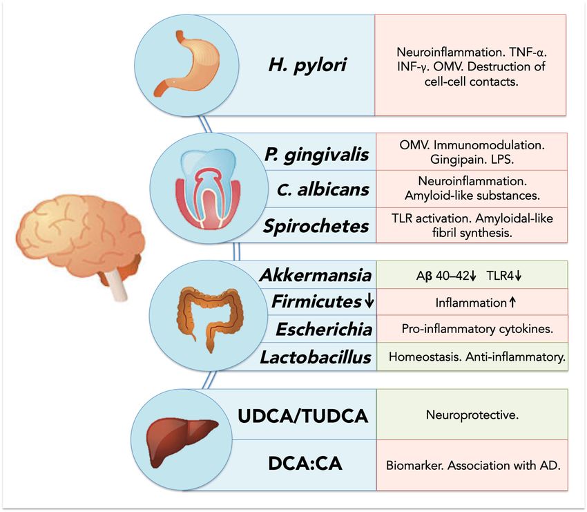
Cells 2020, 9, 2476                                                                                  3 of 28

systemic chronic inflammatory state. Furthermore, we discuss the involvement of associated systems
such as the oral microbiome and bile, and potential routes of entry for bacteria and toxins into the
central nervous system (CNS).

2. Protein Misfolding and Its Accumulation in Neurodegenerative Diseases
      Neurodegenerative pathologies are commonly characterized by the misfolding, oligomerization
and accumulation of toxic species such as Aβ in AD, α-Syn in PD, and the prion protein in CJD [24,25].
These protein alterations trigger neuronal degeneration and dysfunction and drive the progression of
each particular disease [25]. For instance, there is abundant evidence demonstrating that Aβ peptide
accumulation initiates and promotes AD. Aβ is mainly detected in the extracellular matrix in the
brain and cerebrospinal fluid (CSF) at nanomolar concentrations, and is widely accepted as the main
neurotoxic agent in the disease [26]. It is believed that early manifestations of AD are associated with
the synaptotoxic effects produced by soluble oligomeric forms of Aβ [27]. The existence of mutations in
genes for the amyloid-β precursor protein (AβPP) (chromosome 21) and presenilin 1 (chromosome 14)
and 2 (chromosome 1) have also been reported in some AD patients, providing further evidence that
Aβ is an important factor in the development of AD [28].
      A well-accepted hypothesis for AD generation is that monomers of Aβ oligomerize, first forming
low molecular weight species referred to as oligomers [27], which have been found to be highly
neurotoxic to the membrane [13]. There is no clear consensus about the most toxic species, but there is
an agreement that starting from dimers up to 56 kDa, oligomers are the most important causal agents
in the disease [29,30]. These peptides/proteins can associate and damage the cell membrane, affecting
neuronal function. The semipermeable property of the membrane is critical for cellular homeostasis, and
the resulting Aβ-induced leakage of cellular components, as well as the non-regulated calcium influx
into the cell, will turn into synaptotoxicity [13,31]. All the available evidence points to the idea that Aβ
toxic events are multiple and that one/several of them might serve as a therapeutic target. Likewise,
PD is mainly characterized by the formation of intracellular Lewy bodies in dopaminergic neurons.
These structures are mostly formed by intracellular accumulation of α-Syn [24], a 140-residue protein
encoded by the Synuclein Alpha (SNCA) gene that drives neurodegeneration. Prion diseases, on the
other hand, have spongiform vacuolation, gliosis, neuronal loss and deposition of amyloid molecules
immune-positive for prion protein (PrP) as hallmarks of the disease [24]. Thus, prion disorders
are caused by the misfolded form of the prion protein, denoted prion protein scrapie (PrPSc ) [32].
The toxic misfolded PrPSc has a high content of β-sheet in its secondary structure, which generates
a highly hydrophobic and insoluble protein with a high tendency to aggregate and form amyloid
structures [24,32].

3. Protein-Induced Membrane Damage as a Central and Ubiquitous Player in Neurotoxicity
      As discussed above, it is widely accepted that the accumulation of misfolded proteins is an
important hallmark for AD, PD and CJD. Most importantly, these proteins are capable of inducing
membrane damage in the brain by assembling monomers into non-selective ion pores and subsequently
inserting them into a variety of cell membranes. For example, α-Syn oligomers increase the permeability
of cell membranes in distinct types of neurons [33]. Additionally, α-Syn is also known to form pores in
phospholipid bilayers found in mitochondria, inducing a complex series of multilevel conductance
reminiscent of the effects of Aβ in hippocampal membranes [34]. α-Syn insertion into bilayers
is facilitated by cardiolipin, an important phospholipid present in mitochondrial membranes [34].
As mitochondria are key organelles for cell energetic and ionic homeostasis, membrane alterations by
oligomeric proteins can result in important alterations of cell viability.
      Recent data using nanoelectrospray and mass spectrometry have shown that Aβ42 oligomerizes
and forms β-barrel structure hexamers, which can be stabilized by the addition of lipids [35]. A similar
situation is observed for toxic oligomers of PrP, associated with cellular membranes, where they might
induce fast and prolonged toxic effects [36]. Studies in lipid bilayers, for example, have indicated that
Cells 2020, 9, 2476 4 of 28
PrP oligomers cause a rapid and large increase in the permeability of the membrane, whereas monomeric
forms cause no detectable leakage [36]. More recent studies using calcein-leakage assays showed
that soluble prion oligomers are capable of producing leakage in negatively charged vesicles [37].
Studies at the nanometer level with atomic force microscopy showed that a fragment of the human PrP
spanning residues 106–126 (PrP106–126 ) disrupted the intrachain conformation of phosphatidylcholine
lipids [38]. All these results support the idea that, similar to Aβ and α-Syn, PrP oligomers can disrupt
cell membranes. Further data regarding the relevance of these molecules in disease pathogenesis were
obtained using the PrP27–30 fragment extracted from the brains of terminally ill golden Syrian hamsters
infected with the 263K scrapie strain [39]. Interestingly, the electrophysiological recordings carried
out with PrP resembled membrane responses obtained with Aβ in native neurons, including high
variability on the amplitude of the unitary response and some spontaneous membrane breakages [11].
The responses showed a multistate conductance current, with at least one amplitude near 80 pS,
a reversal around 0 mV and dependency on cation concentration (Na+ and K+ ). In addition, using the
recombinant fragment of PrP (PrP90–231 ) a similar dependence on calcium was shown. In sum, AD,
PD and prion diseases are associated with membrane alterations, increases in calcium permeability
and ionic dyshomeostasis, which contribute to neurodegeneration. Most importantly, potentiation
of local brain factors with other peripheral inflammatory mediators (such as those derived from a
dysbiotic gut) may be associated with the progression of neurodegenerative diseases.
4. Neuroinflammation as a Common Factor across Neurodegenerative Diseases
As previously noted, synaptic and cellular alterations mediated by misfolded protein accumulation
are the main hallmarks across NDDs [40]. However, all these diseases also share the common ground of
displaying an increased inflammatory response in the brain, known as neuroinflammation. This process
involves the activation of resident microglia and astrocytes that produce cytokines, chemokines and
other inflammatory molecules within the CNS. Many of these markers are universal across NDDs,
supporting the idea of a common neuroinflammatory profile across these diseases. Some of these
common neuroinflammation mediators are chitotriosidase 1 (CHIT1), chitinase-3-like protein 1
(YKL-40), the glial fibrillary acidic protein (GFAP) and important pro-inflammatory cytokines, such as
interleukin-1β (IL-1), IL-6 and tumor necrosis factor α (TNF-α) [41,42].
In general, for proteinopathies such as AD, PD and prion diseases, it has been shown that
neuroinflammation can be directly induced by amyloids. In the context of AD, not only do reactive
microglia colocalize with amyloid deposits in situ, but also in vitro Aβ oligomers have been shown
to directly induce microglial activation [43–47]. Characterization of inflammatory molecules in CSF
and plasma from AD patients has shown increased levels of pro-inflammatory cytokines such as
IL-1β, IL-6 and TNF-α [42] and increases in the macrophage colony-stimulating factor, which has
been described as a microglial activator [48]. Similarly, animal models of AD such as TgAPPsw and
PSAPP transgenic mice also show an increase in a pro-inflammatory profile characterized by cytokines
IL-1, IL-6 and TNF-α, and the granulocyte macrophage colony stimulating factor. This observation
is consistent with in vitro studies using microglial cell cultures exposed to Aβ42 [48]. IL-12 and IL-23
were produced by microglia in AD transgenic mice models (APP/PS1), and the genetic ablation of these
cytokines resulted in a decrease in cerebral amyloidosis [49].
In PD, microglia activation in the SNpc and striatum is well documented in murine models [50].
However, most of the microglial activation by α-Syn misfolding has been attributed to a deleterious
pro-inflammatory response that is related to dopaminergic neuron degeneration [50,51]. As for AD,
the cytokine profile in PD brains is characterized by the release of pro-inflammatory molecules IL-1β,
IL-6, IL-12, interferon gamma (IFN-γ) and TNF-α [50,52]. Therefore, microglial response is an early
marker of neuroinflammation in NDDs and seems to be the first mediator in the innate immune
reaction in the CNS in these pathologies.
Studies in human prion diseases indicate that microglial activation correlates with the onset of the
clinical signs and that its magnitude depends on prion strain [53,54]. Nevertheless, clustering analysisCells 2020, 9, 2476 5 of 28
of neuroinflammatory gene expression performed in different brain regions of prion-infected mice
suggested that astrocyte function is altered before microglia activation [55]. In this sense, transient prion
neuroinflammation events show only partial similarity with the microglia degenerative phenotype
reported in animal models of other NDDs, where microglial activation precedes astrogliosis. Regarding
cytokine profiles in prion-induced neuroinflammation, similar markers to AD and PD such as TNF-α,
IL-1β and particularly IL-1α are significantly increased in brain tissue from infected mice and CJD
patients [56,57].
Since microglia are a key element in neuroinflammatory responses, and a predominantly
inflammation-linked cytokine profile is found in AD, PD and prion diseases, microglial activation
in these pathologies is considered to be associated with the pro-inflammatory M1 phenotype [58].
Nevertheless, anti-inflammatory cytokines such as IL-4, IL-10 and IL-13 are increased and have
been detected in the striatum of PD patients [50]. Furthermore, increased levels of IL-4 and IL-10
have been found in CSF samples from AD [59] and CJD patients [60,61]. Due to recently developed
one-cell transcriptome analyses, it has been possible to separately define specific phenotypic changes
in microglia, astrocytes and neurons. In AD, these analyses have revealed microglial subpopulations
with a distinctive molecular signature different from the classical M1 and M2 phenotype, which has
led to the concept of disease-associated microglia (DAM) [62,63]. Two main receptors have been
identified as key regulators in the generation of these particular phenotypes in neurodegenerative
diseases: Toll-like receptors (TLRs) and triggering receptors expressed on myeloid cells-2 (TREM2) [62].
TREM2 interacts with two adaptor proteins, DAP12 and DAP10 [62]. Mutations in these proteins have
been linked to AD, PD and other misfolding-related neurodegenerative disorders [64]. Even though the
role of TREM2 signaling in neurodegeneration has not been defined, since both protective and harmful
responses have been described, TREM2 has a clear role in the induction of the DAM phenotype [62].
For instance, in 5xFAD mice (a transgenic model of AD), single-cell transcriptome analyses revealed
the existence of two DAM microglia clusters. Both clusters exhibited downregulation of homeostatic
genes and upregulation of a particular signature that includes TREM2. In addition, TREM2 can act as a
receptor for Aβ [65,66]. Similar microglial disease-specific phenotypes, distinguishable from the classic
M1 phenotype induced by lipopolysaccharide (LPS), have been observed in other neurodegenerative
disorders such as amyotrophic lateral sclerosis and multiple sclerosis [55,62]. Nevertheless, it is
important to highlight that probably both elements, classical M1 and DAM, might be relevant in the
progression of these diseases, with M1 contributing to the detrimental neuroinflammatory effects [62].
On the other hand, TLRs include 13 members that recognize different molecular patterns
associated with pathogens, with LPS being one of the classical TLR inductors [62]. Besides pathogens,
misfolded proteins may induce TLRs. In this sense, both α-Syn and Aβ have been described
as TLR ligands [67,68]. Furthermore, some bacterial metabolites have also been described as
ligands for TLR2 and TLR4 [69]. TREM2 has been also found to bind LPS, which is the most
well-characterized bacterial-derived molecule in neurodegenerative disease models [70]. LPS is able to
activate pro-inflammatory responses and contribute to detrimental effects in AD, PD and Huntington’s
disease [71]. In the early stages of prion disease in ME7 prion strain-infected mice, LPS injection
leads to exacerbated impairment in locomotor and cognitive functions [72]. Overall, LPS inoculation
experiments suggest that bacteria-derived products could accelerate disease progression and contribute
to neuronal decline.
Overall, activation of microglia is linked to the production of pro-inflammatory cytokines known
to have deleterious effects when increased in tissues, including the brain [73]. IL-1β and TNF-α are
able to reduce synaptic plasticity after acute application in brain slices [73]. Additionally, neurons
express cytokine receptors that stimulate the mitogen-activated protein kinase (MAPK) family and
lead to a reduction in synaptic efficiency [73]. Several calcium signaling mechanisms, including
N-methyl-D-aspartate receptors (NMDARs), inositol trisphosphate receptor, ryanodine receptors and
voltage-sensitive Ca2+ channels (VSCCs), may be modulated by cytokines in neurons. In this sense,
increased levels of TNF-α can trigger calcium release from intracellular compartments and increase theCells 2020, 9, 2476 6 of 28
expression of L-type VSCC. Neurons also express IL-1RAcPb, a neuron-specific IL-1 receptor accessory
protein relevant for IL-1β binding that has been linked to an alternative phosphorylation pathway
through Src phosphorylation, which is able to enhance Ca2+ influx through NMDAR activation [73].
In conclusion, AD, PD and prion diseases show early features of neuroinflammation that
can be directly linked to misfolded protein deposition, which in turn triggers a specific microglia-
and astrocyte-activated phenotype. However, external sources of neuroinflammation, different
from those directly related to misfolded proteins, are also able to increase neuronal damage.
In this sense, systemic inflammation could play an important role in the onset and maintenance
of neuroinflammation; thus, the most recent evidence regarding the association between oral and gut
microbiota and the promotion of an inflammatory state will be discussed, as well as its potential link
with neurodegenerative diseases.
5. Human Microbiome Dysbiosis as a Source of a Systemic Chronic Inflammatory State
It is currently known that humans are inhabited by a wide and diverse range of microorganisms
including bacteria, viruses and fungi, among others. These microorganisms, conjunctively known
as the human microbiome, are compartmentalized in different areas of the human body such as the
oral cavity, skin and gut; thus, each one of these “niches” holds a specific microbial composition.
It is currently believed that we carry around more microbial cells on a daily basis than our own
human cells [74]. Recently, it has been demonstrated that an overall healthy microbiome is crucial
for maintaining homeostasis, and that imbalances in microbiota composition (i.e., dysbiosis) can
lead to disease in many tissues and organs [75]. Systemic diseases such as cardiovascular disease,
diabetes mellitus, rheumatoid arthritis and obesity are all believed to have a direct association with
microbiome dysregulation, either via the direct effect of certain pathologic species or due to modulation
of the host inflammatory response [76].
5.1. Human Biofilms: 3D Microbial Structures in Health and Disease
Most of our microbiome is not found in an unattached form, but instead as part of complex
microbial communities known as biofilms. Biofilms are ubiquitous microbial structures found in most
biological and non-biological environments [77]. In the human body, biofilms consist of surface-bound
polymicrobial communities surrounded by an extracellular matrix that protects the biofilm from external
injury such as mechanical forces or antibiotics [78,79]. Thus, biofilms are crucial for enhancing bacterial
survival within the host. The formation of these biofilms is initiated by the attachment of bacteria
onto surfaces, followed by bacterial division and the formation of a complex community. These biofilms
are widespread throughout skin and mucosal surfaces within the mouth, gut, reproductive tract and
urinary tract. Most importantly, a wide diversity of species within the biofilm is crucial for health,
and dysregulation of residing species or imbalance in the number of organisms can lead to disease
(known as biofilm-mediated infections or diseases) [80]. These biofilm imbalances are known to cause
disease either by increasing the number of specific pathogenic species or by modulating the immune
response towards chronic and/or destructive inflammation.
Oral biofilm-mediated diseases are good examples of the consequences of biofilm dysregulation
within a specific niche. Dental caries, one of the most prevalent causes of dental pain and discomfort,
is caused by a significant rise in the numbers of acid-producing species (such as Streptococcus mutans
and lactobacilli) within the dental biofilm [81]. These acids are able to demineralize dental surfaces,
which subsequently leads to cavitation and disease progression into deeper tissues within the tooth.
One of the key factors behind the bacterial imbalance observed in dental caries is an increase in refined
sugar consumption, and thus specific policies and strategies have been implemented worldwide in
order to reduce sugar use in the population [81–83]. Furthermore, periodontal disease, a destructive
inflammatory disease that affects the supporting tissues of teeth, is believed to arise from an imbalance
of microbial species within the subgingival dental biofilm [84]. In periodontal disease, some specific
pathogenic strains such as Porphyromonas gingivalis are able to increase their number within the dentalCells 2020, 9, 2476 7 of 28
biofilm and trigger a destructive inflammatory response by the release of proteases, enzymes and other
bacterial components, as well as by modulating biofilm composition towards a dysbiotic state [84–86].
Interestingly, bacterial strains involved in oral and gut dysbiosis are known to play key roles in
the development and progression of systemic diseases such as heart valvulopathies, diabetes mellitus,
pre-eclampsia, rheumatoid arthritis and AD, among others [75,87–90]. Thus, local dysbiosis of
oral and gut microbiota is known to not only impact local tissues but also affect distant organs,
and there is mounting evidence that microbial elements may be associated with the development of
neuroinflammation and neurodegeneration within the brain. Therefore, for the purposes of this review,
we will focus on discussing recent evidence associating relevant oral and gut microbiota, as well as
their dysbiosis, with AD, PD and prion disease.
5.2. Resident Oral Microorganisms and Their Association with AD and Neuroinflammation
Until recently, it was mostly believed that resident oral bacteria were only capable of generating
disease confined within the oral cavity. However, current research has demonstrated that oral microbes
are indeed associated with a wide range of systemic diseases and remote infections in other tissues
and organs [88]. Although many oral species have been examined, for the purpose of this review we
will focus on the most relevant organisms believed to be implicated with neurodegeneration.
5.2.1. Porphyromonas gingivalis: Link between Periodontal Disease and Neurodegeneration?
One of the most prevalent oral diseases in adults and the elderly is periodontal disease.
Although highly multifactorial, periodontal disease has an important bacterial component.
Recent theories suggest that periodontal disease arises from dysregulation of the oral microbiome,
which allows the overgrowth of highly virulent bacterial strains, paired with a destructive immune
response from the host [91].
One of the most relevant bacteria in periodontal disease is P. gingivalis, a Gram-negative anaerobic
bacterium that is part of the resident oral microbiome [92]. However, an increase in its proportion relative
to other local microorganisms is associated with periodontal disease and tissue destruction [93–95].
Within periodontal disease pathogenesis, P. gingivalis is considered a “keystone” pathogen, as minor
variations in its number within the biofilm can trigger enormous changes in the local environment [84].
Among others, P. gingivalis is known to modulate the host immune response in a biphasic manner:
initially promoting inflammation to increase nutrient availability and biofilm growth, but subsequently
facilitating bacterial resistance by destroying complement factors [96,97].
There is increasing evidence suggesting an important link between P. gingivalis and AD.
Firstly, there is physical evidence of P. gingivalis components in brain samples of patients with AD.
A recent study found P. gingivalis to be present in the brain of AD patients [98]. These authors also
found gingipain, a toxic endopeptidase produced by P. gingivalis, to be present in AD brains and
correlated with tau protein production. The inhibition of gingipain reduced infection of the brain and
reduced neuroinflammation and Aβ42 production [98]. In another study, P. gingivalis-derived LPS
was found in brain samples from AD patients [99]. These data are in line with previous work linking
the presence of LPS from other Gram-negative bacteria, such as Escherichia coli, with increased Aβ
deposition [100–102]. Recently, Haditsch et al. demonstrated that neurons derived from inducible
pluripotent stem cells can be infected by P. gingivalis in vitro. Bacteria were found within the cytoplasm
and lysosomes of affected neurons, which led to the formation of autophagic vacuoles, cytoskeleton
disruption and loss of synapses [103]. Animal models have also demonstrated that P. gingivalis can
migrate into the brain, as researchers demonstrated that ApoE-/- mice infected with P. gingivalis were
able to develop brain infections with the microorganism [104]. Another study by Ilievski et al. found
that mice exposed to P. gingivalis developed neuroinflammation, neurodegeneration and extracellular
deposition of Aβ [105].
Secondly, P. gingivalis may be linked with AD via its ability to modulate systemic inflammation.
P. gingivalis (and other periodontal bacteria) is also known for promoting chronic inflammatory diseasesCells 2020, 9, 2476 8 of 28
such as diabetes, atherosclerosis and hypertension, which are also believed to be risk factors for
the development of AD [106]. Kamer et al. found increased levels of TNF and antibodies against
periodontal pathogens, including P. gingivalis, in AD patients compared to normal controls, suggesting
an important link between periodontal bacteria and systemic inflammatory levels [107].
Furthermore, outer membrane vesicles (OMVs) may also play an important role in the development
of AD. OMVs are 20–250 nm spherical buddings of the bacterial outer membrane containing lipids,
proteins or nucleic acids [108,109]. For decades they were believed to be mostly a by-product of cell
lysis; however, it is now known that their biogenesis is a deliberate and independent process [110].
Functions of OMVs have been associated with quorum sensing [111], as well as the distribution of
virulence factors [112,113] and antibiotic resistance [114]. Within the gut microbiome, they have been
shown to play a crucial role in gut homeostasis [115], carrying digestive enzymes [116] and modulating
immune responses [117]. In the case of P. gingivalis, OMVs are important for immune response
dysregulation and avoidance, tissue disruption and biofilm co-aggregation [118,119]. P. gingivalis
OMVs are known to contain gingipain, LPS and other bacterial constituents [119–121], and previous
research has demonstrated that OMVs are able to permeate the blood–brain barrier (BBB) [122] and
thus could potentially be an important mechanism for entry into the CNS.
Finally, some recent data have also suggested the potential involvement of P. gingivalis in other
NDDs such as PD. Adams et al. have demonstrated that RgpA protease produced by P. gingivalis is
present in platelet-poor plasma clots from PD patient blood samples, and that P. gingivalis-derived LPS
can induce hypercoagulability [123]. It is also believed that the systemic inflammatory state promoted
by P. gingivalis and periodontal disease may also play an important role in PD pathogenesis [124].
However, further research is necessary to continue to unravel the association between this key bacteria
and PD.
5.2.2. Oral Spirochetes and Brain Infection
Another relevant group of microorganisms that has been associated with AD and brain infection
is the spirochetes, and among these, dental spirochetes. Spirochetes are helical-shaped motile bacteria,
with a remarkable ability to penetrate into tissues and disseminate infection [125]. Among these,
Treponema denticola is regarded as an important periodontal pathogen, as its overgrowth is observed
in periodontal disease sites and associated with tissue destruction. Spirochetes have been observed
in the blood, CSF and brain tissue of AD patients [126], and recent investigations have identified
numerous oral spirochetes as potential key players in brain infection in AD.
Riviere et al. found evidence for the presence of six oral Treponema species, namely, T. amylovorum,
T. denticola, T. maltophilum, T. medium, T. pectinovorum and T. socranskii, in the frontal cortex of AD patients.
Of 16 analyzed AD brains, 14 were positive for Treponema, versus only 4 out of 18 non-AD patients [127].
Authors also found evidence of oral Treponema within the trigeminal nerves and ganglion, and thus
suggested that the microorganism is able to reach the brain directly via the peripheral nervous system
instead of through the bloodstream. Furthermore, by employing a mouse model, Foschi et al. detected
DNA from T. denticola in the brain and spleen of mice after dental pulp infection, further strengthening
the idea that oral spirochetes can disseminate into the brain through both vascular and peripheral
nerve routes [128].
The mechanisms behind the association between brain spirochetosis and AD remain debated.
Spirochetes are believed to activate TLR on glial cells via CD14 and induce cytokine and
pro-inflammatory molecule production, suggesting a potential mechanism of involvement in
neuroinflammation and neurodegeneration [129,130]. Moreover, some authors suggest that some
species of spirochetes are capable of synthetizing amyloidal-like fibrils [131], and previous research has
suggested that Aβ itself may be produced in the brain as an antimicrobial peptide against invading
pathogens [132]. A combination of bacterial-derived amyloid-like fibrils with local Aβ deposition
and misfolding could potentiate neuroinflammation and potentially explain the association between
spirochetes and neurodegeneration seen in some patients.Cells 2020, 9, 2476 9 of 28
5.2.3. Oral Fungi and Brain Infection and Inflammation
Another important component of the oral microbiome is fungi. Species such as Candida albicans are
found ubiquitously on oral surfaces and are part of commensal biofilms. However, due to imbalances
such as antibiotic usage or immunosuppressive conditions, they can overproliferate and cause local
diseases such as oral candidiasis [133]. Interestingly, research has suggested that fungal infections
can also migrate into the bloodstream and disseminate to distant tissues and organs. Recent studies
have found the presence of fungal infection in the brains of AD patients [134]. Alonso et al. found
evidence of fungal invasion in blood serum of AD patients, including C. albicans [135], and in a further
study observed the presence of both fungal and microbial species in AD brain samples [136]. Similarly,
Pisa et al. reported the presence of fungal material in the frontal cortex of AD patients, which was
also found intracellularly [136], and further found fungal strains such as Candida spp., Malasezzia
spp. and Sacharomyces cerevisae both intra and extracellularly in brain samples from AD patients [137].
Therefore, it is believed that the presence of C. albicans and other fungi inside the brain is associated
with the development of AD [138]. There are many potential mechanisms explaining how fungal
infection of the brain may promote AD. Most notoriously, Soscia et al. have shown that Aβ has
an antimicrobial peptide behavior against C. albicans [132]; thus, Aβ deposition may be part of a
neuroinflammatory response to clear the fungi from the brain. It is also known that disseminated
fungal invasion can increase systemic cytokine production and activate both innate and adaptative
immunity [139], which could potentiate neuroinflammation in the brain. Furthermore, some fungi
such as Candida have the ability to secrete amyloid-like substances that may serve a similar function
to Aβ inside the brain [140]; however, the effect of these fungal amyloid-like molecules on neuronal
viability remains to be explored.
Interestingly, a recent study by Wu et al. employed a mouse model to generate C. albicans
intravenous infections and observed the development of neuroinflammation, and accumulation of
activated glia cells and Aβ around yeast cells. Within the brain, activation of transcription factor NF-κB
and increases in IL-1β, IL-6 and TNF-α were also observed as a result of C. albicans invasion, which
activated the local innate immune response. As a result of this neuroinflammation, infected mice
showed mild memory impairment associated with Candida infection, which cleared after antifungal
treatment [141]. Overall, fungal invasion of the brain appears to induce local neuroinflammation
via similar molecules to the ones traditionally described in NDDs, and thus may potentiate
neurodegeneration in some patients.
5.3. Resident Gut Bacteria and Their Association with Neuroinflammation and Neurodegeneration
Similar to the oral microbiota, the gut is home to trillions of resident microorganisms that are
essential for our health and well-being, and that are able to influence health and disease locally and
systemically. The resident gut microbiota participates in numerous important processes such as
nutrient digestion and local gene expression and immune system regulation [142]. Most importantly,
alterations in gut microbiota composition are associated with the onset and progression of many chronic
inflammatory diseases in humans (reviewed by [143] and [90]). Among these remote effects, it has
been shown that there is an intimate bidirectional connection between the gut and brain, known as the
“gut-brain axis”, which is believed to regulate behavior, anxiety and pain [144,145]. There are currently
many potential explanations associating alterations in the gut microbiome with NDDs, including the
passage of microbial cells and products into the brain as well as potentiation of neuroinflammation via
inflammatory mediator production (Figure 1).Cells 2020, 9, 2476 10 of 28
Cells 2020, 9, x FOR PEER REVIEW 10 of 29
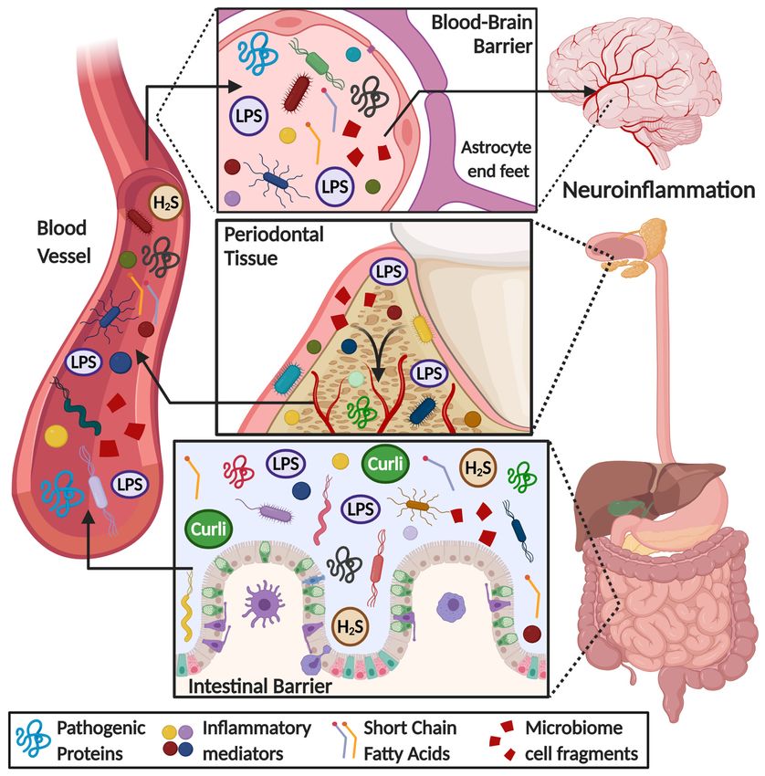
FigureFigure 1. Overview
1. Overview of of
thetherole
roleofofgut
gutand
and oral
oral microbiome
microbiomeininthe onset
the of of
onset neuroinflammation
neuroinflammation in in
neurodegenerative diseases. Some microbial products, such as lipopolysaccharide (LPS),
neurodegenerative diseases. Some microbial products, such as lipopolysaccharide (LPS), short chain short chain
fatty acids, hydrogen sulfide (H2S), amyloid-like substances (i.e., curli protein), bacteria cell fragments
fatty acids, hydrogen sulfide (H2 S), amyloid-like substances (i.e., curli protein), bacteria cell fragments
and pro-inflammatory mediators (i.e., cytokines, chemokines, ROS species), are released into the
and pro-inflammatory mediators (i.e., cytokines, chemokines, ROS species), are released into the
bloodstream because of an increase in gut-blood barrier permeability. These metabolites flow through
bloodstream because of an increase in gut-blood barrier permeability. These metabolites flow
the circulatory system reaching the brain, where they can permeate a weakened blood–brain barrier,
through the circulatory
triggering system reaching
a neuroinflammatory the brain,
response where
and they canthe
worsening permeate a weakened
pathological blood–brain
hallmarks of
barrier, triggering a neuroinflammatory
neurodegenerative diseases. response and worsening the pathological hallmarks of
neurodegenerative diseases.
5.3.1. Gut Microbiota Dysbiosis Generates a Pro-Inflammatory State
5.3.1. Gut Microbiota Dysbiosis Generates a Pro-Inflammatory State
Observational studies in recent years have suggested an association between gut microbiota
Observational studies
alterations and AD. Vogtinetrecent years have
al. observed suggested
that AD an association
patients have reduced gut between
microbial gut microbiota
diversity
alterations
compared andtoAD. Vogtasetwell
controls, al. asobserved that changes
compositional AD patients
such ashave reduced
decreased gut microbial
Firmicutes diversity
and increased
Bacteroidetes compared to control patients [146]. In a recent study, Sanguinetti et
compared to controls, as well as compositional changes such as decreased Firmicutes and increased al. showed that
mice in a pre-dementia state have reduced microbial gut diversity and altered bacterial
Bacteroidetes compared to control patients [146]. In a recent study, Sanguinetti et al. showed that proportions
mice compared to control mice
in a pre-dementia state[147].
haveAnother
reduced study by Minter
microbial gutetdiversity
al. utilizing thealtered
and APPSWE/PS1 DE9 AD mouse
bacterial proportions
model demonstrated that shifting gut microbiome composition with antibiotic treatment decreased
compared to control mice [147]. Another study by Minter et al. utilizing the APPSWE /PS1DE9 AD mouse
Aβ plaque deposition and alterations in cytokine and chemokine levels in circulation, such as the
model demonstrated that shifting gut microbiome composition with antibiotic treatment decreased Aβ
increase in CCL11 believed by the authors to lead to Aβ phagocytosis in the brain [148]. Interestingly,
plaque deposition and alterations in cytokine and chemokine levels in circulation, such as the increase
in CCL11 believed by the authors to lead to Aβ phagocytosis in the brain [148]. Interestingly, germ-free
APP transgenic mice show a significant reduction of Aβ pathology in the brain, strengthening the
notion of microbial involvement in AD pathogenesis [149].Cells 2020, 9, 2476 11 of 28
There is also mounting evidence of a correlation between gut microbiota and PD. Forsyth et al.
found an association between increased gut leakiness and the presence of PD, which was also
accompanied by an increase in E. coli and α-Syn in the intestine [150]. The authors suggested that this
local increase in α-Syn may be a consequence of the pro-inflammatory state in the region generated
by microbial components such as LPS. Research by the same group found significant differences in
the composition of fecal microbiota between PD and healthy patients, as PD patients had decreased
amounts of Firmicutes compared to controls [151], similar to what is observed in AD patients [146].
In the same study, authors also noted that PD duration was positively correlated with Bacteroidetes and
negatively correlated with Firmicutes. Interestingly, they also observed that genes involved in pathways
such as LPS biosynthesis and bacterial secretion were increased in PD patients compared to controls.
Recently, Sampson et al. observed that bacteria that produce curli, a bacterial aggregating amyloid,
were found to promote α-Syn pathology in both the gut and brain and potentiate motor abnormalities
in a mouse model [152]. Although there are still doubts as to how intestinal α-Syn and amyloid
formation may impact the brain in PD, some research has suggested the possibility that α-Syn may
spread via the vagus nerve to the brainstem [153].
Furthermore, in a recent clinical study, Cattaneo et al. found that cognitively impaired patients
with brain amyloidosis expressed a decreased abundance of the anti-inflammatory Eubacterium rectale
and a higher abundance of inflammatory strains such as Escherichia and Shigella compared to healthy
controls and amyloid-free cognitively impaired patients [154]. These microbiota alterations were
associated with increased levels of pro-inflammatory cytokines such as IL-6, NLRP3, CXCL2 and IL-1β
in the amyloid-positive group and correlated with the overabundance of Escherichia/Shigella. Overall,
it seems that the gut microbiota composition is crucial in maintaining inflammatory homeostasis,
and alterations of diversity or relative proportions between species can trigger or maintain chronic
inflammatory states by the modulation of pro-inflammatory cytokine production, among others.
Further strengthening this hypothesis is the fact that probiotic treatments that regulate imbalances in
the gut microbiota have shown an important protective effect against inflammation, cognitive decline
and AD development [155–160]. Administration of probiotic strains such as Lactobacillus plantarum P8
was recently shown to improve cognition, learning and memory in a group of stressed adults [161].
Regarding potential mechanisms behind the neuroprotective effect of probiotic administration,
Bonfili et al. observed that a probiotic formulation of lactic acid and bifidobacteria was able to potentiate
the proliferation of anti-inflammatory species, which in turn modulated gut hormones and peptides
that reduced Aβ load and improved cognitive function [162]. Authors believe that this effect was
mediated by the SIRT1 pathway, a strong neuroprotective and antioxidant molecule in the brain of
treated mice that reduces Aβ and tau accumulation. Furthermore, Wang et al. found that the combined
administration of Bifidobacterium bifidum TMC3115 and Lactobacillus plantarum 45 improved spatial
memory in an AD mouse model, and was associated with the regulation of gut homeostasis via an
increase in microbiota diversity and a reduction of the abundance of Bacteroides species [163].
5.3.2. Helicobacter pylori: A Crucial Species for Chronic Inflammation and AD
Within the gut microbiome, one microbe believed to be a key player in chronic inflammation is
Helicobacter pylori. For years, it has been known that H. pylori is a causative agent of local pathologies
such as stomach ulcer and gastric cancer, mainly due to protease and cytotoxin production [164], as well
as local immune modulation via TNF-α and IL-1β [92–95]. Recent clinical studies have observed a
correlation between H. pylori infection and many chronic inflammatory diseases including AD [165,166].
Furthermore, H. pylori eradication has been associated with reduced progression of dementia [167,168].
Shen et al. found that APP/PS1 mice expressing AD had an increased abundance of Helicobacter within
their gut microbiota compared to healthy mice [169].
Similar to the effect of other microorganisms, the mechanisms behind the link between H. pylori
and AD seem to be multifactorial but mostly mediated by a sustained chronic inflammatory response
with systemic effects. H. pylori infection increases the production of pro-inflammatory mediators suchCells 2020, 9, 2476 12 of 28
as TNF-α, IFN-γ and interleukins that are believed to be important in neuroinflammation [170,171].
Some reports have found an increase in IL-8 and TNF-α in the CSF in H. pylori-infected patients [172].
Furthermore, a H. pylori-derived peptide known as Hp(2-20) was found to alter the expression of
77 AD genes, many of which are known to modulate inflammatory pathways [173].
Questions remain as to whether H. pylori can effectively invade and infect the CNS and trigger
AD by direct brain colonization [166]. However, a possible mechanism might be found within
the known interplay of H. pylori and its OMVs in modulating cell–cell contacts on several levels.
Secretion of serin protease HtrA leads to the cleavage of occludin and claudin-8 (tight junctions) and
E-cadherin (adherens junction). Furthermore, virulence factor CagA (cytotoxin-associated gene A)
acts on apical-junctional complexes, activates β-catenin and, in its phosphorylated form, can induce
cell scattering and morphological changes (reviewed by [174]). In addition, H. pylori OMVs have
been found to carry CagA and to strongly associate with tight junctions, adding another route of
modulation [175]. Although the mechanisms mentioned above have mostly been explored in the
context of gastric cancer, this gut barrier destruction could promote the migration of microorganisms
into other tissues, such as the brain. Nevertheless, the importance of H. pylori in neurodegeneration is
not fully known, and future work is needed to explore the potential mechanistic explanations behind
this association.
5.3.3. Akkermansia muciniphila: An Important Regulator of Inflammation in the Gut
Akkermansia muciniphila appears to be one of the key regulators of inflammation in the gut.
A. muciniphila is part of the phylum Verrucomicrobia, a relatively understudied phylum due to its
difficult cultivation in laboratory conditions. In an attempt to associate microbial involvement with the
development of Aβ pathology, Harach et al. found that a decrease in A. muciniphila was correlated with
the progression of Aβ in the brain [149]. These findings were confirmed by Ou et al. who found that
increasing A. muciniphila resulted in a reduction in Aβ40 and Aβ42 levels in the cerebral cortex of AD
model mice (APP/PS1), and improved learning and completion rates in maze tests [176]. A possible
pathway could be via the involvement of TLR4 in AD as aggregated Aβ can bind TLR4 and subsequently
activate microglia, resulting in increased cytokine production (reviewed by [177]). Furthermore, several
studies by Ashrafian et al. demonstrated that A. muciniphila OMVs have the ability to decrease TLR4,
resulting in decreased inflammation [178,179]. Moreover, the absence of A. municiphila has also been
noted recently in other inflammatory diseases such as autistic disorders [180,181] and depression [182].
Interestingly, the abundance of A. muciniphila has been shown to have the reverse correlation
in PD. Several studies found an increase in fecal A. muciniphila with the progression of symptoms
(reviewed by [183]). To date, the discrepant effect of A. muciniphila in AD and PD has not been discussed.
However, one potential explanation is that alterations in A. municiphila abundance may actually be a
consequence of neurodegeneration, as one of the main symptoms in PD is a reduction in gut motility
due to the involvement of the vagus nerve and the enteric nervous system. Supporting this idea is the
fact that several studies in chronic constipation patients reported a microbiota profile similar to the
one in PD: an increased abundance of A. municiphila together with a decrease in Prevotella [184–189].
Nevertheless, further research is needed to determine the exact association between this bacterial strain
and neurodegenerative diseases.
5.3.4. Bile Acids and Their Potential Role in Neurodegenerative Diseases
The role of bile acids in inflammation has become an emerging topic in recent years. Synthesized
by the liver, bile acids (BAs) are stored in the gallbladder, released into the small intestine, and play a
key role in emulsifying dietary fats as well as in the absorption of lipids and lipophilic vitamins. Overall,
BAs can be divided into primary BAs and secondary BAs. While primary BAs are produced by the liver,
the gut microbiome modulates and metabolizes these primary BAs into secondary BAs [190]. Hence,
the gut microbiome and BAs are strongly interconnected. One the one hand, BAs act against overgrowth
of specific bacteria (e.g., lactobacilli or bifidobacteria [191]), and on the other hand, the microbiome hasCells 2020, 9, 2476 13 of 28
been shown to affect BA composition and metabolism in the liver (reviewed by [192]). Previous sections
have described how gut dysbiosis itself can alter neuroinflammation, which can be extended to BAs and
their antibacterial effect on known inflammation-regulating bacteria such as bifidobacteria. However,
serum BAs also appear to play physiological roles in the brain, displaying a neuroactive potential in
several neurotransmitter receptors in the brain such as γ-aminobutyric acid type A (GABAA ) receptor
and NMDARs [193]. Furthermore, they also act as agonists for the G-protein coupled bile acid receptor
1 (Gpbar1 or TGR5), mediating cyclic adenosine monophosphate (cAMP) signaling [194], and have
been shown to be ligands for farnesoid X receptor (FXR), a nuclear transcription factor [195].
The BA receptor FXR has been associated with a number of AD-related mechanisms.
FXR overexpression appears to play a role in Aβ-triggered neuronal apoptosis. It has been
speculated that interaction with the cAMP-response element-binding protein (CREB) leads to its
decrease, as well as a decrease in brain-derived neurotrophic factor (BDNF) protein levels [196].
Furthermore, Vavassori et al. demonstrated that FXR in the gut is associated with intestinal immunity.
Activation of FXR by LPS-activated macrophages results in a downregulation of NF-κB-dependent
genes IL-1β, IL-2, IL-6, TNF-α and IFN-γ [197].
Several BAs have been associated with neurodegenerative diseases (e.g., deoxycholic acid DCA),
and among these is tauroursodeoxycholic acid (TUDCA), a secondary bile acid that has displayed
a neuroprotective effect in PD [198], AD [199,200] and prion disease models [201]. Interestingly,
TUDCA appears to act on several levels. In PD, TUDCA was shown to decrease degeneration
of dopaminergic neurons caused by 1-methyl-4-phenyl-1,2,3,6-tetrahydropyridine (MPTP) [198].
Furthermore, as PD has been associated with impaired mitochondrial function and an increase in
oxidative stress, Rosa et al. found a TUDCA-associated upregulation of the mitochondrial turnover [202].
In AD, Nunez et al. demonstrated that TUDCA reduced amyloid plaques in the frontal cortex and
hippocampus, and improved memory retention [203]. Wu et al. assessed the effect of TUDCA
in LPS-induced cognitive impairment and discovered that TUDCA reverses LPS-induced TGR5
downregulation, and therefore prevents hippocampal neuroinflammation by NF-κB signaling [204].
In prion diseases, TUDCA has been found to act on yet another mechanism, namely by blocking or
interfering with the conversion of prion protein (PrPc ) into its misfolded form PrPSc , therefore reducing
neuronal loss [201].
The ratios between different BAs appear to be important in the development of neurodegenerative
diseases (Figure 2). For example, an increased ratio of the secondary BA deoxycholic acid compared to
the primary BA cholic acid has been associated with cognitive decline [205]. Interestingly, Firmicutes
such as Clostridiaceae, Lachnospiraceae and Ruminococcaceae, responsible for 7α-dehydroxylation of
cholic acid (CA), have been found to be significantly decreased in AD [206,207], and the association
between the increased DCA:CA ratio has not yet been elucidated. However, increased levels of DCA
have been associated with increased permeability of the BBB through phosphorylation of occludin [208],
and thus may also play an important role in disrupting barriers and facilitating the entry of other
microorganisms into the brain.Cells 2020, 9, 2476 14 of 28
Cells 2020, 9, x FOR PEER REVIEW 14 of 29
Figure 2. Schematic overview of microbiome-associated factors influencing neurodegenerative diseases.
Figure are
Factors 2. displayed
Schematicaccording
overviewtoof microbiome-associated
their factors
organ of origin (stomach, influencing
oral cavity, neurodegenerative
colon and liver); positive
diseases.
effects Factors are displayed
on neurodegenerative according
diseases to their organ
are displayed in lightofgreen,
originnegative
(stomach, oralincavity,
effects colon and
light red.
liver); positive effects on neurodegenerative diseases are displayed in light green, negative effects in
6. Direct
lightand
red. Indirect Effects of Microbiota on the Brain: Role of Barrier Evasion
and Permeability
6. Direct and Indirect
The CNS is one ofEffects of Microbiota
the tissues on the
that benefits fromBrain: Role of Barrier
a degree antigenEvasion
tolerance,and Permeability
also known as
immune Theprivilege.
CNS is one This
of characteristic
the tissues that is mainly
benefitsdue fromto the presence
a degree of the blood–brain
of antigen tolerance, also barrier
known(BBB),
as
which separates the CNS from the systemic immune response and protects
immune privilege. This characteristic is mainly due to the presence of the blood–brain barrier (BBB), the brain and spinal
cord
which from acute inflammatory
separates the CNS from the mediators,
systemicwhich
immune could induceand
response more damage
protects thethan
brainimmune
and spinal control.
cord
This
fromcontrol is not only exerted
acute inflammatory by thewhich
mediators, BBB but couldalsoinduce
by themore
blood-cerebrospinal
damage than immune fluid barrier
control.(BCSB)
This
and the is
control arachnoid
not onlybarrier
exerted[40]. These
by the BBBbarriers
but alsoalsoby explain why antigen emergence
the blood-cerebrospinal within
fluid barrier the brain
(BCSB) and
or spinal cord does not generate a peripheral immune response. The BBB
the arachnoid barrier [40]. These barriers also explain why antigen emergence within the brain oris a complex and highly
regulated
spinal cord exchange
does not interface,
generate composed of pericytes,
a peripheral immune astrocytic
response.processes
The BBB andisnearby neurons
a complex and adjacent
highly
to capillaries. It works as a carrier, an enzymatic barrier, a paracellular
regulated exchange interface, composed of pericytes, astrocytic processes and nearby neurons barrier (due to endothelial
junctions)
adjacent toand a cerebral
capillaries. It endothelium [209,210].
works as a carrier, During systemic
an enzymatic barrier,inflammation,
a paracellularboth barrierdisruptive
(due to
and non-disruptive changes in the BBB can be observed. Although no
endothelial junctions) and a cerebral endothelium [209,210]. During systemic inflammation, visible changes are produced
both
with non-disruptive BBB damage, the changes in BBB physiology might
disruptive and non-disruptive changes in the BBB can be observed. Although no visible changes are alter astrocyte function
and cytokine
produced production,
with and higher
non-disruptive levels of the
BBB damage, pathogen
changes invasion
in BBBcan be produced
physiology might [210].
alterMoreover,
astrocyte
under non-disruptive alterations, very few molecules can cross the
function and cytokine production, and higher levels of pathogen invasion can be produced barrier. On the other hand,
[210].
during disruptive events such as those induced by bacteria-derived LPS, histological
Moreover, under non-disruptive alterations, very few molecules can cross the barrier. On the other and anatomical
changes can bedisruptive
hand, during observed events
with strong
such as alterations
those inducedin permeability. In several
by bacteria-derived neurodegenerative
LPS, histological and
diseases such as AD, the BBB is also affected and its role
anatomical changes can be observed with strong alterations in permeability. in CNS permeability is compromised.
In severalIn
the case of AD, abnormal
neurodegenerative diseasesclearance
such asof Aβthe
AD, andBBBan increased BBB permeability
is also affected and its role in allowing the entrance
CNS permeability is
compromised. In the case of AD, abnormal clearance of Aβ and an increased BBB permeability
allowing the entrance of pro-inflammatory molecules into the brain are observed [210]. In a globalYou can also read