Relevance of Porcine Stroke Models to Bridge the Gap from Pre-Clinical Findings to Clinical Implementation - MDPI
←
→
Page content transcription
If your browser does not render page correctly, please read the page content below
International Journal of
            Molecular Sciences

Review
Relevance of Porcine Stroke Models to Bridge the
Gap from Pre-Clinical Findings to
Clinical Implementation
Marc Melià-Sorolla 1 , Carlos Castaño 2 , Núria DeGregorio-Rocasolano 1 ,
Luis Rodríguez-Esparragoza 3 , Antoni Dávalos 3 , Octavi Martí-Sistac 1,4,5, * and Teresa Gasull 1,5, *
 1    Cellular and Molecular Neurobiology Research Group, Department of Neurosciences,
      Germans Trias i Pujol Research Institute, 08916 Badalona, Catalonia, Spain; mmelia@igtp.cat (M.M.-S.);
      ndgregorio@igtp.cat (N.D.-R.)
 2    Neurointerventional Radiology Unit, Department of Neurosciences, Hospital Germans Trias i Pujol,
      08916 Badalona, Catalonia, Spain; ccastanod@gmail.com
 3    Stroke Unit, Department of Neurology, Hospital Germans Trias i Pujol, 08916 Badalona, Catalonia, Spain;
      luisale555@hotmail.com (L.R.-E.); adavalos.germanstrias@gencat.cat (A.D.)
 4    Department of Cellular Biology, Physiology and Immunology, Universitat Autònoma de Barcelona,
      08916 Bellaterra, Catalonia, Spain
 5    Fundació Institut d’Investigació en Ciències de la Salut Germans Trias i Pujol (IGTP), Carretera del Canyet,
      Camí de les Escoles s/n, Edifici Mar, 08916 Badalona, Catalonia, Spain
 *    Correspondence: octavi.marti@uab.cat (O.M.-S.); tgasull@igtp.cat (T.G.); Tel.: +34-930330531 (O.M.-S.)

 Received: 18 August 2020; Accepted: 7 September 2020; Published: 8 September 2020

 Abstract: In the search of animal stroke models providing translational advantages for biomedical
 research, pigs are large mammals with interesting brain characteristics and wide social acceptance.
 Compared to rodents, pigs have human-like highly gyrencephalic brains. In addition, increasingly
 through phylogeny, animals have more sophisticated white matter connectivity; thus, ratios of
 white-to-gray matter in humans and pigs are higher than in rodents. Swine models provide the
 opportunity to study the effect of stroke with emphasis on white matter damage and neuroanatomical
 changes in connectivity, and their pathophysiological correlate. In addition, the subarachnoid space
 surrounding the swine brain resembles that of humans. This allows the accumulation of blood and
 clots in subarachnoid hemorrhage models mimicking the clinical condition. The clot accumulation
 has been reported to mediate pathological mechanisms known to contribute to infarct progression
 and final damage in stroke patients. Importantly, swine allows trustworthy tracking of brain damage
 evolution using the same non-invasive multimodal imaging sequences used in the clinical practice.
 Moreover, several models of comorbidities and pathologies usually found in stroke patients have
 recently been established in swine. We review here ischemic and hemorrhagic stroke models reported
 so far in pigs. The advantages and limitations of each model are also discussed.

 Keywords: stroke; animal models; pig; swine; gyrencephalic brain; white matter damage; connectivity;
 translational research

1. Introduction
      Stroke is a life-threatening disease that causes neuronal loss and subsequent high rates of mortality
or permanent disability. Around 17 million stroke cases occur each year worldwide, causing 6 million
fatalities and leaving around 6 million patients with serious disabilities. There are two major types of
strokes: The ischemic type, which is associated with the occlusion of a cerebral artery and accounts for
85% of all strokes, and the hemorrhagic stroke, which results from blood spill because of an arterial

Int. J. Mol. Sci. 2020, 21, 6568; doi:10.3390/ijms21186568                             www.mdpi.com/journal/ijms
Int. J. Mol. Sci. 2020, 21, 6568 2 of 30
wall rupture. Clinically, stroke treatment is limited to interventions that restore blood flow in the
ischemic stroke type, either pharmacologically or via mechanical thrombectomy, and only a small 15%
of all stroke patients might benefit from these therapies. Many of the pathophysiological mechanisms
of stroke (e.g., excitotoxicity, the core and penumbra concepts, cortical spreading depolarization,
excess of free radical production, or inflammation) have been primarily identified as drivers of neuronal
death in rodent models with gray matter (GM)-rich brains before being confirmed in human stroke.
In contrast, the pathways that contribute to the complex pathology of neuronal soma-devoid areas of
white matter (WM) following a stroke event are relatively understudied, and mostly use rodent models
or perinatal hypoxic models of cerebral palsy. However, damage to the WM areas is increasingly
recognized as a cause of long-term cognitive and motor disabilities in most stroke survivors. This is
why research, development, and characterization of swine models of stroke, which combine both
the mechanistic knowledge gained in rodent research and a greater degree of neuroanatomical and
connectivity similarities with humans, may play a key role to bridge the gap from pre-clinical findings
to clinical implementation.
2. Pig Brain to Model Human Stroke Pathophysiology
Pigs are big mammals with interesting characteristics for their use in biomedical translational
research as compared to dogs and non-human primates (NHPs) [1]. There are ethical challenges
associated with the fact that NHPs are highly close to humans, and canine models are not as
socially accepted as other models due to their setting as companion animals in western cultures [1,2].
Conversely, due to their generalized use in the meat industry, pigs are higher-order animals that do
not arise the same ethical issues as other animals do in a vast majority of the population [3].
In contrast with lissencephalic brains of rodents—and some commonly used NHPs like the
marmoset [1,4]—pigs have human-like highly gyrencephalic brains (Figure 1), resembling lobes,
gyri, and sulci/fissures of the human brain anatomy [5–7] with a comparable organization of motor
and somatosensory areas to other mammals [8]. This resemblance with humans has also been found
in several brain structures such as the limbic system, the brainstem, as well as other subcortical
and diencephalic nuclei [5]. Swine brain mass is comparable to or greater than that of commonly
used NHP models [5,9] with a well-developed prefrontal cortex in terms of cytoarchitecture and
connectivity [10]. The development of the brain is remarkably similar to that in humans, including
the myelination process [6,10–12], showing remarkable similarities in the resting-state networks and
connectivity [13]. Due to fibrous dura mater, the increase in intracranial pressure (ICP) in large animal
models of stroke is similar to that observed in humans suffering stroke [14]. Importantly, swine models
allow a trustworthy tracking of brain damage evolution using the same non-invasive multimodal
imaging sequences and instruments used in clinical practice [15,16] (Figure 1). Pigs also display
complex individual behaviors and social interactions that can be analyzed using a number of validated
tests [17,18]. Additionally, pigs have some neurovascular characteristics [19,20], including the diameter
of their cerebrals vessels, which make them suitable for pre-first-in-human validation of endovascular
devices or new neurosurgical techniques [15].
As a general rule, larger brains require longer fibers to connect distant cerebral areas. Across species,
brain connectivity through white matter increases more rapidly than brain size [1,21]. The ratios of
white-to-gray matter in humans and pigs are similar and much higher than those of mice and rats:
Rodents have a WM brain composition of 10% compared to the >60% of both humans and swine [1,22,23]
(Figure 1). The failure in stroke clinical trials of many neuroprotectants that are beneficial in rodents
is thought to be in part due to the fact that the study of treatments with potential protection against
ischemic axonal damage in the preclinical phase are seldom considered [24,25]. Hence, using larger
gyrencephalic animals with a WM volume similar to that in humans could be pivotal in the preclinical
study of stroke pathophysiology and of new clinically effective treatments [26,27].Int. J. Mol. Sci. 2020, 21, 6568 3 of 30
Int. J. Mol. Sci. 2020, 21, x FOR PEER REVIEW 3 of 30
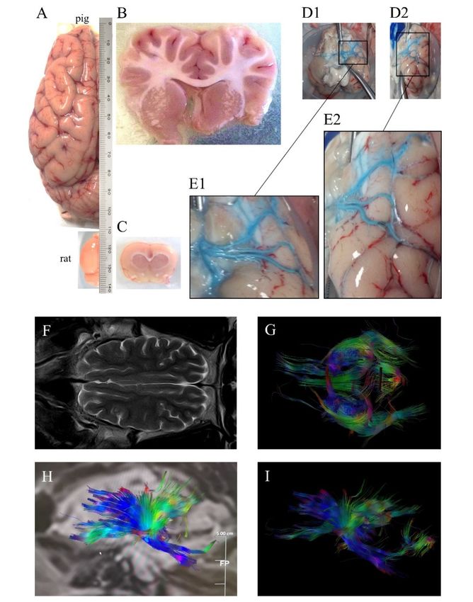
Figure 1. (A) Dorsal view of pig and rat brain hemispheres showing differences in size and
Figure 1. (A) Dorsal view of pig and rat brain hemispheres showing differences in size and gyrencephaly.
gyrencephaly. Coronal (B) pig and (C) rat sections showing differences in white matter. (D1) Ventral
Coronal (B) pig view
and of
(C)
pigrat sections
brain showing
(without differences
cerebellum) in white i.a.
injected unilaterally matter. (D1)
ex vivo Ventral
a blue view
dye, (D2) of pig brain
ventrolateral
view ofinjected
(without cerebellum) the same unilaterally
brain, (E1) andi.a.
(E2)
exmagnifications
vivo a blue of the(D2)
dye, insetsventrolateral
in (D1) and (D2), respectively,
view of the same
showing three main middle cerebral arteries. (F) Horizontal pig brain section obtained by magnetic
brain, (E1) and (E2) magnifications of the insets in (D1) and (D2), respectively, showing three main
resonance T2 sequence, (G) tractography using diffusion tensor imaging (DTI) and (H,I) sagittal pig
middle cerebralbrain
arteries. (F) Horizontal
tractography, pigdenoting
with colors brain section obtained
directionality. by were
Images magnetic resonance
obtained T2 sequence,
at the Comparative
(G) tractography Medicine
usingand Bioimage Centre
diffusion tensorofimaging
Catalonia (CMCiB).
(DTI) and (H,I) sagittal pig brain tractography,
with colors denoting directionality. Images were obtained at the Comparative Medicine and Bioimage
Another aspect of the swine that should not be disregarded in stroke research is its size (Figure
Centre of Catalonia (CMCiB).
1), as large animal species offer some advantages compared to smaller species. To begin with,
depending on the strain and at sexual maturity, pigs can have a weight similar to that of humans,
Another aspect of the
allowing theuse
swine that should
of equipment not be disregarded
and procedures inhumans
specialized for stroke in research is its
the clinical size
arena (Figure 1),
[2,3,28].
as large animal species offer some advantages compared to smaller species. To begin with, depending on
Additionally, their size allows repeated sampling of body fluids with minimal physiological
disturbance,
the strain and at which eases
sexual maturity, pigsperforming
can havelongitudinal studies [3,29].
a weight similar to thatMoreover,
of humans,pigs allowing
are much closer
the use of
genetically to humans than mice [30,31], and have the potential to offer a better modeling of human
equipment andgeneprocedures specialized for humans in the clinical arena [2,3,28]. Additionally,
regulation and function [32]; also, several models of pathologies and comorbidities usually
their size
allows repeated sampling
found of body
in stroke patients, suchfluids with
as obesity minimal physiological
or atherosclerosis, disturbance,
have been established in swine which
[33]. Theeases
performing longitudinal studies [3,29].
shorter the phylogenetic Moreover,
distance between pigsandare
the model the much
modeledcloser
species,genetically to humans
the better inference of
the results
than mice [30,31], obtained
and have theis potential
anticipated [2];
to moreover, animalmodeling
offer a better models withof comorbidities
human gene couldregulation
potentially and
function [32]; also, several models of pathologies and comorbidities usually found in stroke patients,
such as obesity or atherosclerosis, have been established in swine [33]. The shorter the phylogenetic
distance between the model and the modeled species, the better inference of the results obtained is
anticipated [2]; moreover, animal models with comorbidities could potentially improve that inference.
In sum, all these features make the pig an excellent research animal to obtain results with more
translation to the clinic.
In this article we review adult swine stroke models only, not the neonatal hypoxic-ischemic
encephalopathy model, as they better mimic the more prevalent human stroke pathology. In Figure 2,Int. J. Mol. Sci. 2020, 21, x FOR PEER REVIEW 4 of 30
Int. J. Mol. Sci. 2020, 21, 6568 4 of 30
improve that inference. In sum, all these features make the pig an excellent research animal to obtain
results with more translation to the clinic.
In this article we review adult swine stroke models only, not the neonatal hypoxic-ischemic
we show a world map with the locations where some of the main ischemic and hemorrhagic
encephalopathy model, as they better mimic the more prevalent human stroke pathology. In Figure
stroke
models in swine mentioned
2, we show a worldin this
map withreview have
the locations been
where somedeveloped.
of the main ischemic and hemorrhagic stroke
models in swine mentioned in this review have been developed.
Figure 2. World map showing the locations where some of the main ischemic (yellow) and
Figure 2. Worldhemorrhagic
map showing the models
(red) stroke locations where
in swine some
mentioned of the
in this main
review have ischemic (yellow) and hemorrhagic
been developed.
(red) stroke models in swine mentioned in this review have been developed.
3. Importance of White Matter Injury in Stroke
3. Importance of White Matter
WM injuries Injuryfound
are normally in Stroke
in elderly people in the general population [34,35]. In strokes
caused by the occlusion of large cerebral vessels, WM axonal fibers are often affected [36–38]. WM
WM injuries arewith
damage normally found has
axon degeneration in elderly
been foundpeople in the
in post-mortem general
human brains population
from ischemic stroke[34,35]. In strokes
patients [39,40]. Additionally, lacunar stroke, a stroke subtype that involves WM damage caused by
caused by the the occlusion of large cerebral vessels, WM axonal fibers are
impairment of small vessels supplying subcortical areas of the brain, accounts for 20–30% of all
often affected [36–38].
WM damage with axon degeneration has been found in post-mortem human brains from ischemic
acute ischemic strokes [41,42]. Despite being potentially silent at the beginning, lacunar stroke causes
progressive and cumulative damage, and increases the risk of stroke recurrence [42]. The presence
stroke patients [39,40]. Additionally, lacunar stroke, a stroke subtype that involves WM damage caused
and progression of WM injuries have been correlated with a worse clinical outcome in stroke [43,44],
by the impairment of small vessels
as well as with cognitive supplying
[37,40,45–48] subcortical
and motor areasbyof
deficits caused thisthe brain,
disease accounts for 20–30% of all
[40,46,49,50].
In adult swine models of experimental ischemic stroke
acute ischemic strokes [41,42]. Despite being potentially silent at the beginning, lacunarand intracerebral hemorrhage (ICH) stroke causes
significant damage and/or edema in the WM have been reported [16,27,51]. In fact, a recent study
progressive andwith cumulative
a pig model of damage,
ischemic strokeand increases
causing theofrisk
deterioration tractsof
fromstroke recurrence
the internal [42]. The presence
capsules showed
and progressionimpaired
of WM injuries
motor functionhave
[16]. been
Moreover,correlated with pig
a lacunar stroke a worse clinicalprogressive
model showed outcome WMin stroke [43,44],
damage, including the corticospinal tract [52]. These effects occur partly due to the differential
as well as with composition
cognitiveof[37,40,45–48] and motor deficits caused by this disease [40,46,49,50].
WM as compared to GM. While GM mainly consists of neuronal somas, dendrites,
In adult swine
and axonsmodels of experimental
for local signaling, ischemic
supported by glial cells, WMstroke and bodies
lacks neuronal intracerebral
and is composed hemorrhage
of (ICH)
long-tract axons and the axon-supportive glial cells (singularly oligodendrocytes and fibrous
significant damage and/or edema in the WM have been reported [16,27,51]. In fact, a recent study
astrocytes). These tracts associate different cortical areas within the same hemisphere or between
with a pig model of ischemic
hemispheres, stroke causing
and interconnect cortical anddeterioration
subcortical structuresof tracts
[37]. Thefrom
waterthe internal
content is lowercapsules
in showed
impaired motorthe WM compared
function [16]. with the GM, whereas
Moreover, a lacunar the lipid
strokecomposition
pig modelis higher, mostly due
showed to myelin. WM damage,
progressive
Oligodendrocytes (OLs) in the CNS are in charge of wrapping axons to form segments of myelin
including the corticospinal
sheath along the tract [52]. thus
axon length, These effects
allowing occur partly
the saltatory conduction due to the
of action differential
potentials [53] andcomposition of
WM as compared to GM.
providing Whilesupport
metabolic GM mainly consists
to axons [54]. of neuronal
OL precursor cells (OPCs) somas, dendrites,
are ubiquitous in the and
adult axons for local
brain parenchyma, and proliferate to maintain a fairly constant density of mature OLs [55,56].
signaling, supported by glial cells, WM lacks neuronal bodies and is composed of long-tract axons
and the axon-supportive glial cells (singularly oligodendrocytes and fibrous astrocytes). These tracts
associate different cortical areas within the same hemisphere or between hemispheres, and interconnect
cortical and subcortical structures [37]. The water content is lower in the WM compared with the
GM, whereas the lipid composition is higher, mostly due to myelin. Oligodendrocytes (OLs) in the
CNS are in charge of wrapping axons to form segments of myelin sheath along the axon length,
thus allowing the saltatory conduction of action potentials [53] and providing metabolic support to
axons [54]. OL precursor cells (OPCs) are ubiquitous in the adult brain parenchyma, and proliferate to
maintain a fairly constant density of mature OLs [55,56].
Most of the in vivo studies addressing the effect of ischemia on WM brain damage and its
pathophysiological mechanisms have been performed in rodent models of perinatal hypoxic/ischemic
cerebral palsy that do not describe the mechanisms of stroke in the adult brain. Especially in the
neonatal period, the brain is consolidating the myelination process with most of the OPCs switching
to myelin-synthesizing mature OLs and, thus, perinatal damage in WM results mainly from the
impairment of the perinatal myelination process. Moreover, recent evidence suggests that fetal OPCs
are more vulnerable than adult ones to oxygen and glucose deprivation [57].Int. J. Mol. Sci. 2020, 21, 6568 5 of 30
Regarding the differential mechanisms involved in GM and WM damage, peri-infarct depolarizations
and spreading depolarization have been observed to propagate in the GM but not through the WM [58].
In addition, it is well known that WM, especially deep WM, receives less blood flow and less collateral
circulation compared to GM, in both humans and swine [47,59]. Despite GM has been traditionally
considered to have lower tolerability to infarction in ischemic stroke patients [60–62], WM is found
injured in most strokes, and there are studies reporting the high sensitivity to ischemia of the latter,
possibly due to its reduced collateral blood supply [22,37]. In addition, differential expression of
relevant receptors (e.g., glutamate receptors of NMDA or AMPA subtype, or hemoglobin receptors)
in the specific cell types of GM (e.g., neurons) or WM (e.g., OLs) has an impact on their differential
susceptibility to excitotoxicity in ischemic stroke [25,45,63–67] or to cytotoxicity by hemoglobin or
heme iron molecules in hemorrhagic stroke [68–70].
Notwithstanding WM susceptibility to ischemic damage, this structure also has singular
mechanisms of repair. There are reports of an increase of OPCs in ischemic brains of mice [71,72],
maturing into OLs and migrating in response to demyelination or injury [56,60,73] to repair myelin
around axons [72,74,75], restoring to some extent neurological function, but failing to reach a complete
WM regeneration [76–79].
Studies of WM injury after stroke should be a research priority as they would help to develop
strategies for neuroprotection and repair [80]. In fact, the Stroke Treatment Academic Industry Roundtable
(STAIR) recommends that in order to obtain clinically relevant therapeutic agents, not only should
these protect all the cellular components of the WM in addition to neurons in the GM, but also that their
testing in large gyrencephalic animals with increased WM connectivity should be considered [26,81].
4. Interventions to Induce Ischemic Strokes in Pigs
According to the American Heart Association, 87% of all strokes are ischemic [82], caused mainly
by the obstruction of major cerebral arteries, especially the middle cerebral artery (MCA) or its
branches [83]. Typically, the study of ischemic stroke in rodent models has focused in the MCA
territory [84]. The modeling of middle cerebral artery occlusion (MCAO) in larger animals has
implied some additional complications. To start with, several large mammals typically used in research,
in contrast to humans, have a network of small vessels at the base of the brain prior to the internal carotid
artery (ICA), termed the rete mirabile, which has reportedly impeded the occlusion of intracranial
vessels such as the MCA through endovascular approaches [85,86]. The attempts to circumvent this
hindrance through the occlusion of the major vessels irrigating towards the rete mirabile, such as the
common carotid or the pharyngeal artery, have so far rarely been successful due to the well-established
collateral flow in the brain [85,87]. Additionally, compared to the single MCA in each brain hemisphere
of humans, pigs have between two and up to four MCAs on each side, with anatomical variations
found even within the same breeds [7,88,89] (Figure 1). Therefore, these differences between animals
could complicate infarction reproducibility.
As mentioned above, for this section we excluded the neonatal piglet models of hypoxia/ischemia
that have been commonly used for decades [2]. These piglet models achieve global cerebral ischemia
through different mechanisms. Usually, it is performed by blocking carotid artery blood flow bilaterally,
either by external compression [90] or through a surgical approach [91]. There are other strategies such
as performing a generalized circulatory arrest [92,93] or obstructing brain blood flow by increasing
ICP [94]. However, these approaches in neonatal or very immature piglets are more useful as models
of cerebral palsy, which is caused by prenatal hypoxic-ischemic events that damage the brain’s WM
and is the most frequent birth disorder [95].
Here we address the different techniques that have achieved generating partial ischemic strokes in
pigs. In total, five different approaches (Table 1), including electrocoagulation, clip/ligature occlusion,
endovascular embolization, photothrombosis, and endothelin-1 (ET-1) injection, have been used so far
to prevent the blood supply, especially to the MCA territory, and to induce focal brain ischemia in
adult/pediatric swine.Int. J. Mol. Sci. 2020, 21, 6568 6 of 30
Table 1. Models tested to generate ischemic stroke in swine. Abbreviation; ET-1, endothelin-1; MCA, middle cerebral artery; ICA, internal carotid artery; AChA,
anterior choroidal artery; APA, ascending pharyngeal artery; CCA, common carotid artery.
Type of
Approach Method Target Advantages Disadvantages References
Approach
(1) Temporary ischemia (1) Invasive approach [87]
ET-1 injection MCA
(2) Partial ischemia
(1) Temporary
MCA (1) Invasive approach [88,96–100]
Photothrombosis (2) Partial ischemia
(2) Reproducible
MCA
Craniotomy (1) Partial ischemia (1) Invasive approach
ICA [16,52,101–107]
Electrocoagulation (2) Reproducible (2) Not temporary
AChA
MCA + ICA
(1) Temporary
Surgical MCA (1) Invasive approach [86,108]
Arterial clip (2) Partial ischemia
(3) Reproducible
MCA (1) Temporary
(1) Relatively invasive approach [59,109,110]
ET-1 injection (2) Partial ischemia
Cranial burr hole Striatum
Cortex
(1) Partial ischemia (1) Very invasive approach
Electrocoagulation MCA [89,111–115]
(2) Reproducible (2) Not temporary
Transorbital (1) Temporary
MCA (1) Very invasive approach [7,111,113,116]
Arterial clip (2) Partial ischemia
(3) Reproducible
(1) Minimally invasive
Mechanical embolization Extracranial arteries (1) No infarction [117]
(2) Temporary
Extracranial arteries
(1) Minimally invasive
(1) Difficult infarction [118–131]
Blood clot injection CCA (2) Temporary
APA
(1) Minimally invasive (1) Difficult infarction [132–134]
Endovascular Endovascular Polymer injection APA-rete mirabile
(2) Not temporary
(1) Minimally invasive (1) No infarction [135]
CO2 injection CCA
(2) Temporary
(1) Minimally invasive (1) No infarction [136]
Air injection ICA
(2) Temporary
Rete (1) Minimally invasive (1) No infarction [137]
DMSO injection
mirabile (2) TemporaryInt. J. Mol. Sci. 2020, 21, 6568 7 of 30
4.1. Electrocoagulation
Whereas the general term of electrocautery is the use of electricity to generate enough heat to
destroy tissue, electrocoagulation is the use of this technique on blood vessels to achieve its permanent
occlusion [138]. This method, together with microvascular clipping, was the first used to produce
MCAO in swine in 2000. It was likewise the first model achieving a focal cerebral infarction reported
in pigs. Sakoh et al. used a transorbital approach to reach the left MCAs, who reported that there
are generally two, and occluded them proximally together with the ICA distally [111]. They used
the same model in subsequent studies [112–115]. Interestingly, in one of such studies they described
inter-individual differences in the model in terms of collateral blood flow to the MCA territory,
and correlated differences in blood flow with the infarct size [112]. The same method has also been
used by Zhang et al., also occluding the proximal MCAs unilaterally [89]. Subsequently, Imai et al.
were the first to use craniotomy instead of a transorbital approach to expose the left MCAs, and then
occluded their 2 branches from the origin of the lenticulostriate artery to past the olfactory tract, as well
as the ICA by electrocoagulation in miniature pigs [139]. Since that first use, craniotomy has been
the surgical procedure of choice to expose MCA area prior to electrocoagulation, probably to avoid
potential complications related to the removal of an eyeball. Particularly, West’s group has used this
swine model in multiple studies, reportedly occluding the distal MCA [16,101–106]. This method has
allowed the production of consistent strokes with high survival rates in minipig strains after occluding
different intracranial arteries [52,101,102,107,139].
With electrocoagulation, vessels of interest can be accurately targeted, which translates into a
higher reproducibility of the focal brain ischemia models generated. On the other hand, this technique
causes an irreversible occlusion of the vessels, impeding the reperfusion of the infarcted tissue and
there is still the need to use invasive surgical interventions to expose intracranial vessels prior to the
electrocoagulation. Particularly, eye enucleation is considered more severe due to the obvious loss in
the field of vision of the animal and possible post-surgical complications as the infection of the orbital
cavity [140]. However, a less invasive approach as craniotomy is also mediated by a surgical approach
that could have additional effects on the brain such as increased blood-brain barrier permeability due
to ICP changes [141].
4.2. Microvascular Clip
As mentioned above, Sakoh et al. used this technique in the first pig model of MCAO in a
complementary manner to electrocoagulation. This method allowed a transient arterial occlusion as
reperfusion was performed simply by removing the micro-clips occluding the target arteries [111].
As with electrocoagulation, arterial clipping also requires the exposure of the MCA surgically, which can
be performed either with a transorbital approach or craniotomy. The first case of the combination
of craniotomy and microvascular clipping was performed by Mattingly et al., in which only one of
the 2 to 3 branches of the MCA running beneath the posterior frontal lobe was clipped for 3 h using
the Imai et al. procedure, generating variable and small strokes [108]. This combination has been
used in further studies to induce malignant strokes in domestic swine, showing that larger infarct
volumes are generated if the occlusion of the 2 unilateral MCAs is maintained for longer periods [86].
The transorbital approach has persisted in recent papers for experimental procedures not requiring
post-surgical recovery [7,116]. In this latter study, they observed the variability in the MCAs between
pigs from the same breed, finding between 2 and 4 MCAs in each hemisphere, and proposed that
variability of infarct size in previous studies that report the occlusion of all the MCAs could be explained
by the ineffective occlusion of some of such arteries. They also suggested that the frontotemporal
craniotomy to expose the MCA area, despite being less invasive, could be the reason for missing some
of the MCA branches in previous studies, a problem that is not seen with the transorbital approach [7].
The use of microvascular clips to achieve MCAO has the added benefit of allowing the infarction
to be temporary. However, the drawback of the need of surgery to allow its use is still present.Int. J. Mol. Sci. 2020, 21, 6568 8 of 30
4.3. Endovascular Embolization
With the development of thrombolytic therapies, and particularly of thrombectomy devices,
pigs have been used as models due to the anatomical and physiological similarities of their
cardiovascular system with that of humans [3]. However, the objective of these studies has generally
not been to generate a cerebral infarction, but to occlude extracranial vessels anatomically similar to
human intracranial arteries and to study thrombolytic techniques instead [130]. The main reason is the
presence of the previously mentioned rete mirabile in pigs, which has impeded reaching intracranial
vessels with endovascular approaches to date [85,86].
The main method to achieve endovascular embolization of arteries in pigs has been the injection of
autologous blood clots to extracranial vessels [118–131]. The embolization of an artery implies the reduction
of its blood flow [142]. For instance, bilateral thromboembolism to reduce blood flow in both ascending
pharyngeal arteries caused multiple and variable focal areas of stroke damage, although consistent
temporo-parietal infarcts were not generated as seen by diffusion-weighted imaging [127].
Other methods of endovascular embolism used in this species are mechanical embolization [117],
through diverse embolic agents including collagen microbeads [132], dimethyl sulfoxide (DMSO) [137],
air [136], CO2 [135], Eudagrit polymer [133], and sodium alginate [134]. The use of embolic agents in
pigs arose from the use of the rete mirabile as an arteriovenous malformation model. Arteriovenous
malformations are an abnormal formation of blood vessels that shunt arterial blood directly into
veins without passing through the capillaries. These malformations are clinically treated through
endovascular embolization [142]. Nevertheless, using embolic agents as a method to produce brain
ischemia in pigs has seldom been performed. Unilateral embolization of brain-irrigating arteries
produced little ischemic damage to the brain. The only study that reports achieving infarcted areas in
the brain this way is with the injection of sodium alginate into the ascending pharyngeal artery in
Bama minipigs through the unilateral occlusion of the rete mirabile [134]. This embolization caused
scattered damage throughout the brain, infarcted areas being mainly observed at the temporal and
parietal lobes, and/or basal ganglia, one week after the injection.
Endovascular embolization is a minimally invasive technique used to achieve ischemia in target
regions. Additionally, depending on the method used, transient ischemia with its consequent reperfusion
can be performed. However, despite the study of Cui et al. [134], the presence of the rete mirabile
seems to avoid the possibility of reaching intracranial vessels through an endovascular approach.
4.4. Photothrombosis
This method was first used in swine in 2007 as a model of acute ischemic stroke in pediatric
animals, and required enucleation of the eye to reach the MCAs [88]. Since then, similar models have
been generated by Armstead et al. [96,97,100,143], but to our knowledge, it has not been replicated in
mature pigs and it is not as widely used as in other species such as NHPs and rats. Photothrombosis
is the formation of a stable thrombus of aggregating platelets, fibrin, erythrocytes, and other blood
components, due to endothelial peroxidative damage. This is generated with the photochemical reaction
caused by the interaction of intravenous photosensitizing dye erythrosine B and the focused beam
of a laser [88,96]. When used in pigs, photothrombosis of all the MCAs unilaterally, with reportedly
2–3 main and 1–3 smaller arteries supplying the MCA territory, produced a moderate infarct affecting
both GM and WM [88].
In rodents, photothrombosis of the MCA can be generated without craniotomy due to the thinness
of their cranial bones [144], differently from the markedly thick ones in pigs [28]. Thus, photothrombosis
of intracranial arteries in pigs requires exposure of the target vessels by transorbital access or
craniotomy [1], which implies a more invasive surgery. Overall, this model permits the occlusion of
intracranial vessels with its exposure to reperfusion by using common thrombolytic methods.Int. J. Mol. Sci. 2020, 21, 6568 9 of 30
4.5. Endothelin-1 Injection
This is the most recent model of porcine focal ischemic stroke, used for the first time by Elliott et al.
and published in the year 2014 [109]. In this study, ET-1 was pumped into the cortical tissue to achieve
cerebral ischemia, but infarction was not completely generated in the whole region of interest. ET-1 was
firstly described as a vasoconstrictor factor present in the conditioned media of cultured bovine
endothelial cells in 1985. For that reason, it was used primarily in rats as a novel method to achieve
MCAO [145]. Although it was not used to cause a stroke, the effect of ET-1 on the vasoconstriction of
intracranial vessels had been assessed in pigs [146] as a consequence to the observation that the levels
of this molecule increased upon ICH in patients, and that this event entailed vasoconstriction in the
affected area [147].
ET-1 injection is a method with which transient focal ischemia can be achieved; it is reproducible
in terms of the possibility of accurate positioning of the injection and volume/concentration injected.
Studies in rodents and NHPs show that infarction severity can be modulated through the concentration
of injected ET-1 [23,148]. Despite it having been performed without approaches as invasive as
craniotomy or eye enucleation, it still requires a surgical exposure of the target area, the common
approach being through a computer tomography (CT)-guided burr hole [59,109,110], allowing the
administration to virtually any cerebral region. For instance, the anatomically-driven location of the
MCA through a craniotomy between the zygoma and orbit allows the occlusion of the MCA branches
resulting in significant brain parenchyma lesion [89]. The use of invasive surgery might further
affect the animal’s overall well-being in a stroke-independent manner. Additionally, the post-mortem
analysis of human brains showed the expression of ET-1 receptors in non-endothelial cell types such
as neurons [149], and the cerebral expression of ET-1 and its receptors seem to be altered in ischemic
conditions [150,151]. Thus, the injection of ET-1 to induce ischemia through vasoconstriction could
have unwanted effects that do not occur in the human pathology [152]. In addition, a study using
this pig model reported variations between animals in the cerebral blood flow in response to ET-1
injection to the MCA territory, and in some of the animals, ischemia was not extended for enough
time to generate an irreversibly infarcted region [110]. Finally, it is unclear if this model allows the
control of the exact duration of ischemia and, thus, to set up reperfusion with as much control as other
mechanisms of arterial occlusion.
5. Interventions to Induce Hemorrhagic Strokes in Pigs
Around 10–15% of all strokes are non-traumatic intracranial hemorrhages [153,154], which are
classified according to the intracranial compartment in which the bleeding occurs. Within the meninges,
hemorrhage can be subarachnoid, subdural, and epidural [154], the former being the most common of
the three and accounting for 3% of all strokes [82]. Subdural and epidural bleeding are more typically
caused by trauma [154] and the existent pig models of hemorrhage within these spaces are generally
used to model traumatic brain injury [155,156] rather than spontaneously occurring hemorrhagic
stroke. Most ICH events, accounting for 10% of all strokes [82], occur directly in the brain parenchyma,
whereas hemorrhages into the ventricular system are less common [154].
The subarachnoid space of the meninges surrounding the swine brain resembles that of
humans [157]. The porcine brain allows for the accumulation of larger amounts of blood and clots
in subarachnoid and intracranial hemorrhage models compared to rodents due to its morphological
features [153,157,158], mimicking better what happens in the clinical arena. As explained above,
blood accumulates in the brain parenchyma after the onset of the bleeding, disrupting the local
brain anatomy and increasing local pressure rapidly. In a second phase, compounds and blood cells
entrapped in the brain parenchyma promote cytotoxicity. Accordingly, blood experimentally injected
either into the subarachnoid sulcal spaces or intracerebrally has been reported to mediate cortical
spreading depolarizations (CSD) in swine as well as to induce ischemic cascades and perihematomal
cortical infarcts [157,159]. Importantly, swine models allow a trustworthy tracking of hemorrhageInt. J. Mol. Sci. 2020, 21, 6568 10 of 30
volume and brain damage evolution using the same non-invasive multimodal imaging sequences used
in the clinical practice [15].
As with ischemic stroke models, we excluded neonatal piglet models of ICH in this section.
However, we found it convenient to include models in immature pigs, especially considering that
some techniques have only been reported in such models. These techniques include the intracerebral
injection of collagenase and the more recent sonographic disruption of brain vessels (see Table 2).
To better mimic specific types of hemorrhagic stroke, the injection of blood can be performed in different
intracranial regions.Int. J. Mol. Sci. 2020, 21, 6568 11 of 30
Table 2. Models of hemorrhagic stroke generated in swine. Abbreviation; MRgFUS, magnetic resonance-guided focused ultrasound.
Type of Approach Approach Method Target Advantages Disadvantages References
MRgFUS Brain parenchyma (1) Reproducible (1) Invasive [160]
(1) Reproducible (1) Invasive [161–164]
Craniotomy Collagenase injection Brain parenchyma
(2) Only used in juvenile pigs
Single blood injection Brain parenchyma (1) Reproducible (1) Invasive [165]
(1) Reproducible
Transcranial MRgFUS Brain parenchyma (1) Target limitation [166,167]
(2) Minimally invasive
Intracranial Double blood injection Brain parenchyma (1) Reproducible (1) Relatively invasive [51,69,168–171]
hemorrhage
Balloon catheter
dilation and double Brain parenchyma (1) Reproducible (1) Relatively invasive [172–182]
blood injection
Cranial burr hole Balloon catheter
dilation and single Brain parenchyma (1) Reproducible (1) Relatively invasive [183–186]
blood injection
Ventricle (1) Reproducible (1) Relatively invasive [158,159,187–212]
Single blood injection
Brain parenchyma
(1) Reproducible
Transorbital Single blood injection Subarachnoid space (1) Technically difficult [213]
(2) Minimally invasive
Cranial burr hole Single blood injection Subarachnoid space (1) Reproducible (1) Relatively invasive [214,215]
Single blood injection Subarachnoid space (1) Reproducible (1) Invasive [157,216–220]
Meningeal Craniotomy Balloon catheter
hemorrhage dilation and single Epidural space (1) Reproducible (1) Invasive [221,222]
blood injection
Cisterna magna (1) Reproducible [223–226]
Intrathecal Single blood injection
(subarachnoid space) (2) Minimally invasive
Pontine cistern
Laminectomy Single blood injection (1) Reproducible (1) Invasive [227–229]
(subarachnoid space)Int. J. Mol. Sci. 2020, 21, 6568 12 of 30
5.1. Autologous Blood Injection in Meningeal Spaces
The first studies using pigs as models of hemorrhagic strokes were performed in the 1980s,
in which subarachnoid hemorrhage (SAH) was achieved through the injection of autologous blood in
the pontine subarachnoid cistern after exposing it through a C-2 laminectomy [227–229]. Similarly to
the occurrence of hemorrhagic stroke, the subarachnoid space has been the preferred target within
the porcine meninges to model hemorrhagic stroke. The use of epidural injections was reported
shortly thereafter [221], but this model is unusual today. The subdural space has been targeted to
inject autologous blood; however, this approach is used as a traumatic brain injury model [155].
New models were developed to expose the subarachnoid space and infuse it with autologous blood
to simulate SAH (Table 2). Besides the aforementioned laminectomy of the C-2, autologous blood
has been injected directly to the cisterna magna, with a puncturing technique also used to obtain
cerebrospinal fluid [223–226]. Likewise, craniotomy has been used to reach the MCA territory [216,217],
the suprasellar subarachnoid cistern [218–220], and between the crests of the superior frontal and
motor gyri [157]. Basal subarachnoid cisterns have also been reached through burr holes directed
to the anterior skull base [214,215], in studies that exhibit the impact of transient ischemia derived
from the SAH. Finally, a recent study showed that the subarachnoid space could be achieved
precluding craniotomy, through the direct transorbital injection of blood into the interpeduncular
cistern, without eye enucleation [213].
5.2. Intracerebral Autologous Blood Injection
ICH was first modeled in pigs by Farstad et al. in 1994. In this study, they injected autologous
blood in the lateral ventricles of neonatal piglets [230]. An analogous technique was used in juvenile
pigs in 1997 [195], but intraventricular hemorrhage models remain rarely used. The approach used
to access the ventricles to inject blood has been the cannulation of the lateral ventricles through a
stereotaxically- [195,196] or magnetic resonance imaging (MRI)-guided [198] cranial burr hole.
The first reported parenchymal hemorrhage porcine model using autologous blood injection
was performed in piglets in 1996 [158]. In that model, the blood was slowly infused into the WM of
the frontal lobe of the brain through a catheter inserted by stereotaxic surgery. The following ICH
models of autologous blood injection are based on that first study. In the year 2000 the technique
was refined: A balloon catheter was introduced through a cranial burr hole to generate a space in
order to prevent reflux from the intraparenchymal injection of autologous blood in the frontal lobe of
pigs [183]. Subsequently, the same group adapted a double-injection method from rats to pigs, together
with the balloon catheter technique, to further avoid the reflux effect of the injection [172,173,176,177].
This model has also been replicated by other groups [174,175,179–182], as well as using the double
injection method without the previous balloon catheter [168,170]. A similar method has been used by
Bimpis et al.; in their approach a balloon is inflated and, while decompressed, the autologous blood is
injected [184–186]. In a recent study, injection of blood to the target region of the brain was performed
after a craniotomy, but this was only performed to apply focused ultrasounds to liquefy induced
intracerebral blood clots [165]. However, the main method remains to be the surgical access to the
target area and direct injection of the blood through a catheter or needle [158,159,187–194,197,199–212].
5.3. Intracerebral Collagenase Injection
The first study using collagenase as an ICH model in juvenile pigs was done by Mun-Bryce et al.
in 2001 [161]. The authors injected this compound, capable of disrupting the blood-brain barrier, into
the primary somatosensory cortex after exposing it through a craniotomy, and reported recurring
episodes of spreading depolarization. The first in vivo use of this enzyme was performed in
rats by the same group. In that study, collagenase was injected in the caudate nucleus with
the aim of degrading the collagen from the basal lamina of blood vessels to cause ICH [231].
To our knowledge, only this group has used the collagenase (juvenile) swine model to induceInt. J. Mol. Sci. 2020, 21, 6568 13 of 30
hemorrhagic stroke in successive studies. Interestingly, in such studies Mun-Bryce et al. show a
rapid depression of cortical excitability in the ipsilesional hemisphere accompanied by a gradual
depression in the functionally-related contralateral region, whereas callotomized animals—lacking
multiple interhemispheric axonal connections—showed a gradual increase in the excitability of the
contralesional site within the acute phase. This could be indicative that the ICH damages brain areas
connected to the injured region [163]. Furthermore, they observed a depressed cortical excitability in
functionally-related areas of the contralesional hemisphere after this acute phase, and this impairment
was preceded by a rise of the inflammatory and extracellular matrix remodeling marker MMP-9 in
the ipsilesional and contralesional regions, especially in the WM [162]. However, a subsequent study
reported spreading transient depolarizations during acute ICH in disperse brain areas including the
contralateral hemisphere, but with no differences caused by callosotomy [164]. All these observations
are indicative of the consequences of WM damage in brain connectivity caused by ICH, and the
translational advantages of using the pig in neuroscience research due to the higher development of its
WM. Therefore, it would be of interest to further study the WM connectivity in a collagenase adult
pig model.
5.4. Sonographic Blood-Brain Barrier Disruption
The initial report of sonographic disruption of the blood-brain barrier in swine was performed by
Aviv et al. in 2014. In this study, they targeted the vessels within the basal ganglia of young swine using
MRI-guided sonography with the objective of generating a model of ICH that allowed them to quantify
hematoma through the MRI detection of extravasated contrast [166]. Rupture of intracerebral vessels
is thus performed with the magnetic resonance-guided focused ultrasound (MRgFUS), a technique
used to perform minimally invasive targeted tissue surgeries based on the thermal ablation caused
by focused ultrasounds at determined frequencies. The low invasiveness is due to both the precise
ablation of the target tissue and the possibility of a transcranial approach, i.e., without removal of
skull bones [232]. MRgFUS was applied in pigs by the same group in a following study to further
characterize this ICH model [167]. Finally, hemorrhagic lesions were caused in the swine brain using
the same technique, not to generate an ICH model per se, but to determine the non-lesional frequencies
and durations of the ultrasound pulses to the brain [160]. Curiously, this technique has been used
as a potential therapy for ICH in swine models. Using intracerebral autologous blood injection
models in pigs, MRgFUS can be directed to the blood clots within the brain to cause its thrombolysis,
showing positive results [165,198,212].
Overall, MRgFUS is a minimally invasive technique with which a targeted ICH can be precisely
generated. Nonetheless, it has not been widely implemented so far, perhaps due to the lack of access
to this relatively modern technology. Another problem of this technique is that its transcranial use
can be intricate. Firstly, as the swine skull thickness is variable and generally big, transmission of
the different ultrasound beams through this structure can make deep brain structures impossible
to target. Secondly, lesions directed to regions excessively near the skull bones can cause regional
overheating [232]. Actually, in the study of Xu et al. a craniotomy is performed to prevent these
issues [160]. As with the collagenase model, more research needs to be performed using this promising
model, although this might be hampered until a refinement of the technique is achieved.
6. Neurological Function Assessment in Pigs
Using in vivo models of stroke pathology gives the advantageous possibility of evaluating the
neurological outcomes associated with the disease and, with more complex organisms, more detailed
resemblance to the human pathology is expected. The cognition of pigs has been studied throughout
the decades using different approaches. Several tasks and maze-based tests have been developed
and adapted to pigs to assess distinct cognitive and behavioral aspects such as memory and learning,
affective behavior, or social interactions, among others (reviewed in [18]). Despite this, there is not a
current consensus about the assessment of neurological deficits in pig stroke models. In the clinicalInt. J. Mol. Sci. 2020, 21, 6568 14 of 30
arena, there are different scales and scoring systems validated to determine neurological function,
but none has been established as ideal. The first assessment performed in a stroke clinical trial was the
modified Rankin scale and is the most commonly used for functional evaluation [233]. This scoring
system has shown to correlate with other measures of stroke pathology as the infarct volume, as well
as other neurological function scales [233,234]. In swine, the modified Rankin score has been recently
adapted to the species in a study from West’s group [106], which has developed numerous studies using
MCAO swine models. However, a validated scale adapted to the swine is still needed to better assess
the neurological outcome. So far, the first studies that evaluated neurological function in ischemic
and hemorrhagic stroke swine models adapted scales from the dog, such as Purdy et al.’s [235] and
Tibbs et al.’s [236] canine stroke models.
The first neurological examination used in a pig model of focal ischemic stroke was performed
by Imai et al. in 2006. They observed that permanent MCAO caused hemiparesis in the forelimbs
and hindlimbs, whereas ICA occlusion caused less severe deficits with occasional hemiparesis 24 h
after the surgery [139]. Subsequently, Tanaka et al. adapted a modification of the scale used for
the neurologic evaluation of dogs by Tibbs et al. to their pig model of lacunar stroke, which was
generated to target subcortical WM [52]. Their 25-point scoring system remains the more widely
used in pig stroke models. This scale has been used in the ET-1 MCAO model of Zhang et al. [87]
and—with variations—in the bipolar coagulation of the MCA model by Lau et al. from West’s
group [105]. Cui et al. developed a 100-point neurological assessment scale for their ET-1 injection
model, based on Purdy et al.’s system [134]. Similarly, Platt et al., also from West’s group, mention the
use of a standardized neurological examination adapted from canine models to evaluate pigs after
bipolar coagulation of the MCA [102]. These scales analyze different aspects of the general neurologic
function of the animals including the level of consciousness (e.g., response to stimuli), motor function
(e.g., limb paralysis), sensorial capacity (e.g., visual field defects), and behavior (e.g., utterance). On the
other hand, a totally different approach was used almost simultaneously by the same group in the
same model. In this case, they analyzed various gait parameters by video recording the animals
after provoking their walk, and found a dysfunction in the limbs contralateral to the MCAO using
parameters as step height (i.e., maximum height reached by the hoof when walking), swing time
(i.e., time the hoof does not touch the ground when walking), and stance time (i.e., time the hoof is
touching the ground when walking) [101]. This method of gait analysis has been used again recently
in the same model together with the open field test to assess exploratory behavior and motor activity.
In this task, the animal enters a relatively wide area and several aspects of its exploratory behavior
and motor activity can be recorded by a tracking software [16,104]. Finally, the same group recently
adapted the modified Rankin scale to their pig model [106].
Many of the studies with pig hemorrhagic stroke models have obviated the use of neurological
tests to assess the impact of the damage generated. Rohde et al. mention that they observe the
general neurological status and behavior of the pigs to rule out anomalies caused by the intracerebral
autologous blood injection surgery [173]. However, it was not until 2013 that Zhu et al. used an
adaptation of the previously mentioned scale from Purdy et al. to assess the evolution of neurological
damage after intracerebral autologous blood injection [168], and again in 2015 [170]. Together with the
latter, only the recently published article by Gerhardson et al. clearly describes the use of a scaling
system to evaluate the neurological state of pigs after ICH. Specifically, they use the Tanaka et al.
25-point scale previous to and daily after the lobar WM injection of autologous blood [165].
Neurological assessment in pigs has been performed in other disease models different than
stroke. The relation of such models with stroke models is that damage is caused to the brain either
directly or indirectly, e.g., in models of traumatic brain injury (reviewed in [237]) or in generalized
circulatory arrest [238]. Whereas circulatory arrest models have mainly used neurological scoring
systems similar to stroke models, traumatic brain injury models have used varied tests and tasks
to assess the impairment of different cognitive processes in pigs. One of such tests used somewhat
repeatedly is the open field testing, which has also been used in the pig MCAO model [16,104,237].Int. J. Mol. Sci. 2020, 21, 6568 15 of 30
7. Evaluation of Stroke Damage in Swine by Neuroimaging
Non-invasive in vivo imaging techniques allow a longitudinal estimation of the brain damage
location and extent and contribute to our understanding of the evolution of damage after stroke.
Although the extent of damage is a critical determinant, the functions served by the brain areas damaged
(e.g., language, motor, or cognition) and the nature of damage (e.g., necrosis, edema, axonal damage)
determine the final disabilities [239].
Magnetic resonance (MR)-based diffusion and perfusion imaging, positron emission tomography
(PET), computed tomography perfusion (CTP), or a combination of them, have been used to
characterize animal stroke models. These imaging techniques allow to observe parameters in the
models such as post-stroke acute midline shift, which is associated with decreased survival and
recovery [16,103,104,106]. Moreover, early reports allowed to assess hypoperfusion of the brain areas
affected using either: (1) Apparent diffusion of water or apparent diffusion coefficient (ADC) obtained
from diffusion-weighted imaging (DWI) MR combined with metabolic parameters of oxygen and
glucose obtained by PET, showing that ADC below 75% of the normal tissue value indicates irreversible
infarction in a transient MCAO model [113], or (2) CTP and/or PET to determine cerebral blood flow and
the hypoperfusion threshold to develop infarct [59,110], or determining areas of infarct core, penumbra,
or oligemia [109], as used in the swine model of induced cerebral ischemia by endothelin injection.
Conventional MRI/T2 images were used to depict infarct volume in the basal ganglia infarct
model [134] and early infarct volume and neuroprotection by hypothermia [108] in an MCA model.
ADC maps have been used to reveal cerebral swelling and cytotoxic edema. Reduced territorial
signal intensity on ADC maps were consistently observed with the restricted diffusion that is
typical of infarctions at 24 h and hydrocephalus ex vacuo at 90 days in a permanent ischemic
model [101,102]. Infarct in the MCA territory is hyperintense in T2-weighted imaging (T2WI) and T2
fluid-attenuation inversion recovery (FLAIR) relative to normal GM and hyperintense in DWI [139],
with corresponding hypointensity in the ADC map, indicative of cytotoxic edema [104,105]. T1, T2,
and DWI hyperintensities were observed at 24 and 72 h in the ET-1 model [59,87].
WM and GM can also be accurately characterized using conventional MRI sequences as
T1-weighted imaging (T1WI), T2WI, and FLAIR, based on their differences in water and lipid
content. In vivo imaging tools to detect WM abnormalities, axonal loss, and demyelination are still
underexplored, but new advanced MRI techniques, including quantitative susceptibility mapping
(QSM) and DTI have been used recently in humans and rodents. Only a few studies have evaluated
so far these MRI parameters in swine stroke. Fractional anisotropy (FA) maps obtained by DTI
analysis showed loss of WM integrity in the ipsilateral hemisphere (in the internal capsule and corpus
callosum) [16,240] in a pig model of stroke. In addition, a stem cell vesicle-based treatment was found to
preserve the integrity and functionality of WM [103,104].When focusing on the effects of hemorrhagic
stroke, leakage of blood into the brain parenchyma of swine has been mostly measured using
CT [180,206,208,214] or MRI FLAIR images or in T2*-weighted images [165,173,174,178,187,213,214,241].
To overcome possible artefacts, the iron from hemoglobin can be indirectly measured using the rate
of transverse magnetization relaxation, R2*(1/T2*). A recent work shows multi-contrast anatomical
and quantitative parametric maps using FLAIR, T1WI, gradient echo (GRE), R2*, QSM sequences that
allowed to determine the edema and hematoma areas in a swine ICH model, providing longitudinal
information of the heterogeneity and evolution of the damage [191]. A dynamic contrast-enhanced
(DCE) sequence allowed a real-time MRI model to study the extravasation and the acute hematoma
growth [59,193]. Metabolic evaluation of the perihematoma areas by non-invasive MR spectroscopy to
find salvageable tissue within the perihematoma has been recently published [193].
The imaging of other structural biomarkers, which are currently used mostly at research level,
have a huge potential in the future development of personalized medicine. Precisely, characteristics
of the peri-infarct structural integrity of brain around the ischemic areas as microstructural damage
measured by magnetization transfer imaging (MTI) or the identification of different metabolites
(e.g., glutamate/lactate/N-acetylaspartate) by MR spectroscopy will provide significant informationYou can also read


