Guidelines for Prevention and Management of Stroke
←
→
Page content transcription
If your browser does not render page correctly, please read the page content below
National Programme for Prevention and Control of Cancer,
   Diabetes, Cardiovascular Diseases & Stroke (NPCDCS)

                 Guidelines for
Prevention and Management of Stroke

         Directorate General of Health Services
         Ministry of Health and Family Welfare
                  Government of India
                          2019

List of Contributors
Dr. Kameshwar Prasad, Professor & Head, Neurology, All India Institute of Medical
Science, New Delhi
Dr. M. V. Padma Srivastava, Professor, Neurology, All India Institute of Medical
Science, New Delhi
Dr. Sunil Narayan, Professor & Head, Neurology, JIPMER, Puduchery
Dr. M. Veerendra Kumar, Professor, NIMHANS, Bengaluru
Dr. Vivek Lal, Professor & Head, Neurology, PGIMER, Chandigarh
Dr. Rohit Bhatia, Professor, Neurology, All India Institute of Medical Sciences, New
Delhi
Dr. Sudhir Sharma, Neurology, IGMC, Shimla
Dr. Nonica Laisram, Professor & Head, Physical Medicine and Rehabilitation,
Vardhman Mahavir Medical College and Safdarjung Hospital, New Delhi
Dr. Ritu Majumdar, Associate Professor, Physical Medicine and Rehabilitation, LHMC
& Associated Hospitals, New Delhi
Dr. Mohammed Shaukat Usta, Advisor (NCD), Dte. GHS
Dr. Prabha Arora, Additional Director, NCDC
Mr. Nilambuj Sharan, Economic Advisor, MoHFW
Mr. Rajeev Kumar, Director (NCD), MoHFW
Dr. Manas Pratim Roy, DADG (NCD), Dte. GHS
Dr. Chinmoyee Das, DADG (NCD), Dte. GHS
Dr. Rajeev Aggarwal, In-charge, Neuro Physiotherapy unit, AIIMS, New Delhi
1
Abbreviations

ACEI – Angiotensin Converting Enzyme Inhibitors
AF –Atrial Fibrillation
AMI – Acute Myocardial Infarction
ANM–Auxiliary Nurse Midwife
aPTT – activated Partial Thromboplastin Time
ASHA–Accredited Social Health Activists
ARB – Angiotensin Receptor Blocker (ARB)
CAS – Carotid Artery Stenting
CBAC – Community Based Assessment Checklist
CCU – Cardiac Care Unit
CEA – Carotid Endarterectomy
CLOTS trial – Clots in Legs OrsTockings after Stroke trial
CPAP – Continuous Positive Airway Pressure
CSCU – Cardiac and Stroke Care Unit
CSF – Cerebro Spinal Fluid
CTA – CT Angiography
CVD – Cardiovascular Disease
CVE – Cerebrovascular Event
DALY – Disability Adjusted Life Year
DBP – Diastolic Blood Pressure
DH – District Hospital
DVT – Deep Venous Thrombosis
ECG – Electrocardiography
ED –Emergency Department
ESUS – Embolic Stroke of Undetermined Source
FFP – Fresh Frozen Plasma
GCS – Glasgow Coma Scale
HbA1C – GlycatedHemoglobin
HIV – Human Immunodeficiency Virus
HDL – High-Density Lipoprotein
HSC – Health Sub Centre
HWC – Health Wellness Centre
ICH – Intra Cerebral Hemorrhage
ICP – Intracranial Pressure
INR–International Normalized Ratio
LDL – Low-Density Lipoprotein
LFT – Liver Function Test

                                             2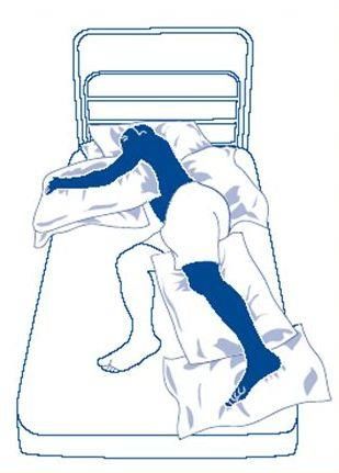
LMWH –Low Molecular Weight Heparin
MAP – Mean Arterial Blood Pressure
MCA – Middle Cerebral Artery
MRA – MR Angiography
MRI – Magnetic Resonance Imaging
MRV – Magnetic Resonance Venography
NCCT – Non-Contrast Computerized Tomography
NCD – Non-Communicable Diseases
NIHSS – National Institute of Health Stroke Scale
NOAC – Newer Oral Anticoagulants
NPO – Nil Per Oral
NVAF – Non valvular AF
OSA – Obstructive Sleep Apnea
PCC –Prothrombin Complex Concentrate
PHC – Primary Health Centre
PR – Pulse Rate
PT – Prothrombin Time
RBS – Random Blood Sugar
rtPA – recombinant tissue Plasminogen Activator
SAH – Sub ArchnoidHemorrahage
SBE –Sub Acute Bacterial Endocarditis
SBP – Systolic Blood Pressure
SCD – Sickle Cell Disease
STEMI – ST-Elevation Myocardial Infarction
TIA – Transient Ischemic Attack
TNK – Tenecteplase
VDRL – Venereal Disease Research Laboratory
VHSNC – Village Health, Sanitation and Nutrition Committee (VHSNC)
VKA – Vitamin K Antagonists
WSO – World Stroke Organization

                                         3
Table of Contents
Sl. No Chapter Page No
1. Introduction 5-6
2. Major risk factors of stroke 7
3. Primary Prevention 8-15
4. Early identification of symptoms of stroke and management 16
of TIA
5. Management of established acute stroke care 17-32
6. Secondary Prevention 33-40
7. Rehabilitation 41-50
8. Pattern of assistance for integrating Stroke care services in 51-60
District Hospital
9. Annexes 61-89
41. Introduction
Stroke, a major Non-Communicable Disease (NCD), is responsible for 3.5%
ofdisabilityadjusted life year (DALY) in India.Apart from risk factors like hypertension,
diabetes, heart diseases and positive family history, other lifestyle related factors such as
unhealthy diet, obesity, lack of physical activity, stress and tobacco use account for its
occurrence. Changes in lifestyles, behavioural patterns, demographic profile (aging
population), socio-cultural and technological advancements are leading to sharp increases in
the prevalence of stroke. The disease by and large can be prevented by making simple
changes in the way people live their lives or simply by changing our lifestyle.
1.1.Magnitude of Stroke burden in India
Stroke is the second leading cause of death worldwide and was responsible for an estimated
6.5 million deaths and 113 million DALYs in 2013. More than 2/3 of these deaths occurred in
developing countries. By 2050, more than 80% of the predicted global burden of new strokes
of 15 million will occur in low and middle-income countries. In India, studies estimate that
incidence of stroke population varies from 116 to 163 per 100,000 population.Recently,
ICMR has come out with a report entitled “India: Health of the Nation’s States”, according to
which stroke was 4thleading cause of death and 5th Leading cause of Disability Adjusted Life
Years (DALY) in 2016.
1.2.National guideline for stroke management and prevention
This guideline has been prepared for health care providers involved in management of
patients with stroke. The aim is to help them, at primary and secondary levels of health care
delivery system to make the best decisions for each patient, using the evidence currently
available. The focus is on the more common clinical questions faced in day-to-day practice.
1.3.Care provided at different levels of health care
In view of complex managements of stroke, the role of PHC (Primary Health Centre) is
limited to risk assessment, early recognition of the symptoms, stabilization and quick referral
to higher centres where facilities for managements are available.
Primary health care –primary prevention, early recognition and referral,
rehabilitation.
5 Secondary health care – acute stroke management, secondary prevention and
follow up, rehabilitation.
Tertiary health care – complex and higher level management of acute cases,
follow up of stroke for enablement and support services, rehabilitation of residual
impairment.
1. Stroke definition:
In 1970, the World Health Organization definedstroke as ‘rapidly developed
clinical signs of focal(or global) disturbance of cerebral function, lastingmore than
24 hours or leading to death, with noapparent cause other than of vascular origin’.
2. Transient Ischemic Attack(TIA)definition:AHA/ASA (2009)
A transient episode of neurologicaldysfunction caused by focal brain, spinalcord,
or retinal ischemia without acuteinfarction.
Presenting features of stroke:
Sudden numbness or weakness in the face, arm, or leg, especially on one
side of the body
Sudden confusion, trouble speaking, or difficulty understanding speech
Sudden trouble seeing in one or both eyes
Sudden trouble walking, dizziness, loss of balance, or lack of coordination
Impairment or loss of consciousness
Presenting features of TIA:
Transient weakness, numbness or paralysis of face, arm or leg, typically on
one side of your body
Transient slurred or garbled speech or difficulty understanding others
Transient blindness in one or both eyes or double vision\
Curtain like appearance in front of eye (Amaurosisfugax)
Transient dizziness or loss of balance or coordination
62. Major risk factors of stroke
For prevention, it is important to identify risk factor for stroke.
Some recognized risk factors are:
1. Well documented Modifiable Risk Factors:
Hypertension
Diabetes Mellitus
Dyslipidemia
Obesity and Body fat distribution
Physical inactivity
Tobacco use
Structured cardiac diseases such as rheumatic valve disease
Atrial fibrillation
Sickle cell disease
Carotid stenosis
Excessive Alcohol consumption
Unhealthy diet and nutrition
2. Less – well documented Modifiable Risk Factors:
Migraine
Metabolic Syndrome
Drug Abuse
Obstructive Sleep apnea
Hyperhomocysteinemia
Hypercoagulability
Elevated Lp (a)
Inflammation and Infection
3. Non-modifiable risk factors:
Genetic factors
Increasing age
Low birth weight
Race/ethnicity
Low socio-economic status
Male gender
73. Primary prevention of stroke
1. Goal: Primary Stroke prevention aims at reducing the likelihood of having a stroke by
either reducing the chances of developing risk factors or controlling various risk factors that
increase the chance of having a stroke.
2. Methods of Primary Prevention of Stroke:
2.1 Mass (population-wide) strategy
2.2 High Risk Strategy
3.What are the Gaps in Primary Stroke/ cardiovascular disease (CVD) prevention?
Lack of awareness
Under usage of population-wide strategies
False reassurance of low risk
Management of blood pressure
Lack of local stroke/CVD prediction algorithms:Most the currently used CVD/stroke
prediction algorithms are based on the Framingham study of a primarily white
population of North America, which may not be accurate enough for other
racial/ethnic groups.
Cost Barrier
4.Assessing the Risk of First Stroke:
An ideal stroke risk assessment tool that is simple, widely applicable and accepted, and takes
into account the effects of multiple risk factors does not exist. Based on some Indian studies,
Framingham Risk Scoring – Cardiovascular Disease (FRS-CVD)may be used to predict risk
for stroke over 10-20 years for an individual, subject to further validation in Indian patients
with stroke. Research is needed to validate risk assessment tools across age sex, and regional
groups; to evaluate whether any of the more recently identified risk factors add to the
predictive accuracy of existing scales; and to determine whether the use of these scales
improves primary stroke prevention.
5. Recommendations
5.1 Hypertension and diabetes mellitus
8Please refer to Operational Guidelines: Prevention, Screening and Control of Common Non-
Communicable Diseases: Hypertension, Diabetes and Common Cancer (Oral, Breast,
Cervix) [part of Comprehensive Primary Health Care], Ministry of Health & Family
Welfare, Govt. of India.
5.2 Tobacco use:
Counselling, in combination with drug therapy using nicotine replacement or
Bupropion is recommended for active smokers to assist in quitting.
Abstention from cigarette smoking is recommended for persons who have never
smoked.
Community wide or state-wide bans on smoking in public places are reasonable for
reducing the risk of stroke and MI.
5.3 Atrial Fibrillation:
For patients with valvular AF at high risk for stroke, long-term oral anticoagulant
therapy with warfarin at a target INR of 2 to 3 is recommended.
For patients with non-valvular AF, a CHA2DS2-VASc score (Annexure I)of > 2 and
acceptably low risk for hemorrhagic complications, oral anticoagulants (either
vitamin K antagonists or newer anticoagulants) are recommended.
Screening for AF in the primary care setting in patients >65 years of age or persons
of any age with irregular pulse followed by ECG as indicated can be useful.
For patients with non-valvular AF and CHA2DS2-VASc score of 0, it is reasonable
to omit antithrombotic therapy.
For patients with non-valvular AF, a CHA2DS2-VASc score of 1, aspirin therapy
may be considered.
5.4 Mitral stenosis
Anticoagulation is indicated in patients with mitral stenosis and a prior embolic
event, even in sinus rhythm.
Anticoagulation is indicated in patients with mitral stenosis and left atrial thrombus.
5.5 Asymptomatic carotid artery stenosis:
9 Patients with asymptomaticextracranial carotid artery stenosis (>50%) should be
prescribed daily aspirin and a statin. Patients should also be screened for other
treatable risk factors for stroke and appropriate medical therapies and lifestyle
changes should be instituted.
Asymptomatic patients who have >70% stenosis of the internal carotid artery should
be referred for evaluation to consider need (or otherwise)of carotid intervention to a
centre with risk of peri-procedural stroke, myocardial infarction and deathif sufficiently high for the benefits to outweigh the risk associated with treatment. Its
use should be clinically monitored regarding bleeding tendency.
5.9 Life-style Management Recommendations:
As mentioned above,healthy dietary habits, regular exercise schedule, yogaand
avoidance of weight gain are recommended.
Population wide strategy
To operationalize this strategy,Population Based Screening has also been launched in 2017 in
100 districts (now expanded to 215 districts) for finding suspected cases of hypertension and
diabetes at community level.All women and men over 30 years in the population would be
screened. On a fixed day in a week at Village or Sub centre, depending upon the distance/
terrain, the ANM, assisted by the ASHA and members of the Village Health, Sanitation and
Nutrition Committee(VHSNC), would screen for hypertension and diabetes, two known risk
factors for stroke.
With the launch of Ayushman Bharat in 2018, it is envisaged that all existing Health Sub
Centres and Primary Health Centres would be upgraded as Health and Wellness Centres to
deliver a comprehensive range of primary health care services. This includes preventive,
promotive, curative and rehabilitative aspects of a wide range of services that encompass care
for the entire population. The HWC at the SHC level would be staffed by an appropriately
trained primary health care team, comprising of Multi-Purpose Workers (M and F), ASHAs
and led by a Mid-Level Health Provider. A Primary Health Centre (PHC) would also be
strengthened to serve as the HWC for the population in its geographical vicinity.
As the Health and Wellness centres are being operationalized, the screening, prevention,
control and management of NCDsare being rolled out at HWCs, as a first step to expand the
range of services. Key components include –
1. Population enumeration, community based risk assessment and mobilization for NCD
screening by ASHAs at community level.
2. Provision of screening services at HSC/ HSC- HWC and PHC/ PHC- HWC level at the
centre or at the village level, depending upon the context and availability of suitable venue
for screening.
113. Referral of screened individuals to PHC / PHC- HWC for confirmatory diagnosis and
initiation of treatment
4. Regular monthly check up and provision of medicines as per treatment plan at the SHC/
SHC- HWC level
5. Health Promotion to create awareness about risk factors and promote life style
modification
12Roles and responsibilities of the primary healthcare team in Prevention, Early detection and Management of Stroke
Activity Role of ASHA Role of ANM Role of PHC team (MO,
Lady health visitor,
Laboratory Technician
Early detection of risk Complete a Community Based Derive the score based on Technical support for the
factors for stroke Assessment Checklist (CBAC)for CBAC to highlight risk ANM/ ASHA
all women and men over 30 years factors Maintain records,
in their population, as per NCD Undertake blood analyze and submit to
guidelines pressure and blood district.
Raise awareness and mobilize the glucose measurement; Supportive supervision
community to attend weekly fixed Refer cases with high on NCD Day.
day’ NCD screening at the HWC/ BP and blood glucose Plan review of select
village States to the appropriate cases during routine
facility for confirmation visits.
and initiation of Confirmation of
treatment plan. Diagnosisand
Provide follow-up initiation of a
management for treatment plan for
patients (monthly people with diabetes
drug supply, periodic and hypertension
BP/ blood sugar at PHC/CHC/DH.
measurement, referral Provide one-three
for complications) months’ supply of
Supportive supervision drugs.
for ASHAs conducting Manage and/or
NCD screening refer complications
Referring those who are and cases requiring
suspected of any of the risk diagnostic work-up
factors to MO of PHC referred by
the ANM
Consider annual
referral to specialist
for HT/diabetes
Referral of complicated
cases of DM/HTN to
CHC/DH.
Early identification of a Identify warning signs of stroke and Identify warning signs of stroke Urgent referral of
possible stroke case in referral. and referral. suspected strokes to
the community In case of suspicion of stroke, refer the In case of suspicion of stroke, CHC/DC.
patient to nearest stroke-ready refer the patient to nearest Communication to
healthcare facility stroke-ready healthcare facility stroke-ready CHC/DH
or any healthcare
facility.
13Prevention-IEC for Create awareness among the Create awareness among the Urgent referral of
stroke community on the warning signs of community on the warning suspected strokes to
heart attack and stroke and including signs of heart attack and stroke CHC/DC
delivering the message that these are and including delivering the
both preventable and treatable. message that these are both
preventable and treatable.
Prevention-IEC on risk Imparting education to women, Imparting education to Raising awareness on
factors for stroke and men, adolescents, on what is women, men, adolescents, stroke during planned
healthy lifestyle stroke, what are the risk factors for on what is stroke, what are IEC activities i.e. on
stroke the risk factors for stroke VHSND
How can someone decrease one’s How can someone
risk of having a stroke decrease one’s risk of
Patient support groups facilitated having a stroke
by the ASHA/ASHA facilitator to Patient support groups
improve motivation and share facilitated by the
challenges and success ASHA/ASHA facilitator to
Lifestyle counselling for people improve motivation and
with diabetes and hypertension. share challenges and
Counseling on: success
Stop Smoking (all forms) Individual and family
Increase regular physical activity counseling will be needed
Dietary reduction of sodium intake for those who are started
Reduction of alcohol intake on treatment for
Weight management compliance to treatment
Individual and family counselling and for lifestyle
will be needed for those who are modifications
started on treatment for
compliance to treatment and for
lifestyle modifications
Those already with AF Emphasize that if there are any Emphasize that if there are Ensure that all patients
signs of heart attack or stroke, any signs of heart attack or on anticoagulation
she/he should seek immediate stroke, she/he should seek undergo monthly INR
medical attention by a qualified immediate medical monitoring (available at
health professional at the higher attention by a qualified district level).
facilities. health professional at the
higher facilities.
Community based To motivate the patient to comply To motivate the patient to To monitor the
Physiotherapy for with home exercise program as comply with home exercise improvement in physical
stroke survivors prescribed by physiotherapists. program as prescribed by activities, participation
Encourage care givers to comply physiotherapists. in self care and society.
with safety instructions and Encourage care givers to To identify appropriate
prescribed home exercise program. comply with safety patients to constrain the
Advice the patient and caregivers instructions and prescribed use of non affected
to maintain a diary for daily home exercise program. upper limb and thereby
14exercise and report any adverse Advice the patient and forced use of affected
event. caregivers to maintain a upper limb.
Motivate the patient to use the diary for daily exercise and To monitor physical
affected side in activities of daily report any adverse event. activity status and any
living. Motivate the patient to use adverse events.
Identify the barriers to indoor and the affected side in To identify the need of
outdoor mobility. activities of daily living. referral to higher center
Identify the need of home for revision of
modifications to facilitate patient’s prescription for exercise
participation in self care and and home modification.
mobility.
154. Early management of stroke and/or TIA
Patients who are first seen after fully resolved (TIA) or rapidly resolving neurological
symptoms need diagnosis to determine whether in fact the cause is vascular (about
50% are not) and then to identify treatable causes that can reduce the risk of stroke
(greatest in first 2 to 14 days).
Any patient who presents with transient symptoms suggestive of a cerebrovascular
event should be considered to have had a TIA, unless neuroimaging reveals an
alternative diagnosis.
All such patients except those with transient monocular blindness should be referred
forimaging of brain, either CT scan or MRI with vascular imaging using CT
angiography (CTA) or MR angiography (MRA) or carotid ultrasound.
Patients presenting with transient monocular blindness (amaurosisfugax) must have a
complete ophthalmological examination to exclude primary disorders of the eye
before diagnosis of TIA.
All patients with diagnosis of TIA should be started on aspirin 150mg daily and
clopidogrel (300 mg loading dose and 75mg daily dose) for first three weeks followed
by either aspirin 75mg daily or clopidogrel 75mg daily and should be assessed as
early as possible by a specialist physician.
Patients with crescendo TIA (two or more TIAs in a week) should be treated as being
at high risk of stroke, and should be assessed as soon as possible within 24 hours.
Patients who have had a TIA but who present more than one week after their last
symptom has resolved should be assessed as soon as possible but not later than one
week by a specialist physician.
All patients with TIA should be managed promptly as indicated on the lines of
‘Secondary Prevention’ (see the section on secondary prevention).
“Dekhna, dikhna, hath, paer, bol, chal” – could be an easy mnemonic. Any sudden
onset disturbance in dekhna, dikhana, hath, pair, bol or chal should raise suspicion of
Cerebrovascular event, and indicate prompt medical consultation.
Another commonly used acronym for stroke is FAST (F – Face drooping, A – Arm
numb/ weak, S – Speech slurred, T – Time) [Time not to be wasted, should act fast for
the treatment]
165. Management of established acute stroke care
The aims of emergent evaluation are to:
(a) Distinguish stroke (a vascular event) from other causes of rapid-onset
neurological dysfunction (stroke mimics);
(b) To determine its pathology (hemorrhage vs. ischemia)
(c) Obtainclues about the most likely etiology
(d) Predict the likelihood of immediate complications, and
(e) Plan appropriate treatment.
It should be recognized that ‘stroke’ is primarily a clinical diagnosis and that the
diagnosis should be made with special care
i. In the young
ii. If the sensorium is altered inthe presence of mild to moderate hemiparesis,
iii. If the history is uncertain, or
iv. If there are other unusual clinical features such as gradual progression over
days, unexplained fever or papilloedema.
1. Admission to Hospital
Most patients with acute stroke should be admitted to a hospitalto:
(i) Perform urgent CT scan of brain
(ii) Detect worsening and perform urgent intervention and management.
(iii) Evaluate, monitor and control risk factors for hemorrhage and ischemia
(iv) Prevent and treat non-neurological complications like aspiration pneumonia
(v) Plan rehabilitation
(vi) Educate the caregiver
Generally, patients with acute stroke (onset within last 72 hours or altered consciousness due
to stroke) should be admitted to hospital for initial care and assessment. Circumstances where
a physician might reasonably choose not to admit selected patients with stroke include the
following:
a) Individuals with severe pre-existing irreversible disability (e.g. severe untreatable
dementia), or terminal illnesses (e.g. cancer), who or whose caregivers opt to be cared
at home or at lower level healthcare facility.They may be treated forgeneral
conditions like diabetes, hypertension and any other co-morbidity.
b) Alert patients with mild neurological deficits (not secondary to ruptured saccular
aneurysm) who present more than 72 hours after onset of symptoms, who can be
17evaluated and treated expeditiously as outpatients, and who are unlikely to require
surgery, invasive radiological procedures, or anticoagulation;
2. Stroke Unit
There is strong evidence to support a geographically defined (minimum fourbeds)unit in
the hospital where patients with stroke are managed by a multidisciplinary team
(minimum two physicians/doctors trained in stroke care, four nurses and a
physiotherapist) with written standard protocols.
3. History, Physical Examination and common investigations
History should follow usual routine. Special attention should be paid to time of onset of
symptoms, recent stroke, myocardial infarction, seizure, trauma, surgery, bleeding,
pregnancy, vegetarianism and use of anticoagulation/ insulin / antihypertensive, history of
modifiable risk factors: hypertension, diabetes, smoking, non-smoking tobacco use, heart
disease, hyperlipidemia, migraine, and history of headache or vomiting, recent child birth,
risk of dehydration.
Physical examination should be on usual lines with special attention to ABC (airway,
breathing, and circulation), temperature, oxygen saturation, sign of head trauma
(contusions), seizure (tongue laceration), evidence of petechiae, purpura or jaundice,
carotid bruits, peripheral pulses and cardiac auscultation.
Glasgow coma scale (GCS) score (separately for eye opening, best motor and best verbal
responses) should be recorded for each patient. (Annexure III)
Validated stroke scales like National Institute of HealthStroke scale (NIHSS) may be used
to determine the degree of neurological deficit. (Annexure IV)
All patients should have neuroimaging including vascular imaging, complete blood count
including ESR, blood glucose, urea, serum creatinine, serum electrolytes, ECG and
transthoracic echocardiography (in cryptogenic stroke transesophageal echocardiography
should be done). Selected patients may require markers of cardiac ischemia,liver function
tests, serum homocysteine, chest radiography, arterial blood gases, EEG, lumbar
puncture, blood alcohol level, toxicology studies, or pregnancy test, anticardiolipin
antibody. In patients with cryptogenic stroke, protein C, protein S, and anti-thrombin III
should be tested three months after the onset of stroke. A list of indicative investigations
is given at Annexure V.
All patients should have their clinical course monitored and any patient whose clinical
course is unusual for stroke should be reassessed for possible alternative diagnosis.
18 Brain imaging should be performed urgently inall patients with suspected stroke to
determine the type of stroke (ischemic or hemorrhagic), rule out stroke mimics like
chronic subdural haematoma, brain tumors and to plan for treatment.
Every district hospital should have CT scan facility available 24x7.
4. Immediate Specific Management of Ischemic Stroke
4.1 Immediate specific management depends on the time of arrival of the patients after
onset of symptoms and severity of stroke.
All patients with measurable neurological deficit after acute ischemic stroke who can
be treated within 4.5 hours after symptom onset should be evaluated without delay to
determine their eligibility for treatment with a thrombolytic agent (please see
Annexure VI).
Patients with acute ischemic stroke should be considered for combination intravenous
thrombolysis and intra-arterial clot extraction (using stent retriever and/or aspiration
techniques) if they have internal carotid or proximal middle cerebral artery occlusion
causing a disabling neurological deficit and patient can be referred to a tertiary
healthcare facility, where the procedure can begin (arterial puncture) within 24hours
of last known well.
Patients with acute ischemic stroke and a contraindication to intravenous thrombolysis
but not to thrombectomy should be considered for intra-arterial clot extraction (using
stent retriever and/or aspiration techniques) if they have internal carotid or proximal
middle cerebral artery occlusion causing a disabling neurological deficitand patient
can be referred to a tertiary healthcare facility, where the procedure can begin (arterial
puncture) within 24 hours of last known well.
All acute stroke patients should be given at least 150 mg of plain aspirin immediately
after excluding intracranial haemorrhage with neuroimaging (in patients receiving
thrombolysis, aspirin should be delayed until after the 24-hour post-thrombolysis).
Patients with acute ischemic stroke who are allergic to or intolerant of aspirin should
be given an alternative antiplatelet agent (e.g. clopidogrel). (drug details given at
Annexure VII)
In patients with large hemispheric infarct [malignant middle cerebral artery (MCA)
territory infarct], aspirin may be delayed until surgery. Aspirin may be started after
decision is made not to operate.
In dysphagic patients, Aspirin may be given by enteral tube.
19 For non-cardio embolic stroke, aspirin (at least 75 mg) should be continued as
indicated in ‘secondary prevention’.
Any patient with acute ischemic stroke who is known to have dyspepsia with aspirin
should be given a proton pump inhibitor in addition to aspirin (also see ‘secondary
prevention’).
Patients with indication for neurosurgery should be referred to a centre with
neurosurgical facility.
4.2 Indication of surgery for ischemic stroke:
Patients with large middle cerebral artery infarctmaybe referred to higher centres
equipped with provision for neurosurgery.
Patients with middle cerebral artery territory infarction should be considered for
decompressivehemicraniectomy and operated as early as possible, preferably within
48 hours who meet the criteria below[decision to be individualised]:
o Decrease in the level of consciousness GCS score (total between 6 and 13,
eye-motor score4 mmwith or without infraction in the territory of
anterior or posterior cerebral artery on the same side or diffusion-weighted
MRI showing infarct volume >145cm3.
Patients with large cerebellar infarct causing compression of brainstem and altered
consciousness should be surgically managed with suboccipitalcraniectomy.
Symptomatic hydrocephalus should be treated surgically with cerebrospinal fluid
(CSF) diversion procedure.
4.3 Hemodilution therapy is not recommended for the management of patients with acute
ischemic stroke.
4.4 No neuroprotective drug is recommended outside the setting of randomized clinical
studies.
4.5 Acute carotid or vertebral artery dissection
Carotid or vertebral artery dissection should be suspected if patient has neck pain, history
of neck/neurotrauma or is young without any apparent risk factors.
Any patient suspected of having arterial dissection should be investigated with
appropriate imaging (Brain MRI and MRA/CTA).
20 People with stroke secondary to arterial dissection should be treated with either
anticoagulants or antiplatelet agent. In selected patients, stenting may be indicated.
In case facilities are not available at DH, the patient may be referred to higher centres.
4.6 Cardioembolic stroke
Patients with disabling ischemic stroke (i.e. large infarction) who are in atrial fibrillation
should be treated with aspirin 150 mg for the first one to two weeks before starting
anticoagulation.
In patients with prosthetic valves who have disabling cerebral infarction and who are at
significant risk of hemorrhagic transformation, anticoagulation treatment should be
stopped for one week and aspirin 150 mg should be substituted.
Heparin may be started within 48 hours of cardioembolic stroke except in large
infarctions. However, evidence to support this is lacking.
In patients with suspected embolic stroke of undetermined source (ESUS), 24-48 hour
Holter monitoring is indicated.
5. Immediate specific management of intracerebralhemorrhage (ICH).
5.1 ICH-related to anti-thrombotic or fibrinolytic therapy
5.1.1 ICH related to intravenous heparin requires rapid normalization of aPTT by
protamine sulphate with adjustment of dose according to time elapsed since
the last heparin dose (for 120
minutes 0.25 to 0.3mg/100 units of heparin). Protamine sulphate is given by
slow intravenous not to exceed 5 mg/min (maximum of 50 mg). Protamine
sulphate may also be used for ICH related to use of subcutaneous low
molecular weight heparin.
Monitor for signs of anaphylaxis; the risk is higher in diabetics who have
received insulin.
Follow-up with STAT aPTTeveryonehour for the next 4 hours, then every
4 hours through 12 hours of hospitalization.
5.1.2 ICH related to acenocoumarol/warfarin should be managed with vitamin K,
fresh frozen plasma (FFP) and wherever available prothrombin complex
concentrate (PCC).
Vitamin K (10 mg IV) should be used but with FFP/PCC.Vit. K alone
takes at least 6 hours to normalize the INR.
21 FFP (15 to 20 ml/kg) is an effective way of correcting INR, but there is
risk of volume overload and heart failure. Both PCCand factor IX complex
concentrate require smaller volumes of infusion than FFP (and correct the
coagulopathy faster but with greater risk of thromboembolism).
5.1.3 Symptomatic haemorrhage after thrombolysis, administration of IV alteplase
should be stopped if still infusing until NCCT head has been done. (If CT
shows no evidence of bleeding, theninfusion can be resumed).
If the CT shows hemorrhage, then immediately check values of: CBC, PT,
a-PTT, platelets, fibrinogen and D-dimer. If fibrinogen is 3 cm in diameter) who are deteriorating
neurologically or who have signs of brain stem dysfunction should have
suboccipitalcraniectomy and surgical evacuation of hematoma.
Patients with supratentorial ICH causing midline shift and/or herniation with impairment
of consciousness or deteriorating neurologically should have surgical evacuation of
hematoma within 72 hours of onset of symptoms, unless they were dependent on others
for activities of daily living prior to the event or their GCS is6.Immediate specific management of cerebral venous thrombosis
Patients suspected to have stroke due to cerebral venous thrombosis should be first
investigated by CT scan alone. If diagnosis remains unclear, then MRI/MR or CT
venogram (MRV/CTV) should be done.
Patients diagnosed with stroke due to cerebral venous thrombosis (with or without
hemorrhagic infarct or secondary cerebral hemorrhage) should be given full-dose
anticoagulation (initially heparin and then oral anticoagulants: warfarin or acenocoumarol
for at least six months(longer therapy may be required for patients with
diagnosedprothromboticstate) unless there are contraindications. Target INR should be
between 2 to 3.
General Management
Management of general conditions, any co-morbidity and routine work up should be done by
a general physician on a daily basis.
1. Physiological Homeostasis (Oxygen, Temperature, Blood Pressure, Blood
Glucose)
1.1 Supplemental Oxygen therapy:
Patients should receive supplemental oxygen if their oxygen saturation drops below 95%.
1.2 Management of Body Temperature:
Body temperature should be measured:
(i) 4-hourly in ICU or if patient is non-ambulatory
(ii) twice a day as long as patient is hospitalised
(iii)as and when patient reports / is reported to have fever
Fever (>99.50F) should be treated with paracetamol. The search for possible infection
(site and cause) should be made.
Temperature1.3 Management of Blood Pressure:
1.3.1 Ischemic stroke
In acute ischemic stroke, parenteralantihypertensive medication should be
recommended only if there is a hypertensive emergency with one or more of the
following serious concomitant medical issues:
o hypertensive encephalopathy
o Malignant Hypertension
o hypertensive cardiac failure/myocardial infarction
o aortic dissection
o pre-eclampsia/eclampsia
Antihypertensive medication should be withheld in ischemic stroke patients
unless systolic blood pressure/diastolic blood pressure(SBP/DBP) >220/120
mmHg or the mean arterial blood pressure (MAP) is >120mmHg. Lowering by
15% during the first 24 hours is recommended.
Except in hypertensive emergency, lowering of blood pressure should be slow
and with use of oral medications.
Sublingual use of antihypertensive is not recommended.
Blood pressure reduction to 185/110 mmHg or lower should be considered in
people who are candidates for thrombolysis.
For management of hypertension, please see Annexure VIII and hypotension –
Annexure IX.
1.3.2 Pre-Thrombolysis
If BP is >185/110 mm of Hg, Inj. labetolol 10-20mg I.V. should be given over 1-2min
and may be repeated every 10 min to a maximum dose of 300mg or labetolol infusion
can be started at 1-8mg/min.
If labetolol is not available, nitroglycerin infusion at 5µg/min or nicardipine infusion
at 5mg/hour is an alternative to labetolol. Nitroglycerin dose may be increased by 5
µg/min every 3–5 minutes to a maximum rate of 200 µg/min. Nicardipine can be
increased by 2.5mg/hour every 5min up to a maximum dose of 30 mg/hour.
Aim is to continue treatment till target BP BP should be monitored every 15 min for 2 hours, then every 30min for next 6 hours
and finally every hour for next 16 hours.
BP goal is 200 mmHg or MAP is >150 mmHg (recorded twice,
two or more minutes apart), then blood pressure should be aggressively treated
with parenteral antihypertensive (e.g. labetolol, nitroglycerin or nicardipine or
sodium nitroprusside).
If systolic blood pressure is >180 mmHg or MAP is >130 mmHg (up to 150 mm
Hg), use of rapidly acting oral or parenteral medication or nitroglycerin patch is
advised.
Target SBP should be 140mmHg – 150mmHg for at least 7 days (see secondary
prevention guidelines for subsequent days).
1.4 Management of Blood Glucose:
Oral hypoglycemic agents (OHAs) should be discontinued andbasal bolus or sliding
scale insulin should be started.
The blood glucose level should be maintained between (140-180 mg/dL). Elevated
blood glucose >150 mg/dL should be managed with insulin administration using the
sliding scale in the first week of stroke onset.
Blood glucoseshould be monitored in case of hypoglycaemia and accordingly 20%
glucose (50 ml bolus) should be administered.
1.5 Management of renal function
As the renal clearance of most of the medicines is important,specially when the patient is
on thrombolytics, management and monitoring of renal function is important for
optimum levels of medicine in blood. Renal clearance is delayed when KFTs are
abnormal, it may result in generalhemorhage.
1.6 Cerebral edema and Increased Intracranial Pressure (ICP)
Until more data are available, Corticosteroids are not recommended for the management
of cerebral edema and increased intracranial pressure following stroke.
In patients whose condition is deteriorating secondary to increased ICP, including those
with herniation syndromes, various options include: hyperventilation, mannitol,
furosemide, CSF drainage and surgery. If CT scan (first or repeat one after deterioration)
25suggests hydrocephalus as the cause of increased ICP, then CSF diversion procedure can
be used.
Initial care includes mild restriction of fluids, elevation of head end of the bed by 30
degrees and correction of factors that might exacerbate increased ICP (e.g. hypoxia,
hypercarbia and hyperthermia).
Hyperventilation acts immediately (reduction of the pCO2 by 5 to 10 mmHg lowers ICP
by 25% to 30%) and may be used as a temporary measure to lower ICP but should be
followed by another intervention to control brain edema and ICP. Hyperventilation can
cause vasoconstriction that might aggravate ischemia.
An intravenous bolus of 40 mg furosemide may be used in patients whose condition is
rapidly deteriorating. If required, furosemide20 mg (once daily) may be continued for the
first week.3% hypertonic salineor acetazolamide 250 mg (BD) may be added in those not
responding to other treatment methods.
Strict intake-output chart must be maintained to avoid dehydration.
In those with altered consciousness, mannitol(0.5 gm/kg intravenously given over 20
minutes) can be given every 6 to 8 hours. If clinically indicated, dose frequency may be
increased to every 4 hoursonly if the central venous monitoring is possible. Central
venous pressure should be kept between 5 and 12 mm Hg to prevent hypovolemia.This
may be continued for three to five days.
2. General Early Supportive Care
2.1 Position
Patients should be advised to undertake activities like sitting, standing or walking only
with caution. An occasional patient, who deteriorates neurologically on assuming sitting
or standing posture, should be advised bed rest for at least 24 hours and then gradual
assumption of upright position.
Non-ambulatory patients should be positioned to minimize the risk of complications such
as contractures, respiratory complications, and shoulder pain. Unconscious patients
should be placed in recovery position. Change of position every two hours during the day
(and also during the night for unconscious patients but for conscious patientsevery four
hours during the night) is recommended to avoid pressure sores.
Non-ambulatory patients should preferably be nursed on air-mattresses.
2.2 Swallowing
26 All conscious patients should have assessment of the ability to swallow. A water swallow
test performed at the bedside is sufficient (e.g. 50 ml water swallow test)
Testing the gag reflex is invalid as a test of swallowing.
Patients with normal swallow should be assessed for the most suitable posture and
equipment to facilitate feeding. Any patient with abnormal swallow should be fed using a
nasogastric tube.
Patients who require nasogastric tubefeedingfor more than three weeksmay be referred
for gastrostomy.
Patients with altered sensorium should be given only intravenous fluids (Dextrose saline
or normal saline) for at least 2-3 days, followed by nasogastric tube feeding.
2.3 Oral care
All stroke patients should have an oral / dental assessment including dentures, signs of
dental disease etc. upon or soon after admission.
For patients wearing a full or partial denture, it should be determined if they have the
neuromotor skills to safely wear and use the appliance(s). If not, the denture should be
removed.
The oral care protocol should address areas including frequency of oral care (twice per
day or more), types of oral care products (toothpaste and mouthwash) and specific
management for patients with dysphagia.
If concerns are identified with oral health and/or appliances, patients should be referred to
a dentist for consultation and management as soon as possible.
2.4 Early mobilization
All patients should be referred to a physiotherapist/rehabilitation as soon as possible,
preferably within 24to 48 hours of admission.
Passive full-range-of-motion exercises for paralyzed limbs can be started during the
first 24 hours.
The patient’s need in relation to moving and handling should be assessed within 48
hours of admission.
2.5 Nutrition
Physician/nurse/dietician should do nutrition assessment at bedside and nutritional
support should be considered in any malnourished patient.
2.6 Management of Seizures
27 Patients with seizure, even single should be treated with loading and maintenance
dose of a suitable anticonvulsant.Statusepilepticus should be treated as per its
guidelines. At present there is insufficient data to comment on the prophylactic
administration of anticonvulsants to patients with recent stroke.
2.7 Deep venous thrombosis (DVT)
2.7.1 Prophylaxis against DVT:
Patients with paralyzed legs (due to ischemic stroke) should be given standard
Heparin (5000 units subcutaneous twice daily) or low-molecular weight heparin (with
appropriate prophylactic doses as per agentonce a day) to prevent DVT.
In patients with paralyzed legs (due to ICH), DVT pump,routine physiotherapy and
early mobilization should be carried out to prevent leg vein thrombosis.
Early mobilization and optimal hydration should be maintained for all acute stroke
patients.
CLOTS (Clots in Legs OrsTockings after Stroke) trial data does not support the
routine use of thigh length graduated compression stockings for prevention of deep
vein thrombosis.
2.7.2 Treatment of DVT
Standard heparin (5000 U IV) or low molecular weight heparin (with appropriate
therapeutic doses as per agent)should be started initially. When standard heparin is
used, a prior baseline complete blood count and aPTT(activated partial
thromboplastin time) should be done and a rebolus (80 U/kg/h) and maintenance
infusion (18 U/kg/h) should be given (target aPTT of 1.5 times the control value). For
using low molecular weight heparin, aPTT monitoring is not required.
Anticoagulation (warfarin 5 mg once dailyoracenocoumarol 2 mg) should be started
simultaneously unless contraindicated and the dose should be adjusted subsequently
to achieve a target INR of 2.5 (range 2.0-3.0), when heparin should be stopped.
(Annexure X)The relation with diet and such drugs are given in Annexure XI.
Baseline INR must be done before starting anti-coagulation with warfarin or
acenocoumarol.
2.8 Bladder Care
An indwelling catheter should be avoided as far as possible and if used, indwelling
catheters should be assessed daily and removed as soon as possible.
Intermittent catheterization should be used for urinary retention or incontinence.
282.9 Bowel care
Patient with bowel incontinence should be assessed for other causes of incontinence
including impacted faeces with spurious diarrhea. Appropriate management with diapers
may be considered.
Patients with severe constipation should have a drug review to minimize use of
constipating drugs, be given advice on diet, fluid intake and exercise (as much as
possible), be offered oral laxatives and be offered rectal laxatives only if severe problems
remain.
2.10 Infections
Development of fever after stroke should prompt a search for pneumonia, urinary tract
infection or deep venous thrombosis.
Prophylactic administration of antibiotics is not recommended.
Appropriate antibiotic therapy as per national guidelines on antibiotic use should be
administered early (after taking relevant culture specimens).
2.11 Eye care
Eye complications in patients with stroke are common especially in patients who are
unconscious or sedated. Eye complications can range from mild conjunctival infection to
serious corneal injury. Permanent ocular damage may result from ulceration, perforation,
vascularization, and scarring of the cornea.Eye care should be part of the care provided to
all people upon admission to stroke unit. To prevent dry eye, polyethylene film,
methylcellulose drops, or methylcellulose ointment may be used. Polyethylene film
covers are more effective at reducing the incidence of corneal abrasions than are
ointments and drops.
III. Discharge planning
Discharge planning should be initiated as soon as a patient is stable
Patients and families should be prepared and fully involved
Care givers should receive all necessary training in caring for it including
physiotherapy.
Patients should be given information about all issues including secondary
prevention and explained the need for and timing of follow up after discharge.
29Stroke (CVE) Patients
Persistent stroke
Stroke symptoms resolved or rapidly
symptoms
resolving (within hours)
Conscious andAlert– Arrival >24 hours, OR
arrival within 24 hrs Altered consciousness/
Severe Headache/
Neck stiffness /
1. Take history of Smoking, Excess
alcohol consumption, Diabetes,
Hypertension NCCT Head & Thrombolysis
2. Assess BP, Pulse Rate, Peripheral forStroke (CVE) Patients
Stroke symptoms Persistent stroke
resolved (within hours) symptoms
Altered consciousness/
5. Take history of Smoking, Excess Conscious andAlert within Severe Headache/
alcohol consumption, Diabetes, 24 hours of onset Neck stiffness
Hypertension
6. Assess BP, Pulse Rate, Peripheral
Refer urgently to a
pulses, ECG, RBS. Airways, Breathing,
centre Circulation,
approachable Temperature
within 4.5 hours of O2 Saturation
stroke where Random Blood Glucose
NCCT Head PR, BP
Refer to District hospital for
&Thrombolysis Inj. Mannitol 150 ml
CT scan Brain and further intravenous stat
and/ or
management.
angiography Patient Positioning properly
facility is Refer to higher centre for
further management as
available.
indicated
If there is history of seizure,
load IV anticonvulsant
Algorithm for Stroke Patients at
Primary Health Centre
31NCCT Head done
Stroke mimic
(Chronic subdural haematoma, Consistent with Stroke
Brain tumor and others)
Intracranial blood present No Intracranial blood present
ICH SAH Venous stroke Normal Dense MCA or basilar No dense artery sign
artery sign with or but subtle changes of
without changes in cerebral infarct
brain parenchyma
Notes
ASHA should refer suspected cases of stroke to nearest stroke ready hospital directly
within 3 hours of onset of symptoms
Stroke ready hospital is a facility equipped with CT Scan,thrombolytics (alteplase/
tenecteplase) and trained doctor/ nurse round the clock
Blood glucose, if readily available, should be checked before referral
Physiotherapist may be called in from District NCD clinics
326. Secondary prevention
1. Introduction
This includes measures to reduce the risk of recurrence of stroke in patients who have had
TIA or stroke. These guidelines apply to vast majority of patients with TIA or stroke,
although some of the recommendations may not be appropriate for those with unusual causes
of stroke, like trauma, infections, etc.
2. Risk stratification
All stroke and TIA patients must undergo a risk assessment for recurrent stroke and
categorized accordingly by a physician trained in stroke care to initiate appropriate
investigations and management strategies.
However, secondary prevention should be addressed at all appropriate healthcare encounters
on an ongoing basis following a stroke or transient ischemic attack.
3. Evaluation for modifiable risk factors
Every patient should be evaluated promptly for modifiable risk factors but certainly within
one week of onset. This includes:
Hypertension
Diabetes
Dyslipidemia
Lifestyle risk factor - diet, daily sodium intake, exercise, weight,
smoking and alcohol
Carotid artery stenosis (for those with non-disabling stroke)
Atrial fibrillation or other arrhythmias
Structural cardiac disease
Obstructive sleep apnea (OSA)
4. Basic investigations
Basic investigations include:CT brain preferably with CTA or brain MRI with MRA,
carotid ultrasound, ECG, Echocardiography,complete blood count, serum electrolytes,
creatinine, fasting lipid profile, fasting glucose level, HbA1C, coagulation profile,
liver function test.
In selected patients, when basic investigations are inconclusive, Holter monitoring for
24-48 hoursshould be done, especially in suspected arrhythmia cases.
33 In patients below 45 years of age without apparent cause, additional tests like serum
VDRL, HIVand anti-phospholipid antibodies, protein C, S and anti-thrombin III,
antinuclear antibodies, anti-cardio lipid antibodies should be done.
For those investigations not available in district hospitals, patient may be referred to
higher centres.
5. Interventions
5.1 Antiplatelet therapy
All patients with ischemic stroke or TIA should receive antiplatelet therapy or
anticoagulation as per indication.
All patients with diagnosis of TIA should be started on aspirin 150mg daily and
clopidogrel (300 mg loading dose and 75mg daily dose) for first three weeks followed by
either aspirin 75mg daily or clopidogrel 75 mg daily and should be assessed as early as
possible by a specialist physician
In children, the maintenance dose of aspirin is 3 to 5 mg/kg per day.
Combined aspirin-extended release dipyridamole as well as clopidogrel are marginally
more effective than aspirin in preventing vascular events.
The combination of aspirin (75 mg per day) and clopidogrel(300 mg loading dose
followed by 75 mg per day) should be given for first three weeks in patients with TIA and
minor stroke but not beyond three weeks because it increases the risk of hemorrhage and
is not recommended unless there is indication for this therapy (i.e. coronary stent or acute
coronary syndromes).
Addition of proton pump inhibitor/ H2 receptor blocker should not be routine and should
only be considered when there is dyspepsia or other significant risk of gastro-intestinal
bleeding with Aspirin.
5.2 Anticoagulation
Anticoagulation should be started in every patient with atrial fibrillation (valvular or non-
valvular) unless contraindicated, if they are likely to be compliant with the required
monitoring (for VKA) and are not at high risk for bleeding.
If there are constraints to the use of oral anticoagulation, then aspirin should be used.
(Table-1).
With nonvalvular AF, paroxysmal or permanent, VKA (warfarin or acenocoumarol) and
newer oral anticoagulants [NOAC (apixaban, rivaroxaban or dabigatran)]are all equally
34effective and are to be given on individual basis, and should be initiated within 14 days of
event.
Combination of antiplatelet and anticoagulation is not recommended, except in cases of
acute coronary syndrome or stent placement.
Anticoagulation should be considered for all patients who have ischemic stroke
associated with mitral valve disease, prosthetic heart valves, or within 3 months of
myocardial infarction.
For patients with rheumatic valvularheart disease developing stroke / TIA while on VKA,
an anti-platelet drug can be added.
For patients with mechanical aortic/mitral valve with history of ischemic stroke/TIA prior
to its insertion, VKA therapy with target INR of 2.5 and 3.0 respectively, is
recommended. Addition of aspirin along with VKA is recommended in those patients
who are at low risk of bleeding.
Anticoagulation should not be started until brain imaging has excluded haemorrhage, and
7 to14 days have passed from the onset of a disabling ischemic stroke (except when a
demonstrable intracardiac thrombus is present).
Anticoagulation should not be used for patients in sinus rhythm (excluding intermittent
atrial fibrillation) unless cardiac embolism is suspected.
For effective anticoagulation target, INR is 2.5 (range 2.0 to 3.0) except for mechanical
cardiac valves (3.0: range 2.5 o 3.5).
5.3 Blood Pressure Lowering
Blood pressure lowering treatment is recommended for all patients with history of TIA or
stroke. The benefit extends to persons with or without a history of hypertension.
After acute period is over, an optimal target for stroke patients is 130/80 mmHg, but for
patients known to have bilateral severe (>70%) internal carotid artery stenosis, systolic
BP of 150 mmHg may be appropriate.
Optimal drug should be used for blood pressure management considering co-morbidity.
5.4Carotid Intervention
Patients with TIA or non-disabling stroke and ipsilateral 70-99% internal carotid artery
stenosis (measured by two concordant non-invasive imaging modalities or on a catheter
angiogram) should be offered carotidintervention (see below) within two weeks of the
incident event unless contraindicated.
35 Carotid intervention is recommended for selected patients with moderate (50-69%)
stenosis in symptomatic patients.
Carotid ultrasound / angiogram should be performed on all patients who would be
considered for carotidintervention.
Carotid endarterectomy or carotid angioplasty should be performed by a surgeon or
interventionist with a known perioperative morbidity and mortality of In case of presence of LA/LV mural thrombus, VKA therapy is recommended for 3
months. Patients should also be under care of a cardiologist.
In case of presence of mural thrombus or EF 200 mg%, or LDL cholesterol > 100 mg%.
Treatment with high dose statin therapy should be avoided or if used, should be with
caution in patients with history of haemorrhagic stroke.
5.9 Lifestyle measures:
All patients who smoke should be advised to stop smoking and to avoid environmental
smoke.
All patients who can do regular exercise should be advised to do so for at least 30
minutes each day. They should be advised to start with low intensity exercise and
gradually increase to moderate levels (sufficient to become slightly breathless).
All patients should be advised yoga,use of low fat dairy products and products based on
vegetables, fruits and whole grains and plant oils, and reduce intake of sweets and red
meat.
Patients’ body mass index or waist circumference should be measured, and those who
are overweight or obese should be offered advice and support to lose weight.
All patients, but especially those with hypertension, should be advised to reduce their
salt intake by not adding extra (table) salt to food, using as little as possible in cooking,
and avoiding preserved foods, pickles etc. and choosing low salt foods.
All patients should be screened for diabetes and treated to achieve target HbAICYou can also read