Neurocritical Care Guide 2020-2021 - University of Pennsylvania - University of ...
←
→
Page content transcription
If your browser does not render page correctly, please read the page content below
1

Neurocritical Care
 Guide 2020-2021

 University of Pennsylvania
2

                    Mission Statement

   The Penn Neurocritical Care Program aims to serve patients
   with severe acute injury to the nervous system through
   provision of exceptional, compassionate, cutting-edge care, to
   make important discoveries through innovative research that
   lessens the burden of suffering, and to train the next
   generation of international leaders in Neurocritical Care
   through a rigorous, multifaceted and comprehensive fellowship
   program.

Editors:
Ramani Balu, MD PhD
Atul Kalanuria, MD FACP MHCI

Contributors:
Jonathan Ji, MD
Sok Lee, MD
Margaret Huynh, DO
Jessy Walia, MD
Francisco Gomez, MD
Kiruba Dharaneeswaran, MD
Aleksandra Yakhkind, MD
Jason Yoon, MD
James Lee, MD
James Soh, MD
Katherine Kessler, MSN CRNP

Advisors:
Joshua Levine, MD
Monisha Kumar, MD
John Chandler, MD
3

Contents
       THE NEUROLOGIC EXAMINATION ................................................................................................................. 5

       EXAMINATION OF MENTAL STATUS .............................................................................................................. 6

       MANAGING ELEVATED ICP ............................................................................................................................ 7

       Stepwise management of acute intracranial hypertension ............................................................................ 9

       Combined ICP and brain tissue oxygen monitoring in TBI Patients ............................................................... 11

       ACUTE ISCHEMIC STROKE ........................................................................................................................... 13

       Endovascular thrombectomy for acute ischemic stroke ............................................................................... 14

       Guidelines for recombinant tPA administration .......................................................................................... 15

       Management of tPA associated intracranial hemorrhage ........................................................................ 17

       Decompressive Hemicraniectomy for Malignant MCA Territory Ischemic Stroke .......................................... 18

       Protocol for cerebellar stroke/posterior fossa syndrome ............................................................................. 19

       Secondary Stroke Prevention...................................................................................................................... 20

       Stroke Syndromes ...................................................................................................................................... 21

       PRIMARY INTRACEREBRAL HEMORRHAGE .................................................................................................. 22

       Anticoagulant ReversalGuidelines............................................................................................................... 24

       ANEURYSMAL SUBARACHNOID HEMORRHAGE ........................................................................................... 26

       Intraventricular Nicardipine for SAH associated vasospasm ......................................................................... 32

       Antifibrinolytic Therapy to Prevent Aneurysm Re-bleeding ......................................................................... 34

       PERIMESENCEPHALIC SUBARACHNOID HEMORRHAGE ................................................................................ 35

       SEIZURES/STATUS EPILEPTICUS ................................................................................................................... 38

       Seizure Evaluation ...................................................................................................................................... 39

       CONTINUOUS EEG (LTM) GUIDELINES ......................................................................................................... 40

       NEUROMUSCULAR EMERGENCIES ............................................................................................................... 44

       ACUTE SPINAL CORD COMPRESSION ........................................................................................................... 46

       Acute Traumatic Spinal Cord Injury ............................................................................................................. 47

       LUMBAR PUNCTURE/CSF ANALYSIS ............................................................................................................ 48
4
NEURORADIOLOGY ..................................................................................................................................... 50

Criteria for Portable CT Scans ..................................................................................................................... 51

Steroid Preperation of Patients with Contrast Dye Allergy ........................................................................... 51

SCALES AND FORMULAE ............................................................................................................................. 52
5
         THE NEUROLOGIC EXAMINATION
MENTAL STATUS:
  • Attention: Alert, Sleepy, Lethargic, Stuporous, Coma
         o Does patient open eyes to voice vs sternal rub vs deep pain
         o Can pt. spell WORLD backwards, count from 20 to 1, perform serial 7s
  • Language: Fluent/nonfluent, repetition, naming, comprehension
         o Can p. repeat a phrase (e.g., no ifs ands or buts)
         o Can pt. name high frequency objects (e.g., watch, pen) and low frequency objects
         o Can pt. follow 1-step and 2-step commands, midline and appendicular commands?
  • Memory: can pt. recall 3-5 objects at 1 minute, forward digit span (avg 7), story details
  • Visuospatial: Organization of space (i.e., ability to draw clock, complex figure), Neglect
  • Executive: organization of knowledge
         o Ask pt. to name as many animals as possible in 1 minute (>20 is normal)
         o Ask pt. to complete oral trails (A1, B2, 3C. .. )
         o Ask pt. to perform go/no go tasks
CRANIAL NERVES:
  • Pupils OD/OS, size, reactivity, ptosis
  • Visual acuity: Snellan eye chart (can use pinhole to correct refraction), color testing
  • Visual fields: Test all four quadrants, central vision, neglect
  • Fundus: Assess disc/vasculature/venous pulsations/retina
  • Extraocular movements: Ab/adductions-both eyes, versions-one eye, alignment
  • CN V / Face sensation: LT/PP/temp, V1-V3 dist, corneal reflex
  • CN VII / Face strength— assess upper and lower facial symmetry, hyperacusis,
     dysgusia, corneal dehydration
  • CN VIII: Hearing— hi/lo pitch, VOR, vestibular testing (past pointing, Fukuda step
     test—march in place with eyes closed, Dix-Hallpike, Frenzel lenses—nystagmus)
  • Palate elevation—Aaaah, gag, uvula position, myoclonus
  • CN XI: Sternocleidomastoid strength/bulk, trapezius strength/bulk
  • CN XII -Tongue: position, bulk, fasciculations, strength (tongue against cheek)
MOTOR:
  • Bulk: atrophy, fasciculations
  • Tone: flaccid, spastic/clasp knife, rigid/lead pipe, cogwheeling, etc.
  • Abnormal movement -- tremor, dystonia, chorea, athetosis, etc. Note frequency, amplitude
     and triggers (i.e., rest, postural, action) for tremors
  • Strength: Pronator drift, 0-5 isolated muscles (3=antigravity), gait testing
SENSORY:
  • Extremities/trunk/back/perineum, Romberg
  • Anterolateral system/Spinothalamic tract (pain, temperature, some fine touch)
  • Dorsal Columns (light touch/vibration/2 point discrimination/proprioception)
CEREBELLAR:
  • Distal coordination: Finger-nose-finger, heel-knee-shin, titubation, optic ataxia,
      nystagmus, rhythm testing / rapid alternating movements
  • Midline/vermis: Appendicular coordination
REFLEXES:
  • DTRs: 0 to 4+ - note that 3 indicates spread of reflex, not amplitude of response
  • Jaw jerk (can be used to distinguish between cord & brain lesions)
  • Frontal release signs: glabellar, rooting, snouting, palmomental, grasp, perseveration
  • Hoffman and Babinski
GAIT:
  • Ability to arise and sit (proximal movements), base of stance, stability of stance
  • Also assess posture, initiation of gait, stepping, base of gait, arm swing, turning strength
      and balance (i.e., with tandem walk, heel/toe, deep knee bend)
6
EXAMINATION OF MENTAL STATUS
1. General Observations
o Arousal Affect Communication
2. Attention
o Count backwards:20 19 18 17 16 15 14 13 12 11 10 9 8 7 6 5 4 3 2 1 0
o Digit span: 539 9703 59128 7681302 52081643
o Vigilance: GFALTRNAJRPOANAGKILOPAWERZMALAKWOPTHAKFLA
3. Orientation:
o Date , Time , Place
4. Speech/Language
o Speech: dysarthric/hypophonic/spastic/scanning
o Fluency: fluent/non fluent//paraphasias//circumlocutions
o Repetition:
▪ No ifs ands or buts
▪ Methodist Episcopal
o Comprehension:
▪ one step
▪ two-step
▪ imbedded
o Naming:
▪ high frequency
▪ low frequency
o Reading/Writing:
▪ irregular words
▪ non-words
5. Memory:
o Short term: Ball/flag/tree repeat: remember:
o Long term: Presidents, historic events, current events
6. Praxis:
o Oro buccal
o Limb
o Tool use
7. Visual spatial/Perceptual
o Line bisection
o Extinction: visual/auditory/tactile
o Object recognition: Visually guided reaching
8. Emotion
o Prosody
o Expression
o Comprehension
o Executive/frontal function
▪ Verbal fluency: Category (animals): Letter (F):
▪ Oral trails:Al B2 C3 D4 E5 F6 G7 H8 19 JIO KI I LI2 Ml3 NI4
▪ Go no go or contrasting programs
▪ Crossed motor inhibition7
MANAGING ELEVATED ICP
Elevated ICP is bad because:
• increased intracranial pressure reduces cerebral perfusion pressure (remember: CPP =
MAP-ICP)
• increased intracranial pressure can lead to herniation.
The Monro-Kellie doctrine states that the skull has a fixed volume, so that any increase in
volume of one intracranial compartment (e.g. blood, mass, etc.) will result in an increase in ICP.
The increase in volume of one intracranial compartment can initially be offset by reductions in
volume of other compartments, but after a certain point the brain’s volume buffering capacity is
exceeded and disastrous increases in ICP can occur. The absolute ICP number is less important
than the rate of rise and the pressure gradient between compartments. Patients can have a
normal ICP and still herniate if their baseline is low, and patients with slow, longstanding
increases in ICP may be asymptomatic such as patients with slow-growing tumors or idiopathic
intracranial hypertension (IIH).
All measures used to reduce ICP either reduce the volume of one of the intracranial
compartments or change the total volume of the cranium
• CSF drainage → reduces CSF volume
• Hyperosmolar therapy → reduces brain tissue edema and brain tissue volume
• Hyperventilation → reduces cerebral blood flow (CBF), in turn reducing cerebral blood
volume (CBV) - transient effect, can lead to rebound increase in CBF
• Metabolic therapy (e.g. barbiturate coma or hypothermia) → primary reduction in cerebral
metabolic rate (CMRO2), secondary reduction in CBF and CBV
• Removal of mass lesion/blood → reduces volume of mass/blood compartment
• Craniectomy → increases fixed volume of cranium
Symptoms of elevated ICP include:
• Headache
• Diminished level of consciousness
• VI nerve palsies
• Impaired upgaze
• Cushing’s response (bradycardia, elevated BP, respiratory depression)
Herniation Syndromes
• Uncal herniation: ipsilateral pupilary dilation--> IIIrd nerve palsy --> contralateral or
ipsilateral hemiparesis (Kernohan's notch phenomenon)
• Subfalcine herniation: weakness/increased tone in ipsilateral leg (due to contralateral ACA
compression)
• Tonsillar herniation: respiratory arrest, downbeat nystagmus8
9

Stepwise management of acute intracranial hypertension
    A. Universal Measures:

            1. HOB elevated above 30 degrees
            2. Maintain head facing straight, consider exchanging c-collar for bolsters to stabilize c-
               spine (goal is to optimize jugular venous drainage)
            3. Avoid hypotonic fluids
            4. Minimize CNS metabolic needs: adequate sedation, control fevers, treat subclinical
               seizures
            5. Appropriate monitoring of ICP and other physiologic parameters
            6. Treat elevated intraabdominal and intrathoracic pressure

    B. Hyperosmolar therapies

        Mannitol:
        Effects peak at 1 hour and last 4 – 24 hours. Complications include hypovolemia due to osmotic
        diuresis, hyperkalemia, acute renal failure
        Relative Contraindications: renal failure/insuff., hypovolemia
        To administer:

            1. Give Mannitol 20% 1 g/kg bolus; maximum 150g
            2. Give additional 1 g/kg in 6 hours
            3. Check serum osmolality and basic chemistry in 5 hours:
            4. If Posm < 320 give additional 1 g/kg
            5. If Posm > 320, calculate the osmolar gap (measured Posm – [2 x plasma Na + Glc/18 +
               BUN/2.8]); if < 20 AND Na 20 and mannitol is contraindicated or ineffective, AND HTS has not been
                    administered within the past 4 hrs
                 2. Contraindications to HTS?
                 3. Obtain stat serum Na if none available in last two hours
                 4. Make sure Na20 and 5% NaCl ineffective
                 2. Contraindications to HTS?
                 3. Obtain stat serum Na
                 4. Make sure Na
10
HTS Infusion Sliding Scale
Serum Na+ ICP 3% NaCl Rate
20 Increase rate by 20ml/hr not to exceed 100ml/hr.
20 Increase rate by 10ml/hr not to exceed 100ml/hr.
20 Increase rate by 5ml/hr not to exceed 100ml/hr.
160 Stop infusion; recheck serum Na+ in 2 hrs. Call MD.
C. Other measures
CSF Drainage: Consider ventriculostomy for monitoring and controlling ICP when appropriate.
Hyperventilation: Short-lived effects11
Combined ICP and brain tissue oxygen monitoring in TBI Patients
ICP < 20 ICP > 20
PbtO2 > 20 A B
PbtO2 < 20 C D
Treatment is triggered by ICP > 20mmHg and/or PbtO2 < 20 mmHg for > 5 min: Choose at
least 1 intervention from a tier before progressing toward the subsequent tier.
Scenario B (good PbtO2, high ICP)
• Tier 1 (begin within 15 minutes of episode)
o Increase angle of HOB, straighten neck, loosen ETT tape, C-collar
o Ensure core temp < 38°C
o Treat agitation and pain with lowest necessary dose of sedative and analgesic
o Adjust minute ventilation for target PaCO2 35 – 45 torr
o CSF drainage
o Mannitol per protocol
o Hypertonic saline per protocol
Tier 2 (begin within 60 minutes of episode)
o Adjust minute ventilation for target PaCo2 30 – 35 torr
o Higher dose of mannitol or administer more frequently
o Repeat HCT – look for increased size of intracranial mass lesions
o Treat surgically remediable lesions - craniotomy
o Lower core temp to 35 – 37°C while treating rigors
Tier 3
o Decompressive craniectomy
o Therapeutic hypothermia (32 – 34°C) per protocol
o Barbiturate (pentobarbital) coma (if possible try test dose of thiopental)
o Trial of neuromuscular blockade
Scenario C (good ICP, low PbtO2)
• Tier 1 (begin within 15 minutes of episode)
o Increase angle of HOB, straighten neck, loosen ETT tape, C-collar
o Ensure core temp < 38°C
o Increase CPP to maximum of 70 mmHg with fluid boluses (goal euvolemia)
o Increase PaO2 by increasing FiO2 to maximum of 60 torr
o Increase PaO2 by increasing PEEP
o Add EEG monitoring and treat seizures if present
Tier 2 (begin within 60 minutes of episode)
o Increase CPP to maximum of 70 mmHg with pressor
o Transfuse PRBC for goal hgb > 10
o Decrease ICP to < 10 mmHg through CSF drainage and/or increased sedation
o If significant hypoxemia then increase PaO2 further by increasing FiO2 to 100%
and/or increasing PEEP
o Adjust minute ventilation for goal PaCO2 45 – 50 torr12
Scenario D (low PbtO2, high ICP)
• Tier 1 (begin within 15 minutes of episode)
o Increase angle of HOB, straighten neck, loosen ETT tape, C-collar
o Ensure core temp < 38°C
o Treat agitation and pain with lowest necessary dose of sedative and analgesic
o Mannitol per protocol
o Hypertonic saline per protocol
o Increase PaO2 by increasing FiO2 to maximum of 60 torr
o Add EEG monitoring and treat seizures if present
Tier 2 (begin within 60 minutes of episode)
o Higher dose of mannitol or administer more frequently
o Increase CPP to maximum of 70 mmHg with pressor – however, consider
evaluating for hyperemia as cause
o If significant hypoxemia then increase PaO2 further by increasing FiO2 to 100%
and/or increasing PEEP
o Transfuse PRBC for goal hgb > 10
o Repeat HCT – look for increased size of intracranial mass lesions
o Treat surgically remediable lesions - craniotomy
o Lower core temp to 35 – 37°C while treating rigors
Tier 3
o Barbiturate (pentobarbital) coma (if possible try test dose of thiopental to see if
ICP and PbtO2 respond appropriately)
o Decompressive craniectomy
o Lower core temp to 32 – 34.5°C while treating rigors
o Trial of neuromuscular blockade13
ACUTE ISCHEMIC STROKE
GENERAL MEASURES
• Assess if patient is eligible for endovascular thrombectomy or tPA (see below)
• CALL THE STROKE TEAM (215-452-2793)
Neuro and BP checks q 1-2 hrs x 24 hrs if unstable or in unit bed; q 4 hrs if stable (Not the
same frequency if s/p tPA or endovascular thrombectomy; see separate section)
Head of bed flat especially if perfusion dependent exam.
IV fluids- Normal Saline or LR at 80-100 cc/hr, NO D5 solutions. Watch for fluid
overload. Allow BP autoregulation: goal MAP > 100 (Mean Arterial Pressure =
Diastolic BP + 1/3 (Systolic BP - Diastolic BP); MAP 120-140 is not uncommon after
large MCA strokes- unless on t-PA, do not aggressively manage for first 10 days.
For MAP > 140, SBP > 220 or signs of end-organ damage, try labetalol prn first, then
nicardipine gtt. Can also try low-dose IV enalaprilat.
For MAPs < 100 and fluctuating symptoms, give IV fluid bolus and increase rate if
tolerated by cardiac status. May also consider pressors.
Keep NPO if perfusion-dependent or low level of arousal. Otherwise can start on
appropriate diet if speech and swallowing intact. If unsure, keep NPO and order speech
and swallow evaluation for AM. Patient will need NG tube in a few days if still NPO.
Keep on sliding scale with FSBG checks q 6 (regardless of whether pt has DM).
Avoid fevers - if febrile, pan-culture, then start on round-the-clock tylenol for 24 hours.
Start antiplatelet therapy (usually aspirin) if ischemic stroke and not on t-PA or heparin.
ALMOST NEVER use IV heparin in acute stroke. (See exceptions below).
WORKUP
Order fasting lipids, CBC, coags, LFTs, basic metabolic panel, and U/A.
If appropriate, consider ordering ESR, RPR, TSH (for new onset a-fib), type and screen,
cardiac enzymes, or CXR. Remember, if RPR+, patient WILL need a lumbar puncture to
r/o neurosyphilis.
• Consider ordering D-dimer (elevated in associated cancerhypercoagulability)
Order TTE for AM; keep NPO past midnight (in case of TEE).
Order CUS if anterior circulation stroke and pt is a CEA candidate (even if known afib).
If posterior circulation stroke, order MRA or CTA of head and neck to evaluate
vertebral/basilar/PCA vessels.
Order TCDs if not ordering MRA or CTA to evaluate for intracranial stenosis.
If stroke in an unusual location/posterior circulation/hemorrhage, t/c MRI as an inpatient.
May consider cerebral angiogram to evaluate vessels, esp if exam deteriorating despite
maximal medical therapy
T/C labs for hypercoaguability state if all above studies negative and no stroke etiology
identified yet.
Heparin
When may IV heparin be possibly indicated in an acute stroke setting?
Central venous sinus thrombosis
Extracranial carotid or vertebral artery dissection (although no randomized study
to support this) - anticoagulation in intracranial dissections can lead to
subarachnoid hemorrhage
Stuttering TIA (although no randomized study to support this)
Basilar artery thrombosis (although no randomized study to support this)
Stump emboli from carotid occlusion (based on TOAST subgroup analysis)14
Endovascular thrombectomy for acute ischemic stroke
• Many studies have demonstrated superior survival and functional outcomes with
endovascular therapy with or without standard care (i.e. intravenous thrombolysis)
• The DEFUSE-3 and DAWN trials showed that mechanical thrombectomy can improve
outcomes in select patient populations up to 16 and 24 hours, respectively
• Inclusion criteria:
o DEFUSE-3 (up to 16 hours after last known well)
➢ Age 18-90, NIHSS >6, occlusion of proximal MCA or ICA, infarct
volume 1.8, penumbra
volume >15cc
o DAWN (up to 24 hours after last known well)
➢ NIHSS >10, pre-stroke mRS 10, 0-30cc if age 10, and 31-50cc if age 20)
• In any patient with suspected acute ischemic stroke, CALL THE STROKE TEAM (215-
452-2793). They will decide on need for Endovascular thrombectomy.
Sequence of events on recognition of potential endovascular AIS patient: www.pennstroke.org
1. Stroke fellow will contact stroke attending, Neuro IR fellow, R2 clinical lead nurse,
Neuro consult JAR, CT, transfer center, NCC team to coordinate care
2. Patient will undergo endovascular therapy, if applicable (see inclusion criteria above)
3. Patient will be transported to Neuro ICU, possibly after dual energy CT en route
4. Neuro IR fellow, Stroke fellow, and Anesthesia resident will provide handoff to NCC
team via telephone or at bedside
5. Begin post-endovascular therapy care. Use EPIC order set "Acute Ischemic Stroke
Post Thrombolysis and Endovascular Thrombectomy Order Protocol."
Supportive measures following endovascular therapy
• Vital signs: q15 min x8 times, then q30 min x12 times, then q1 hour x16 times
o Temperature q4 hours
• Neurochecks: q15 min x8 times, then q30 min x12 times, then q1 hour x16 times
• Bed rest with head of bed flat x24 hours
• If femoral access, leg immobilization per protocol and groin checks (pulse and visual
assessment) q15 min x4 times, then q30 min x2 times, then q1 hour x4 times.
o If bleeding appears significant or loss of pulses, call NCC or Neuro IR fellow
• Hemodynamic parameters and interventions:
o Goal SBP < 180 and > 110, goal DBP < 105 and > 60
➢ Use labetalol 20mg IVP q2 hours PRN as 1st line
➢ If continues to be hypertensive, use nicardipine gtt
o SpO2 > 92%
o Avoid fevers, culture if temp > 101.4
• Accuchecks q4 hours, goal normoglycemia
• STAT CXR for endotracheal tube placement if patient not extubated post EVT
• Standard acute ischemic stroke work up (see previous page)
• Repeat head CT in 24 hours or sooner if clinical change
• No antithrombotics including chemical DVT prophylaxis x24 hours or longer at
discretion of NCC/Stroke team
• NPO, dysphagia screen with consult to Speech if concerns
• Physical/occupational therapy15
Guidelines for recombinant tPA administration
CALL THE STROKE TEAM AT 215-452-2793 FOR ANY rt-PA QUESTIONS and do
NIH stroke scale on all tPA patients!
Indication/ eligibility: Age >/= 18; clinical diagnosis of ischemic stroke with measurable
neurological deficit; onset of symptoms less than 4.5 hours ago
Strong Contraindications:
Symptoms minor or rapidly improving
Other stroke or serious head trauma within past 3 months
Major surgery within last 14 days
Known history of intracranial hemorrhage
Uncontrolled hypertension at the time of treatment (>185 mm Hg systolic or >110 mm Hg
diastolic)
Aggressive treatment needed to lower BP
Suspicion of subarachnoid hemorrhage
Gastrointestinal or urinary tract hemorrhage within 21 days
Arterial puncture at noncompressible site within 7 days
Administration of heparin within 48 hours preceding the onset of stroke and an elevated
aPTT at presentation
Platelet count < 100,000
INR > 1.7
Relative Contraindications:
Additional indications for extended
Seizure at the onset of stroke window tPA
Serum glucose 400 mg/dL • Age < 80 years
Hemorrhagic eye disorder • No history of prior stroke or diabetes
• No active anticoagulation
Myocardial infarction in the prior six weeks • NIHSS16
After t-PA Treatment:
*Use EPIC orderset "Acute Ischemic Stroke Post Thrombolysis and Endovascular
Thrombectomy Order Protocol"
Monitor BP (maintain 140 mm Hg:
o Start an IV infusion of sodium nitroprusside; begin at 0.25-0.5 mcg/kg/min and
titrate until diastolic decreases by 20%
If systolic BP>230 mm Hg and/or diastolic BP 121-140 mm Hg:
o Give labetalol 20 mg IV over 1-2 min. The dose may be repeated and/or doubled
every 10 minutes up to 150 mg. Alternatively, after the first bolus of labetalol, an
IV infusion of 2-8 mg/min of labetalol may be initiated and continued until the
desired BP is reached. If satisfactory response is not achieved, use nitroprusside
If systolic BP 180-230 m Hg and/or diastolic BP 105-20 mm Hg on two readings 5
minutes apart:
o Give labetalol 10 mg IV over 1-2 min. The dose may be repeated or doubled every
10-20 min, up to 150 mg. Alternatively, after the first bolus of labetalol, start an
IV infusion of 2-8 mg/min of labetalol and continue until the desired BP is
reached.
Monitor BP every 15 min during the antihypertensive therapy. Observe for hypotension.
Management of suspected intracranial hemorrhage: (Suspicion of intracranial hemorrhage
prompted by neurologic deterioration, new headache, acute hypertension, new onset or increase in
nausea/vomiting. If suspect intracranial bleed:)
Discontinue rt-PA infusion if still on-going.
Obtain a STAT CT scan; take patient down yourself.
If CT shows bleed: draw blood: PT, aPTT, platelet count, fibrinogen
Prepare to give 6-8 units cryoprecipitate containing Factor VIII and/or 6-8 units platelets.
Consult neurosurgery if indicated.17
Management of tPA associated intracranial hemorrhage
(See Pennstroke.org - password = silver9)
rimary goal is to give back fibrinogen and clotting factors, which are found in plasma and
cryoprecipitate. tPA has a poorly understood antiplatelet effect, so we also give platelets.
One unit of cryoprecipitate has the same amount of clotting factors as one unit of FFP.
Cryoprecipitate is much more concentrated than FFP and so has a much smaller volume. As
a result, cryoprecipitate can be used to deliver a large amount of clotting factors in a short
period of time. HOWEVER, cryoprecipitate must be thawed prior to use, which takes time.
FFP also has to be thawed and takes longer since the volume is greater.
HUP will almost always have several units of thawed plasma ready to be infused, so
although it has a lower concentration of clotting factors, plasma can be given much faster
than cryoprecipitate. You have to specifically ask for "thawed plasma."
Our suggested protocol for reversal of tPA- ssociated ICH is to give:
2 units of thawed plasma (available from blood bank in 5- 5 minutes)
Can give unmatched if T&S not back
2 bags (5 units per bag) of cryoprecipitate
If the cryo becomes available after the first unit of plasma, stop the plasma
and give the cryo
2 doses of platelets
One dose is equivalent to 4 units of pooled donor platelets
These blood products can be ordered verbally by calling the blood bank at 215-662- 448
and informing them that you need blood for the "tPA Reversal Protocol"
You will need the name of the neurology attending, patient name, patient MRN, and patient
location to order.
f there is a problem, call the transfusion medicine attending on call at 215-838-8449.
onsider checking fibrinogen after completion of cryo infusion - if persistently< 100 mg/di,
give addit18
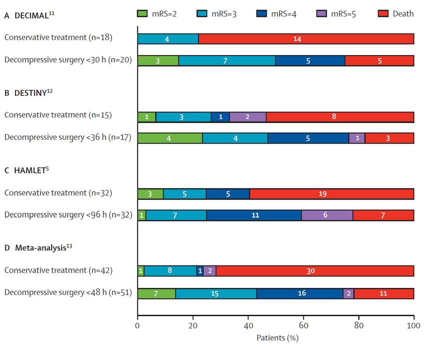
Decompressive Hemicraniectomy for Malignant MCA Territory
Ischemic Stroke
Complete MCA territory stroke has a high (80%) mortality because cytotoxic edema can cause
life threatening herniation. Decompressive hemicraniectomy can be a life-saving (and function
saving) procedure for patients with malignant MCA territory infarction.
Consider early (< 48 hrs) decompressive hemicraniectomy if
Patient age less than 60
o > ½ MCA territory infarcted
o DWI infarct volume > 145 cm3 on MRI
Results of randomized trials evaluating decompressive hemicraniectomy:
Important points when talking to families:
Decompressive hemicraniectomy is life saving
A significant proportion of patients who get DHC and survive have Modified Rankin
Scales (MRS) of 3 or better (able to walk without assistance)
However, a significant proportion of patients who get DHC and survive have MRS of
4 or worse (unable to walk, dependent on others for assistance with bodily needs)
Left versus right hemispheric stroke does not impact outcome19
Protocol for cerebellar stroke/posterior fossa syndrome
Cerebellar ischemic stroke
No
Mass effect? Observe
Yes
• Hydrocephalus with 4th ventricle obliteration
• Neurologic deterioration referable to brainstem compression in opinion of
treating physician
• Neurologic deterioration suspected due to brainstem compression that
improves with osmotic therapy
Yes to ANY No to ALL
of above of above
Increasing edema on serial scans
Urgent decompressive surgery over 3-5 days post stroke onset?
Yes No
Consider prophylactic Observe
decompressive surgery
Notes:
• Above applies only to patients who are suitable candidates for surgery
• Involvement of vermis is associated with increased risk of neurologic deterioration and should
lower threshold for surgery
•Seizure
For uncomplicated cases, single anti-platelet therapy or IV heparin may be started 3 days post-
op if there is a strong clinical indication
10/13/2011
Created by:
Brett Cucchiara, MD
Approved by:
Sean Grady, MD (Neurosurgery)
Scott Kasner, MD (Stroke)
Josh Levine, MD (NeuroICU)20
Secondary Stroke Prevention
• Antiplatelet agent
o ASA 81-325 mg or Plavix 75mg daily as soon as it is safe
o ASA and Plavix ONLY if patient has acute coronary syndrome (CURE) or has just
been stented.
• Statin
o Preferably Lipitor 80 mg daily or simvastatin 40 mg daily if total cholesterol > 135
(Heart Protection Study, 2004)
• ACE-inhibitor (i.e. ramipril) or ARB
o Within 1-2 weeks after D/C (HOPE, PROGRESS, LIFE)
• Hypertension:
o Patients with flow dependent lesions may need higher BP acutely
o Chronically hypertensive patients may have a shifted cerebral autoregulation curve
and rapid lowering > 25% may compromise cerebral perfusion
• Long-term anticoagulation:
o For A fib, EF < 30%, or cardiac thrombus, hypercoagulable state, mechanical valve
in the setting of acute ischemic stroke (multiple refs).
o No good data exist about when to start--our practice is generally to start ASA
within 48 hours, and warfarin in 1-2 weeks.
o No benefit to warfarin over aspirin for symptomatic intracranial stenosis (WASID,
2005).
• Carotid endarterectomy or stent:
o Remember, even lacunes may be due to emboli (20%), so rule out carotid disease.
o Consider CEA in >70% stenosis & +/- 50-69% (NASCET, 1997).
o Symptomatic patients with poor surgical risk patients benefit from carotid stenting
(SAPPHIRE), but more widespread use is not yet established.
• Intracranial stenting and interventional neuroradiology:
o Limited evidence, but option for patients with multiple events due to symptomatic
stenoses > 50% despite medical therapy.
• Intracranial hemorrhages:
o If ICH, no antiplatelet/anticoagulant therapy x 3 months in general.
o If ICH in the face of atrial fibrillation/ cardiac thrombus, can try ASA in 2 weeks,
reconsider warfarin in a month after deep ICH if BP controlled, and monitor VERY
closely.
o Do not resume anticoagulation in amyloid angiopathy or most lobar ICHs.
• PFO and IASA
o About 25% of people have PFOs.
o Ensure there is no other source for a stroke before attributing it to the PFO. Get a
complete hypercoag workup.
o Recent studies show long term outcomes support PFO closure in cryptogenic stroke
in patients < 60 yo (RESPECT, CLOSE, REDUCE studies)
o There are no FDA approved PFO closure devices although cardiologists will close
them with devices approved for other purposes. The decision is largely a matter of
personal preference for the patient. Offer them enrollment in a trial if appropriate.
• Atrial flutter vs. fib:
o Some cardiac studies suggest that patients in atrial flutter may be going in and out
of atrial fibrillation.
o Consider outpatient Holter monitor if suspicion of AF.
o CHADS2VAS2C scoring system
• Glucose:
o Screen for diabetes with fasting glucose, HbA1c
• Smoking cessation:
o Screen and counsel on smoking cessation
o Consider nicotine replacement products21
Stroke Syndromes
Eponym/artery Anatomy Signs and symptoms
Contralateral leg > arm weakness
ACA Medial frontal and parietal
Abulia
Anterioinferior caudate
Recurrent artery of Huebner Contralateral face weakness (Huebner)
Putamen
(A branch off A1 segment) Contralateral leg weakness (A1 segment)
Anterior limb of internal capsule
Face and arm > leg weakness / numbness
MCA - Superior M2 (anterior) Expressive aphasia (dominant, Broca's)
Hemineglect (nondominant)
Homonymous hemi / upper quadrantinopsia
MCA - Inferior M2 (posterior) Receptive aphasia (dominant Wernicke's)
Constructional apraxia (non-dominant)
Alexia / agraphia
Gerstmann syndrome Dominant inferior parietal lobe Finger agnosia
(Partial MCA) (angular gyrus) R-L confusion
Acalculia
Occipital and infero-medial Homonymous hemianopsia with macular sparing +/-
Unilateral PCA
temporal lobes, posterior thalamus alexia without agraphia / anomia
Optic ataxia
Balint syndrome (bilateral PCA) Bilateral parieto-occipital lobe Ocular apraxia
Simultanagnosia
Dominant occipital lobe with
PCA Callosal branch Alexia without agraphia, or "pure word blindness"
splenium of corpus callosum
Dejerine-Roussy or "thalamic pain Contralateral hemisensory loss
Thalamus
syndrome" (PCA branches) Contralateral hemibody pain
Weber (PCA penetrators) Midbrain, anterior Contralateral weakness, ipsilateral CN III palsy
Contralateral rubral tremor, ipsilateral CN III palsy +/-
Claude (PCA penetrators) Midbrain, tegmentum
contralateral weakness and numbness
Contralateral rubral tremor, ipsilateral CN III palsy,
Benedikt (PCA penetrators) Midbrain, tegmentum
ipsilateral ataxia, contralateral hemisensory loss
Ipsilateral CN VI (spares CN VII)
Raymond (basilar paramedian branches) Pons, ventral-medial
Contralateral weakness
Millard-Gruber (basilar short and Pons, basis pontis and VI and VII Ipsilateral CN VI and VII palsies
paramedian branches) fascicles Contralateral weakness
Foville (basilar shorts and paramedian Ipsilateral VI / PPRF (gaze) and VII palsies
Pons, tegmentum and caudal third
branches) Contralateral weakness and sensory loss (ML)
Marie-Foix (basilar/AICA) Pons / lateral Ipsilateral ataxia, contralateral weakness and numbness
Bilateral face/arm/leg weakness
Locked-in syndrome (basilar) "de-
Bilateral ventral pons Bilateral VI palsies
efferented state"
Aphonia
Ipsilateral facial sensory loss (CN V)
Wallenberg of lateral medullary Ipsilateral ataxia, nystagmus, N/V
Medulla, lateral
syndrome (vertebral artery > PICA) Vertigo, hoarseness, dysphagia
Ipsilateral Horner's, contralateral body sensory loss
Contralateral weakness
Anterior spinal artery (Dejerine
Medulla, medial Contralateral vibration/proprioceptive loss (ML)
syndrome)
Ipsilateral tongue deviation (CN XII nucleus)22
PRIMARY INTRACEREBRAL
HEMORRHAGE
Despite aggressive efforts, mortality from ICH is high. Scoring systems have been devised to
calculate in-hospital mortality risk and functional outcome based on patient age, initial neurologic
examination, hematoma size and site of bleeding (see appendix). It is, however, important to use
these scores judiciously when making treatment decisions, since they can provide a “self-fulfilling
prophecy”.
Most ICHs are secondary to hypertension. These tend to be in deeper structures (thalamus, basal
ganglia, brainstem, cerebellum). The (distant) second most common cause in the elderly is
secondary to cerebral amyloid angiopathy. Amyloid bleeds tend to be superficial and lobar.
Don’t forget other potential causes:
• Iatrogenic secondary to systemic anticoagulation
• Hemorrhagic conversion of an ischemic infarct
• AVM or aneurysm rupture
• Trauma
• Cerebral Venous Sinus Thrombosis
• Vasculitis
• Cavernous Malformations
• Hemorrhagic Metastases/Primary Tumor
• Infection/endocarditis
The principles of ICH management involve:
• management of intracranial pressure (ICP) and cerebral perfusion pressure (CPP)
• correction of underlying coagulopathies that may exacerbate bleeding
• aggressive blood pressure control to limit hematoma expansion
• prevention of other medical complications
General Approach to ICH Management
• CT scan shows acute blood: is it in a pattern that suggests a hypertensive hemorrhage or
amyloid hemorrhage, or something else atypical?
• Consider CTA to look for underlying vascular malformation and for “spot sign”
o The “spot sign” is a bright spot within the hematoma on contrast enhanced CT
angiography
o It suggests active bleeding and is strongly associated with hematoma expansion
• Are there any correctable coagulopathies? (Also refer to reversal guidelines on page 82)
o Aspirin – no clear benefit from platelet transfusion (Sansing et al., Neurology
72:1397, 2009). Do not transfuse platelets.
o Plavix – unclear if platelet transfusion beneficial. Do not transfuse platelets.
o Warfarin – For ICH or life threatening hemorrhage with elevated INR consider
activated Prothrombin Complex Concentrate (PCC). First dose can be given
without hematology approval (50 units/kg). Also give intravenous 10 mg
vitamin K stat. (Watch for anaphylactoid reaction) FFP is a lot of volume and
takes time to thaw.
o Heparin – follow PTT and correct with protamine sulfate. Dose for protamine is
1 mg for every 100 units unfractionated heparin given in the last 2 hours. Dose23
changes depending on when heparin infusion stopped. Pharmacy can help you calculate the
dose (maximum = 50mg).
o Low Molecular Weight Heparin (e.g. enoxaparin, dalteparin) – cannot followPTT effectively
but can use protamine for partial reversal. Pharmacy will help you calculate the dose (1mg per
100 anti-Xa units).
o Dabigatran and newer oral direct thrombin inhibitors (e.g. rivaroxaban, apixaban). PTT
can be elevated, but not always. SEE REVERSAL PROTOCOLS BELOW.
o tPA – reverse with 2 bags cryoprecipitate (5 units/bag), thawed plasma (2 units – must call blood
bank) and platelets (2 doses to start). Follow fibrinogen q6 hr
o Uremic Platelet Dysfunction – Can give one time desmopressin (ddAVP) (0.3 - 0.4 ug/kg IV bolus)
and conjugated estrogens (0.6 mg/kg IV slow bolus over 30 min daily X 5 days). Hemodialysis is
definitive treatment. (Nat Clin Pract Nephrology, 3:138, 2007)
o Note: Recombinant Factor VIIa limits hematoma expansion but should not be used due to
increased thrombotic complications! (Mayer et al., NEJM, 358:2127, 2008)
o Keep platelet count greater than 50,000 or >100K if neurosurgical candidate.
• Aggressive blood pressure control – target systolic < 160, MAP < 110 at least, lower BP target if patient
has lower baseline BP. Use labetalol IV bolus (10-20 mg) first and then nicardipine infusion if needed.
Avoid nitroprusside and nitroglycerin as these can increase ICP due to prominent venodilation
• Consider surgical evacuation for large superficial lobar hemorrhage. STICH I and II trials give evidence
that earlier intervention may be life-saving especially in those who are clinically declining.
• Place EVD if evidence for early hydrocephalus
• If prominent intraventricular hemorrhage, consider Intraventricular tPA . This should be a discussion with
attending, pharmacy, neurosurgery, etc., since this is still investigational. Data for this extends from
MISTIE and CLEAR-IVH trials. Consider if prominent intraventricular hemorrhage with
intraparenchymal hematoma < 30 cc, and patient has existing EVD for treatment of hydrocephalus.
Current protocol at HUP:
o 1 mg tPA followed by saline flush into EVD
o Clamp EVD for 1 hour
o Open EVD at 0 cm above tragus, can repeat every 12 hours until resolution of clot for 5 days
o Get CT scan 8 hours after each tPA treatment24
Anticoagulant Reversal Guidelines
All guidelines can be assessed from Penn Medicine Formulary link in the Upenn homepage
Dabigatran (Pradaxa)
• Hold dabigatran dosing
• Assess nature and severity of hemorrhage; timing and dose of last taken; assess need for surgery to
control bleeding; presence or absence of dialysis access
• Collect aPTT, thrombin time (TT), PT, dilute thrombin time (dTT), fibrinogen, CBC, serum Cr, LFTs
• The presence of severe bleeding is the primary determinant of therapeutic intervention
o The aPTT, when prolonged, tends to predict residual dabigatran effect well
o Thrombin time (TT) is highly sensitive to Pradaxa. A normal TT level rules out clinically
significant drug level.
• In mild to moderate bleeding, use general measures to control bleeding. Follow renal function
closely
• Fresh frozen plasma (FFP) should not be used
• Activated charcoal can be used (12.5g x1) if ingestion was within 2h
• Praxbind (idarucizumab) 5mg IV (2 infusions of 2.5g IV within 15 minutes of each other) is the
preferred first reversal agent and does not need Hematology approval for severe or life threatening
bleeding
• In severe bleeding, hemodialysis should be considered. Consult renal fellow for hemodialysis
o If bleeding persists in spite of multiple doses of Praxbind, consider hemodialysis
• Hematology consult is required for multiple doses of Praxbind
• Do not give Activated Prothrombin Complex Concentrate (APCC - FEIBA), rFVIIa (Factor VIIa-
NovoSeven RT), or Prothrombin Complex Concentrate (PCC – Kcentra)
Anti Xa drugs: Rivaroxaban (Xarelto), and Apixaban (Eliquis)
• Hold Factor Xa inhibitor
• Assess nature and severity of hemorrhage; timing and dose of last taken; need for surgery to control
bleeding
• Collect PT, INR, Anti-Xa level, aPTT, fibrinogen, CBC, serum Cr, LFTs
• A prolonged PT/INR may suggest residual Factor Xa inhibitor effect
• Normal anti-Xa assay rules out significant anti-Xa drug levels. Anti-Xa levels are available at HUP
weekdays from 8:00 am to 4:00 pm. Contact hematology for guidance
• Activated oral charcoal (12.5g x1) can be used if drug was ingested within 2 hours
• Reversal of anti-coagulation has increased risk for thrombosis; always balance risk-benefit ratio
• Andexanet alfa (Andexxa) is the preferred agent for patients on rivaroxaban or apixaban with intra-
cranial or immediate life-threatening or limb/organ-threatening bleeding
• Prothrombin Complex Concentrate (PCC-Kcentra) (50U/kg x1) is an alternative agent that may be used
for Factor Xa inhibitor reversal in patients who do not meet criteria for Andexanet alfa.
• Fresh frozen plasma (FFP) should not be used
• Hematology consult required for Activated Prothrombin Complex Concentrate (APCC- FEIBA), rFVIIa
(Factor VIIa-NovoSeven RT), or subsequent doses of PCC and should be considered only if bleeding
persist after first dose of PCC
o aPCC dosing: 50-100 U/kg x1
o RFVIIa dosing 20-40 mcg/kg or weight-based (2 mg if weight 100kg) x125
Antithrombotic Reversal agent
Vitamin K antagonists If INR >1.5, no signs of bleeding and reversal required in >24h:
for urgent surgery or Vit K 2.5-5 mg PO or, 0.5-1mg IV
other invasive
procedures If INR >1.5, no signs of bleeding and reversal required in 12 h from dosing
Dalteparin, Nadroparin and Tinzaparin:
Dosed within 3–5 half-lives of LMWH: Protamine 1 mg IV per 100
anti-Xa units of LMWH (up to 50 mg in a single dose)
OR
rFVIIa 90 mcg/kg IV if protamine is contraindicated
Danaparoid rFVIIa 90 mcg/kg IV
Pentasaccharides Activated PCC (FEIBA) 20 units/kg IV or rFVIIa 90 mcg/kg IV
Thrombolytic agents Cryoprecipitate 10 units IV OR
(plasminogen Antifibrinolytics (tranexamic acid 10–15 mg/kg IV over 20 min or e-
activators) aminocaproic acid 4–5 g IV) if cryoprecipitate is contraindicated
Antiplatelet agents DDAVP 0.4 mcg/kg IV 9 1
If neurosurgical intervention: Platelet transfusion (one apheresis unit)26
ANEURYSMAL SUBARACHNOID
HEMORRHAGE
I. INTRODUCTION:
Subarachnoid hemorrhage (SAH) is a disorder where bleeding occurs between the
arachnoid membrane (the middle membrane covering the brain) and the brain itself, with
bleeding onto the surface of the brain. This is usually caused by a “weak spot” in a blood
vessel, also known as an aneurysm. Twenty percent of individuals affected have multiple
aneurysms. Subarachnoid hemorrahages can also be non-aneurysmal in cause. Examples
of this would be a traumatic SAH, rupture of an AVM, or other unidentified causes. The
following guidelines address management of aneurysmal subarachnoid hemorrhage
II. RISK FACTORS FOR ANEURYSMAL SAH
• Hypertension
• Cigarette Smoking (greatest risk occurring 3 hours after smoking)
• Binge alcohol drinking
• Drug abuse
• Use of stimulants
• Gender: Incidence is greater in females than in males
• Age (range 20-65; mean age 50; most common between 35-60)
• Disorders associated with weakened blood vessels: 55
▪ Fibromuscular dysplasia (FMD)
▪ Aneurysms in other blood vessels
▪ Polycystic kidney disease
▪ Ehler-Danlos syndrome
▪ Marfan’s syndrome
▪ Alfa-one antitrypsin deficiency
III. PRIOR TO SECURING THE ANEURYSM (Surgical Clip/Neurovascular Coil):
Admission labs, tests and procedures
• Admit to the NeuroICU
• Assess Hunt and Hess Scale57 (Clinical Grade) and Fisher Grade55 (CT Grade) (See
Appendix A)
• Obtain routine labs, including:
• BMP, Ca2+, Mg2+, PO4-2, CBC, PT, INR, PTT, serial troponins, type and screen
• Obtain the following studies:
o 12-lead EKG, portable chest X-ray, non-contrast head CT, CT angiogram
(CTA; unless contraindicated due to allergy or renal dysfunction)
• Alert research coordinator to determine eligibility for clinical studies
Neurologic
• Place EVD immediately in all patients with hydrocephalus that is symptomatic or
have GCS27
the EVD should generally not be lowered prior to securing of the aneurysm as
this may predispose to rebleeding.
• Start seizure prophylaxis
• Leviteracitam load: 1000mg PO/IV on admission
• Leviteracitam 500 mg PO/IV q12 hrs x 7 days for seizure prophylaxis
▪ If evidence of renal dysfunction, dose accordingly
▪ It is preferable to administer leviteracitam orally or
enterally when able
▪ Discontinue AED POD #7 if there have been no
clinical or electrographic seizures
• Anti-Fibrinolytics
• Consider in patients who cannot be secured in < 24 hours
Cardiovascular
• Place Arterial line in all patients
• Maintain SBP within 10% of baseline SBP if known. If baseline SBP cannot be
determined, then maintain SBP < 160 mm Hg.
o If SBP > 160 mmHg, optimize analgesia and treat with IV anti-
hypertensives
o Titratable anti-hypertensives (drips) are favored to control blood pressure
to avoid large blood pressure fluctuations.
• Start nimodipine 60 mg PO/NGT q4hrs. If BP drops after administration, then
decrease nimodipine dose to 30 mg q2hrs.
• Statins
o Statins taken as home medications should be continued upon admission for
SAH.
• Vascular access
• Place a triple lumen subclavian or IJ venous catheter in selected patients.
• Avoid IJ if evidence of severe intracranial hypertension exists or
patient is felt to be at high risk for developing intracranial
hypertension.
• Indications for central line placement:
• High grade patient (HH≥3)
• Hemodynamic instability
• Need for vesicant medications (sedation or vasopressors)
• Venous access difficulty
• Obtain a transthoracic echocardiogram when:
• Unexplained hypotension
• Ischemia or ST changes are present on EKG
• Response to vasopressor medications is suboptimal
• Initial cardiac enzymes are abnormal
Renal (Fluids and Electrolytes)28
• Goal is to maintain euvolemia.
• Hypotonic IVF should be avoided.
• Maintain Na+ within normal range.
• Maintain Mg+2 within normal range.
Endocrine (Glycemic Control)
• Blood glucose monitoring and sliding scale insulin q4 hrs in all patients
• Target glycemic threshold 200mg/dL x 2 on SSI coverage
Prophylaxis
• Stress ulcer prophylaxis with either H2 blocker or PPI
• Venous thromboembolism prophylaxis
• Pneumatic compression boots upon admission
• Prophylactic dose anticoagulation should be withheld until after the aneurysm
is secured due to risks of re-rupture. After craniotomy, anticoagulants can be
started 24h after the procedure.
• Alcohol abuse/withdrawal
• Thiamine, folate, MVI in patients with alcohol abuse history/concern
• For patients who exhibit sign/symptoms of withdrawal, or who have a history
of alcohol withdrawal, consider starting a taper of long-acting
benzodiazepines.
Other issues
• Analgesia
• Pain should be controlled with lowest effective dose of IV or PO
medications that do not limit clinical neurological examination. Overly
sedating medications should be avoided.
• Anti-emetics
• Anti-emetics that are unlikely to cause sedation or have CNS side effects
are preferred (ie: ondansetron, trimethobenzamide) if necessary
IV. ANEURYSM OCCLUSION:
There are two main methods for obliterating aneurysms: surgical clipping and
endovascular coiling. The feasibility of both methods is assessed and a collaborative
decision between neurosurgery and interventional neuroradiology is made to determine
optimal treatment based on aneurysm morphology and patient characteristics. 55 Certain
aneurysms are better suited to one technique or another. Endovascular treatment is often
the preferred technique for posterior circulation aneurysms and ruptured aneurysms in
elderly patients (>70 years of age) given higher surgical risks. Aneurysms in the middle
cerebral artery, ruptured aneurysms with large intraparenchymal hematomas are commonly
approached surgically. Combined endovascular and surgical techniques may be required
for some very large or complex aneurysms. The advent of Guglielmi platinum detachable
coils (GDC)30 in the 1990s introduced endovascular therapy for cerebral aneurysms.
Alternative methods for obliterating the aneurysmal sac include stent-assisted coiling,
balloon-assisted coiling, flow diverters and embolic agents.
The optimal timing of surgery is unclear. The International Cooperative Study on the
Timing of Aneurysm Surgery31, 32 demonstrated that although pre-operative re-bleeding
rates were lower with early surgery, timing of surgery had no effect on overall outcome.29
Early surgery (1 – 3 days post SAH) has become common practice, in part because it is
associated with lower rates of re-bleeding and allows for more aggressive treatment of
vasospasm.
• Attempts are made to obliterate the aneurysm within 3 days of SAH, either by
endovascular or open surgical techniques.
V. POST ANEURYSM OCCLUSION MANAGEMENT:
Neurologic
• Intracranial Hypertension
• Maintain ICP < 20 mmHg, CPP > 60 mmHg (see NeuroICU Protocol).
• Vasospasm detection
• Continuous EEG monitoring:
• Consider monitoring in patients with Fisher Grade III SAH
and/or GCS < 8 with cEEG.
• In all other patients, consider cEEG to detect early ischemic
changes. In awake patients, changes in alpha variability can be
used to trigger a trial of hypertensive therapy (see below) followed
by vascular imaging with a CTA or conventional angiogram (see
cEEG Protocol).
• TCD monitoring:
• Obtain daily TCD. This must include MCA velocity/extracranial
ICA velocity (Lindegaard ratio). See appendix B for interpretation.
• Algorithm for approach to TCD and clinical examination data: See
appendix C
• Treatment of Vasospasm or DCI
• Triggers for treatment:
• TCD elevation (Lindegaard ratio >3)
• Clinical exam change (DCI)
• Clinical concern for vasospasm or DCI should be followed by immediate
diagnostic testing with imaging. Previous studies on hypervolemic therapy
have failed to show any benefit.
• Minimize risk of volume overload and avoid hypovolemia
• Prophylactic therapy with hypervolemia, hemodilution is
no longer recommended
• Induced hypertension continues to remain effective in increasing
cerebral blood flow; the loss of innate autoregulation during
vasospasm makes cerebral perfusion pressure more dependent on
systemic blood pressure
• This strategy is not recommended for prophylaxis but
during vasospasm to reduce the risk of ischemia in
patients developing vasospasm
• Use of norepinephrine, dopamine and phenylephrine have
all been shown to be beneficial in improving neurological
outcome
• If nimodipine administration results in hypotension, dosing
intervals should be changed to more frequent, lower doses.30
• Vascular imaging with a CTA should be performed when
clinically feasible.
• CTA should be obtained in all patients with suspected
vasospasm who do not have contraindications to the
procedure.
• If CTA is indeterminate or shows evidence of vasospasm,
proceed to cerebral angiography.
• Perfusion imaging may be considered in appropriate
patients.
• Advanced cerebral monitors (Licox, microdialysis, etc…) may be considered on a
case-by-case basis.
Cardiovascular
• For patients undergoing craniotomy for aneurysm coiling, SBP should be
maintained < 160 mmHg for 24 hours after surgery to minimize risk of
postoperative bleeding. Thereafter, SBP parameters should be liberalized to 100 –
200 mmHg.
• In aneurysms that are endovascularly coiled, blood pressures can be liberalized to
100-200 mmHg immediately post-procedure.
• A higher blood pressure may be needed to achieve adequate regional brain
perfusion and these parameters may be adjusted in individual patients (refer to
treatment of vasospasm/DCI above).
• The presence of other, unruptured aneurysms should not influence hemodynamic
management.
• Target euvolemia with administration of isotonic or hypertonic IVF as needed.
Fluid and Electrolyte Management
• Maintain Na+ > 135 mmol/L (see CSW Protocol).
• Treat hyponatremia with IV normal saline and oral salt tabs.
• For refractory cases, consider the use of fludrocortisone 0.3mg/day or
hypertonic saline bolus/infusion to raise serum Na to the target range.
• Maintain Mg+2 within normal range
Endocrine (Glycemic Control)
• Blood glucose monitoring and sliding scale insulin q4 hrs in all patients
• Target glycemic threshold 200mg/dL x 2 on SSI coverage
Temperature Control
• Maintain strict normothermia (see Normothermia Protocol).
Nutrition
Early enteral nutrition benefits critically-ill patients.53 In the neurologically injured
patients, continuous enteral feeding via small bore tube or NGT/OGT is safe.54
• Continuous enteral feeding should begin on POD #1 (see NeuroICU Feeding
Protocol).
• TPN should only be considered in patients who have contraindications or intolerance
to enteral feedings.31
Prophylaxis
• See above recommendations for GI and DVT prophylaxis.
• Subcutaneous heparin 5000U TID should be added on POD #1.
VII. TRANSFER/DISCHARGE PLAN:
• Low grade (I-II) patient criteria to transfer out of NeuroICU
▪ Day 10 if no evidence of vasospasm for 48hrs
• High grade (III-V) patient criteria to transfer out of NeuroICU
▪ Day 14 and no evidence of vasospasm for 48hrs.
• Nimodipine stop after 21 days, not discharged on nimodipine.
• Acute brain injury rehab
Approach to TCD and clinical examination data
EXAM TCD ACTION
Normal Normal None
Normal Abnormal CTA, Perfusion scan
Deteriorating Normal Hypertensive rx, CTA, Perfusion scan
Deteriorating Abnormal Hypertensive rx, Angiogram
Comatose Normal Consider surveillance CTA, cEEG
Comatose Abnormal Non-contrast CT, Angiogram Consider cEEG
Risk factors associated with or predictive of vasospasm55
Younger age IVH on admission CT Dehydration
Cigarette smoking Hydrocephalus Hypotension
Poor admission clinical grade Hyponatremia Hypoxia
Admission systolic blood pressure Anti-fibrinolytic agents Fever
Thick or diffuse SAH on admission CT Increased ICPYou can also read