Update on uveal melanoma: Translational research from biology to clinical practice (Review) - Spandidos Publications
←
→
Page content transcription
If your browser does not render page correctly, please read the page content below
INTERNATIONAL JOURNAL OF ONCOLOGY 57: 1262-1279, 2020

                  Update on uveal melanoma: Translational research
                      from biology to clinical practice (Review)
              MIGUEL A. ORTEGA1‑3*, OSCAR FRAILE‑MARTÍNEZ1*, NATALIO GARCÍA‑HONDUVILLA1‑3,
              SANTIAGO COCA1‑3, MELCHOR ÁLVAREZ‑MON1‑4, JULIA BUJÁN1‑3 and MIGUEL A. TEUS5,6

          1
          Department of Medicine and Medical Specialties, Faculty of Medicine and Health Sciences, University of Alcalá,
            Alcalá de Henares, 28871 Madrid; 2Ramón y Cajal Institute of Sanitary Research (IRYCIS), 28034 Madrid;
    3
      University Center for The Defense of Madrid (CUD‑ACD), 28047 Madrid; 4Internal and Oncology Service (CIBER‑EHD),
                University Hospital Príncipe de Asturias, Alcalá de Henares, 28805 Madrid; 5Department of Surgery,
          Medical and Social Sciences, Faculty of Medicine and Health Sciences, University of Alcalá, Alcalá de Henares,
    28871 Madrid; 6Ophthalmology Service, University Hospital Príncipe de Asturias, Alcalá de Henares, 28805 Madrid, Spain

                                     Received May 10, 2020; Accepted September 24, 2020

                                                   DOI: 10.3892/ijo.2020.5140

Abstract. Uveal melanoma is the most common type of intra-         microRNAs as a target for disease control. In the present
ocular cancer with a low mean annual incidence of 5‑10 cases       study, the main epidemiological, clinical, physiopathological
per million. Tumours are located in the choroid (90%), ciliary     and molecular features of this disease are reviewed, and the
body (6%) or iris (4%) and of 85% are primary tumours. As in       associations among all these factors are discussed.
cutaneous melanoma, tumours arise in melanocytes; however,
the characteristics of uveal melanoma differ, accounting
for 3‑5% of melanocytic cancers. Among the numerous                Contents
risk factors are age, sex, genetic and phenotypic predisposi-
tion, the work environment and dermatological conditions.          1. Introduction
Management is usually multidisciplinary, including several         2. Risk factors
specialists such as ophthalmologists, oncologists and maxil-       3. Clinical manifestations
lofacial surgeons, who participate in the diagnosis, treatment     4. Anatomopathological study of uveal melanoma
and complex follow‑up of these patients, without excluding the     5.	Molecular classification of uveal melanoma: Genes
management of the immense emotional burden. Clinically,                involved
uveal melanoma generates symptoms that depend as much              6.	Uveal vs. cutaneous melanoma: Similarities and differences
on the affected ocular globe site as on the tumour size. The       7. Biology of uveal melanoma
anatomopathological study of uveal melanoma has recently           8. Blood biomarkers for uveal melanoma
benefited from developments in molecular biology. In effect,       9. Proteomics and metabolomics in uveal melanoma
disease classification or staging according to molecular profile   10. Clinical management of uveal melanoma
is proving useful for the assessment of this type of tumour.       11. Conclusions and future directions
Further, the improved knowledge of tumour biology is giving
rise to a more targeted approach to diagnosis, prognosis and
treatment development; for example, epigenetics driven by          1. Introduction

                                                                   Although relatively rare, uveal melanoma is the most common
                                                                   type of intraocular tumour with a mean annual incidence of
                                                                   5‑10 cases/1,000,000 individuals. Among all cancers of the
Correspondence to: Dr Miguel A. Ortega, Department of              eye, 85% are primary tumours of this type and occur in indi-
Medicine and Medical Specialties, Faculty of Medicine and Health
                                                                   viduals with a mean age of 60 years. The remaining 15% cases
Sciences, University of Alcalá, Ctra, Madrid‑Barcelona km 33.6,
Alcalá de Henares, 28871 Madrid, Spain                             are non‑Hodgkin lymphomas, retinoblastomas and medullo-
E‑mail: miguel.angel.ortega92@gmail.com                            epitheliomas. Despite these figures, the most frequent tumours
                                                                   affecting the eye are metastases of other types of cancer,
*
    Contributed equally                                            mainly lung cancer in males and breast cancer in females (1,2).
                                                                   Uveal melanoma is also a melanocytic cancer, representing
Key words: uveal melanoma, cell transduction pathways,             approximately 3‑5% of all of these cancers, although its char-
epigenetics, miRNA, immunotherapy                                  acteristic features differ from those of the cutaneous form.
                                                                   Tumours are mainly located in the choroid 85‑90%, followed
                                                                   by the ciliary body (6%) and iris (4%). Several studies have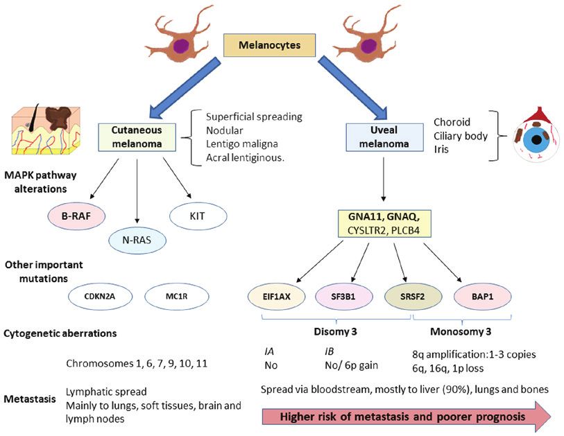
ORTEGA et al: UPDATE ON UVEAL MELANOMA                                                1263

demonstrated that both its cell mutation pattern and aetiology       Sex as a risk factor is related to age. For example, in individ-
have their own characteristics, unrelated in a large measure to      uals
1264                                INTERNATIONAL JOURNAL OF ONCOLOGY 57: 1262-1279, 2020

the presence of symptoms and orangey colour, among others.            biology. This has meant that currently, classification according
It should be underscored that these risk factors generally lead       to the molecular profile of uveal melanoma has proven more
to an earlier appearance of uveal melanoma (11,12).                   useful than its histological classification, in line with the
                                                                      concept of individualized precision medicine for these patients.
3. Clinical manifestations
                                                                      5. Molecular classification of uveal melanoma: Genes
Uveal melanoma generates symptoms depending on the ocular             involved
site involved, meaning that most clinical signs are determined
by both tumour size and location. Usually patients present            Uveal melanoma is often divided into two categories according
with blurred vision, photopsia and/or myodesopsia or are              to its gene expression profile and to its metastasizing capacity.
asymptomatic and the uveal melanoma is detected incidentally          Hence, class 1 uveal melanomas are associated with a low risk
during a routine ophthalmological examination (7). When the           of metastasis and have been linked to a better prognosis, while
tumour affects the macula, patients exhibit a gradual painless        class 2 tumours feature a high risk of spread and a worse prog-
decline in visual acuity. It should also be mentioned that if         nosis. In addition, there is significant variation in cytogenetics
there is involvement of the iridocorneal angle, signs may be          and expression levels of some genes in the different subtypes;
those of acute glaucoma, namely the loss of visual acuity,            for example, chromosome 3 monosomy is characteristic of
pain, photopsia and increased intraocular pressure. These             class 2 tumours (35). However, this initial classification is
symptoms can lead to permanent blindness and are therefore,           insufficient to explain, for example, why some class 1 tumours
constitute an ophthalmological emergency. By contrast, the            show a higher risk of metastasis than others.
involvement of the iris is usually asymptomatic and presents               For this reason, uveal melanoma classification has been
as a dark growing, invasive hyperpigmented lesion. If the             extended to include 4 groups: 2 subclasses characterized by
ciliary body is involved, this can compromise the natural lens,       chromosome 3 monosomy (M3) with a worse prognosis, and
causing its subluxation and impaired accommodation, thus              a further 2 subtypes that lack this chromosome abnormality;
interfering with the patient's vision (21). It should be noted that   i.e., with chromosome 3 disomy (D3), with a better prognosis.
infrequently, intraocular progression can give rise to haemor-        The first 2 subclasses are associated with a higher metastasis
rhage within the ocular cavity presenting as haemorrhage              risk and exhibit a loss of or mutation of the gene encoding
and exophthalmos. Up to 22% of patients may have systemic             BRCA‑associated protein 1 (BAP1) located on 3p21.1
manifestations as a consequence of metastatic spread mainly           (NCBI), and conferring a different methylation state to those
to the liver, and almost 90% succumb to the disease before            without this monosomy. Between both M3 subtypes, there is
5 years following diagnosis (22).                                     a series of genomic, transcriptional and clinical variations,
                                                                      such as the amplification of 1 to 3 copies of the long arm of
4. Anatomopathological study of uveal melanoma                        chromosome 8 (36).
                                                                           In turn, the D3 subtypes are divided into IA and IB.
Callender (23) was the first to establish an anatomopatholog-         The former exhibits no aneuploidy, the least risk of spread
ical classification of these tumours, which was later modified        and is characterized by a mutation in eukaryotic translation
by McLean et al (24), who distinguished between type A                initiation factor 1A X‑linked (EIF1AX). Subtype IB, char-
fusiform cell, type B fusiform cell, epithelioid cell and mixed       acterized by the possible presence of a total or partial gain
tumours. Fusiform type A followed by B tumours were associ-           of 6p and a higher metastasis risk, features mutations in the
ated with a higher survival rate, and epithelioid cell tumours        splicing factor 3b subunit 1 (SF3B1) gene (37). Furthermore,
were associated with the worse prognosis. Mixed tumours               Field et al (38,39) highlighted the role of gene expression of
were associated with an intermediate outcome (25,26).                 preferentially expressed antigen in melanoma (PRAME) as
Another series of histopathological criteria has proven useful        an independent biomarker of metastasis frequently found in
to assess disease prognosis in a patient with uveal melanoma.         tumours with a mutation in SF3B1. This marker may also
For instance, an elevated microvascular density (MVD) related         appear in M3 tumours and is also inversely related to muta-
to tumour irrigation and the presence of a network vascular           tions in EIF1AX. Mutations in the genes EIF1AX, SF3B1 and
pattern have been associated with a worse prognosis (27,28).          BAP1 are mutually exclusive, as well as being key prognostic
High IGF‑1R levels and mean nucleolar diameter have been              markers to understand the behaviour of each uveal melanoma
also related to a lower survival (29,30). The role of some of the     subtype (40). Of note, both in D3 uveal melanomas which
more important cell proliferation markers, such as Ki‑67 or           do not exhibit mutations in SF3B1 or EIF1AX and in M3,
proliferating cell nuclear antigen (PCNA), have been assessed         which exhibit gain of chromosome 8q, mutations in serine and
in uveal melanoma cells, their presence indicating a worse            arginine rich splicing factor 2 (SRSF2) have also been found,
prognosis (31). Finally, localizing some immune system cells,         indicating a role for this marker in the metastasis of uveal
such as lymphocytes or infiltrating macrophages, or the detec-        melanoma and its functional analogy with SF3B1 (36).
tion of markers like HLA‑A have been also associated with
a worse prognosis in patients with uveal melanoma (32,33).            6. Uveal vs. cutaneous melanoma: Similarities and
Notably, the presence of HLA‑B has been associated with the           differences
epithelioid subtype, which is the histological class exhibiting a
lower survival (34).                                                  While cutaneous and uveal melanoma both arise from mela-
    The anatomopathological study of uveal melanoma has               nocytes, their molecular profiles, cytogenetic alterations,
recently benefited from developments in the field of molecular        prognosis and dissemination capacity vary appreciably (Fig. 1).
ORTEGA et al: UPDATE ON UVEAL MELANOMA 1265
For example, it is known that approximately 50% of cases of of class 1 and 2 HLA. This phenotype usually appears in M3
uveal melanoma progress to metastasis and the mean survival tumours as a sign of a worse prognosis (56).
rate of these patients is 6 to 12 months (13). The most frequent This type of information also provides access to new more
site of spread of these tumours is the liver, though lung and effective therapeutic tools for the treatment of uveal melanoma.
bone metastases are also common (41,42) whereas cutaneous However, although several studies have shown the efficacy
melanoma metastasizes with the same frequency to the lungs, of the key immune response regulators PD‑1 and CTLA‑4
bone, brain and soft tissues and mainly spreads via the lymph inhibitors (57,58) in patients with cutaneous melanoma, the
system (43). response to these molecules in patients with uveal melanoma
As in cutaneous melanoma, in uveal melanoma, the has not been the same, suggesting the need to gain further
overexpression of the MAPK pathway is observed. However, insight into the evasive mechanisms of the immune system
mutations found in both types of melanoma differ. In the skin in uveal melanoma (59). In effect, Mougiakakos et al (60)
form, most frequent abnormalities are found in molecules demonstrated how high levels of cyclooxygenase (COX)‑2, a
directly involved in this pathway especially the B‑RAF muta- marker of a worse prognosis in these tumours, were associ-
tion (in 40‑60% of cases). In this type of mutation, particularly ated with elevated Treg levels in uveal melanoma and how
in residue V600, a worse prognosis has been described (44). In this could explain the poor efficacy of antitumour therapies.
addition, are mutations in other genes, such as NRAS (15‑25%) However, there is a need for further research in this area, as
and KIT (39%) are frequent (45). However, it is known that other authors have found no such link between Treg levels
these polymorphisms seldom occur in uveal melanoma (46). and survival in this type of tumour (61,62). Recently, the study
The mutations found in this tumour type appear mainly in the conducted by Petralia et al (63) demonstrated how levels of
genes that code for the α subunit of G, mainly G protein CD47 exhibit a better correlation with elevated levels of Treg
subunit alpha (GNA)11 or GNAQ, detected in up to 90% of and of other inflammatory cells. These results were also
cases of uveal melanoma. Furthermore, these mutations seem reported by Basile et al (64), who also noted that in uveal
to play an important role in the onset and progression of uveal melanoma, CD200 and HVEM are significantly reduced and
melanoma as it has been observed that both abnormalities are that there is an inverse association between the PDL1 levels
not associated with a worse prognosis (47,48). Mutations in and mean overall survival (OS), progression‑free survival
other genes have also been observed, such as cysteinyl leukot- (PFS) and tumour thickness. Notably, the PD‑1/PD‑L1 levels
riene receptor 2 (CYSLTR2; 4%) or phospholipase C beta 4 have been shown to regulate the levels of non‑coding RNA
(PLCB4; 2.5%) (49,50). The mechanisms through which all in a number of types of cancer, whose importance in uveal
these alterations affect tumour biology are described below. melanoma will be subsequently discussed (65). While PD‑L1
In some cases of uveal melanoma, mutations in the expression has been reported at the primary tumour site,
telomerase reverse transcriptase (TERT) gene have been metastatic uveal melanoma exhibits a low expression of this
described. However, the frequency of this mutation is low, marker (66). Importantly, the presence of T cells expressing
having been found in 1 of 50 uveal melanoma specimens LAG3 rather than CTLA‑4 or PD‑1 also plays a role in the
examined by Dono et al (51). Furthermore, this mutation inflammatory pattern in the microenvironment of primary
appeared to be associated with a tumour with variations in uveal melanoma (67). Equally, liver metastasized tumours
GNA11 and EIF1AX, that is, it appeared in the least aggres- show infiltration of clonally expanded plasma cells, suggesting
sive profile. Nonetheless, this TERT variant has been detected antibody‑mediated immunity. The importance of hepatic stel-
at a higher frequency in both sporadic and familiar cutaneous late cells in liver metastasis has also been reported (68). The
melanoma (52). The greatest utility of this marker could be in paracrine signalling of these cells affects the transcriptional
identifying ocular melanoma type as indicated by the study activity of uveal melanoma cells, linked to inflammation and
conducted by Griewank et al (53). These authors found that interleukin production. Hence, inflammatory conditions in the
up to 32% of conjunctival melanomas had a mutated TERT primary tumour seem very different to metastasis locations.
promotor, while this polymorphism was absent in 47 uveal Collectively, these data provide direction for future treatments
melanomas examined. Their findings indicate that the pres- pursuing these targets to improve treatment outcomes.
ence or absence of this mutation is able to distinguish between
both ocular melanomas and may help explain the different Signalling pathways. As described above, the most frequent
behaviour shown by each one. mutations that appear in the early development of uveal mela-
noma are those affecting GPCR receptors, particularly variants
7. Biology of uveal melanoma of GNA11 or GNAQ. These last 2 genes code for subunit G‑α
of G proteins and are activated by the serotonin receptor
Roles of inflammation and immune system in uveal melanoma. 2A and 2B in the melanocyte (5‑HT2A and 5‑HT2B (69).
Hanahan and Weinberg (54) described the main characteristics Receptor 5‑HT2B mutations are often found in a wide
or hallmarks of tumour cells that form the basis of our under- variety of tumours and have been linked to a greater metas-
standing of cancer biology along with the targets of current tasis risk (70). Furthermore, GNAQ and GNA11 mutations
cancer therapies. The inflammatory response represents one trigger a wide range of cell signalling cascades, including the
of these hallmarks and its important role in uveal melanoma PI3K/Akt/mTOR, YAP/TAZ, Wnt/β‑catenin, Rac/Rho, Notch
was reviewed by Bronkhorst and Jager (55). Among other and MAPK pathways (71‑73). The modification of so many
characteristics, the presence of an inflammatory phenotype cell signalling pathways notably hinders treatments targeting
has been described comprised of different types of lympho- their inhibition owing to their possible interactions. An
cytes and macrophages, along with the increased expression example is YAP/TAZ, whose activation occurs independently1266 INTERNATIONAL JOURNAL OF ONCOLOGY 57: 1262-1279, 2020
Figure 1. Diagram illustrating the diagnostic features of cutaneous melanoma and uveal melanoma. Although both melanomas arise from melanocytes, each
one shows its own characteristics while sharing the feature of an altered MAP kinase signalling pathway. Presently, these abnormalities are one of the most
promising targets of the treatment of these patients, such as the inhibition of B‑RAF for cutaneous melanoma. Nevertheless, as illustrated in the diagram, the
mutations that activate this pathway differ and are accompanied by another set of modifications that are also different. In the case of uveal melanoma, these
mutations serve to classify tumours into subtypes according to their molecular profile. The four mutations described are mutually exclusive. This molecular
classification is also associated with metastasis risk and disease prognosis. The spread of both tumours also differs as cutaneous melanomas usually spread via
the lymph while uveal melanomas usually spread via the bloodstream. Uveal melanoma exhibits a high predisposition to spread to the liver, which occurs in
90% of cases. By contrast, cutaneous melanoma may metastasize to the lungs, brain, lymph nodes and soft tissues with almost equal probability. Cytogenetic
aberrations are also common in both types of melanoma, although these also differ. GNA, G protein subunit alpha; EIF1AX, eukaryotic translation initiation
factor 1A X‑linked; SF3B1, splicing factor 3b subunit 1; SRSF2, serine and arginine rich splicing factor 2; BAP1, BRCA‑associated protein 1.
of HIPPO through its interaction with Rac/Rho, as reported mechanism has not been detected in melanocyte lines and it
by Feng et al (74). Thus, efforts in therapies targeted at inhib- has been described that the loss of BAP‑1 leads to defective
iting these pathways need to assess the cell dynamics of these DNA repair, thus favouring later mutations and cytogenetic
tumours to increase their efficiency. aberrations, promoting the metastasis and aggressiveness of
tumour cells (81). Matatall et al (82) also examined the role of
Mechanisms involved in metastasis. As previously described, BAP1 in the differentiation of uveal melanocytes and found
one of the most important mutations found in uveal melanoma that its lack of expression induces a progenitor phenotype
and a key point for understanding its biology, particularly its in these melanocytes. Furthermore, it has been proposed
metastasis, is BAP1. BAP1 is a tumour suppressor gene that that the loss of BAP1 can lead to an inflammatory tumour
appears mutated in up to 84% of cases of metastasized uveal microenvironment (83). Finally, the location of BAP1 also
melanoma and in 38% of primary uveal melanomas (36,75). seems to be crucial for metastasis. Szalai et al (84) reported
BAP1 codes for an enzyme with deubiquitinating capacity no nuclear immunodetection of BAP1 in approximately 50%
that binds to other suppressor proteins, such as BARD1 or of patients with metastatic uveal melanoma, hence supporting
BRCA1, generating heterodimers that act as tumour suppres- the relevance of BAP1 mutations in metastasis.
sors (76). It has been observed that mutations in the BAP1 Another key mutation in uveal melanoma progression
germline are associated with a large variety of tumours, is that detected in SF3B1. SF3B1 encodes a component of
including lung adenocarcinoma, menangioma and uveal the spliceosome and its gaining function mutations affect
melanoma (77). Somatic mutations mainly affect premature the splicing of several transcripts with effects at different
protein termination or ubiquitin carboxy‑terminal hydrolase levels (85,86). Yavuzyigitoglu et al (87) confirmed that SF3B1
domains. Among other functions, BAP1 is a key regulator of mutations were important in late metastasis, due to their
cell cycle control and transcription, whereby it interacts with effects on splicing, which in turn has been associated with a
histone H2A (78,79). BAP‑1 deubiquitinates H2A and its wide range of carcinogenic processes in a number of tumours,
loss has been associated with the death of cells which enter including invasion and metastasis (88). In uveal melanoma,
an RNF‑2 apoptotic‑dependent program (80). However, this SF3B1 splicing defects may play an important role in differentORTEGA et al: UPDATE ON UVEAL MELANOMA 1267
processes, probably sharing common oncogenic mechanisms this pathway by cMET has also been described as a mecha-
with BAP1 and EIF1AX (89). Mutant SF3B1 is considered to nism of resistance to MEK inhibitors (103). A lack of PTEN
recognise intronic sequences in the bromodomain containing 9 is also frequent in these tumours, affecting up to 40% of uveal
(BRD9), degrading them and affecting the non‑canonical melanomas (104).
barrier‑to‑autointegration factor complex (ncBAF), thus Once again, these data suggest the importance of a wide
resulting in the development of myelodysplastic syndrome perspective when treating uveal melanoma based on the
and uveal melanoma (90). In addition, mutations in SRSF2, combination of different therapies to improve their efficacy.
U2AF1 and ZRSR2 have also been linked to defective splicing
in uveal melanoma. Furthermore, in tumours with mutations in Hypoxia and oxidative stress. Another mechanism which
both BAP1 and SF3B1, elevated levels may appear of PRAME, plays a significant role in the development of uveal melanoma
which act as a repressor of retinoic acid signalling and of its is hypoxia. This situation appears in tumours as a consequence
receptor, two known tumour suppressors, whose inhibition of their rapid growth and has been attributed to their metabolic
has been incriminated in a wide variety of cancers (91,92). reprogramming (54). Hypoxia is an essential mechanism for a
Mutations affecting EIF1AX, which participate in the onset number of carcinogenic processes and is an important factor to
of translation, has no influence on metastases and more work consider when designing more effective therapies for various
is needed to establish possible relations between both (86). tumours (105). As a response to this setting of hypoxia, factors
Of note, EIF1AX mutations seem to exert a synergistic effect induced by hypoxia (HIF) will drive a large variety of cell
on Ras mutations in certain types of tumours, such as ovary responses among which we find the control of genes and
and thyroid (93,94). The low proportions of uveal melanoma molecules involved in anaerobic metabolism. This is a crucial
cell mutations in these genes may explain why EIF1AX is not process in tumour cells (known as the Warburg effect), in
associated with a greater metastasis risk in the tumours. metastasis, in cell motility and in angiogenesis (106,107).
Another interesting signalling pathway associated with Hypoxia‑induced factors consist of 2 heterodimer subunits
a number of tumours is that of endothelin 2 and its receptor formed by an α subunit (HIF‑1 α, HIF‑2 α or HIF‑3 α) and a
endothelin receptor type B (EDNRB) associated with a β subunit expressed constitutively. In conditions of normoxia,
large number of tumours (95,96). EDNRB is a G protein α subunits are degraded by the proteasome following a process
coupled receptor (GPCR) and these proteins play a role in of hydroxylation and ubiquitination. In hypoxia, the α subunit
the differentiation of melanocytes (97). Certain studies have joins to the β subunit, recruiting p300/CBP coactivators to bind
found that a lower expression of this receptor in metasta- the hypoxia response element (HRE) present in approximately
sized uveal melanomas indicates a poor prognosis (35,98). 100 genes (108). Although the functions of HIF‑1 or HIF‑2 are
However, the mechanism responsible for this remains still under investigation, they seem more implicated in cancer
unclear. As a GPCR, the EDNRB receptor seems capable of than the HIF‑3 isoform (109).
activating protein G α subunits, such as GNAQ and GNA11. In uveal melanoma, hypoxia has been associated with
Urtatiz and Van Raamsdonk (99) proposed that reduced numerous alterations. Asnaghi et al (110) detected increased
EDNRB receptor expression causes signalling dysregula- signalling mediated by Notch and the phosphorylation levels
tion mediated by Wt variants and GNAQ/GNA11 mutants. of Erk1‑2 and Akt. These authors also noted that the inhibition
However, in the study by Van Raamsdonk et al (47), it was of the Notch pathway partially reduced Erk and Akt phosphor-
observed that patients without GNAQ or GNA11 mutations ylation, suggesting a need to gain further insight into these
exhibited a worse prognosis. Thus, lower EDNRB expression targets to delay or avoid tumour dissemination. Furthermore,
could be beneficial for patients with mutations in both proteins an increased HIF‑1α expression was directly associated with
through their interference with the cell signalling cascade. increased levels of markers of cell proliferation (MIB‑1),
Further insight into the mechanisms of action of G proteins in vessel growth (CD31 and VEGF‑A) and necrosis; however, it
cancer and the role of EDNRB in uveal melanoma is required. was found to have no effect on patient survival (111).
The mechanisms whereby uveal melanoma exhibits high In a later study, Hu et al (112) assessed the role of hypoxia
tropism for the liver remain elusive. Some authors propose the in the angiogenic phenotype of uveal melanoma by examining
bloodstream as the dissemination route from the eye to the liver another key component, angiopoietin‑like 4 (ANGPTL4). In
aided by the fenestrated structure of hepatic capillaries (43). In their study, the inhibition of this molecule and of VEGF was
parallel, it has also been hypothesized that it may be the result found to reduce the angiogenic potential of these tumours.
of increased expression of cMET, a tyrosine kinase inhibitor Furthermore, HIF‑1α has been demonstrated to contribute
that is activated by binding to the hepatic growth factor (HGF) to the expression of c‑MET and CXCR4. Inhibition with aryl
receptor produced in the liver that appears elevated in primary sulphonamide 64B interrupts the interaction between the
uveal melanomas (70,100). Other authors suggest that it is due HIF‑1 complex and its coactivators, and therefore reduces
to the increase in IGF‑1/IGF‑IR previously described in uveal its binding to HRE present in the promoters of these genes,
melanoma (30). diminishing their expression (113).
Recent studies have revealed a role of cytokine CXCL12 Recently, Brouwer et al (114,115) observed that in tumours
and its receptor CXCR4, which also interacts with vascular exhibiting M3 and a lack of BAP1 expression, the expression
endothelial growth factor (VEGF), potentiating its role of HIF‑1 α was elevated, as was microvascular density and
in metastasis (101). Furthermore, both this pathway and the angiogenic phenotype, while VEGF‑B expression was
cMET/HGF have been described to contribute to activation of reduced. This suggests a need to address the mechanisms of
the pathway PI3K/Akt/mTOR, indicating a worse prognosis angiogenesis in these tumours. HIF‑1 α expression could not
for patients with this type of cancer (102). The activation of be associated with tumour size, but was related to the presence1268 INTERNATIONAL JOURNAL OF ONCOLOGY 57: 1262-1279, 2020
of T cells and macrophages. Tumour hypoxia also promotes of the RASSF1a (Ras association domain family 1 isoform A)
the metabolic programming that tumour cells undergo. gene promotor region in uveal melanoma tumours (125). This
Collectively, these data identify hypoxia as an impor- gene also appears methylated in a wide variety of tumours,
tant factor to consider in the treatment of uveal melanoma, such as cutaneous melanoma, and lung, liver, breast or head
warranting further investigation. Notwithstanding, the and neck cancer, among others, and is a factor for a worse
mechanisms involved in hypoxia and its possible associa- prognosis directly correlated with tumour progression (126).
tion with different carcinogenic processes need to be further Maat et al (127) examined the role of Ras and EF‑hand
examined. Some of the more important interactions of the domain containing (RASEF) as a tumour suppressor gene
hypoxia‑induced factor are summarized in Fig. 2 along with in 11 uveal melanoma cell lines and 35 samples of primary
the different biological mechanisms involved in this disease. uveal melanoma, and found that homozygosity in conjunction
Oxidative stress is a cell condition that arises from an with hypermethylation was the mechanism whereby RASEF
imbalance of oxidizing molecules produced mainly via mito- expression was lost, which was associated with a lower survival
chondrial respiration, and of reducing molecules, also known rate. Similarly, it has been reported that in both cutaneous and
as antioxidants. The main oxidising molecules are reactive uveal melanoma, the hypermethylation of promotor sequences
oxygen species (ROS) or nitrogen reactive species (NRS), of the genes p16, DcR1 and DcR2 is often observed, directly
which have been incriminated in a wide variety of diseases, involved in regulating cell processes, such as senescence and
such as Alzheimer's and other neurodegenerative diseases, apoptosis (128,129). Of note, it has been observed that this
or cardiovascular diseases, among others (116,117). The role hypermethylation of p16 leads to the phosphorylation of the
of oxidative stress in the development of cancer is, however, retinoblastoma protein, which is key for controlling the cell
still a somewhat controversial issue. To date, it has been estab- cycle (130). Other important components of the cell cycle
lished that oxidative stress can induce a carcinogenic process that exhibit an upregulated expression in uveal melanoma are
in early disease stages. For example, it is known that, as with Bcl‑2, MDM2 and CD1 (102).
malignant melanoma of the skin, the pheomelanin pigment Gene hypomethylation is a less frequent epigenetic
pathway, which is associated with fairer skin tones and lighter mechanism than hypermethylation and yet has been related to
eye colours, may lead to the development of uveal melanoma increased gene expression involved in these PRAME mecha-
through a carcinogenesis mechanism independent of UV nisms or those of the gene deleted in split hand/split foot 1
radiation that eventually gives rise to a process of oxidative (DSS1) (39,131). Notably, it is known that the DNA methylation
damage (43,118). Furthermore, oxidative stress is directly patterns present in M3 tumours with abnormal BAP1 differ
related to an inflammatory response, which can promote the from those of D3, which, in turn, also differ between each
process of carcinogenesis (119). other according to whether their mutation affects EIF1AX or
In more advanced disease stages, this mechanism may SF3B1/SRFR2 (132). This could indicate the importance of
block or impair certain key events for tumour development. these genes in epigenetic regulation mechanisms and is also
Accordingly, it is currently proposed that adaptation to oxidative considered an interesting topic of further investigation.
stress is one of the main mechanisms involved in the develop- Histone modification is another process with an important
ment of the different cancers (120). Piskounova et al (121) role in epigenetic control affecting events, such as methylation,
demonstrated that antioxidants, whose function is to minimize phosphorylation or acetylation. The dysregulation of these
oxidative stress in cells, promoted the metastasis of melanoma mechanisms can lead to the inappropriate activation of onco-
cells. Recently, Dithmer et al (122) assessed the effects of the genes or in the inactivation of tumour suppressor genes making
VEGF antagonist, bevacizumab, on the survival and prolifera- this an important line of study in the field of cancer (133). In
tion of 5 uveal melanoma tumour lines, simulating a possible uveal melanoma, the overexpression of transcription factors,
complication of the use of ionising radiation to treat primary such as HES1 has been directly involved in the metastatic
tumours. The results indicated that this inhibitor exerted a capacity of uveal melanoma, suggesting the methylation of the
protective effect against the oxidative stress induced by the promotor region of histone H3K4 is an inducer of this over-
ionising radiation, highlighting the need for detailed studies expression (134). In effect, this is another interesting issue to
designed to unveil the role of oxidative stress in this disease. explore in terms of increasing the efficacy of current therapies,
especially for metastatic uveal melanomas.
Role of epigenetics in the development of uveal melanoma.
Epigenetics is another key issue for understanding the factors Role of miRNAs in uveal melanoma. Advances in molecular
underlying cancer. Epigenetic mechanisms are varied and biology have identified an important role of miRNAs in a
include processes, such as DNA methylation, the modifica- wide variety of diseases, particularly cancer. Over the past
tion of histones or regulation by non‑codifying RNAs, such 10 years, the number of studies addressing these molecules
as microRNAs (miRNAs or miRs), as interesting therapeutic has increased exponentially, enhancing the knowledge of their
targets for diverse types of cancer (123). In this first section, function (135). For example, it is known that miRNAs are a
we focus on the two former mechanisms. The miRNA control key epigenetic mechanism for the control of gene transcrip-
of gene transcription is discussed in the subsequent section. tion and may act in some cancer types as tumour suppressors
The methylation state is one of the main epigenetic mecha- and in others as oncogenes (136,137). In effect, miRNAs
nisms. The hypermethylation of the most significant CpG are emerging as promising therapeutic targets in various
islands through tumour gene suppressor inactivation takes types of cancer and as ever more reliable prognostic factors
place in numerous cancers including uveal melanoma (124). in individualized precision medicine (138,139). The roles of
For example, it is common to observe the hypermethylation miRNAs in uveal melanoma as important prognostic andORTEGA et al: UPDATE ON UVEAL MELANOMA 1269
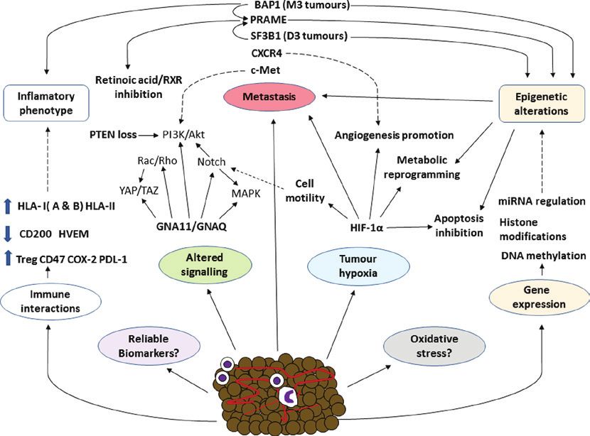
Figure 2. Overall schematic diagram of some of the most significant factors involved in the biology of uveal melanoma. In different types of cancer, asso-
ciations among the different components of the tumour process are complex explaining the non‑success of therapies such as PD‑1 inhibition. Some factors
involved in uveal melanoma, such as the role of oxidative stress, have been well established. In a cancer as aggressive as uveal melanoma, knowledge of the
mechanisms involved and their interactions is essential to develop more effective treatments, predict tumour behaviour and identify new more reliable and
accurate biomarkers. BRCA‑associated protein 1; PRAME, preferentially expressed antigen in melanoma; SF3B1, splicing factor 3b subunit 1; GNA, G protein
subunit alpha; HIF‑1α, hypoxia inducible factor α.
diagnostic markers of tumour onset and progression have been tumour suppressors expressed in normal uveal tissue, but not
confirmed (140). Some of the miRNAs playing a significant in uveal melanoma following their downregulation of other
role in uveal melanoma are discussed below. molecules, such as c‑Met, Akt and proteins involved in the
Yang and Wei (141) compared the expression profiles cell cycle (146,147). Recently, Serocki et al (148) confirmed
of miRNAs in 4 uveal melanoma tissues and 4 normal the role of miR‑17 family miRNAs in controlling HIF expres-
uveal tissues. Their results revealed increased expression sion under conditions of hypoxia. Some miRNAs also reduce
levels of miRNAs of the miR‑17 family (miRNA‑20a, the expression of phosphatase and tensin homolog (PTEN),
miRNA‑106a and miRNA‑17) and significant increases in promoting PI3K/AKt/mTOR pathway activation (149,150). In
miRNA‑21 expression in 4 uveal melanoma cell lines, along addition, Liu et al (151) described the role of miR‑216a‑5p as
with diminished miRNA‑145 and miRNA‑204 expression. an indicator of a better prognosis due to its inhibitory effect
Wang et al (142) elucidated the role of miRNA‑21 in uveal on hexokinase 2, an enzyme overexpressed in a wide array of
melanoma cell metastasis. The results obtained revealed tumours that is directly related to induction of the Warburg
miRNA‑21 overexpression following inhibition of p53 expres- effect. Therapy, pursuing the dysregulation of these miRNAs
sion, which via a series of effector molecules may promote is a promising approach for the treatment of this type of cancer.
tumour metastasis in vitro. In vivo, its inhibition also leads to Of note, miRNAs represent one of the various mechanisms
a reduced tumour size. Radhakrishnan et al (143) identified involved in the pathogenesis of uveal melanoma, as summa-
19 miRNAs expressed in metastasized and not in metastatic rized in Fig. 2. The interactions between all these factors are
uveal melanoma, while up to 11 miRNAs were detected only undoubtedly complex, and future studies will be crucial for a
in the metastasized phenotype. Recently, other miRNAs with better understanding of such a complex cancer.
oncogenic effects have been identified, such as miR‑155 (144).
Among the uveal melanoma tumour suppressor miRNAs, 8. Blood biomarkers for uveal melanoma
miRNA‑145 should be mentioned. Li et al (145) found that the
insulin 1 receptor substrate (IRS‑1) could serve as a therapeutic Despite scientific and technological advances that have
target to increase the levels of miR‑145, which is essential for improved our understanding of various types of cancers, the
uveal melanoma cells to enter into apoptosis. Similarly, the incidence of uveal melanoma and patient survival has not
members of the miR‑34 family of miRNA precursors, miR‑34a, markedly altered over the past 30 years (152,153). This has
miR‑34b and miR‑34c, have been identified as important determined that the most effective measure against this type1270 INTERNATIONAL JOURNAL OF ONCOLOGY 57: 1262-1279, 2020
of cancer is its early detection. As uveal melanoma tumours most used for this type of study, as they are minimally invasive
spread via the bloodstream, blood biomarkers may be useful to and of great clinical value (165). However, these studies still
detect metastases early on and to monitor disease progression have some limitations, such as a need for greater refinement in
or the response to treatment (154). the measurement systems used and analytical variations in the
Circulating tumour cells (CTCs) and circulating free DNA data obtained and difficulties in their translation from bench
(cfDNA) are among the components that may be detected to bedside (166).
in blood, indicating the presence of a tumour and both are The proteins melanoma inhibitory activity (MIA) and
prognostic markers of a variety of cancers (155,156). In uveal OPN (osteopontin) are among the most tested as biomarkers of
melanoma, both the detection of CTCs or cfDNA has proven a uveal melanoma and have been directly associated with metas-
reliable indicator of a worse prognosis. Of note, the detection tasis (167,168). Another biomarker examined is S100‑β (169).
of melanocytic CTCs has exhibited efficacy in arterial blood, These latter studies determined that all 3 of these proteins
but not in veins (157), whereas cfDNA seems more useful in (MIA, OPN and S100‑β) combined were able to detect with
this tumour type, particularly in patients with easily detectable a high sensitivity the presence of metastases in the liver.
known mutations (158). In effect, today there is an ongoing However, in the study conducted by Missotten et al (170), no
clinical trial designed to assess the detection and variations association was observed between this combined biomarker
produced in blood levels of cfDNA in patients followed and any clinical or pathological feature of the tumour, ques-
before and after undergoing surgery for liver metastasis tioning its actual prognostic value. Of note, Strobel et al (171)
(NCT02849145). found elevated serum S100‑β concentrations in patients with
Characterizing different CTC populations is also crucial liver metastases from cutaneous melanoma compared to uveal
for the understand of the biological mechanisms underlying melanoma in which no association was noted. In patients
this type of cancer. Schuster et al (159) examined CTCs in with liver metastasis, increased levels of the oncoprotein,
68 patients with uveal melanoma and the gene expression in DJ‑1/PARK7, the soluble marker, c‑Met, and the glycoprotein,
these cells of tyrosinase and MelanA/MART1. Their results ME20‑S, have been observed (172‑175). Notably, through cell
indicated that the presence of CTCs was directly related to the culture techniques, Angi et al (176) compared the proteins
metastatic process and that the detection of these transcripts secreted by uveal melanoma tumours with a high and low
points to a worse prognosis. Tura et al (160) demonstrated that metastasis risk with those secreted by choroidal melanocytes.
FISH could be used to examine CTCs in patients with primary These authors detected the presence of OPN, MIA, GDF15,
uveal melanoma and thus detect the status of chromosome 3. PARK7 and ME20, and only recorded significant differences
Following a 4‑year follow‑up period, the results revealed the in MIA and GDF15 secretion between cells of uveal melanoma
high reliability of this method to predict the metastases that and normal choroidal melanocytes. No differences emerged
these patients could develop. between the tumours with a high and low risk of metastasis.
miRNAs can also represent important blood biomarkers Advances in omics‑related technologies are proving helpful
detectable in uveal melanoma. Achberger et al (161) identified in the identification of the proteins and metabolites involved in
an association between plasma miRNAs and their variation uveal melanoma and in elucidating their roles. In the study
in a setting of metastasis. Compared to the controls, the by Crabb et al (177), iTRAQ technology was used to examine
levels of miR‑20a, ‑125b, ‑146a, ‑155, ‑181a and ‑223 were large numbers of proteins present in 8 samples of metastasized
elevated, while those of miRNA‑181a were reduced when and 7 of non‑metastasized uveal melanoma. Their findings
metastasis appeared. Along these lines, Russo et al (162) found identified a need for further investigation into proteins, such
significantly higher blood and tissue levels of miRNA‑146a. as heat shock protein (HSP)β ‑1 and collagen α 3 (VI) as
Furthermore, Eldh et al (163) detected higher levels of possible biomarkers of these tumours. Shi et al (178), using
exosomes and miRNAs in patients with hepatic metastasis mass spectrometry and fractioning techniques with magnetic
from uveal melanoma compared to patients without metastasis. pearls, detected up to 49 differentially expressed peptides
Based on these data, Stark et al (164) measured the serum in patients with uveal melanoma and healthy controls. Their
levels of up to 17 miRNAs in 65 patients with uveal nevus, data indicated that peptides of 1,467 to 9,289 kDa were able
localized uveal melanoma and metastasized uveal melanoma. to differentiate between patients with uveal melanoma and
The results served to define a panel of 6 miRNAs (miR‑16, healthy individuals with a specificity of 100%. These authors
miR‑145, miR‑146a, miR‑204, miR‑211 and miR‑363‑3p) that also identified precursors of the fibrinogen α chain as possible
could be used for a precision diagnosis of uveal melanoma markers of uveal melanoma. Also, recently Song et al (179)
with 93% sensitivity and 100% specificity. Collectively, these conducted a multiplex immunoassay on serum samples from
data indicate a need for advancements in the field of miRNAs, 48 patients diagnosed with uveal melanoma and 36 healthy
given their great diagnostic and therapeutic value in a disease controls. Once again, HSPβ‑1 and OPN levels proved useful to
as complex as uveal cancer. distinguish between patients and healthy control individuals.
Apart from these biomarkers, other blood indicators
have proven useful in uveal melanoma, such as proteins, 10. Clinical management of uveal melanoma
glycoproteins and tumour metabolites.
Risk and prognosis of uveal melanoma. The general prognosis
9. Proteomics and metabolomics in uveal melanoma is that 50% of patients will present metastasis within the first
15 years of diagnosis. Once this occurs, the mean life expec-
In the study of cancer, interest in proteomics and metabolo- tancy is between 6 months to 1 year. However, it should be
mics continues to mount. Tissue and blood samples are the highlighted that the latency period from locoregional diseaseORTEGA et al: UPDATE ON UVEAL MELANOMA 1271
control until the onset of metastasis can be >25 years, such implications of knowledge regarding prognosis could be essen-
that patients require exhaustive follow‑up over a long period tial to establish guidelines for the follow‑up of patients when
of time. The preferred sites of presenting metastasis are the the metastatic risk is low and opt for more aggressive treatment
liver (~60%), lungs (~25%), skin and soft tissues (~10%) and options if the risk is high. For instance, the presence of M3 or
bones (~8%) (13). The genetic analysis of melanocyte lesions D3 is critical for the clinical management of uveal melanoma.
has identified that extraocular invasion is related to both the The detection of the commonly found M3 in small tumours
inactivation of the tumour suppressor gene, BAP1 (detected prompts the use of more aggressive treatments in these patients,
in 85% of cases), and to monosomy 3, as the main risk factors especially to prevent metastasis (191). If M3 were detected, this
for disease spread (180). Currently, there are no established could mean the tumour has spread to other organs, and hence,
criteria for the long‑term follow‑up of patients diagnosed with local therapy would not be effective (192). Surveillance in these
uveal melanoma. Recommended approaches are imaging high‑risk patients may be hepatic imaging and liver function
techniques conducted every 3 to 12 months. An MRI is the tests every 3‑6 months (193). The biopsy method must also be
best option both for the detection of liver and extrahepatic considered in the study of M3 in uveal melanoma. Whereas fine
metastases, such as those affecting bones or retroperitoneal needle aspiration (scleral approach) obtains a tumour sample
nodes. A CT scan is also useful for lung node manifestations from the base, the transvitreal approach collects the biopsy
and larger liver metastases and in patients for whom MRI through the apex returning different results. Because of tumour
is not recommended. Ultrasonography exclusively reveals heterogeneity, the scleral approach is the best method to detect
hepatic metastases and PET cannot detect small lesions, the M3 (194). Similarly, BAP1 tumours may have a significant
high radiation dose being another major drawback of this clinical impact in uveal melanoma management, particularly in
technique (181). the development of targeted therapy.
Tumour size, extraocular extension, mitotic activity and
epithelioid cell type are considered important risk factors for Current and potential therapies. A close association exists
melanoma (182). As previously stated, genetic mutations and between metastatic disease, prognosis and response to therapy.
chromosome abnormalities are also directly associated with This is due to the fact that considerable advances have been
patient outcomes and shed light into the prognosis of uveal made in the locoregional control of the disease through both
melanoma. To examine all these chromosome and molecular conservative techniques (e.g., brachytherapy, external beam
features during the management of uveal melanoma, a wide radiation therapy or laser photodynamic and photocoagula-
range of methods can be used. The most common approaches tion therapy) and more aggressive approaches (enucleation)
are karyotyping, fluorescent in situ hybridisation (FISH) or rendering an overall 5‑year survival of approximately 80% (4).
comparative genome hybridisation (CGH). Further techniques, This survival rate has remained stable over the past 30 years,
such as microsatellite analysis, multiple ligation‑dependent and developments have therefore consisted mainly of more
probe amplification (MLPA) and genome‑wide single nucleo- effective and less aggressive surgical techniques.
tide polymorphism can also be used for the genomic study of Similar to the association existing between the prog-
uveal melanoma. Karyotyping is useful for the detection major nosis and metastasis of uveal melanoma, immunotherapy
chromosome gains or losses. However, minor genetic altera- is one of the main pillars of the treatment of disseminated
tions are not identified. FISH, such as CGH is more accurate disease. Systemic chemotherapy barely improves the overall
in detecting chromosome aberrations in uveal melanoma; prognosis of a patient and the response rate to conventional
however, it is still insufficient for the detection of all chromo- chemotherapy is1272
Table I. Ongoing clinical trials targeting the treatment of metastatic uveal melanoma.
Name Identifier Status Population Phase Purpose
ENSIGN: Phase II window of opportunity NCT02831933 Recruiting 25 participants with lung squamous Phase 2 Determine the efficacy and safety of in situ gene
or body radiation therapy and in situ gene cell carcinoma stage IV and therapy and stereotactic body radiation therapy
therapy followed by nivolumab in metastatic non‑squamous non‑small cell cancer
squamous or non‑squamous non‑small cell metastatic uveal melanoma trial of
lung carcinoma and metastatic uveal melanoma stereotactic
Ipilimumab and nivolumab in combination with NCT03472586 Recruiting 35 participants with uveal melanoma Phase 2 Test the use of the monoclonal antibodies ipilimumab
immunoembolization for the treatment of and liver metastasis and nivolumab and immunoembolization to treat
metastatic uveal melanoma patients with liver metastasis
A Phase 1/2 dose‑finding study to evaluate the NCT02743611 Active, not 28 participants with AML, MDS and Phase 1 Assess the effect of BPX‑701 in tumours showing
safety, feasibility, and activity of BPX‑701, a recruiting uveal melanoma Phase 2 high PRAME expression
controllable PRAME T‑cell receptor therapy,
in HLA‑A2+ subjects with AML, previously
treated mds, or metastatic uveal melanoma
Phase1b/2 study combining hepatic percutaneous NCT04283890 Recruiting 88 participants with metastatic uveal Phase 1 Assess the use of immunotherapy (ipilimumab with
perfusion with ipilimumab plus nivolumab in melanoma Phase 2 nivolumab) plus chemotherapy (melphalan)
advanced uveal melanoma
Phase Ib Study of cellular adoptive NCT03068624 Active, not 19 participants with metastatic uveal Phase 1 Determine the maximum tolerated dose (MTD) of
immunotherapy using autologous Cd8+ recruiting melanoma adoptively transferred SLC45A2‑specific cytotoxic
antigen‑specific T cells and anti‑Ctla4 for T‑lymphocytes (CTL) and its combination with
patients with metastatic uveal melanoma cyclophosphamide, aldesleukin and ipilimumab
A phase 2 study to evaluate the efficacy and NCT03467516 Recruiting 59 participants with metastatic uveal Phase 2 Assess the use of TIL in conjunction with TIL high
safety of adoptive transfer of autologous tumour melanoma dose aldesleukin
infiltrating lymphocytes in patients with
INTERNATIONAL JOURNAL OF ONCOLOGY 57: 1262-1279, 2020
metastatic uveal melanoma
Phase I vaccination trial in metastatic uveal NCT04335890 Recruiting 12 participants with metastatic uveal Phase 1 Assess the effects of vaccination with IKKb matured
melanoma using IKKb‑matured dendritic cells melanoma dendritic cells loaded with autologous tumour‑RNA +
loaded with autologous tumour‑RNA + RNA RNA coding for defined antigens and driver mutations
coding for defined antigens and driver mutations
A phase II study of BVD‑523 in metastatic NCT03417739 Active, not 13 participants with metastatic uveal Phase 2 Assess the targeting of the MAPK signalling pathway
uveal melanoma recruiting melanoma using BVD‑523 in advanced uveal melanoma
Efficacy study of pembrolizumab with entinostat NCT02697630 Active, not 29 participants with metastatic uveal Phase 2 Assess the potential combination of entinostat (HDAC
to treat metastatic melanoma of the eye recruiting melanoma inhibitor) and pembrolizumab (immunotherapy)Table I. Continued.
Name Identifier Status Population Phase Purpose
Intravenous and intrathecal nivolumab in NCT03025256 Recruiting 30 participants with brain metastasis, Phase 1 Compare intrathecal nivolumab and examine how
treating patients with leptomeningeal disease among them uveal melanoma well it acts in combination with intravenous nivolumab
when treating patients with leptomeningeal disease
Trial of nivolumab in combination with NCT02626962 Active, not 48 participants with metastatic uveal Phase 2 Assess the impact of nivolumab combined with
ipilimumab in subjects with previously recruiting melanoma ipilimumab in subjects with previously untreated,
untreated metastatic uveal melanoma unresectable or metastatic uveal melanoma
A study to assess PV‑10 chemoablation of NCT00986661 Recruiting 78 participants with liver metastasis Phase 1 Examine the safety, tolerability, pharmacokinetics and
cancer of the liver including those with uveal melanoma effect of a single intralesional injection of PV‑10 on
tumour growth in subjects with primary or metastatic
liver cancer
IN10018 monotherapy and combination NCT04109456 Recruiting 52 participants with metastatic Phase 1 Assess the safety, tolerability and antitumor properties
therapy for metastatic melanoma cutaneous or uveal melanoma of IN10018 as monotherapy and in combination with
cobimetinib in subjects with metastatic uveal
melanoma and NRAS‑mutant metastatic melanoma
Modified virus VSV‑IFNbetaTYRP1 in NCT03865212 Recruiting 72 participants with stage III‑IV Phase 1 Confirm the efficacy, side effects and best dose of a
treating patients with stage iii‑iv melanoma cutaneous and uveal melanoma modified virus VSV‑IFNbetaTYRP1
Yttrium90, ipilimumab, and nivolumab for NCT02913417 Recruiting 26 participants with liver metastatic Phase 1 Examine the synergistic effects of SirSpheres
uveal melanoma with liver metastases uveal melanoma Phase 2 Yttrium‑90 selective internal hepatic radiation
followed by immunotherapy combined with
ipilimumab and nivolumab
ORTEGA et al: UPDATE ON UVEAL MELANOMA
Iodine I 131 monoclonal antibody 3F8 in NCT00445965 Active, not 78 participants with brain metastasis Phase 2 Assess iodine I 131 monoclonal antibody 3F8 used to
treating patients with central nervous system recruiting including those with uveal melanoma treat patients with central nervous system or
cancer or leptomeningeal cancer leptomeningeal cancer
Neoadjuvant and adjuvant checkpoint NCT02519322 Recruiting 53 participants with stage III‑IV Phase 2 Check the performance of nivolumab with or without
blockade melanomas ipilimumab or relatlimab before surgery in patients
with resectable stage IIIB‑IV melanoma
Cabozantinib‑S‑malate compared with NCT01835145 Active, not 47 participants with recurrent/stage Phase 2 Compare cabozantinib‑s‑malate with temozolomide
temozolomide or dacarbazine in treating recruiting III‑IV uveal melanoma or dacarbazine in patients with unresectable metastatic
patients with metastatic melanoma of the melanoma of the eye
eye that cannot be removed by surgery
1273You can also read