The Microbiome and Food Allergy - Annual Review of Immunology - Cathryn Nagler Lab
←
→
Page content transcription
If your browser does not render page correctly, please read the page content below
IY37CH16-Nagler ARjats.cls April 4, 2019 12:57
Annual Review of Immunology
The Microbiome and
Food Allergy
Access provided by University of North Carolina - Chapel Hill on 04/27/19. For personal use only.
Onyinye I. Iweala1 and Cathryn R. Nagler2,3
Annu. Rev. Immunol. 2019.37:377-403. Downloaded from www.annualreviews.org
1
UNC Food Allergy Initiative and Thurston Arthritis Research Center, Division of
Rheumatology, Allergy, and Immunology, The University of North Carolina at Chapel Hill,
North Carolina 27599-7280, USA; email: onyinye.iweala@med.unc.edu
2
Department of Pathology, Biological Sciences Division, University of Chicago, Chicago,
Illinois 60637-1824, USA; email: cnagler@bsd.uchicago.edu
3
Committee on Immunology, Biological Sciences Division, University of Chicago, Chicago,
Illinois 60637-1824, USA
Annu. Rev. Immunol. 2019. 37:377–403 Keywords
The Annual Review of Immunology is online at
oral tolerance, microbiome, food allergy, dysbiosis
immunol.annualreviews.org
https://doi.org/10.1146/annurev-immunol-042718- Abstract
041621
The gut-associated lymphoid tissue (GALT) faces a considerable challenge.
Copyright © 2019 by Annual Reviews.
It encounters antigens derived from an estimated 1014 commensal microbes
All rights reserved
and greater than 30 kg of food proteins yearly. It must distinguish these
harmless antigens from potential pathogens and mount the appropriate host
immune response. Local and systemic hyporesponsiveness to dietary anti-
gens, classically referred to as oral tolerance, comprises a distinct comple-
ment of adaptive cellular and humoral immune responses. It is increasingly
evident that a functional epithelial barrier engaged in intimate interplay with
innate immune cells and the resident microbiota is critical to establishing
and maintaining oral tolerance. Moreover, innate immune cells serve as a
bridge between the microbiota, epithelium, and the adaptive immune sys-
tem, parlaying tonic microbial stimulation into signals critical for mucosal
homeostasis. Dysregulation of gut homeostasis and the subsequent disrup-
tion of tolerance therefore have clinically significant consequences for the
development of food allergy.
377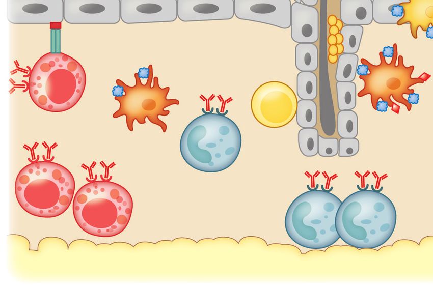
IY37CH16-Nagler ARjats.cls April 4, 2019 12:57
INTRODUCTION
Food allergies are a major public health concern and represent an unmet clinical need (1, 2). An
individual can become allergic to any food, at any time, but eight foods cause most reactions: milk,
eggs, peanuts, tree nuts, soy, wheat, fish, and shellfish. Reactions can range from swelling and ur-
ticaria to life-threatening anaphylactic shock. No treatment other than strict avoidance is currently
available for the 15 million Americans who suffer from food allergies. These numbers represent a
marked generational increase in disease prevalence that has been noted in industrialized societies
worldwide. The rising rate of food allergies parallels increases in what have been called the diseases
of Western society, including obesity, diabetes, asthma, autism, and inflammatory bowel disease
(among others). How do we account for this kind of generational change? Food allergies often
present as part of a constellation of allergic diseases, referred to as the atopic (or allergic) march
Access provided by University of North Carolina - Chapel Hill on 04/27/19. For personal use only.
(3). Atopic dermatitis appears first in infancy, followed by food allergies between ages 2 and 5 years.
Asthma and allergic rhinitis typically arise at school age. The increased prevalence of food allergy
Annu. Rev. Immunol. 2019.37:377-403. Downloaded from www.annualreviews.org
has followed an earlier rise in asthma and other allergic diseases in heavily industrialized nations
(4, 5). A number of theories have been proposed to explain the rising prevalence of allergic disease
(Table 1). The hygiene hypothesis initially linked the environment to allergic disease (6). It posited
that improved cleanliness and household conditions reduced exposure to infectious disease and in-
creased susceptibility to allergy. In particular, infection in early childhood due to “unhygienic con-
tact” with older siblings or transmitted prenatally from mothers infected by their older children
protected against the development of hay fever (6). We and others first suggested a critical role for
Table 1 Theories to explain the development and rising prevalence of food allergy
Purpose of theory Hypothesis Reference Summary
To explain the rising Hygiene 6 In large families, infection in early childhood due to “unhygienic
prevalence of food hypothesis contact” with family members prevents the development of
allergy atopic disease. Shrinking family size and decreased infection
exposure lead to increases in atopic disease development.
Balanced 7, 8 Antibiotic use and dietary differences in industrialized countries
microbiota and have upset normal commensal gastrointestinal microbial
microbial communities, disrupting tonic microbial signaling through
stimulation microbial pattern recognition receptors and preventing the
hypotheses development of tolerogenic mucosal immune networks that
protect against allergic hyperreactivity.
Old friends and 9–11 Changes in living environment, diet, and lifestyle associated with
biodiversity industrialized, Westernized countries impact commensal
hypotheses microbial diversity in gut and on skin, disrupting the
immunoregulatory function of the microbiota at these sites, thus
predisposing to allergic sensitization.
To explain the failure of Dual-allergen 18 Low-dose cutaneous exposures to food predispose to allergic
oral tolerance and exposure sensitization while early ingestion of higher doses of food
development of hypothesis proteins leads to oral tolerance.
allergic sensitization Barrier 19 Twenty-first-century lifestyle factors have depleted
regulation allergy-protective bacterial populations in the intestinal mucosa
hypothesis of that are required to maintain epithelial barrier integrity and limit
allergic allergen access to the systemic circulation.
sensitization
378 Iweala • Nagler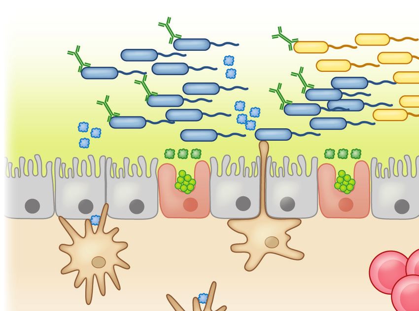
IY37CH16-Nagler ARjats.cls April 4, 2019 12:57
immunoregulatory signals from commensal bacteria in the regulation of allergic hyperreactivity
(7, 8). This led to a reformulation of the hygiene hypothesis as the “old friends” or the “biodi-
versity” hypotheses of allergy, which propose that changes in the environment, diet, and lifestyle
associated with Westernized, industrialized countries have altered the diversity of the gut and
skin microbiomes (9–11). Industrialized populations are exposed to both prescribed antibiotics,
which dramatically alter the microbiome (12), and residual antibiotics used in agribusiness to en-
hance livestock growth (reviewed in 13). Modern Western diets contain large quantities of highly
processed, high-fat, low-fiber foods which also cause shifts in microbial communities (14, 15).
Epidemiologic studies point to a decreased prevalence of food allergies among populations where
parasitic helminth infection is endemic compared to populations where helminth infection is rare
(16). Increased microbial exposure in rural (particularly farming) populations suggests that higher
Access provided by University of North Carolina - Chapel Hill on 04/27/19. For personal use only.
biodiversity contributes to protection against disease (17). The available evidence therefore indi-
cates that the loss of beneficial symbiotic relationships between humans, parasites, bacteria, and
Annu. Rev. Immunol. 2019.37:377-403. Downloaded from www.annualreviews.org
other microbes acquired throughout human evolution has increased the risk for developing atopic
diseases (Figure 1). The timing and route of first exposure to food allergens also seem to play a
role. The dual allergen hypothesis suggests that sensitization is promoted by allergen contact with
skin and prevented by ingestion of food allergens early in life (18). We have proposed that tolerance
to dietary antigen (and prevention of food allergy) requires both a food-antigen-specific regulatory
response and a commensal-bacteria-induced intestinal-epithelial-barrier-protective response (19).
In this review, we explore the many ways in which the microbiome regulates the response to dietary
antigens.
Modern lifestyle factors
Food allergy
Antibiotics
Genetic
predisposition
High-fat, low-fiber
diet
Altered
commensal microbial
biodiversity
Eradication of
Transition from enteropathogens
rural to urban and (gastrointestinal
suburban living helminths,
Helicobacter pylori)
Vaccine-enhanced
immunity and
reduced
exposure to
infection
Figure 1
Elements of a modern, industrialized lifestyle trigger shifts in the commensal microbiota, predisposing to the
development of food allergy in genetically susceptible individuals. Adapted from Reference 20 with
permission from Springer Nature.
www.annualreviews.org • The Microbiome and Food Allergy 379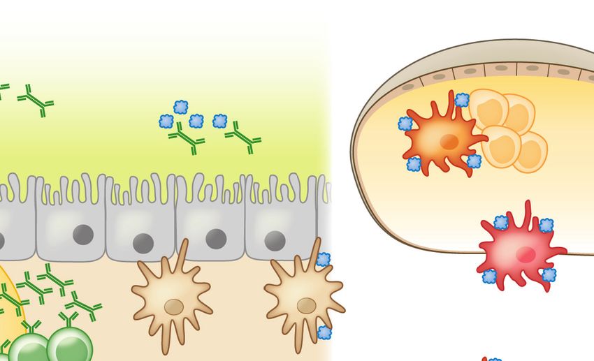
IY37CH16-Nagler ARjats.cls April 4, 2019 12:57
COMMENSAL BACTERIA PROTECT AGAINST FOOD ALLERGY
Commensal Bacteria Regulate Antigen Presentation in the Gut
The paradigmatic view has been that the primary mechanism regulating tolerance to dietary anti-
gen is the induction of food-antigen-specific regulatory T cells (Tregs) (21). Tolerogenic responses
to luminal antigens depend on the translocation of these antigens across the gut epithelial bar-
rier. M (microfold) cells, specialized intestinal epithelial cells located above collections of small
intestinal submucosal lymphoid follicles called Peyer patches (PPs), are critical for the transcyto-
sis of particulate antigens and macromolecules from the gut lumen into the submucosa (22, 23).
Recent work has shown that goblet cell–associated antigen passages (GAPs) also play an impor-
tant role in transferring luminal antigens to antigen-presenting cells (APCs) (24, 25). Multiple,
functionally distinct subsets of APCs, including dendritic cells (DCs) and mononuclear phago-
Access provided by University of North Carolina - Chapel Hill on 04/27/19. For personal use only.
cytes (MNPs), capture these transported antigens in the subepithelial dome (SED) of the follicle-
associated epithelium (26). Antigen-loaded DCs migrate from the SED to the mesenteric lymph
Annu. Rev. Immunol. 2019.37:377-403. Downloaded from www.annualreviews.org
nodes (MLNs), which drain the small intestine. Presentation of antigen to naive T cells in the
presence of the vitamin A metabolite retinoic acid (RA) favors TGF-β−dependent conversion to
Foxp3+ Tregs (27–29). Concomitant upregulation of the gut-homing receptors CCR9 and α4 β7
(30) allows these committed Tregs to home back to the lamina propria (LP) and expand under the
influence of IL-10 produced by resident MNPs (31). Some Tregs exit the mucosa via the lymph
or bloodstream to promote systemic tolerance (Figure 2).
What is driving allergic, rather than tolerogenic, responses to food antigens? The role of the
microbiota in regulating both antigen uptake and antigen presentation is beginning to be unrav-
eled. In the small intestine, where nutrients (and food antigens) are absorbed, MNPs resident in the
LP express the chemokine receptor CX3 CR1; some form transepithelial dendrites that penetrate
gut epithelia and sample luminal antigens (32, 33). Microbial stimulation of intestinal epithelial
cell Toll-like receptors (TLRs) and signaling through the adaptor molecule MyD88 upregulate
the numbers of these DC extensions in the small intestine (34). Both antigen presentation and
IL-10 production by CX3 CR1+ MNPs are required for oral tolerance (35); genetic ablation of
either MHC-II or IL-10 in CX3 CR1+ MNPs abrogates the induction of tolerance, although it
is not yet clear whether the same cell must perform both functions. In the absence of MHC-II
expression on CX3 CR1+ MNPs, the induction of dietary-antigen-specific Tregs is hampered (35).
Interestingly, the commensal microbiota regulates the tolerance-inducing capabilities of this MNP
subset. CX3 CR1+ MNPs from antibiotic-treated mice lose the ability to produce IL-10 (35).
The enteric microbiota therefore appears to be critical to the antigen presentation function of
CX3 CR1+ APCs in the gut.
Notably, rather than migrating directly to lymphoid tissues, CX3 CR1+ MNPs transfer their
antigenic cargo to CD11c+ CD103+ DCs in a manner dependent on gap junction proteins like
Connexin-43 (36). Gut epithelial cells that produce TGF-β and RA create a microenvironment
that drives DCs to express CD103 (37). These CD103+ DCs then migrate from the LP through
afferent lymphatic vessels in a CCR7-dependent manner to present antigen to naive T cells in
the MLNs. MLN stromal cells express higher mRNA transcript levels of the enzyme critical for
RA production, RALDH, compared to peripheral lymph nodes; RA derived from these cells trig-
gers expression of the gut-homing molecules α4 β7 and CCR9 on activated T cells (38, 39). Mice
deficient in CCR9, or the integrins α4 β7 or MAdCAM-1, fail to develop tolerance to orally de-
livered antigen (31, 40). Multiple DC subsets have been described in the MLNs. Elegant lineage-
depletion studies confirm that CD103+ DCs are critical for oral tolerance (41). CD103+ CD11b−
DCs expressing IRF8 seem to be the most potent producers of TGF-β and RA (41).
380 Iweala • Nagler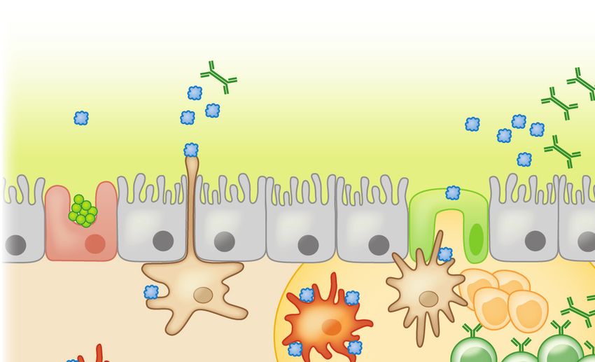
IY37CH16-Nagler ARjats.cls April 4, 2019 12:57
Stromal cells
IgA
Mucous Food Ag
layer TGF-β
RA 7
1 DC
3 MLN
2
GAP M cell T cells Tregs
TGF-β
IL-10
pDC
Goblet MNP MNP T cells 8
IgA MNP MNP
cell CD103+ pDC
DC ↓CD40/CD80/CD86
CX3CR1+ 4 10 ↑IDO
6
MNP CX3CR1+
CD103+ DC CX3CR1+
Access provided by University of North Carolina - Chapel Hill on 04/27/19. For personal use only.
MNP 5
DC ↑IDO IgA+ B cells MNP
DC
CD103+ pDC pDC Peyer patch IL-10 9
↓CD40/CD80/CD86
Annu. Rev. Immunol. 2019.37:377-403. Downloaded from www.annualreviews.org
CD103+ DC CD103+
↑IDO TGF-β
DC
cs
ati
11 Bloodstream mph
Tregs Ly
Tregs Lymphatics CCR9+α4β7+
Systemic tolerance
Figure 2
Tolerance to dietary antigen: the food-antigen-specific Treg paradigm. Luminal antigens are translocated across the gut epithelial
barrier via M cells (1 ) or GAPs ( 2 ). CX3 CR1+ lamina propria–resident MNPs sample luminal antigens; some form transepithelial
dendrites that penetrate gut epithelia ( 3 ). Different populations of antigen-presenting cells, including tolerogenic IDO-expressing
CD103+ DCs and pDCs, reside in the subepithelial dome below M cells ( 4 ). CX3 CR1+ MNPs transfer luminal antigens to CD103+
DCs and pDCs ( 5 ) that traffic to the draining MLNs ( 6 ) and display antigen to naive T cells in the presence of TGF-β and RA,
favoring the generation of food-antigen-specific Tregs ( 7 ) that traffic through lymphatics ( 8 ) back to the lamina propria in a CCR9-
and α4 β7 -dependent manner. MNP-derived IL-10 promotes Treg proliferation ( 9 ). TGF-β produced by Tregs regulates B cell
antibody class switching to IgA; IgA is transported across the epithelial barrier via the polymeric immunoglobulin receptor (pIgR, not
shown) into the intestinal lumen, where it acts to exclude luminal food antigens ( 10 ). Proliferating Tregs eventually exit the gut and
enter the circulation to promote systemic tolerance to dietary antigen ( 11 ). Dashed lines represent the movement/trafficking of cells
from one area to another. Solid lines represent the production of factors by a cell; the transfer of materials from one cell to another; or
how chemical factors or cells influence the behavior of other cells. Abbreviations: Ag, antigen; DC, dendritic cell; GAP, goblet
cell–associated antigen passage; IDO, indoleamine 2,3-dioxygenase; M, microfold; MLN, mesenteric lymph node; MNP, mononuclear
phagocyte; pDC, plasmacytoid DC; RA, retinoic acid; Treg, regulatory T cell.
TGF-β is critical to the regulation of both cellular and humoral immunity in the gut (42, 43).
It is produced by multiple gut-associated immune cells, including DCs and Tregs, as well as by
intestinal epithelial cells (44). Interestingly, inflammation can induce expression of the integrin
αv β8 by Foxp3+ Tregs (45). This integrin, in addition to other metalloproteinases and integrins,
cleaves the latency-associated peptide that binds the inactive form of TGF-β, converting it to an
active form that binds to TGFβRII in the TGF-βR complex and regulates host immunity (44).
LP CD103+ DCs express high levels of αv β8 integrin, which can activate TGF-β to generate
Foxp3+ Tregs. Expression of αv β8 integrin depends on RA and TGF-β as well as bacterial sig-
naling through the TLR adaptor protein MyD88. Boucard-Jourdin and colleagues (46) showed
that mice deficient in TGF-β signaling or fed a vitamin A–deficient diet had reduced β8 inte-
grin expression. β8 integrin expression was similarly reduced in mice treated with antibiotics and
in MyD88-deficient mice. In a chemical-injury-based colitis model, the gut microbiota drives
proinflammatory cytokine production to modulate retinaldehyde dehydrogenases, which are crit-
ical to the synthesis of RA (47). Although this has yet to be shown in an oral tolerance or food
www.annualreviews.org • The Microbiome and Food Allergy 381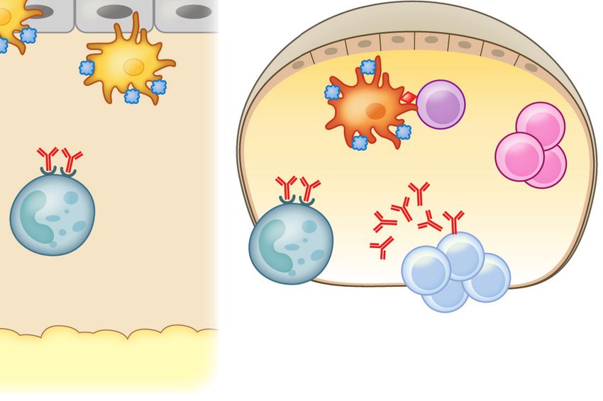
IY37CH16-Nagler ARjats.cls April 4, 2019 12:57
allergy model, this finding raises the intriguing possibility that microbes influence colonic LP Treg
numbers by modulating RA and TGF-β levels in the enteric microenvironment.
Other work has shown that CD103+ DCs expressing indoleamine 2,3-dioxygenase (IDO), an
enzyme involved in tryptophan metabolism, can push CD4+ T cells toward a Foxp3+ regulatory
phenotype. Blocking IDO expression in vivo hinders the development of antigen-specific Tregs
and impairs oral tolerance induction (48). IDO digests tryptophan into the metabolite kynurenine,
and the kynurenine:tryptophan ratio can be used as a marker of IDO activity. In subjects with food
allergy, the serum kynurenine:tryptophan ratio is significantly reduced compared to that in healthy
controls (49), suggesting that dysregulated IDO activity may hinder the development of tolerance
to dietary antigens. Tryptophan is also a substrate for trillions of gut microbes that synthesize
serotonin and other metabolically active compounds. Multiple studies have demonstrated reduc-
Access provided by University of North Carolina - Chapel Hill on 04/27/19. For personal use only.
tions in tryptophan metabolism and diminished diversity of tryptophan and indole metabolites in
germfree and antibiotic-treated mice compared to specific-pathogen-free (SPF) controls (50–52).
Annu. Rev. Immunol. 2019.37:377-403. Downloaded from www.annualreviews.org
One could postulate that gut dysbiosis and the resultant alterations in host serum concentrations
of tryptophan and indole-containing compounds impact IDO expression in DCs, which in turn
may hinder DCs’ ability to induce antigen-specific Foxp3+ Tregs. Another DC subset, plasma-
cytoid DCs (pDCs), has also been shown to express IDO (50) and varying levels of gut-homing
receptor molecules like α4 β7 , CCR9, and CD103 whether they reside in the GALT or in sys-
temic tissues like spleen and peripheral blood (53). In contrast to CD103+ DCs, pDCs express
low levels of costimulatory molecules like CD40, CD80, and CD86, hampering their ability to
stimulate antigen-specific CD4+ T cell proliferation and suggesting a tolerance-promoting role
for pDCs in the gut (53). Using a mouse model, Uto and colleagues (53) showed that MLN pDCs
induced antigen-specific Foxp3+ Tregs in the presence of TGF-β and upregulated expression of
the Aldh1a2 gene that encodes RALDH, an enzyme needed to generate RA. Foxp3+ Tregs that
have homed to the intestines can differentiate into Foxp3− T follicular helper cells in the PPs
and promote the generation of IgA-producing B cells (54). The role of IgA antibodies in facilitat-
ing oral tolerance is not well defined, but feeding dietary antigen does generate food-specific IgA
antibodies that may promote oral tolerance through immune exclusion (55). Interestingly, recent
work has shown that much of the commensal microbiota is coated with IgA, particularly in the
small intestine, where IgA is predominantly produced (56, 57). Whether and how IgA coating of
commensal bacteria contributes to tolerance to dietary antigens have not yet been determined.
Commensal microbes also influence Treg development directly through microbial fermen-
tation products, like butyrate, a short-chain fatty acid (SCFA) produced from dietary fiber pre-
dominantly by Clostridia, a class of mucosa-associated Firmicutes (58, 59). Comparative nuclear
magnetic resonance–based metabolome analysis showed a positive correlation between colonic
Treg cell numbers and luminal concentrations of SCFAs (59). In particular, butyrate stimulated
Treg differentiation under a number of conditions, which were associated with histone H3 acety-
lation at the Foxp3 promoter and conserved, noncoding sequences (58, 59). These findings suggest
that a class of commensal bacteria can promote Treg development through epigenetic regulation
of the signature Treg transcription factor Foxp3. Moreover, colonic-microbiota-induced Foxp3+
Tregs also express RORγt, the transcription factor that canonically controls the Th17 pathway
(60, 61). Mice deficient in RORγt+ Foxp3+ Tregs show increased Th2 responses in both oxa-
zolone colitis and helminth infection models, suggesting that these type 3 Tregs are required for
the regulation of Th2 immunity (61). What, however, is the specificity of these Tregs, and how
do they regulate tolerance to food antigens? Bacteria-induced Tregs with bacteria-specific T cell
receptors have been described in the colonic LP (62, 63). Although Foxp3+ Tregs are depleted
in the colons of germfree mice, they are found in numbers comparable to SPF mice in the small
intestines. Elegant studies with germfree mice fed an elemental (antigen-free) diet demonstrated
382 Iweala • Nagler
IY37CH16-Nagler                                                                                               ARjats.cls   April 4, 2019    12:57

                                                                                                    that Foxp3+ Tregs with specificity for dietary antigens dominate in the small intestine (64). Inter-
                                                                                                    estingly, most of these small intestine Tregs develop after weaning to solid food and are essential
                                                                                                    to suppress immunity to dietary antigens. Taken together the data suggest that both bacteria- and
                                                                                                    food-antigen-induced Foxp3+ Tregs cooperate to prevent allergic responses to food.

                                                                                                    Allergic Effector Cells Are Responsive to Bacterial Stimuli
                                                                                                    Classical food allergy (and atopic disease in general) is characterized by the generation of antigen-
                                                                                                    specific IgE antibody responses. Allergen activation at epithelial barrier surfaces elicits the pro-
                                                                                                    duction of epithelial alarmins including TSLP, IL-33, and IL-25, which stimulate type 2 innate
                                                                                                    lymphoid cells (ILC2s) to produce Th2 cytokines and prime DCs to elicit allergen-specific im-
Access provided by University of North Carolina - Chapel Hill on 04/27/19. For personal use only.

                                                                                                    munity (65). B cells are licensed to class switch to IgE in a Th2 cytokine microenvironment rich in
                                                                                                    IL-4, IL-13, and IL-5 (19). Class switching to allergen-specific IgE leads to sensitization and is a
       Annu. Rev. Immunol. 2019.37:377-403. Downloaded from www.annualreviews.org

                                                                                                    requirement for the development of IgE-mediated food allergy. Subsequent exposure to allergen
                                                                                                    cross-links IgE bound to the FcεRI on allergic effector cells, like mast cells and basophils, that be-
                                                                                                    come decorated with IgE generated during sensitization. The symptoms associated with an acute
                                                                                                    allergic reaction arise following this cross-linking and subsequent degranulation, with the release
                                                                                                    of inflammatory mediators like histamine, other vasoactive amines, lipid mediators, and cytokines.
                                                                                                    This response encompasses fast-acting chemical mediators of the immediate hypersensitivity re-
                                                                                                    sponse and the synthesis of additional cytokines and chemokines that contribute to later phases
                                                                                                    of the allergic inflammatory response (66).
                                                                                                        Commensal bacteria regulate allergic effector cell numbers at sites of allergic inflammation. In
                                                                                                    an allergic airway inflammation model, increased numbers of basophils accumulated in the airways
                                                                                                    of germfree mice compared to SPF mice. Germfree mice also demonstrated increased airway hy-
                                                                                                    perresponsiveness following intranasal ovalbumin challenge compared to their SPF counterparts
                                                                                                    (67). Antibiotic-treated and germfree mice have higher resting serum IgE levels and an increased
                                                                                                    frequency of circulating basophils (68). Moreover, Hill and colleagues (69) have shown that the
                                                                                                    absence of MyD88 in B cells triggers a rise in IgE and circulating basophils. Human subjects with
                                                                                                    hyper-IgE syndrome due to loss-of-function mutations in the gene encoding dedicator of cytoki-
                                                                                                    nesis 8 (DOCK8) experience frequent infections, increased risk of atopic dermatitis, on average
                                                                                                    tenfold increases in serum IgE levels, and higher frequencies of circulating basophils compared
                                                                                                    to the general population (69). A correlation between elevated serum IgE and elevated circu-
                                                                                                    lating basophil numbers in the setting of impaired commensal microbial signaling has led some
                                                                                                    to postulate the existence of a commensal bacteria–IgE–basophil axis of allergic inflammation,
                                                                                                    particularly since IgE has been shown to shape elements of granulocyte homeostasis (70). Con-
                                                                                                    sistent with this model, Hill et al. (69) demonstrated that in anti-IgE-treated mice, antibiotic
                                                                                                    treatment did not increase circulating basophil numbers as it did in control animals. In addi-
                                                                                                    tion, commensal microbial signals influenced basophil development by limiting the proliferation
                                                                                                    of bone marrow–derived basophil precursor populations in a manner dependent on the IL-3 re-
                                                                                                    ceptor (IL-3R) and IgE. Thus, commensal microbial signaling, in part through IgE, modulates
                                                                                                    the development of allergic effector cells like basophils and the generation of the Th2 cytokine
                                                                                                    environment.

                                                                                                    A Bacteria-Induced Barrier-Protective Response Is Required to Prevent
                                                                                                    Allergic Responses to Food
                                                                                                    Physiologically, tolerance to dietary antigens results in the development of neutralizing, nonin-
                                                                                                    flammatory IgG or IgA humoral immune responses. IgA, the most abundant isotype at mucosal

                                                                                                                                                        www.annualreviews.org   •   The Microbiome and Food Allergy   383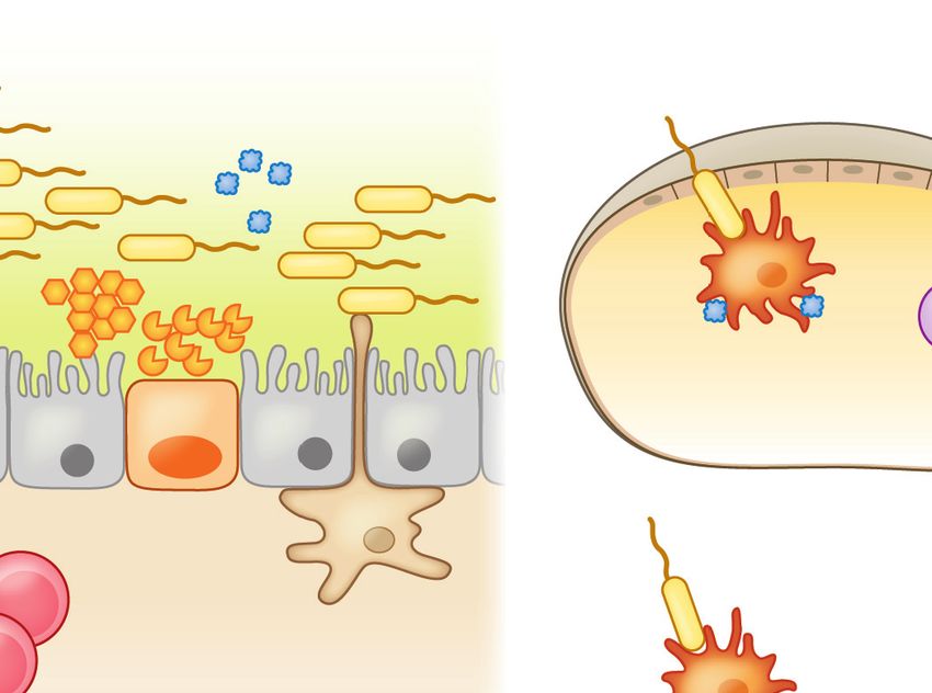
IY37CH16-Nagler ARjats.cls April 4, 2019 12:57
surfaces, is critical to the maintenance of homeostasis. Its functions include regulating bacterial
adherence or translocation, sampling luminal antigens, immune exclusion, and influencing the
composition of the intestinal microbiota (56). Early studies in germfree mice suggested that in-
testinal bacteria or microbial products like lipopolysaccharide (LPS) were important for the gen-
eration of tolerance to dietary antigens (71–73). In particular, Kiyono et al. (72) showed that oral
tolerance could not be induced in LPS-hyporesponsive C3H/HeJ mice. After Sampson and col-
leagues described a model of systemic anaphylaxis to peanut in C3H/HeJ mice (74), we showed
that this strain’s susceptibility to food allergy was linked to its inability to signal via TLR4 (75). We
hypothesized that the TLR4 ligand originated from commensal bacteria and demonstrated that
reducing the commensal bacterial load in TLR4-sufficient neonatal mice using a cocktail of broad-
spectrum antibiotics induced an allergic response equivalent to that seen in TLR4-mutant mice
Access provided by University of North Carolina - Chapel Hill on 04/27/19. For personal use only.
(75). Subsequent murine model studies from our laboratory have shown that both germfree mice
and mice treated with a cocktail of antibiotics, beginning preweaning, are highly susceptible to al-
Annu. Rev. Immunol. 2019.37:377-403. Downloaded from www.annualreviews.org
lergic sensitization to food (76). To identify allergy-protective bacterial populations, we selectively
colonized germfree mice with representatives of bacterial orders (Bacteroidales and Clostridiales) nu-
merically predominant in the murine gut. All mice were intragastrically sensitized with peanut plus
the mucosal adjuvant cholera toxin. Using this approach, we identified mucosa-associated spore-
forming Firmicutes in the Clostridia class as the taxa responsible for protection against allergic
sensitization (76). Sensitization to a food allergen was blocked in antibiotic-treated mice that re-
ceived a Clostridia-containing microbiota (76). Microarray analysis of intestinal epithelial cells iso-
lated from the colonized gnotobiotic mice identified a novel innate mechanism by which Clostridia
protect against sensitization to dietary antigens. We found that Clostridia colonization stimulated
ILC3s in the colonic LP to produce the barrier-protective cytokine IL-22. IL-22 fortified epithe-
lial barrier function by regulating the secretion of mucus by goblet cells and the production of
Paneth cell antimicrobial peptides (76, 77). These IL-22-dependent responses reduced the access
of orally administered dietary antigen to the systemic circulation and protected against allergic
sensitization (76). In mice lacking a Clostridia-induced barrier-protective response, the immun-
odominant peanut allergens Ara h 2 and Ara h 6 were readily detectable in serum by ELISA post-
gavage. Their detection by ELISA indicates that these food proteins resisted proteolytic degrada-
tion in the gut; this may be a feature common to the dominant food allergens (78). In the barrier
regulation hypothesis of allergic sensitization to food we proposed that a bacteria-induced barrier-
protective response is required to reduce allergen access to the systemic circulation and prevent
allergic responses to food (19) (Figure 3). Clostridia, however, do not signal directly via TLR4
since they do not bear LPS. What, then, does LPS have to do with it (79)? Interestingly, other
work has identified a role for LPS variants in protection against autoimmune and allergic disease
(80). Finnish and Estonian children have a higher incidence of autoimmune and allergic disease
than children with similar genetic ancestry in Russian Karelia. Examination of fecal samples from
each group from birth until three years of age showed that the microbiota of the Russian children
was significantly less diverse than that of the Finnish or Estonian children during the first year
of life. Escherichia coli, with its hexa-acylated form of lipid A (one component of LPS) dominated
in the healthy Russian children and promoted endotoxin tolerance (80). By contrast, the allergy-
prone Finnish and Estonian children had a higher abundance of Bacteroides, which carry a penta- or
tetra-acylated form of lipid A and are unable to effectively mediate endotoxin tolerance, promot-
ing inflammatory responses in later life (80). These data suggest that multiple distinct bacteria-
induced immunostimulatory pathways are involved in the prevention of allergic responses to
food.
384 Iweala • Nagler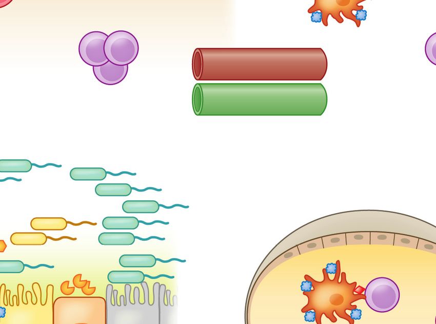
IY37CH16-Nagler ARjats.cls April 4, 2019 12:57
a
Stromal cells RALDH
Food
IgA Clostridia
TGF-β
RA
Mucous MLN DC
layer SCFAs AMPs
Mucin (butyrate)
CD103+ IDO+ Tregs
TGF-β
IL-10
Goblet MNP Paneth MNP
cell IL-22 cell
MNP
CX3CR1+ IL-22 CX3CR1+
IL-10 ILC3
CX3CR1+ DC
Access provided by University of North Carolina - Chapel Hill on 04/27/19. For personal use only.
MNP IgA
CD103+
Annu. Rev. Immunol. 2019.37:377-403. Downloaded from www.annualreviews.org
Bloodstream
CX3CR1+ Tregs Tregs
Lymphatics
IgA+ B cells TGF-β IL-10 Systemic tolerance
b IgA
Stromal cells
SCFAs (butyrate) Clostridia
Mucous Food
layer Mucin Stress AMPs
MLN DC
IgE Th2
Goblet MNP TSLP CX3CR1+
cell IL-33 MNP IL-4
IL-25
CX3CR1+
IgA
IgE+ B cells
ILC2 IL-13
IL-5 OX40L
IgA+ B cells DC
Th2
Tregs IgE CD103+
IL-10 MNP
TGF-β
IL-4
IgE+ B cells
Figure 3
An epithelial barrier in equilibrium with commensal bacteria is required for protection against allergic sensitization. (a) In healthy
individuals, Clostridia (and possibly other allergy-protective commensal bacteria) maintain epithelial barrier integrity by stimulating
ILC3s to produce IL-22. IL-22 promotes the production of mucus from goblet cells and antimicrobial peptides from Paneth cells and
reduces the ability of food allergens to gain access to the systemic circulation. Clostridial metabolites, including SCFAs, directly induce
Treg development. Treg-derived TGF-β favors local IgA antibody production, and circulating Tregs promote systemic tolerance.
(b) Twenty-first-century lifestyle factors like antibiotics and high-fat, low-fiber diets promote microbial dysbiosis, predisposing to
allergic sensitization. Depletion of Clostridia leads to loss of IL-22-dependent barrier functions and decreased concentrations of SCFAs,
impairing barrier integrity and increasing allergen access. The stressed epithelium (yellow arrow) produces alarmins like TSLP, IL-33,
and IL-25, which promote the generation of a Th2 immune response in part by stimulating ILC2s to produce Th2 cytokines. CD103+
DCs in a TSLP-rich microenvironment upregulate OX40L expression and traffic to MLNs or interact with lamina propria–resident T
cells, where they stimulate the generation of antigen-specific Th2 cells producing IL-4 that triggers antibody class switching to IgE.
Adapted from Reference 19 with permission from Cell Press. Abbreviations: AMP, antimicrobial peptide; DC, dendritic cell; IDO,
indoleamine 2,3-dioxygenase; ILC2/3, type 2/3 innate lymphoid cell; MLN, mesenteric lymph node; MNP, mononuclear phagocyte;
RA, retinoic acid; SCFA, short-chain fatty acid; Treg, regulatory T cell.
www.annualreviews.org • The Microbiome and Food Allergy 385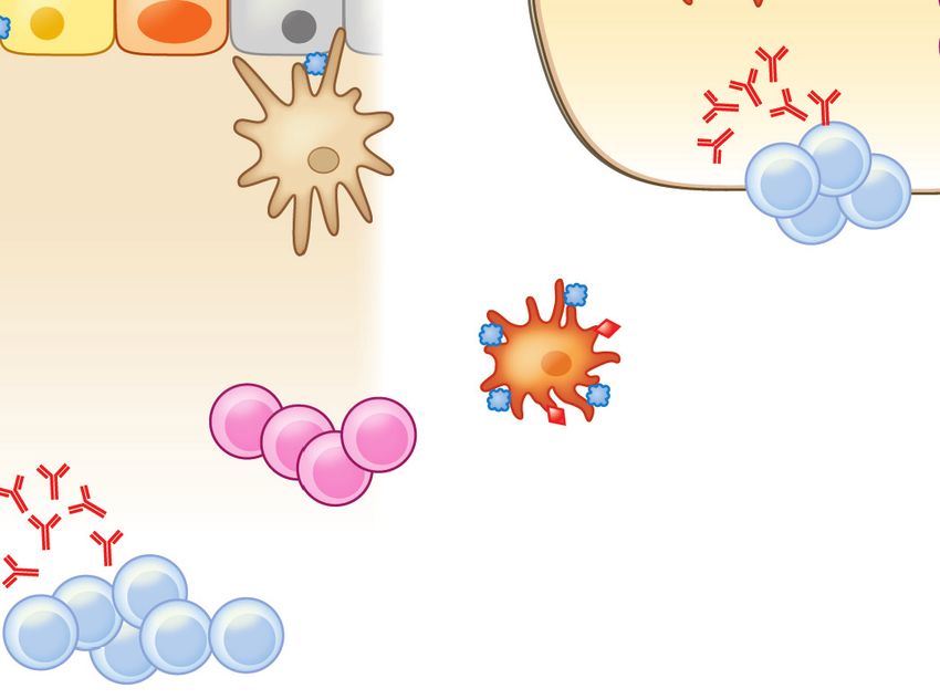
IY37CH16-Nagler ARjats.cls April 4, 2019 12:57
Healthy Infants Harbor Intestinal Bacteria That Protect Against Food Allergy
Humans have coevolved with their microbiota for millennia. Colonization with a founder mi-
crobiota occurs at birth; lactobacilli from the mother’s vaginal tract are the dominant founder
bacteria (81, 82). A highly ordered ecological succession then follows, with taxa emerging later
dependent on the metabolites and environment created by the founder bacteria (83). Cesarean
delivery disrupts this process and introduces founder bacteria derived largely from the skin (81);
some data suggest that this effect may be short-lived (84). It is clear, however, that the maternally
acquired microbiota profoundly affects innate immune system development after birth (85, 86).
Other work has hinted at a role for gut dysbiosis in the pathogenesis of food allergy (87–89). To
begin to understand the influence of intestinal bacteria on the development of food allergies, we
compared the composition and diversity of fecal samples collected from 4- to 5-month-old in-
Access provided by University of North Carolina - Chapel Hill on 04/27/19. For personal use only.
fants at the time of diagnosis with IgE-mediated cow’s milk allergy (CMA) with those of samples
collected from age- and gender-matched healthy infants attending a vaccination clinic (90). All
Annu. Rev. Immunol. 2019.37:377-403. Downloaded from www.annualreviews.org
infants belonged to the same Neapolitan cohort. The microbiota of the healthy infants was dom-
inated by taxa from the orders Lactobacillales, Bifidobacteriales, and Enterobacteriales (90), as shown
in other reports on the infant microbiome (84). Lactobacilli and bifidobacteria are readily cultur-
able bacteria that have been highly studied for potential probiotic activity (91). To our surprise
the CMA infants had an adult-type microbiota, dominated by Bacteroidales and Clostridiales, as
if the maturation of their microbiota had occurred at an accelerated pace (90). Dietary manage-
ment with an extensively hydrolyzed casein formula (EHCF) containing the probiotic Lactobacillus
rhamnosus GG (LGG) resulted in a higher rate of tolerance acquisition in infants with CMA than
other formulas studied (90, 92, 93). When we examined the influence of this dietary interven-
tion on the composition of the gut microbiota, we found that it did not result in an increased
abundance of lactobacilli detectable in the feces of the treated infants. Instead, treatment with
EHCF plus LGG, but not EHCF alone, was associated with changes in microbial community
structure that included the expansion of particular subsets of butyrate-producing Clostridia (90).
CMA infants treated with EHCF plus LGG also had significantly higher levels of butyrate de-
tectable in their feces and an enhanced acquisition of tolerance to cow’s milk (90). While the
small sample size examined is a limitation of this study, our findings have been corroborated in
a much larger cohort (94). The Consortium of Food Allergy Research (CoFAR) collected fe-
cal samples from 226 children enrolled in an observational study of milk allergy (94). They re-
ported that certain taxa within Clostridia were enriched between ages 3 and 6 months in children
with CMA whose disease had resolved by 8 years of age when compared to those with persistent
disease.
To examine whether commensal bacteria play a causal role in protection against food allergy,
we used human feces from four healthy and four IgE-mediated-CMA infant donors matched
for age, gender, and mode of birth to colonize germfree mice. Stable colonization of germfree
mice with human feces depends, in part, on the animals’ diet (95). The colonized mice were
therefore fed the same formulas consumed by their human infant donors to maintain the
donor-derived bacterial food source (in addition to plant-based mouse chow). All of the mice
were then sensitized to the cow’s milk allergen β-lactoglobulin (BLG). In agreement with earlier
studies (68, 76), germfree mice were highly susceptible to food-induced anaphylaxis. Following
colonization, germfree mice that received bacteria from healthy infants were protected against
sensitization to BLG (96). By contrast, germfree mice colonized with bacteria from CMA infants
had significantly higher serum levels of BLG-specific IgE and experienced anaphylaxis after BLG
challenge. Healthy and CMA-colonized mice exhibited differences in bacterial composition and
386 Iweala • NaglerIY37CH16-Nagler ARjats.cls April 4, 2019 12:57
unique transcriptome signatures in ileal intestinal epithelial cells (96). Correlation of ileal bacteria
with differentially expressed genes in the ileum of healthy-colonized mice allowed us to identify
a butyrate-producing clostridial species, Anaerostipes caccae, as a candidate allergy-protective bac-
terial taxa (96). Strikingly, monocolonization of germfree mice with this single species mimicked
the effect of the healthy microbiota and was sufficient to protect against food allergy. Our findings
demonstrate a causal role for the intestinal microbiota for protection against allergic responses
to dietary antigens and indicate that targeted modulation of specific bacterial communities may
represent a viable therapeutic strategy for food allergy (see below).
THE DUAL ALLERGEN EXPOSURE HYPOTHESIS AND ALLERGIC
SENSITIZATION TO FOOD
Access provided by University of North Carolina - Chapel Hill on 04/27/19. For personal use only.
Atopic children frequently present with evidence of sensitization to a food antigen (typically
Annu. Rev. Immunol. 2019.37:377-403. Downloaded from www.annualreviews.org
peanut or tree nuts) with no previous history of ingestion of the food in question. This observation
suggests that these sensitized children are exposed to food antigens through a route independent
of oral ingestion. In the case of peanut allergy, sources for epicutaneous exposure include peanut
allergen on tabletops, on hands after spreading peanut butter (even after rinsing hands with water)
(97) and as a component in skin oils, and in household dust (98). As exposure to environmental
sources of food allergen appears to be a risk factor, this has led some to hypothesize that while oral
ingestion of food promotes immune tolerance to food antigens, epicutaneous exposure to food al-
lergens promotes allergic sensitization (18) (Table 1). This hypothesis has been referred to as the
dual allergen exposure hypothesis (18) (Figure 4).
As with the intestinal epithelial barrier, it appears that allergen exposure through an intact
skin epithelial barrier promotes tolerogenic responses to the allergen (99). Dioszeghy et al. (100)
found that when ovalbumin (OVA) was applied to intact skin of mice it did not passively cross
the skin epithelial barrier and was not systemically detectable. However, prolonged application
to intact skin led to the internalization and transport of OVA to the draining lymph nodes by
DCs in the stratum corneum and the induction of antigen-specific Tregs (100), providing con-
ceptual support for the use of epicutaneous skin patches as an immunotherapeutic approach to
allergen-specific desensitization (see below). Hair serves as a critical component of the skin’s phys-
ical barrier, and the hair follicle is home to dense populations of specialized APCs, including
CD14+ CX3 CR1+ Langerhans DCs, with the ability to sample high-molecular-weight antigens
applied to the skin (101). The trafficking of these cells is controlled by chemokines generated
by hair follicles (102). Epicutaneous antigen exposure to intact skin generates Foxp3+ Tregs that
traffic between the site of immunization and the draining skin lymph nodes (103, 104). In mouse
models, these Tregs have been shown to suppress inflammatory hypersensitivity responses to epi-
cutaneously applied protein antigens (100, 104). Skin barrier compromise leads to dysregulated
immune responses to antigens introduced epicutaneously (99). Sensitization to food allergens
like egg and peanut is far more common in children with eczematous skin and atopic dermati-
tis (105, 106). Up to 50% of individuals with atopic dermatitis have loss-of-function mutations
in filaggrin (FLG), a protein that modulates the integrity of the stratum corneum and regulates
the skin’s permeability to water and antigens (107). Brough et al. (108) found an increased risk of
peanut sensitization and allergy in children with FLG gene mutations, while Venkataraman and
colleagues (109) also showed an association between FLG mutations and food allergy in older
children due to eczema and food allergen sensitization in early childhood. In mice orally sensi-
tized with peanut and the adjuvant cholera toxin, epicutaneous peanut antigen delivered to intact
skin muted Th2 cytokine production and the IgE response to oral challenge with peanut, whereas
www.annualreviews.org • The Microbiome and Food Allergy 387IY37CH16-Nagler ARjats.cls April 4, 2019 12:57
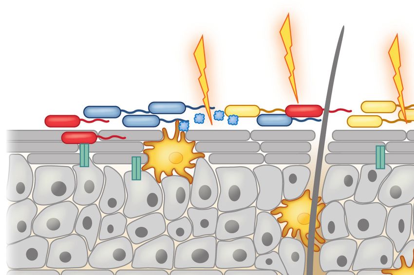
Trauma (tick bite) Infections Atopic dermatitis
TSG factors Genetic mutations
Tick-resident microbes (FLG, DSG1)
Commensal microbes
Pathogenic Food Ag
microbes
LC Stratum corneum
TLR2
Epidermis
Keratinocytes
Draining lymph node
Access provided by University of North Carolina - Chapel Hill on 04/27/19. For personal use only.
LTA TSLP
TLR2 ↑AMP IL-33 OX40L
IL-13
Annu. Rev. Immunol. 2019.37:377-403. Downloaded from www.annualreviews.org
↑SCF IL-25
OX40L DC
DC Th2
Dermis Hair
ILC2 follicle IgE
IL-4
FcεRI
FcεRI IL-4
Mast cells Basophil
Basophils IgE+ B cells
IL-4
Subcutaneous
fat layer
Figure 4
A compromised skin barrier elicits allergic sensitization to foods. A population of skin dendritic cells called Langerhans cells has the
ability to sample high-molecular-weight antigens applied to the skin. Defects in skin barrier function due to trauma (e.g., tick bite, TSG
factors), infection-mediated alterations in skin commensal microbiota, atopic dermatitis, and genetic defects in barrier proteins like
FLG and DSG1 trigger the release of TSLP and other alarmins (IL-25, IL-33) by the skin epithelium. In the epidermis, LTA-mediated
activation of the pattern recognition receptor TLR2 on keratinocytes induces secretion of SCF and TSLP and also upregulates AMP
production by both mast cells and keratinocytes. These cytokines promote Th2 immune responses by stimulating Th2 cytokine
production by ILC2s, upregulation of OX40L by antigen-laden DCs, and IL-4 production by basophils. IL-4 promotes antibody class
switching to IgE and allergic sensitization once IgE binds Fcε receptors (FcεRI) on basophils and mast cells. Abbreviations: Ag,
antigen; AMP, antimicrobial peptide; DC, dendritic cell; ILC2, type 2 innate lymphoid cell; LC, Langerhans cell; LTA, lipoteichoic
acid; SCF, stem cell factor; TLR2, Toll-like receptor 2; TSG, tick salivary gland.
epicutaneous peanut antigen delivered through injured, tape-stripped skin reinforced systemic
Th2 immune responses to peanut (110). When mice had hair removed with depilatory cream fol-
lowed by epicutaneous exposure to peanut extract, they also developed Th2-biased IgE and IgG1
responses and anaphylaxis following intraperitoneal challenge with peanut (111). Skin barrier im-
pairment is associated with increased expression of TSLP, the cytokine that links innate immune
responses at epithelial barriers to Th2-skewed adaptive immune responses (112, 113). Epicuta-
neous food allergen exposure in the setting of increased TSLP production by inflamed skin is
associated with an increase in serum allergen-specific IgE and an expansion of basophils in the
skin that amplify Th2 allergen-specific cytokine responses (114). Recent findings by Hussain and
colleagues (115) suggest that TSLP-expanded basophils work in concert with DCs through direct
cell contact to promote a Th2 polarized microenvironment. This interaction enhances OX40L
expression on DCs and stimulates IL-4 production by basophils critical to generate IgE-mediated
hypersensitivity responses (115). Moreover, compared to wild-type mice, clinical signs of food al-
lergy are significantly reduced after epicutaneous sensitization in mice whose basophils cannot
388 Iweala • NaglerIY37CH16-Nagler ARjats.cls April 4, 2019 12:57
produce IL-4. This underscores the critical role for IL-4 derived from TSLP-expanded basophils
in the development of food allergy (115).
Commensal Bacteria Maintain a Tolerogenic Environment in the Skin
Analogous to their role in the maintenance of homeostasis at mucosal surfaces, commensal bacteria
are critical for creating and maintaining a noninflammatory, tolerogenic environment in normal,
intact skin (116). Skin bacteria in humans are found in distinct niches, like the hair follicles (117)
that are also home to skin-resident DCs, putting skin bacteria proximal to key sentinels of the
skin’s immunologic barrier. In normal human skin, microbial DNA can be amplified from several
different cutaneous compartments including epidermis, hair follicles, dermis, and subcutaneous
Access provided by University of North Carolina - Chapel Hill on 04/27/19. For personal use only.
adipose tissue (118). The skin microbiome is dominated by members of the phylum Proteobacteria
with some representation from Actinobacteria, Firmicutes, and Bacteroidetes (118). In a model sys-
Annu. Rev. Immunol. 2019.37:377-403. Downloaded from www.annualreviews.org
tem mapping out the antigen-specific response to Staphylococcus epidermidis, Scharschmidt et al.
(116) found that microbial colonization of neonatal, but not adult, mice with this skin commensal
within a two-week window was required to establish a healthy host-microbe interface regulated
by highly activated skin Tregs. Colonization with S. epidermidis can also induce IL-17A+ CD8+
T cells, in a manner dependent on CD103+ skin-resident DCs, that migrate to the epidermis to
fortify skin barrier immunity against invasive pathogens (119). Infection with pathogenic microbes
like Staphylococcus aureus, a common complication in individuals with severe atopic dermatitis, can
cause significant skin barrier dysfunction (120). S. aureus produces exotoxins, proteases, and li-
pases that promote skin breakdown. Nakamura et al. (121) provided mechanistic insight into the
relationship between S. aureus colonization and allergic skin disease with the demonstration that
the S. aureus δ toxin activates mast cells and induces their degranulation. In children with atopic
dermatitis, Jones et al. (120) found a link between S. aureus colonization and allergic responses
to peanut, egg white, and cow’s milk. These studies suggest that through direct insult to the skin
epithelium, S. aureus disrupts the tolerogenic presentation of food antigens epicutaneously and
promotes the generation of Th2 immune responses to the antigen that lead to sensitization. In-
terestingly, an increase in the presence of S. aureus (and reduced diversity of other species) in the
skin microbiome has been associated with flares of atopic dermatitis and worsening disease activity
(122). Similarly, FLG-deficient skin has significantly less microbial diversity than FLG-sufficient
skin, with underrepresentation of bacterial taxa that use histidine. Keratinocytes exhibit different
cytokine and antimicrobial peptide responses depending on the composition of the bacteria to
which they are exposed (123). In culture, human keratinocytes can be stimulated with TLR lig-
ands and S. aureus membranes via TLR2 to produce TSLP, bridging innate immune responses at
epithelial barriers to Th2-skewed adaptive immunity (124). In addition, compared with conven-
tional mice, germfree mice have keratinocytes expressing abnormally low levels of stem cell factor
(SCF), a factor critical for mast cell differentiation, and higher numbers of immature dermal mast
cells with impaired ability to degranulate. Dermal mast cell maturation and degranulation can be
rescued when staphylococcal lipoteichoic acid (LTA), absent in the epidermis and hair follicles
of germfree mice, is injected into the skin. The microbe-derived LTA stimulates keratinocytes to
produce SCF through a TLR2-dependent pathway (125). Skin mast cells can also express TLR2
and bind LTA directly, which can increase their activity against vaccinia viruses and enhance their
production of antimicrobial peptides (126). Thus, alterations to microbial skin diversity in atopic
dermatitis patients, by changing the cytokine and antimicrobial peptide makeup at the epithelial
surface, may serve as another factor pushing the skin epithelial barrier to promote Th2-skewed
responses to epicutaneously delivered antigens.
www.annualreviews.org • The Microbiome and Food Allergy 389IY37CH16-Nagler ARjats.cls April 4, 2019 12:57
EOSINOPHILIC ESOPHAGITIS—AN EMERGING
FOOD-RELATED DISEASE
Classic IgE-mediated food allergy is not the only food intolerance associated with dysbiosis.
Eosinophilic esophagitis (EoE) is a chronic inflammatory disease characterized by eosinophilic
inflammation of the esophagus with a peak count of ≥15 eosinophils per high-power field of
esophageal biopsy tissue and an associated Th2 inflammatory response (127). On endoscopy,
linear furrows with little to no vascularity, mucosal rings and exudates, strictures, and a narrow
esophageal lumen can be found in both adult and pediatric patients with this condition, although
up to one-third of pediatric patients may have a normal endoscopy despite clinically active EoE
(128). Clinically, individuals present with symptoms related to esophageal dysfunction. Although
no clear role for IgE has been described in the pathogenesis of EoE (127, 129), there are signifi-
Access provided by University of North Carolina - Chapel Hill on 04/27/19. For personal use only.
cant associations between genetic loci specific for EoE and loci for general atopic disease, which
may act synergistically (130). IgE-mediated sensitization to ingested, inhaled, or epicutaneously
Annu. Rev. Immunol. 2019.37:377-403. Downloaded from www.annualreviews.org
introduced allergens is often present in subjects with EoE, even those with no history of anaphy-
laxis to food (127, 131, 132). Esophageal exposure to food, and in some individuals, to aeroal-
lergens, triggers the disease. As with IgE-mediated food allergy, population-based studies point
to a rise in the incidence of EoE starting in the early 2000s in Westernized societies (133, 134).
The sharp rise in the incidence of EoE in the United States corresponds with widespread intro-
duction of antibiotics in the mid-twentieth century and a decline in the prevalence of Helicobacter
pylori infection (131, 135–137). Recent work shows that, like food allergy, early-life environmental
factors (prenatal, intrapartum, and postnatal) influence susceptibility to EoE; positive risk factors
include antibiotic treatment, cesarean delivery, maternal fever, preterm labor, and acid suppres-
sant use in infancy (138). Postnatal dog exposure is a negative risk factor (138). Topical gluco-
corticoids and dietary therapy, including the six-food elimination diet (in which dairy, wheat, egg,
peanuts/tree nuts, fish/shellfish, and soy, the six most common US allergens, are removed from the
diet) and elemental diets, are the current mainstays of treatment (127). It is tempting to speculate
that the efficacy of elemental diets is due, in part, to diet-induced alterations in the esophageal
microbiome.
Several challenges surround research into the esophageal microbiome and the relationship with
EoE, including the current invasiveness and cost of obtaining endoscopic brushings or esophageal
biopsies (131). While endoscopic techniques have been reported to be most effective at securing
bacterial samples within the esophageal microbiome (139), there are ongoing attempts to make
sampling and analysis of the esophageal microbiome less invasive using nonendoscopic techniques
like the Esophageal String Test (140), a mesh capsule or cytosponge (141), and inflatable balloons
to sample the upper gastrointestinal tract (131). However, contamination of samples obtained
by these methods with the oral microbiota is common (131). 16S rRNA–targeted sequencing
of esophageal biopsies (142) and esophageal mucosa and mucosal secretions collected with the
Esophageal String Test (143) from patients with or without a diagnosis of EoE have identified over
300 bacterial species among the esophageal microbiota. The most common genera detected were
Streptococcus, Prevotella, and Veillonella, with an increased abundance of members of the Haemophilus
genus in patients with untreated EoE (143).
Barrier Dysfunction Underlies the Pathogenesis of Eosinophilic Esophagitis
Similar to IgE-mediated food allergy, a growing body of research has implicated a dysfunctional
epithelial barrier in the pathophysiology of EoE. The esophagus, like the skin, comprises stratified
390 Iweala • NaglerIY37CH16-Nagler ARjats.cls April 4, 2019 12:57
squamous epithelium that protects underlying tissues in areas at high risk for mechanical or chem-
ical injury (144). Subjects with mutations in skin barrier proteins like filaggrin or desmoglein-1
(DSG1) develop atopic dermatitis. DSG1 is an adherens junction protein and a desmosomal com-
ponent linking the cell surface to the keratin cytoskeleton. In the absence of functional DSG1,
allergic dermatitis is particularly severe (145). Interestingly, DSG1 is reduced in esophageal biop-
sies from EoE patients and was shown to be critical for esophageal epithelial integrity (146). Davis
et al. (128) showed that IL-13, present in inflamed esophageal tissue in EoE, triggered the loss
of DSG1 expression by enhancing production of CAPN14, an intracellular calcium-activated
protease in the calpain family of proteases, whose overexpression in the esophageal epithelium
reduced DSG1 levels, impairing epithelial barrier function. Recent work has identified a pos-
sible role for the serine protease inhibitor SPINK7, constitutively generated by differentiated
Access provided by University of North Carolina - Chapel Hill on 04/27/19. For personal use only.
esophageal squamous epithelium, in the pathogenesis of EoE (147). The absence of SPINK7 pro-
moted proinflammatory responses in the esophagus that compromised barrier integrity, including
Annu. Rev. Immunol. 2019.37:377-403. Downloaded from www.annualreviews.org
dilated intercellular spaces and increased epithelial permeability, dysfunctional DSG1, and absent
FLG expression. It also triggered a transcriptome signature consistent with allergic inflammation
and stimulated TSLP release in esophageal cell lines stimulated via TLR3 (147). TLR-mediated
TSLP production in the absence of SPINK7 suggests that esophageal microbial signaling in ge-
netically susceptible individuals may influence esophageal epithelial barrier integrity. Future stud-
ies using germfree and gnotobiotic mouse models of EoE could begin to address the relationship
between the esophageal microbiome and esophageal barrier function.
Human studies and mouse models suggest that invariant natural killer T (iNKT) cells are
also involved in eosinophil recruitment to the esophagus in EoE. Lexmond and colleagues (148)
observed significant upregulation of the human iNKT cell–associated invariant TCR Vα24, the
MHC-I-like molecule CD1d, and the chemokine CXCL16, which has been associated with the
recruitment of iNKT cells to mucosal surfaces, in esophageal biopsies from EoE patients. Studies
in germfree mice have shown enhanced accumulations of iNKT cells in the colonic LP and lung
and heightened morbidity in models of inflammatory bowel disease and allergic asthma in com-
parison to SPF mice (149). Moreover, in a murine model, treatment of neonatal mice with gly-
cosphingolipids derived from the commensal bacterium Bacteroides fragilis protected these mice
against oxazolone-induced colitis in adulthood (150). Colonization of germfree neonatal mice
with a conventional microbiota induces epigenetic regulation of the CXCL16 promoter, decreas-
ing hypermethylation and reducing CXCL16 production and iNKT cell recruitment (149, 150).
These findings in the lower intestines raise the possibility that commensal microbiota in the upper
gastrointestinal tract and esophagus may also regulate iNKT cell levels in the submucosa, which
in turn can modulate eosinophil recruitment to this organ.
Whether the change in esophageal microbiota is predisposing individuals to EoE or whether
the influx of eosinophils triggers alterations in the esophageal microbiota requires additional study.
Treatments used to address dental disease and other gastrointestinal diseases like gastroesophageal
reflux disease (GERD) may impact the esophageal microbiota and predispose patients to EoE.
Alternatively, it is possible that current pharmacologic treatments used to control EoE, such as
proton pump inhibitors and oral corticosteroids (127), also contribute to the quelling of the disease
by altering the esophageal microbiota. Additional larger-scale studies in the vein of Benitez et al.
(142) and Harris et al. (143) will be useful to expand our understanding of how treatments like
antibiotics, proton pump inhibitors, and oral corticosteroids change the microbiota in those with
EoE and those without. Such studies may also identify a microbial class (similar to Clostridia in
food allergy) critical for the maintenance of a normal esophageal epithelial barrier and with the
potential to prevent or treat EoE.
www.annualreviews.org • The Microbiome and Food Allergy 391You can also read