Mucosal Vaccination Primes NK Cell-Dependent Development of CD8+ T Cells Against Pulmonary Brucella Infection - Frontiers
←
→
Page content transcription
If your browser does not render page correctly, please read the page content below
ORIGINAL RESEARCH
                                                                                                                                                 published: 07 July 2021
                                                                                                                                       doi: 10.3389/fimmu.2021.697953

                                            Mucosal Vaccination Primes NK
                                            Cell-Dependent Development of
                                            CD8+ T Cells Against Pulmonary
                                            Brucella Infection
                           Edited by:       Ella Bhagyaraj , Hongbin Wang , Xinghong Yang , Carol Hoffman , Ali Akgul ,
                      Pam Kozlowski,        Zakia I. Goodwin and David W. Pascual *
            Louisiana State University,
                        United States       Department of Infectious Diseases & Immunology, University of Florida, Gainesville, FL, United States

                         Reviewed by:
   Guillermo Hernán Giambartolomei,        Past studies with the live, double-mutant B. abortus (znBAZ) strain resulted in nearly
 Consejo Nacional de Investigaciones
   Cientı´ficas y Técnicas (CONICET),
                                            complete protection of mice against pulmonary challenge with wild-type (wt) Brucella via a
                              Argentina     dominant CD8+ T cell response. To understand the contribution innate immune cells in
               Erika Sousa Guimarães,
                                            priming CD8+ T cell responses, mice were nasally dosed with wt B. abortus, smooth
Federal Institute of Minas Gerais, Brazil
                      Alansana Darboe,      vaccine strain 19 (S19), or znBAZ, and examined for innate immune cell activation. Flow
       Medical Research Council The         cytometric analysis revealed that znBAZ, but not wt B. abortus nor S19 infection, induces
          Gambia Unit (MRC), Gambia
                                            up to a 5-fold increase in the frequency of IFN-g-producing NK cells in mouse lungs. These
                   *Correspondence:
                     David W. Pascual
                                            NK cells express increased CXCR3 and Ki67, indicating their recruitment and proliferation
                     pascuald@ufl.edu        subsequent to znBAZ infection. Their activation status was augmented noted by the
                                            increased NKp46 and granzyme B, but decreased NKG2A expression. Further analysis
                   Specialty section:
         This article was submitted to
                                            demonstrated that both lung caspase-1+ inflammatory monocytes and monocyte-derived
              Vaccines and Molecular        macrophages secrete chemokines and cytokines responsible for NK cell recruitment and
                          Therapeutics,     activation. Moreover, neutralizing IL-18, an NK cell-activating cytokine, reduced the
               a section of the journal
              Frontiers in Immunology       znBAZ-induced early NK cell response. NK cell depletion also significantly impaired
              Received: 20 April 2021       lung dendritic cell (DC) activation and migration to the lower respiratory lymph nodes
             Accepted: 21 June 2021         (LRLNs). Both lung DC activation and migration to LRLNs were significantly impaired in NK
             Published: 07 July 2021
                                            cell-depleted or IFN-g-/- mice, particularly the CD11b+ and monocytic DC subsets.
                            Citation:
       Bhagyaraj E, Wang H, Yang X,
                                            Furthermore, znBAZ vaccination significantly induced CD8+ T cells, and upon in vivo
    Hoffman C, Akgul A, Goodwin ZI          NK cell depletion, CD8+ T cells were reduced 3-fold compared to isotype-treated mice. In
    and Pascual DW (2021) Mucosal           summary, these data show that znBAZ induces lung IFN-g+ NK cells, which plays a critical
Vaccination Primes NK Cell-Dependent
 Development of CD8+ T Cells Against        role in influencing lung DC activation, migration, and promoting protective CD8+ T
          Pulmonary Brucella Infection.     cell development.
           Front. Immunol. 12:697953.
     doi: 10.3389/fimmu.2021.697953         Keywords: Brucella, dendritic cells, macrophages, IFN-g, chemokines, CD8+ T cells

Frontiers in Immunology | www.frontiersin.org                                       1                                           July 2021 | Volume 12 | Article 697953
Bhagyaraj et al.                                                                                         NK Cells Augment Anti-Brucella Immunity

INTRODUCTION                                                                 The present study describes a live, double-mutant DnorD
                                                                          DznuA Brucella abortus (znBAZ) strain that, after mucosal
Brucellosis is one of the most prevalent bacterial zoonotic               vaccination, confers complete protection against pulmonary
diseases worldwide and listed by the World Health                         challenge with virulent B. abortus 2308 by stimulating robust
Organization as one of the “seven most neglected diseases” (1–            CD8+ T cell responses in the lungs (31). To better understand
3). Brucella, a Gram-negative, facultative bacterium, is                  how znBAZ confers protection, the role of innate cells,
responsible for this disease. While commonly thought of as a              particularly, NK cells was examined. A robust, early activation
disease of livestock, the four closely-related Brucella species –         of NK cells was induced following nasal znBAZ infection, but not
B. abortus, B. melitensis, B. suis, and B. canis - can cause human        observed when infected with wild-type (wt) B. abortus 2308 or
disease (4, 5). Brucella is transmitted from infected animals to          B. abortus S19 vaccine. Additional mechanistic studies were
humans through direct contact, ingestion of contaminated foods,           conducted to discern the impact of NK cells has upon znBAZ-
or by inhalation of Brucella-laden aerosols (6). The animal and           induced lung CD8+ T cell responses, and the relevance of IFN-g
human forms of brucellosis tend to differ symptomatically – in            in innate cell activation and migration. These results provide
livestock, brucellosis causes abortion, infertility, mastitis, and        important insights regarding the role of early lung NK cell
lameness, whereas in humans, the effects are incapacitating               activation in znBAZ-mediated immunity.
illness characterized by undulating fever with flu-like
symptoms, which can persist if untreated (5, 7, 8). An estimate
of 500,000 humans are annually infected with Brucella (9, 10).
Moreover, chronic brucellosis can lead to additional                      MATERIALS AND METHODS
complications in humans such as endocarditis, arthritis,
epididymo-orchitis, and sacroiliitis (11–14). The most common             Bacterial Strains and Culture Conditions
routes of infection in humans are oral via consumption of                 The construction of znBAZ, a live attenuated mutant Brucella
contaminated foods or inhalation (15). Thus, induction of                 abortus strain, was previously described, and lacks functional
strong mucosal immunity in the aerodigestive tracts is                    znuA and norD genes (32). All B. abortus vaccine strains, S19,
desirable to provide efficient and long-lasting protection                 RB51, znBAZ, and wt 2308, were inoculated and grown on
against Brucella infection. Given the routes of exposure, a               Potato Infusion Agar (PIA) plates for three days at 37°C under
mucosal vaccine that activates immunity in the lungs and gut              5% CO2. Before infection, bacteria were harvested, washed, and
seems a logical step to advancing Brucella vaccines. Mucosal              diluted in sterile phosphate-buffered saline (sPBS) for use.
vaccination is advantageous because antigen-specific humoral
and cell-mediated immune responses can be induced both in the             Mice
mucosal and systemic compartments (16–18). As such, mucosal               BALB/c and C57BL/6 (female, 6–8 weeks old) mice were obtained
vaccines may provide better coverage against pathogens than               from Charles River Laboratory (Frederick, MD, USA), and
those given parenterally.                                                 IFN‐g−/− mice on C57BL/6 background were bred in-house. All
    Embedded within the mucosal tissues are innate immune                 animal experiments performed with live attenuated Brucella
cells, which play a crucial role in antimicrobial defenses.               vaccine strains S19 and znBAZ were conducted under biosafety
Stimulation of innate immunity by respiratory pathogens is                level-2 (BSL-2) containment; studies involving wt B. abortus 2308
central to generating a pathogen-specific immunity (19, 20).               were done under BSL-3 containment. Mice were maintained in
Among the innate immune cell population in the lungs, NK cells            individually ventilated cages under HEPA-filtered barrier
merit special attention due to their role as a bridge between             conditions with 12 h of light and 12 h of darkness, and food
innate and adaptive immune responses (21, 22). The lungs                  and water were provided ad libitum. All animal care and
harbor NK cells, underscoring their importance in respiratory             procedures were in strict accordance with the recommendations
mucosal immune protection (23). NK cells act as an essential first         in the Guide for the Care and Use of Laboratory Animals of the
line of defense against respiratory infections, and are poised to         National Institutes of Health. All animal studies were conducted
exert effector functions and produce cytokines and chemokines             under protocols approved by the University of Florida
that coordinate innate and adaptive immune responses (24, 25).            Institutional Animal Care and Use Committee.
Early in infection, NK cells are primarily activated by cytokines
such as type I interferons, interleukin-12 (IL-12), IL-15, and            Infection
IL-18, secreted by macrophages and monocytes (26, 27). NK cells           BALB/c or C57BL/6 mice (n = 5/group) were nasally dosed with
activation is also regulated by a wide array of soluble or                30 ml of sPBS, 1×10⁵ CFUs wt B. abortus 2308, 1×10⁸ CFUs S19,
membrane bound ligands on infected cells that interact with               or 1×10⁹ CFUs znBAZ administered into the anterior nares
both activating and inhibitory receptors on the NK cell surface           dropwise using a micropipette under isoflurane anesthesia. The
(28). Activated NK cells protect against invasive respiratory             infected mouse lungs and lower respiratory lymph nodes
pathogens either by direct lysis of infected cells or indirectly by       (LRLNs) were harvested at different time points (2, 5, and 15
activating other innate immune cells such as macrophages or               days) after infection and analyzed for brucellae colonization and
dendritic cells (DCs) (29). In addition, NK cells can indirectly          mononuclear cell composition. Lung and splenic homogenates
modulate adaptive immune responses by its assistance in                   from individual mice were plated on Farrell’s medium (Oxoid
priming T cell responses (30).                                            Ltd, Basingstoke, UK) or PIA for 3–5 days at 37°C in 5% CO2.

Frontiers in Immunology | www.frontiersin.org                         2                                   July 2021 | Volume 12 | Article 697953
Bhagyaraj et al.                                                                                           NK Cells Augment Anti-Brucella Immunity

Bronchoalveolar Lavage Fluid                                              Flow Cytometry Assay
Nasally infected and uninfected mice were euthanized, their               For flow cytometry analysis, cells were washed with PBS plus 2%
tracheas were cannulated, and the lungs were perfused with                FBS, and incubated with Fc blocker (eBioscience) along with Live/
1 ml of cold sPBS three times, and a total of 3 ml BAL fluids were         dead stain (ThermoFisher) for 15 min at 4°C. After an additional
collected from each mouse. BAL fluid samples were centrifuged              wash, the cells were surface stained for 30 min at 4°C with mAbs
at 500 × g for 10 min at 4°C, and recovered fluids were filter-             specific for CD49b (DX5), NK1.1 (PK136), CD4 (GK1.5), CD8 (53-
sterilized using 0.22 µ syringe filters. The sterile BAL fluid              6.7), CCR2 (SA203G11), CCR5 (HM-CCR5), CXCR3 (CXCR3-
samples were stored at −70°C for cytokine analysis.                       173), NKG2D (CX5), NKp46 (29A1.4), NKG2A (16A11), and
                                                                          TCR-b (H57-597) for lymphocyte analysis. For macrophages and
Cytokine ELISAs                                                           dendritic cells, cells were stained with mAbs specific for SiglecF
Cytokines (IFN-g, IL-12p70, IL-15, IL-18) and chemokines (CXCL-           (1RNM44N), CD11b (M1/70), CD11c (N418), CD103 (2E7), F4/80
9 and CXCL-10) levels were measured from BAL fluids (duplicate             (BM8), Ly6C (HK1.4), Ly6G (1A8), CD40 (3/23), CD80 (16-10A1),
samples) collected from mice dosed nasally with wt B. abortus 2308,       CD86 (GL-1), and MHCII (M5/114.15.2). For detection of asialo-
S19, or znBAZ, along with those from uninfected mice by capture           GM1+ T cells, the rabbit polyclonal anti-asialo-GM-1 Ab was used
ELISA using the following antibody pairs: IFN-g (clone R4-6A2 and         (eBioscience). For intracellular staining, the surface stained cells
clone XMG1.2 from BD Pharmingen), IL-12p70 (clone 9A5                     were washed, fixed, and permeabilized using the True-Nuclear
and biotinylated clone C17.8; BD Pharmingen), IL-15 (clone                Transcription Factor Buffer Set (BioLegend, San Diego, CA)
201136 and biotinylated polyclonal goat IgG anti-mouse IL-15;             followed by intracellular staining with mAbs specific for IL-6
R&D Systems), IL-18 (clone 74 and biotinylated clone 93-10C; R&D          (MP5-20F3), IL-12 (C15.6), IL-18 (93-10C), IFN-g (XMG1.2),
Systems), CXCL-9 (polyclonal goat and biotinylated polyclonal goat        TNF-a (MP6-XT22), Ki-67 (SoIA15), iNOS (CXNFT), and
IgG anti-mouse CXCL-9; R&D Systems), and CXCL-10 (clone                   granzyme-B (NGZB). All the fluorescently conjugated mAbs were
134013 and biotinylated polyclonal goat IgG anti-mouse CXCL-              procured from (BioLegend, San Diego USA) or (eBioscience, San
10; R&D Systems). ELISAs were developed using a third step                Diego USA). After staining, cells were acquired on a BD Fortessa
antibody Ab, horseradish peroxidase (HRP) conjugated goat anti-           flow cytometer and analyzed by using FlowJo software.
biotin Ab (Vector Laboratories). After a wash step, ABTS
peroxidase substrate (Moss, Inc., Pasadena, ME, USA) was added            Active Caspase-1 Detection
to each well, and reactions were read at 415 nm using a Bio-Tek           Activated caspase-1 was detected in cells by using FLICA assay
Instruments Epoch Microplate Spectrophotometer (Winooski, VT).            (Immunochemistry Technology) as per manufacturer guidelines.
Cytokine concentrations were extrapolated from standard curves            Briefly, cells were incubated with FLICA reagent (FAM-YVAD-
generated by recombinant murine cytokines and chemokines: IFN-g           FMK) for 30 mins at 37°C. These FLICA stained cells were then
(Peprotech), IL-15 (R&D Systems), IL-12 (R&D Systems), IL-18              washed with PBS containing 2% FBS and subjected to surface
(R&D Systems), CXCL-9 (R&D Systems), and CXCL-10 (R&D                     staining before flow cytometry analysis.
Systems). ELISA methods used were similar to those previously
described (31, 32).                                                       Anti-IL-18 mAb Treatment
                                                                          IL-18 was neutralized in vivo using 200 µg/dose/mouse of anti-
Single-Cell Preparation and                                               mouse IL-18 mAb (clone YIGIF74-1G7; Bio X Cell). Mice were
In Vitro Stimulation                                                      treated i.p with isotype control IgG (Bio X Cell) or anti-mouse
LRLNs and lungs were aseptically removed from euthanized mice             IL-18 mAb on day -1 (one day before infection) and every three
and collected into 2ml tubes containing 1ml of incomplete media           days after that until the termination of the experiment. Mice
(ICM): RPMI-1640, 10 mM HEPES buffer, and 10 mM penicillin/               were infected with 1×10⁹ CFUs znBAZ nasally on day 0.
streptomycin. These tissues were then mechanically homogenized
and filtered through 70-µm cell strainers (Fisherbrand) to obtain          In Situ Carboxy-Fluorescein Succinimidyl
single-cell suspensions. Lung tissue was additionally digested with       Ester Staining and Lung DC Migration
20 mg of Liberase TL research-grade (Roche) and 50 units of RNase-        Lung DC migration was monitored by using in situ CFSE
free DNase I (Promega) for 45 min at 37°C under 5% CO2, followed          staining (33). 25mM CFSE (Molecular Probes) was diluted in
by 0.5M EDTA treatment for 5 mins. Cells were then suspended in           sPBS to a concentration of 5mM. Mice were anesthetized, and
freshly made ammonium-chloride-potassium (ACK) lysis buffer for           nasally instilled with 30 µl of diluted CFSE for labeling lung cells
3 mins, then washed with ICM, and resuspended in complete media           in vivo 6 hr before nasal infection. Five days after infection, the
(CM): ICM plus 10% fetal bovine serum (FBS), 10 mM nonessential           LRLNs were analyzed for CFSE+ DC subsets.
amino acids, and 10 mM sodium pyruvate. For flow cytometry
analysis, single-cell suspensions were either stimulated overnight        NK Cell Depletion Studies
with heat-killed RB51 (HKRB51) followed by 4 hours of 5 ng/ml             NK cells were depleted using 50 µg/dose/mouse of rabbit anti-
phorbol myristate acetate (PMA; SIGMA-ALDRICH), 500 ng/ml                 asialo-GM1 antibody (eBioscience) in BALB/c mice or anti-
ionomycin (SIGMA-ALDRICH) and 10 mg/ml brefeldin A                        NK1.1 (clone PK136; Bio X Cell) in C57BL/6 mice. Mice were
(SIGMA-ALDRICH) before antibody (Ab) staining or                          treated i.p with isotype control IgG or anti-asialo-GM1 Ab or
stained directly.                                                         anti-NK1.1 on day -1 (one day before infection) and every three

Frontiers in Immunology | www.frontiersin.org                         3                                    July 2021 | Volume 12 | Article 697953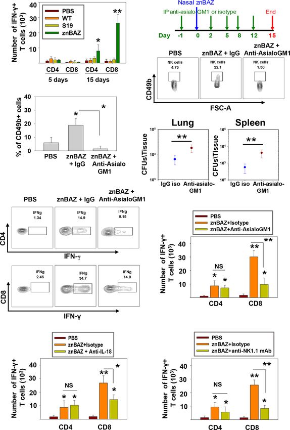
Bhagyaraj et al.                                                                                           NK Cells Augment Anti-Brucella Immunity

days after until the termination of the experiment. Mice were              Levels of granzyme-B and NK cell activation molecule, NKp46,
nasally infected with 1×10⁹ CFUs znBAZ on day 0. NK cell                   increased significantly in znBAZ-dosed mice, in contrast to wt B.
depletion in the lungs and spleens was confirmed by                         abortus or S19. Notably, expression of NK cell inhibitory molecule,
flow cytometry.                                                             NKG2A, was unchanged in wt B. abortus- and S19-infected mice
                                                                           relative to naïve controls, but NKG2a was significantly reduced in
Statistical Analysis                                                       znBAZ-infected mice (Figures 1H, I). These findings were validated
All experiments were conducted two or three times with n=5                 in C57BL/6 mice, showing similar results (Supplementary
mice/group. The statistical significance of the data was calculated         Figures 1C–H). Collectively, these findings support the notion
by using One-way ANOVA followed by Tukey’s multiple                        that nasal znBAZ infection activates NK cells, while nasal
comparison test with SigmaPlot 12.0. All results were                      infection with wt B. abortus or S19 does not alter lung NK cell
discerned to the 95% confidence interval.                                   numbers nor their activation status.

                                                                           Nasal Infection With znBAZ Elicits NK
RESULTS                                                                    Cell-Activating Cytokines and Chemokines
                                                                           to Recruit Lung Monocytes and
Nasal znBAZ Infection Elicits an Early NK                                  Macrophages
Cell Response in Mouse Lungs                                               To identify the cytokines and chemokines that contribute to early
To determine the role of NK cells in establishing znBAZ-induced            lung NK cell activation upon nasal znBAZ infection, groups of
protective immunity in the lungs, groups of BALB/c mice were               BALB/c mice were nasally infected with wt B. abortus, S19, or
nasally dosed with wt B. abortus 2308, B. abortus S19 vaccine, or          znBAZ, and 3 days later, individual BALs and lungs were harvested
znBAZ. Different doses were used since wt B. abortus 2308 and              to measure chemokine and cytokine levels produced locally. Upon
S19 are pathogenic in mice, whereas znBAZ is a highly attenuated           znBAZ infection, CXCR3 ligands, CXCL-9, and CXCL-10 levels
strain (31) compared to these strains. znBAZ showed a modest               were significantly enhanced by 14- and 12-fold, respectively
increase in lung colonization by day 10, but afterwards declined           (Figure 2A). Likewise, NK cell-activating cytokines, IL-12 and IL-
by greater than two-logs by day 15 (Supplementary Figure 1A).              18, were notably elevated by 4- and 10-fold, respectively
S19 retained elevated levels over the entire course, and showed no         (Figure 2B). However, wt B. abortus and S19 showed no
indication of decline. Wt B. abortus 2308 achieved similar levels          significant difference in CXCL-9, CXCL10, IL-12, IL-15, or IL-18
as S19 by day 10, and continued to increase exceeding znBAZ                production compared to PBS-dosed mice (Figures 2A, B).
colonization by greater than two logs (Supplementary                           To determine the cell source of these chemokines and
Figure 1A). The S19 vaccine was selected since it is a smooth              cytokines, flow cytometry analysis was performed on stained
vaccine resembling znBAZ. Lung lymphocyte populations were                 lung mononuclear cells isolated from uninfected and infected
analyzed at different time points to assess when NK cells (gating          mice 3 days after nasal infection. The gating strategy for
strategy provided in Supplementary Figure 1B) became                       identifying myeloid lung cells is presented in Supplementary
involved. Subsequent to znBAZ infection, the frequency of lung             Figure 2. SiglecF+ CD11blo CD11c+ alveolar macrophages (AMs)
NK cells gradually increased and peaked after 5 days, in contrast          were notably reduced, as were SiglecF- CD11b+ Ly6G- F4/80+
to lung NK cell levels in mice infected with wt B. abortus or S19          Ly6C- interstitial macrophages (IMs; Figures 2C, D). In contrast,
vaccine resembled uninfected mice (Figures 1A–C). The early                the SiglecF- CD11b+ Ly6G-, F4/80+, Ly6C+ monocyte-derived
surge in lung NK cells was due to either their recruitment or              macrophages (MDMs) and SiglecF- CD11b+ Ly6G- F4/80- Ly6C+
proliferation of sentinel NK cells. To distinguish between the two         lung monocytes were significantly increased by 1.5- and 3-fold,
possibilities, the expression of the proliferation marker Ki67,            respectively, in the znBAZ-infected mice. The macrophage and
along with chemokine receptors, CCR2, CCR5, and CXCR3,                     monocyte subsets remained unchanged in the lungs from those
which aid with NK cell recruitment, was analyzed on lung NK                infected with wt B. abortus 2308 or S19 (Figures 2C, D).
cells 3 days after nasal instillation. A significant increase in Ki67       Importantly, the lung MDMs and monocytes from the znBAZ-
and CXCR3 expression by NK cells was evident, but CCR2 and                 infected mice produced elevated CXCL-9, CXCL-10, IL-12, and
CCR5 levels remained unchanged, suggesting that NK cell                    IL-18 (Figures 3A–D). Caspase-1 is necessary to activate pro-IL-
infiltration and proliferation occurred after znBAZ infection               18 (34, 35), so as expected, the caspase-1 activity was found
(Figures 1D, E). Additionally, NK cells serve as an important              associated with these mononuclear cells (Figure 3E). During
early sentry and a source of IFN-g, crucial to initiating the              infections, APC-elicited IL-18 stimulates IFN-g production by
adaptive immune response following infection. At day 5 post-               NK cells (36). IL-18 often works synergistically with IL-12
infection, the lung NK cells showed significantly enhanced IFN-g            resulting in elevated IFN-g production by NK cells, and
in znBAZ-infected mice (Figures 1F, G). IFN-g expression by NK             actually, IL-18 alone is sufficient to enhance IFN-g production
cells from wt B. abortus- or S19-infected mice did not vary from           (37, 38). Given the importance of IL-18 in NK cell activation and
naïve levels. Hence, the escalation in NK cells was not evident in         its elevated presence following nasal znBAZ infection, the role of
the lungs from wt B. abortus- or S19-infected mice.                        IL-18 was investigated. Groups of BALB/c mice were treated by
    Lung NK cells from znBAZ-infected mice displayed an activated          the i.p. route with either anti-IL-18 mAb or IgG isotype control
status unlike those from mice infected with wt B. abortus or S19.          one day prior to nasal znBAZ infection and every three days

Frontiers in Immunology | www.frontiersin.org                          4                                    July 2021 | Volume 12 | Article 697953
Bhagyaraj et al.                                                                                                       NK Cells Augment Anti-Brucella Immunity

                                                  A

                                    B                                                    C

                                    D                                                    E

                                    F                                                    G

                                    H                                             I

 FIGURE 1 | Nasal znBAZ infection promotes lung NK cell expansion. Groups of BALB/c mice were nasally infected with wild-type (wt) B. abortus 2308 (1×10⁵
 CFUs), B. abortus S19 vaccine (1×10⁸ CFUs), or znBAZ (1×10⁹ CFUs). (A) Lung NK cell numbers were measured at the indicated days post-infection. (B, C) Flow
 cytometry analysis of NK (gated on TCRb- CD49b+) cells on day 5 post-infection, and examined for (D, E) CCR2, CCR5, CXCR3 and Ki67 expression. (F, G) On day
 3 post-infection, expression of IFN-g-producing lung NK cells is depicted. (H) Lung NK cells for (top row) NKp46, granzyme B, perforin, (bottom row), NKG2A,
 inhibitory molecule, NKG2D, and CD69, and their respective (I) MFIs are shown. The data depict the means ± SEM of 5 mice/group; *p < 0.01 and **p ≤ 0.001,
 compared with PBS-dosed mice. Data are representative of two experiments.

afterwards. Subsequent to in vivo IL-18 neutralization, analysis of               (39). Antigen uptake and presentation by DCs are critical for
lung lymphocytes at day 5 post-infection revealed a significant                    priming T cell responses. NK cells have been reported to interact
decline in the frequency of total NK cells and IFN-g+ NK cells by                 with DCs and modulate their activation (40). Depending on their
2.2-fold for both (Figures 3F–I).                                                 activation status and cytokine profiles, DCs induce distinct T cell
                                                                                  polarization to shape the immune response (41). Under steady-state
znBAZ-Induced Lung NK Cell Response                                               conditions, mouse lungs have two major DC subsets: CD103+ DC
Is Essential for Lung DC Maturation                                               (CD11c+, MHCIIhi, CD103+, CD11b-) and CD11b+ DC (CD11c+
and Migration                                                                     MHCIIhi CD103- CD11b+). During infection and inflammation, a
DCs are involved in the initiation of adaptive immune responses,                  third subset appears, referred to as monocytic DCs (moDC; CD11c+
and are major envoys between innate and adaptive immune systems                   MHCIIhi CD103- CD11b+ Ly6C+) (41). We queried whether

Frontiers in Immunology | www.frontiersin.org                                 5                                         July 2021 | Volume 12 | Article 697953
Bhagyaraj et al.                                                                                                         NK Cells Augment Anti-Brucella Immunity

                                    A                                         B

                                    C                                         D

 FIGURE 2 | Lung monocytes and macrophages produce CXCR3 ligands, CXCL-9 and CXCL-10, and NK cell-activating cytokines, IL-12 and IL-18, subsequent to
 nasal znBAZ infection. Groups of BALB/c mice were nasally dosed with wild-type B. abortus 2308 (WT), B. abortus S19 vaccine, or znBAZ, and three days later,
 (A) CXCR3 ligands, CXCL-9 and CXCL-10, and (B) NK cell activating cytokines, IL-12, IL-15, and IL-18, present in individual bronchoalveolar lavage (BAL) fluids,
 were measured by cytokine-specific ELISA. (C, D) Total mononuclear cells were isolated from the lungs to measure the frequency of lung monocytes and
 macrophages including alveolar macrophages (AMs), interstitial macrophages (IMs), monocyte-derived macrophages (MDMs), and monocytes by flow cytometry. The
 data depict the means ± SEM of 5 mice/group; *p < 0.01 and **p ≤ 0.001, compared with sPBS-dosed mice. Data are representative of two or three experiments.

znBAZ-induced lung NK cells can modulate lung DC phenotypes.                       Depleting NK cells also reduced the DC iNOS levels
A gating strategy for lung DC analysis is provided in                              (Figures 4E, F). Additional analysis was performed examining
Supplementary Figure 3. To determine NK cells’ relevance                           the effect of NK cell depletion on lung DC migration to LRLNs by
following their depletion, groups of BALB/c mice were i.p. treated                 CFSE labeling. Mice were nasally treated with CFSE 6 hr prior to
with either anti-asialo-GM1 or equivalent rabbit IgG control Ab one                znBAZ infection, and 5 days later, the CFSE+ cells in the LRLNs
day prior to nasal znBAZ infection and two days thereafter. On day                 were analyzed for DCs. The gating strategy for migratory DC
5 post-infection, a significant reduction in total lung DCs was                     analysis is provided in Supplementary Figure 4A. Total CFSE+ DCs
observed in NK cell-depleted mice compared to normal rabbit IgG-                   increased in the LRLNs following znBAZ infection relative to naïve
treated mice (Figures 4A, B). CD103+ DCs were absent from any of                   (PBS) controls. The majority of the migratory DCs in the LRLNs
the treated groups (Figure 4B). Both CD11b+ DCs and moDCs                          were split between CD11b+ DCs and moDCs (Figures 4G, H). In
were induced by znBAZ infection, and these were significantly                       those znBAZ-infected mice treated with the anti-asialoGM1 Ab
reduced by 2.1- and 4.3-fold, respectively in NK cell-depleted mice                (Figure 4H), no changes were observed for CD103+ DCs, but
(Figure 4B). Examination of lung DCs from wt B. abortus 2308 was                   CD11b+ DCs and moDCs increased in the LRLNs subsequent to
unrevealing (data not shown), and results were similar to DC                       znBAZ infection. In NK cell-depleted mice, both CD11b+ DCs and
phenotypes previously described for B. abortus-infected lung                       moDCs were reduced by 2- and 4-fold, respectively (Figures 4G, H).
DCs (42).                                                                          Together, these observations underscore an essential role for lung
    To assess the activation state of total lung DCs in znBAZ-                     NK cells in lung DC activation and migration to the LRLNs.
infected mice with or without NK cells, expression levels of co-
stimulatory molecules, CD40, CD80, and CD86, and                                   znBAZ Infection Induces IFN-g-Dependent
inflammatory mediators, IL-6, IL-12p70, TNF-a, and iNOS                             Lung DC Maturation and Migration
were measured. DCs from NK cell-depleted mice exhibited a                          IFN-g secreted from activated NK cells regulates T cell priming
less activated phenotype compared to DCs from IgG-treated mice                     either directly or by modulating the maturation and migration of
noted by reductions in CD80, CD86, and CD40 expression as well                     DCs. To determine the role of NK cell secreted IFN-g in lung DC
as reductions in their corresponding MFIs (Figures 4C, D).                         activation and migration upon znBAZ infection, B6 and IFN-g-/-
Examination of cytokine responses revealed reduction in                            mice were nasally infected with znBAZ, and on day 5 post-
intracellular IL-6, IL-12p70, and TNF-a levels, and MFIs                           infection, the lung DC subsets were analyzed. Fewer total lung
subsequent anti-asialoGM1 Ab treatment (Figures 4D, F).                            DCs were observed in znBAZ-infected IFN-g-/- mice compared

Frontiers in Immunology | www.frontiersin.org                                  6                                          July 2021 | Volume 12 | Article 697953
Bhagyaraj et al.                                                                                                             NK Cells Augment Anti-Brucella Immunity

                                     A

                                     B

                                     C

                                     D

                                     E

                                     F                                                    G

                                     H                                                     I

 FIGURE 3 | The source of CXCL-9, CXCL-10, IL-12, and IL-18 are from monocyte-derived macrophages (MDMs) and monocytes (Mono). (A–E) Flow cytometry
 analysis of lung MDMs and monocytes was performed three days after infection to determine the source and mean fluorescence intensity (MFI) of CXCL-9, CXCL-
 10, IL-12p70, IL-18, and activation of caspase-1 (FLICA-positive) cells. Flow cytometry analysis of lung (F, G) CD49b+ and (H, I) IFN-g-producing CD49b+ NK cells
 on day 5 post-znBAZ infection of BALB/c mice i.p treated with anti-IL-18 mAb or an equivalent amount IgG isotype control on days -1 and +2. The data depict the
 means ± SEM of 5 mice/group; *p < 0.01 compared with PBS-dosed mice or as indicated. Data are representative of two or three experiments.

to those from similarly infected B6 mice (Figure 5A). While no                       infected IFN-g-/- mice (Figure 5C). Using the same in vivo
CD103+ DCs were obtained for either treatment group or                               labeling method as was applied for Figure 3H, the IFN-g-/-
species, the CD11b + DCs and moDCs were significantly                                 DCs were not as effective in recruiting pulmonary DCs to the
induced in IFN-g-/- mice, but not to the degree as observed                          LRLNs following znBAZ infection. DC recruitment to LRLNs
with the B6 mice. In fact, the moDCs was reduced 3.8-fold, the                       was significantly reduced in IFN-g-/- mice (Figure 5D). The
CD11b+ DCs, 1.9-fold (Figure 5A). The lung DC activation                             reductions impacted both CD11b+ DCs and moDCs by 1.7-fold
status was also significantly compromised in IFN-g-/- mice,                           and 3.9-fold, respectively, relative to those obtained in B6 mice.
evidenced by the reduction in MFIs for CD80, CD86, and                               These data point to the relevance of IFN-g produced by lung NK
CD40 (Figure 5B). In a similar fashion, DC-derived IL-6, IL-                         cells mediating lung DC activation and migration to LRLNs
12p70, TNF-a, and iNOS were significantly reduced in znBAZ-                           during the early course of vaccination.

Frontiers in Immunology | www.frontiersin.org                                    7                                           July 2021 | Volume 12 | Article 697953
Bhagyaraj et al. NK Cells Augment Anti-Brucella Immunity
A B
C D
E F
G
H
FIGURE 4 | znBAZ induces early NK cell activation, and enhances lung DC maturation and migration to the lower respiratory lymph nodes (LRLNs). (A–H) Groups
of BALB/c mice were nasally infected with znBAZ (1×10⁹ CFUs) on day 0. One half of znBAZ-infected mice were depleted of NK cells with rabbit anti-asialo-GM1
Ab, and the other half with an equivalent amount of normal rabbit IgG on days -1 and +2. Flow cytometry analysis was performed on day 5 post-infection to
determine the (A, B) various DC subset numbers: total MHC class IIhigh CD11c+ DCs, CD103+ DCs, CD11b+ DCs and monocytic DCs (moDCs); (C, D) their
activation status via expression of (C) CD80, CD86, and CD40 expression and (D) respective mean fluorescence intensity (MFI); and (E) their expression and
(F) MFIs for IL-6, IL-12, TNF-a, and iNOS. (G, H) Anti-asialo-GM1 Ab treatment was performed as described above, and mouse lung mononuclear cells were
labeled in vivo with CFSE 6 hrs before nasal znBAZ infection. On day 5 post-infection, flow cytometry analysis of migratory CFSE+ lung DCs in LRLNs was performed
on individual mice from the three treatment groups, and the (H) specific numbers are shown. The data depict are the means ± SEM of 5 mice/group; *p < 0.01 and
**p ≤ 0.001, #p < 0.01, compared with sPBS-dosed mice or as indicated; NS, not significant. Data are representative of two or three experiments.
Early NK Cell Response Is Crucial for mice nasally dosed with sPBS, wt B. abortus 2308, S19, or znBAZ.
Lung CD8+ T Cell-Priming At 5 and 15 days after infection, lungs were examined for
Data thus far show that lung NK cells act upon lung APCs numbers of CD4+ and CD8+ T cells. Beginning at 5 days post-
influencing their activation status and cytokine production infection, no obvious change in IFN-g-producing CD4+ and
levels. Such interactions are conducive in driving IFN-g- CD8+ T cells was noted. On day 15, the number of IFN-g-
dependent responses. Hence, we queried whether lung NK cells producing CD4+ and CD8+ T cells in the lungs were significantly
can influence pulmonary T cell responses. Groups of BALB/c augmented in mice infected with znBAZ by 4- and 14-fold,
Frontiers in Immunology | www.frontiersin.org 8 July 2021 | Volume 12 | Article 697953Bhagyaraj et al. NK Cells Augment Anti-Brucella Immunity
A C
B D
FIGURE 5 | IFN-g plays a key role in NK cell-mediated lung DC maturation and migration following znBAZ infection. Groups of B6 and IFN-g-/- mice were nasally
infected with znBAZ (1×10⁹ CFUs). (A–C) Flow cytometry analysis was performed on day 5 post-infection to quantify the numbers of lung DC subsets: (A) total lung
MHC class IIhigh CD11c+ DCs, CD103+ DCs, CD11b+ DCs and monocytic DCs (moDCs); and (B) MFI for their expression of CD80, CD86, CD40 and (C) MFI for IL-
12, IL-6, TNF-a, and iNOS. (D) To measure the number of migratory CFSE+ lung DCs to the LRLNs, flow cytometry analysis was also performed on day 5 post-
znBAZ infection of B6 and IFN-g-/- mice. The data depict the means ± SEM of 5 mice/group; *p < 0.01 and **p ≤ 0.001, compared with PBS-dosed mice, #p < 0.01
compared to znBAZ B6 mice; NS, not significant. Data are representative of two experiments.
respectively (Figure 6A). Conversely, neither wt B. abortus- or isotype Ab, were analyzed on day 15 post-infection. A
S19-infected mice showed any appreciable change in their lung significant decline by 1.8-fold in the frequency of IFN-g-
IFN-g-producing CD4+ or CD8+ T cell numbers relative to producing lung CD8+ T cells in the znBAZ-vaccinated group
naïve mice. was noted upon IL-18 neutralization (Figure 6H). To assess
To determine the impact of NK cell depletion in znBAZ- whether similar change occurs for CD8+ T cells in B6 mice,
induced T cell responses, groups of mice were treated with the groups of mice were treated with an anti-NK1.1 mAb or its
anti-asialo-GM1 Ab, and compared to BALB/c mice treated with isotype control Ab. The NK cell depletion in B6 mice lungs was
rabbit IgG control Ab one day prior to nasal znBAZ infection, verified on day 5 post-infection (Supplementary Figure 4B). A
and every 3 days thereafter (Figure 6B). The NK cell depletion in significant 3-fold decrease in znBAZ-induced CD8+ T cells was
the lungs was verified on day 5 post-infection (Figures 6C, D). observed in NK1.1-depleted mice, along with no appreciable
Fifteen days after znBAZ infection, mice lungs were analyzed for difference in CD4+ T cells (Figure 6I). It has been reported that
znBAZ colonization of lungs and spleen and IFN-g-producing infection with some pathogens induces asialo-GM1+ CD8+ T
CD4+ and CD8+ T cell levels. In the NK cell-depleted mice, cells in BALB/c mice and NK1.1+ CD8+ T cells in C57BL/6 mice
significantly more brucellae were harbored in lungs and spleen (43, 44). However, in nasally znBAZ-infected mice, no lung
compared to IgG-treated mice (Figure 6E), implicating that the asialo-GM1 + nor NK1.1 + CD8 + T cells were observed
loss of NK cells compromises znBAZ’s clearance. A 3.1-fold (Supplementary Figures 5A, B). Hence, depletion of NK cells
reduction in the CD8+ T cells was obtained when compared to using anti-asialo-GM1 Ab or anti-NK1.1 mAb was not directly
IgG control-treated mice (Figures 6F, G). No changes in CD4+ T responsible for lysing CD8+ T cells, similar to that found by
cell levels were observed in NK cell-depleted mice. To assess the others (45).
impact of the reduced early lung NK cell activation on znBAZ- Collectively, these results demonstrate that NK cells are
induced T cell response, the lung cells isolated from znBAZ essential for priming IFN-g-producing CD8+ T cells in mice
infected BALB/c mice, treated with anti-IL-18 mAb or IgG nasally dosed with znBAZ (Figure 7). Our data suggest that
Frontiers in Immunology | www.frontiersin.org 9 July 2021 | Volume 12 | Article 697953Bhagyaraj et al. NK Cells Augment Anti-Brucella Immunity
A B
C
D
E
F
G
H I
FIGURE 6 | znBAZ induces an early NK cell response that augments CD8+ T cell response in the lungs. (A) Lung lymphocytes were analyzed on days 5 and 15
post-infection for IFN-g-producing CD4+ and CD8+ T cells from BALB/c mice nasally infected with wt B. abortus (1×10⁵ CFUs), S19 (1×10⁸ CFUs), or znBAZ (1×10⁹
CFUs). (B) Groups of BALB/c mice were nasally infected with znBAZ (1×10⁹ CFUs) or sPBS on day 0. One half of znBAZ-dosed mice was depleted of NK cells with
rabbit anti-asialo-GM1 Ab, and the other half was treated with an equivalent amount of normal rabbit IgG on days -1, 2, 5, 8, and 12. (B, C) NK cell depletion in the
lungs was verified on days 5 and (D) 15 post-infection. On day 15 post-infection, (E) lungs and spleen from mice dosed with znBAZ + IgG or znBAZ + anti-asialo-
GM1 Ab were evaluated for extent of znBAZ colonization, and (F, G) for the number of IFN-g-producing CD4+ and CD8+ T cells. (H) Separate groups of BALB/c
mice were nasally dosed with znBAZ (1×10⁹ CFUs) or sPBS on day 0. One half of znBAZ-dosed mice were treated i.p. with an anti-IL-18 mAb and the other half
with an equivalent amount IgG isotype control on days -1, 2, 5, 8, and 12. Analysis of lung lymphocytes for IFN-g-producing CD4+ and CD8+ T cells was performed
on day15 post-infection. (I) Groups of B6 mice were treated with anti-NK1.1 mAb to deplete NK cells or IgG isotype control Ab, and dosed with znBAZ as described
in (B). sPBS was administered to one group as negative control. Analysis of lung IFN-g-producing CD4+ and CD8+ T cells in control and NK cell-depleted B6 mice is
shown at 15 days post-infection. The data depict the means ± SEM of 10 mice/group (two experiments combined); *p < 0.01 and **p ≤ 0.001, compared with
sPBS-dosed mice or as indicated; NS is not significant.
pulmonary NK cells are activated by IL-18 and IL-12 produced DISCUSSION
by znBAZ-infected MDMs and monocytes. The activated NK
cells in turn co-activate infected or Brucella Ag-bearing CD11b+ Although mucosal exposure is the most common route of Brucella
DCs and moDCs via IFN-g to drive the stimulation of Brucella infection, parenteral vaccination is still the route of choice for live
Ag-responsive CD8+ T cells. vaccine administration (46). It is well established that mucosal
Frontiers in Immunology | www.frontiersin.org 10 July 2021 | Volume 12 | Article 697953Bhagyaraj et al. NK Cells Augment Anti-Brucella Immunity
FIGURE 7 | A schematic representation * highlighting the role NK cells in znBAZ-induced CD8+ T cell response. znBAZ is phagocytosed by lung inflammatory
monocytes and monocyte-derived macrophages, which upon activation releases IL-12 and IL-18. These cytokines induce early NK cell activation. Early activation of
lung NK cells releases granzyme B and IFN-g. Granzyme B induces death (apoptosis/necrosis or both) of infected cells, and IFN-g induces lung DC activation. These
activated lung DCs uptake the bacterial Ags or engulf znBAZ or indirectly by phagocytizing infected host cells. After Ag uptake, DCs then migrate to lower respiratory
lymph nodes (LRLNs), where they prime naïve T cells. These DC-primed CD8+ T cells becomes activated, undergo differentiation, and migrate to the site of infection
(lungs) to provide protection. (Depiction was used generated using a program from BioRender.com).
vaccination elicits both local and systemic immunity (47). (31). For both mouse strains, znBAZ infection increases lung NK
Therefore, adopting a mucosal vaccination should be considered cells’ frequency between 2 to 5 days post-infection and decreases
as an alternate strategy to protect against natural Brucella exposure. thereafter. This early surge in lung NK cells is attributed to both
This study shows that nasal vaccination with a live, attenuated their recruitment and proliferation. NK cell recruitment is
B. abortus znBAZ mutant induces an early NK cell response in primarily mediated by chemokine receptor, CCR2, CCR5, and
the lungs in both BALB/c and C57BL/6 mice. NK cells have an CXCR3 signaling (50). Analysis of lung NK cells two days post-
important role in generating host resistance to various bacterial, infection reveals that these are CXCR3+ and exhibit the Ki67
fungal, viral, and parasitic infections at mucosal tissues by proliferation marker, suggesting that znBAZ infection induces
coordinating innate and adaptive immune responses (48, 49). early NK cell infiltration and then, proliferation. NK cells are also
Studies were performed in both mouse strains to allay concerns an important early source of IFN-g used to modulate adaptive
regarding Th cell bias. In fact, equivalent full protection was immune responses, particularly by driving Th1 cell polarization
achieved upon znBAZ vaccination of either mouse strain, and (51). As demonstrated here, znBAZ induces NK cells to produce
Th1 cell bias associated with B6 mice did not influence efficacy a significant amount of IFN-g. These NK cells also show an
Frontiers in Immunology | www.frontiersin.org 11 July 2021 | Volume 12 | Article 697953Bhagyaraj et al. NK Cells Augment Anti-Brucella Immunity
increased activation status for NKp46 and granzyme B, and the absence of NK cells or in the absence of IFN-g. Hence, NK cells
reduced NK cell inhibitory molecule, NKG2A. In contrast, nasal have an active role in the stimulation of APCs and ultimately the
infection with either wt B. abortus 2308 or with the S19 vaccine APCs’ migration to initiate and establish T cell responses.
failed to elicit the early NK cell response in murine lungs. These Past studies have shown the importance of IL-18-primed NK
latter findings are consistent with that previously reported in that cells to promote recruitment and activation of effector CD8+ T cells
NK cells had minimal to no impact upon infection with wt via DC activation and migration (57, 58). This current study shows
B. abortus 2308 (52). In contrast, brucellae lung colonization that mice nasally dosed once with znBAZ develop a significant
with znBAZ was significantly enhanced in the lungs and spleen increase in the number of IFN-g+ CD4+ and CD8+ T cells in the
by NK cell depletion. Hence, a significant finding is that znBAZ lungs. The CD8+ T cell response is remarkably elevated compared to
augments NK cell numbers and behaves differently from CD4+ T cell response. Similar infection with wt B. abortus 2308 and
wt Brucella. S19 failed to induce any significant T cell response in the lungs, even
In peripheral tissues, the interaction between myeloid cells and when administered via the nasal route. Such evidence suggests that
NK cells is a major first-line defense against pathogenic infections stimulation of CD8+ T cells is dependent on the Brucella strain, not
(25). Recruitment and NK cells’ activation at the site of infection the route of administration. Previous work showed that mucosal
requires chemokines (ligands for CCR2, CCR5, and CXCR3) and znBAZ vaccination protects against pulmonary wt B. abortus
cytokines (IL-12 and IL-18) secreted from myeloid cells, mainly challenge, and this protection is CD8+ T cell-dependent (31). To
monocytes and macrophages. It has been reported that lung verify the role of znBAZ-induced NK cells upon T cell responses,
resident macrophages provide a replicative niche for wt Brucella NK cell depletion was conducted in BALB/c and B6 mice. In both
strains (42). Within alveolar macrophages, Brucella inhibits host cell mouse strains, a significant decrease in znBAZ-induced IFN-g+
apoptosis, evades immune surveillance which in turn makes more CD8+ T cells was observed, and the CD4+ T cell response remained
difficult for antigen processing and presentation (53, 54). In unaffected. As shown, IL-18 is critical for znBAZ-induced lung NK
addition, during pulmonary infection with wt Brucella, alveolar cell activation. Both NK cell depletion and in vivo IL-18
macrophages inhibit pulmonary DC activation. Depletion of lung neutralization studies demonstrated these negatively impacted the
macrophages led to greater brucellae uptake by lung DCs, and stimulation of CD8+ T cell responses.
induced a stronger inflammatory response (42). In contrast to these Collectively, these data show that nasal znBAZ vaccination
findings, nasal znBAZ infection reduced resident alveolar and induces chemokines and cytokines from lung monocytes and
interstitial macrophage populations and strongly induced MDMs that are responsible for NK cell recruitment and
inflammatory monocytes and MDMs into the lungs supporting activation in mouse lungs. Upon activation, these NK cells secrete
the notion that the stealth traits associated with wt Brucella are IFN-g, which in turn modulates lung DC maturation and migration
lessened by the introduction of the genetic mutations in znBAZ. to LRLNs. The early NK cell activation in the lungs is ultimately
Analysis of BAL fluid revealed elevated IL-12 and IL-18, as well as important for znBAZ-induced CD8+ T cell responses in the lungs.
CXCR3 ligands, CXCL-9 and CXCL-10, but not in BAL fluids from These findings provide mechanistic details of how znBAZ
wt B. abortus- or S19-infected mice. Flow cytometry analysis reveals stimulates innate lymphocytes to support CD8+ T cell responses.
that the znBAZ-induced lung monocytes and MDMs act as sources These findings can be further used to aid in the design for improved
for these chemokines and cytokines. Moreover, these cells were also mucosal vaccines against Brucella sp.
FLICA positive, an indicator of caspase-1 activation, required for
active IL-18 secretion. The synergistic action of IL-12 and IL-18 in
the activation and IFN-g secretion by NK cells is well-established DATA AVAILABILITY STATEMENT
(37). IL-18’s relevance in znBAZ-induced responses is particularly The original contributions presented in the study are included in
highlighted in having a major role in mediating NK cell activation. the article/Supplementary Material. Further inquiries can be
In fact, IL-18-deficient NK cells were unable to secrete IFN-g in directed to the corresponding author.
response to IL-12 stimulation (37, 38, 55). Therefore, the
contribution of znBAZ-induced IL-18 for NK cell IFN-g
production was examined. In vivo IL-18 neutralization led to a ETHICS STATEMENT
significant reduction in znBAZ-induced lung NK cell frequency and
IFN-g production. The animal study was reviewed and approved by University of
NK cell-derived IFN-g plays a key role in modulating DC Florida Institutional Animal Care and Use Committee.
function at the site of infection (56). In line with these studies,
our data provide evidence that znBAZ induced early lung NK cells AUTHOR CONTRIBUTIONS
supporting DCs activation and migration to LRLNs. These CD11b+
DCs and moDCs from znBAZ-infected lungs also served as a source Conceptualization: EB, HW, and DP. Formal analysis: EB, HW, AA,
of IL-6, IL-12, and TNF-a, and their activation were significantly and DP. Funding acquisition: DP. Investigation: EB, HW, XY, CH, AA,
abrogated upon NK cell depletion. Likewise, experimental analysis ZG, and DP. Methodology: EB, HW, XY, CH, AA, ZG, and DP.
in B6 and IFN-g-/- mice revealed that IFN-g plays a key role in the Validation: EB, HW, XY, CH, AA, ZG, and DP. Visualization: EB,
NK cell-mediated DC activation. NK cells also contributed to DC HW, CH, AA, and DP. Writing: EB, HW, XY, AA, ZG, and DP. All
migration to draining lung LNs noted by reduced DC migration in authors contributed to the article and approved the submitted version.
Frontiers in Immunology | www.frontiersin.org 12 July 2021 | Volume 12 | Article 697953Bhagyaraj et al. NK Cells Augment Anti-Brucella Immunity
FUNDING SUPPLEMENTARY MATERIAL
This work was supported by National Institute of Allergy and The Supplementary Material for this article can be found online
Infectious Diseases grants AI123244 and AI125516, and NIH at: https://www.frontiersin.org/articles/10.3389/fimmu.2021.
1S10 OD021676 (DP). 697953/full#supplementary-material
21. Sun JC, Lanier LL. Natural Killer Cells Remember: An Evolutionary Bridge
REFERENCES Between Innate and Adaptive Immunity? Eur J Immunol (2009) 39(8):2059–
1. Pappas G, Papadimitriou P, Akritidis N, Christou L, Tsianos EV. The New 64. doi: 10.1002/eji.200939435
Global Map of Human Brucellosis. Lancet Infect Dis (2006) 6(2):91–9. doi: 22. Moretta A, Marcenaro E, Parolini S, Ferlazzo G, Moretta L. NK Cells at the
10.1016/S1473-3099(06)70382-6 Interface Between Innate and Adaptive Immunity. Cell Death Differ (2008) 15
2. Zhou K, Wu B, Pan H, Paudyal N, Jiang J, Zhang L, et al. ONE Health (2):226–33. doi: 10.1038/sj.cdd.4402170
Approach to Address Zoonotic Brucellosis: A Spatiotemporal Associations 23. Cong J, Wei H. Natural Killer Cells in the Lung. Front Immunol (2019)
Study Between Animals And Humans. Front Vet Sci (2020) 7:521. doi: 10:1416. doi: 10.3389/fimmu.2019.01416
10.3389/fvets.2020.00521 24. Ivanova D, Krempels R, Ryfe J, Weitzman K, Stephenson D, Gigley JP. NK
3. Olsen SC, Palmer MV. Advancement of Knowledge of Brucella Over the Past Cells in Mucosal Defense Against Infection. BioMed Res Int (2014)
50 Years. Vet Pathol (2014) 51(6):1076–89. doi: 10.1177/0300985814540545 2014:413982. doi: 10.1155/2014/413982
4. Pappas G, Akritidis N, Bosilkovski M, Tsianos E. Brucellosi. N Engl J Med 25. Lodoen MB, Lanier LL. Natural Killer Cells as an Initial Defense Against
(2005) 352(22):2325–36. doi: 10.1056/NEJMra050570 Pathogens. Curr Opin Immunol (2006) 18(4):391–8. doi: 10.1016/
5. Khan MZ, Zahoor M. An Overview of Brucellosis in Cattle and Humans, and j.coi.2006.05.002
its Serological and Molecular Diagnosis in Control Strategie. Trop Med Infect 26. Freeman BE, Raue HP, Hill AB, Slifka MK. Cytokine-Mediated Activation of
Dis (2018) 3(2):65. doi: 10.3390/tropicalmed3020065 NK Cells During Viral Infection. J Virol (2015) 89(15):7922–31. doi: 10.1128/
6. Kaufmann AF, Fox MD, Boyce JM, Anderson DC, Potter ME, Martone WJ, JVI.00199-15
et al. Airborne Spread of Brucellosis. Ann N Y Acad Sci (1980) 353:105–14. 27. Zwirner NW, Domaica CI. Cytokine Regulation of Natural Killer Cell Effector
doi: 10.1111/j.1749-6632.1980.tb18912.x Functions. Biofactors (2010) 36(4):274–88. doi: 10.1002/biof.107
7. Moreno E. Retrospective and Prospective Perspectives on Zoonotic 28. Long EO, Kim HS, Liu D, Peterson ME, Rajagopalan S. Controlling Natural
Brucellosis. Front Microbiol (2014) 5:213. doi: 10.3389/fmicb.2014.00213 Killer Cell Responses: Integration of Signals for Activation and Inhibition.
8. Hull NC, Schumaker BA. Comparisons of Brucellosis Between Human and Annu Rev Immunol (2013) 31:227–58. doi: 10.1146/annurev-immunol-
Veterinary Medicine. Infect Ecol Epidemiol (2018) 8(1):1500846. doi: 10.1080/ 020711-075005
20008686.2018.1500846 29. O’Connor GM, Hart OM, Gardiner CM. Putting the Natural Killer Cell in Its
9. Dean AS, Crump L, Greter H, Schelling E, Zinsstag J. Global Burden of Place. Immunology (2006) 117(1):1–10. doi: 10.1111/j.1365-2567.2005.02256.x
Human Brucellosis: A Systematic Review of Disease Frequency. PloS Negl 30. Cook KD, Waggoner SN, Whitmire JK. NK Cells and Their Ability to
Trop Dis (2012) 6(10):e1865. doi: 10.1371/journal.pntd.0001865 Modulate T Cells During Virus Infections. Crit Rev Immunol (2014) 34
10. Franc KA, Krecek RC, Hasler BN, Arenas-Gamboa AM. Brucellosis Remains a (5):359–88. doi: 10.1615/CritRevImmunol.2014010604
Neglected Disease in the Developing World: A Call for Interdisciplinary 31. Wang H, Hoffman C, Yang X, Clapp B, Pascual DW. Targeting Resident
Action. BMC Public Health (2018) 18(1):125. doi: 10.1186/s12889-017-5016-y Memory T Cell Immunity Culminates in Pulmonary and Systemic Protection
11. Galinska EM, Zagorski J. Brucellosis in Humans–Etiology, Diagnostics, Against Brucella Infection. PloS Pathog (2020) 16(1):e1008176. doi: 10.1371/
Clinical Forms. Ann Agric Environ Med (2013) 20(2):233–8. journal.ppat.1008176
12. Johansen MV, Welburn SC, Dorny P, Brattig NW. Control of Neglected Zoonotic 32. Yang X, Clapp B, Thornburg T, Hoffman C, Pascual DW. Vaccination With a
Diseases. Acta Trop (2017) 165:1–2. doi: 10.1016/j.actatropica.2016.11.036 DnorD DznuA Brucella abortus Mutant Confers Potent Protection Against
13. de Figueiredo P, Ficht TA, Rice-Ficht A, Rossetti CA, Adams LG. Virulent Challenge. Vaccine (2016) 34(44):5290–7. doi: 10.1016/
Pathogenesis and Immunobiology of Brucellosis: Review of Brucella-Host j.vaccine.2016.09.004
Interactions. Am J Pathol (2015) 185(6):1505–17. doi: 10.1016/j.ajpath. 33. Legge KL, Braciale TJ. Accelerated Migration of Respiratory Dendritic Cells to the
2015.03.003 Regional Lymph Nodes Is Limited to the Early Phase of Pulmonary Infection.
14. Dean AS, Crump L, Greter H, Hattendorf J, Schelling E, Zinsstag J. Clinical Immunity (2003) 18(2):265–77. doi: 10.1016/S1074-7613(03)00023-2
Manifestations of Human Brucellosis: A Systematic Review and Meta- 34. Gu Y, Kuida K, Tsutsui H, Ku G, Hsiao K, Fleming MA, et al. Activation of
Analysis. PloS Negl Trop Dis (2012) 6(12):e1929. doi: 10.1371/journal. Interferon-g Inducing Factor Mediated by Interleukin-1b Converting Enzyme.
pntd.0001929 Science (1997) 275(5297):206–9. doi: 10.1126/science.275.5297.206
15. Demars A, Lison A, Machelart A, Van Vyve M, Potemberg G, Vanderwinden 35. Ghayur T, Banerjee S, Hugunin M, Butler D, Herzog L, Carter A, et al.
JM, et al. Route of Infection Strongly Impacts the Host-Pathogen Caspase-1 Processes IFN-Gamma-Inducing Factor and Regulates LPS-
Relationship. Front Immunol (2019) 10:1589. doi: 10.3389/fimmu.2019.01589 Induced IFN-g Production. Nature (1997) 386(6625):619–23. doi: 10.1038/
16. Neutra MR, Kozlowski PA. Mucosal Vaccines: The Promise and the 386619a0
Challenge. Nat Rev Immunol (2006) 6(2):148–58. doi: 10.1038/nri1777 36. Yasuda K, Nakanishi K, Tsutsui H. Interleukin-18 in Health and Diseas. Int J
17. Pavot V, Rochereau N, Genin C, Verrier B, Paul S. New Insights in Mucosal Mol Sci (2019) 20(3):649. doi: 10.3390/ijms20030649
Vaccine Development. Vaccine (2012) 30(2):142–54. doi: 10.1016/ 37. Nakahira M, Ahn HJ, Park WR, Gao P, Tomura M, Park CS, et al. Synergy of
j.vaccine.2011.11.003 IL-12 and IL-18 for IFN-Gamma Gene Expression: IL-12-Induced STAT4
18. Kim ED, Han SJ, Byun YH, Yoon SC, Choi KS, Seong BL, et al. Inactivated Contributes to IFN-g Promoter Activation by Up-Regulating the Binding
Eyedrop Influenza Vaccine Adjuvanted With Poly(I:C) Is Safe and Effective Activity of IL-18-Induced Activator Protein 1. J Immunol (2002) 168(3):1146–
for Inducing Protective Systemic and Mucosal Immunit. PloS One (2015) 10 53. doi: 10.4049/jimmunol.168.3.1146
(9):e0137608. doi: 10.1371/journal.pone.0137608 38. Gosmann C, Frazer IH, Mattarollo SR, Blumenthal A. IL-18, But Not IL-12,
19. Higgs R, Higgins SC, Ross PJ, Mills KH. Immunity to the Respiratory Induces Production of IFN-Gamma in the Immunosuppressive Environment
Pathogen Bordetella pertussis. Mucosal Immunol (2012) 5(5):485–500. doi: of HPV16 E7 Transgenic Hyperplastic Skin. J Invest Dermatol (2014) 134
10.1038/mi.2012.54 (10):2562–9. doi: 10.1038/jid.2014.201
20. Iwasaki A, Foxman EF, Molony RD. Early Local Immune Defences in the 39. Steinman RM, Hemmi H. Dendritic Cells: Translating Innate to Adaptive
Respiratory Tract. Nat Rev Immunol (2017) 17(1):7–20. doi: 10.1038/ Immunity. Curr Top Microbiol Immunol (2006) 311:17–58. doi: 10.1007/3-
nri.2016.117 540-32636-7_2
Frontiers in Immunology | www.frontiersin.org 13 July 2021 | Volume 12 | Article 697953Bhagyaraj et al. NK Cells Augment Anti-Brucella Immunity
40. Ferlazzo G, Morandi B. Cross-Talks Between Natural Killer Cells and Distinct Leishmania Major in Mice. J Exp Med (1993) 178(2):567–77. doi: 10.1084/
Subsets of Dendritic Cell. Front Immunol (2014) 5:159. doi: 10.3389/ jem.178.2.567
fimmu.2014.00159 52. Fernandes DM, Benson R, Baldwin CL. Lack of a Role for Natural Killer Cells
41. Feili-Hariri M, Falkner DH, Morel PA. Polarization of Naive T Cells Into Th1 or in Early Control of Brucella abortus 2308 Infections in Mice. Infect Immun
Th2 by Distinct Cytokine-Driven Murine Dendritic Cell Populations: Implications (1995) 63(10):4029–33. doi: 10.1128/iai.63.10.4029-4033.1995
for Immunotherapy. J Leukoc Biol (2005) 78(3):656–64. doi: 10.1189/jlb.1104631 53. Ma Z, Li R, Hu R, Deng X, Xu Y, Zheng W, et al. Brucella abortus BspJ Is a
42. Archambaud C, Salcedo SP, Lelouard H, Devilard E, de Bovis B, Van Rooijen Nucleomodulin That Inhibits Macrophage Apoptosis and Promotes
N, et al. Contrasting Roles of Macrophages and Dendritic Cells in Controlling Intracellular Survival of Brucella. Front Microbiol (2020) 11:599205. doi:
Initial Pulmonary Brucella Infection. Eur J Immunol (2010) 40(12):3458–71. 10.3389/fmicb.2020.599205
doi: 10.1002/eji.201040497 54. Zhou D, Zhi FJ, Qi MZ, Bai FR, Zhang G, Li JM, et al. Brucella Induces
43. Kosaka A, Wakita D, Matsubara N, Togashi Y, Nishimura S, Kitamura H, Unfolded Protein Response and Inflammatory Response via GntR in Alveolar
et al. AsialoGM1+CD8+ Central Memory-Type T Cells in Unimmunized Macrophages. Oncotarget (2018) 9(4):5184–96. doi: 10.18632/
Mice as Novel Immunomodulator of IFN-g-Dependent Type 1 Immunity. Int oncotarget.23706
Immunol (2007) 19(3):249–56. doi: 10.1093/intimm/dxl140 55. Chaix J, Tessmer MS, Hoebe K, Fuseri N, Ryffel B, Dalod M, et al. Cutting
44. Ruiz AL, Soudja SM, Deceneux C, Lauvau G, Marie JC. NK1.1+ CD8+ T Cells Edge: Priming of NK Cells by IL-18. J Immunol (2008) 181(3):1627–31. doi:
Escape TGF-b Control and Contribute to Early Microbial Pathogen Response. 10.4049/jimmunol.181.3.1627
Nat Commun (2014) 5:5150. doi: 10.1038/ncomms6150 56. Goldszmid RS, Caspar P, Rivollier A, White S, Dzutsev A, Hieny S, et al. NK
45. van der Touw W, Burrell B, Lal G, Bromberg JS. NK Cells are Required for Cell-Derived Interferon-g Orchestrates Cellular Dynamics and the
Costimulatory Blockade Induced Tolerance to Vascularized Allografts. Differentiation of Monocytes Into Dendritic Cells at the Site of Infection.
Transplantation (2012) 94(6):575–84. doi: 10.1097/TP.0b013e318264d3c4 Immunity (2012) 36(6):1047–59. doi: 10.1016/j.immuni.2012.03.026
46. Yang X, Skyberg JA, Cao L, Clapp B, Thornburg T, Pascual DW. Progress in 57. Clavijo-Salomon MA, Salcedo R, Roy S, das Neves RX, Dzutsev A, Sales-
Brucella Vaccine Development. Front Biol (Beijing) (2013) 8(1):60–77. doi: Campos H, et al. Human NK Cells Prime Inflammatory DC Precursors to
10.1007/s11515-012-1196-0 Induce Tc17 Differentiation. Blood Adv (2020) 4(16):3990–4006. doi: 10.1182/
47. Nizard M, Diniz MO, Roussel H, Tran T, Ferreira LC, Badoual C, et al. bloodadvances.2020002084
Mucosal Vaccines: Novel Strategies and Applications for the Control of 58. Wong JL, Berk E, Edwards RP, Kalinski P. IL-18-Primed Helper NK Cells
Pathogens and Tumors at Mucosal Sites. Hum Vaccin Immunother (2014) Collaborate With Dendritic Cells to Promote Recruitment of Effector CD8+ T
10(8):2175–87. doi: 10.4161/hv.29269 Cells to the Tumor Microenvironment. Cancer Res (2013) 73(15):4653–62.
48. Reid-Yu SA, Small CL, Coombes BK. CD3(-)NK1.1(+) Cells Aid in the Early doi: 10.1158/0008-5472.CAN-12-4366
Induction of a Th1 Response to an Attaching and Effacing Enteric Pathogen.
Eur J Immunol (2013) 43(10):2638–49. doi: 10.1002/eji.201343435 Conflict of Interest: The authors declare that the research was conducted in the
49. Hall LJ, Murphy CT, Hurley G, Quinlan A, Shanahan F, Nally K, et al. Natural absence of any commercial or financial relationships that could be construed as a
Killer Cells Protect Against Mucosal and Systemic Infection With the Enteric potential conflict of interest.
Pathogen Citrobacter rodentium. Infect Immun (2013) 81(2):460–9. doi:
10.1128/IAI.00953-12 Copyright © 2021 Bhagyaraj, Wang, Yang, Hoffman, Akgul, Goodwin and Pascual.
50. Pak-Wittel MA, Yang L, Sojka DK, Rivenbark JG, Yokoyama WM. Interferon- This is an open-access article distributed under the terms of the Creative Commons
Gamma Mediates Chemokine-Dependent Recruitment of Natural Killer Cells Attribution License (CC BY). The use, distribution or reproduction in other forums is
During Viral Infection. Proc Natl Acad Sci USA (2013) 110(1):E50–9. doi: permitted, provided the original author(s) and the copyright owner(s) are credited and
10.1073/pnas.1220456110 that the original publication in this journal is cited, in accordance with accepted
51. Scharton TM, Scott P. Natural Killer Cells are a Source of Interferon g That academic practice. No use, distribution or reproduction is permitted which does not
Drives Differentiation of CD4+ T Cell Subsets and Induces Early Resistance to comply with these terms.
Frontiers in Immunology | www.frontiersin.org 14 July 2021 | Volume 12 | Article 697953You can also read