Influence of Induced Reactive Oxygen Species in p53-Mediated Cell Fate Decisions
←
→
Page content transcription
If your browser does not render page correctly, please read the page content below
MOLECULAR AND CELLULAR BIOLOGY, Dec. 2003, p. 8576–8585 Vol. 23, No. 23
0270-7306/03/$08.00⫹0 DOI: 10.1128/MCB.23.23.8576–8585.2003
Copyright © 2003, American Society for Microbiology. All Rights Reserved.
Influence of Induced Reactive Oxygen Species in p53-Mediated Cell
Fate Decisions
Salvador Macip,1 Makoto Igarashi,1 Petra Berggren,1 Jian Yu,2 Sam W. Lee,3 and
Stuart A. Aaronson1*
Derald H. Ruttenberg Cancer Center, Mount Sinai School of Medicine, New York, New York 100291; University of
Pittsburgh Cancer Institute, Pittsburgh, Pennsylvania 152132; and Cancer Biology Program, Beth Israel Deaconess
Medical Center, and Harvard Medical School, Boston, Massachusetts 021153
Received 22 January 2003/Returned for modification 26 February 2003/Accepted 22 August 2003
Downloaded from http://mcb.asm.org/ on January 14, 2021 by guest
The p53 tumor suppressor gene can induce either apoptosis or a permanent growth arrest (also termed
senescence) phenotype in response to cellular stresses. We show that the increase in intracellular reactive
oxygen species (ROS) associated with the magnitude of p53 protein expression correlated with the induction
of either senescence or apoptosis in both normal and cancer cells. ROS inhibitors ameliorated both p53-
dependent cell fates, implicating ROS accumulation as an effector in each case. The absence of Bax or PUMA
strongly inhibited both p53-induced apoptosis and ROS increase, indicating an important role these p53
targets affecting mitochondrial function genes in p53-mediated ROS accumulation. Moreover, physiological
p53 levels in combination with an exogenous ROS source were able to convert a p53 senescence response into
apoptosis. All of these findings establish a critical role of ROS accumulation and mitochondrial function in
p53-dependent cell fates and show that other ROS inducers can collaborate with p53 to influence these fate
decisions. Thus, our studies imply that therapeutic agents that generate ROS are more likely to be toxic for
normal cells than p53-negative tumor cells and provide a rationale for identifying therapeutic agents that do
not complement p53 in ROS generation to ameliorate the cytotoxic side effects in normal cells.
The p53 tumor suppressor protein can trigger the onset ROS and the release of apoptotic factors resulting from mito-
either of reversible or permanent growth arrest (51, 52) or of chondrial damage (45).
apoptosis (27, 34). However, the mechanisms involved in the An increase in ROS has independently been implicated in
decision between these cellular responses are not well under- cellular senescence (10). Senescence was first observed in nor-
stood. Cell type, the presence of growth factors or oncogenes, mal human fibroblasts in culture, which have a finite replicative
the intensity of the stress signal, and the cellular level of p53 life span and then become permanently arrested (21). Senes-
have been cited as important factors in determining a specific cent cells have higher levels of ROS than normal cells (20), and
p53-induced response (7, 12, 53). Posttranslational modifica- both oncogenic Ras (30) and the cyclin-dependent kinase in-
tions of the p53 gene have also been reported to influence the hibitor p21Waf1/cip1/sdi1 (36) induce senescence in association
response observed. For example, p53 phosphorylation by dif- with increased intracellular ROS. It has also been reported
ferent kinases in response to stress can select for arrest or that oxidative stress caused by sublethal doses of H2O2 (11) or
apoptosis, suggesting the involvement of modifiers upstream of hyperoxia (55) can force human fibroblasts to arrest in a se-
the p53 gene (29). Moreover, p53 mutants that can induce nescence-like fashion (9). Loss of wild-type p53 is sufficient for
growth arrest but not apoptosis, or vice versa, have been iden- these cells to escape senescence (5, 48), and it has therefore
tified (12, 49, 60), which is consistent with the concept that been suggested that senescence acts as a tumor suppressor
certain p53 gene mutations may cause selective loss of the mechanism to avoid the emergence of immortal cells (51, 52).
ability to transactivate certain p53-responsive promoters (35). In view of the implication of ROS in both apoptosis and se-
Several p53-target genes have been reported to be specifi- nescence and the capacity of p53 to elevate ROS levels, we
cally involved in apoptosis. These genes include those encoding studied the involvement of ROS in p53-induced senescence
KILLER/DR5 (56), Bax (39), IGF-BP3 (6), and, more re- and how conditions of p53 expression may modulate ROS
cently, PIG3 (45), PAG608 (24), PERP (1), Noxa (43), PIDD levels to achieve different cell fate outcomes.
(33), p53AIP1 (44), APAF-1 (46), ferredoxin reductase
(FDXR) (23), and PUMA (41, 57). Some of the genes, like the MATERIALS AND METHODS
PIG3 and FDXR genes, are involved in reactive oxygen species
Cell culture. EJ or PC3 cells with a tetracycline (TET)-regulated expression
(ROS)-related pathways (45). Moreover, apoptosis triggered system (17) were maintained in Dulbecco’s modified Eagle’s medium supple-
by p53 has been reported to be dependent on an increase of mented with 10% fetal bovine serum, penicillin-streptomycin (50 units/ml), hy-
gromycin (100 g/ml) and geneticin (750 g/ml), plus 1 g of TET per ml to
repress expression of p53. Fresh medium with TET was added every 3 days. To
induce p53 expression, cells were washed three times with phosphate-buffered
saline (PBS) and seeded in medium in the absence of TET. HCT116 cells,
* Corresponding author. Mailing address: Derald H. Ruttenberg normal human fibroblasts (501T), 293T cells, and mouse embryo fibroblasts were
Cancer Center, Mount Sinai School of Medicine, One Gustave L. Levy maintained in Dulbecco’s modified Eagle’s medium supplemented with 10%
Pl., Box 1130, New York, NY 10029. Phone: (212) 659-5400. Fax: (212) fetal bovine serum and penicillin-streptomycin (50 units/ml). Cells were treated
987-2240. E-mail: Stuart.Aaronson@mssm.edu. by adding to the medium 10 mM NAC (Sigma), 1 mM reduced glutathione
8576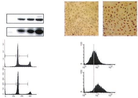
VOL. 23, 2003 ROS INFLUENCES p53-INDUCED CELL FATE DECISIONS 8577
(GSH; Sigma), 10 mM N-acetylalanine (NAA; Sigma), 0.2 g of doxorubicin per three more times with PBS and once with 0.5% Triton X-100, and then incubated
ml (Sigma), or different concentrations of tert-butyl-hydroperoxide (tBH; Sigma) for 5 min in the dark with diaminobenzidine (Sigma).
for the specified time. If chemical was to be removed, cells were washed twice
with PBS before fresh medium was added.
Adenoviral infection. An adenovirus containing p53 (Adp53), a generous gift RESULTS
of B. Vogelstein (Johns Hopkins University, Baltimore, Md.), or LacZ (AdLacZ)
were amplified as previously described (22). Cells were exposed to 10 l of the Cell fate decisions concerning senescence or apoptosis in
appropriately diluted virus stock. the same cell correlate with p53 protein levels. To study the
FACS analysis. Fluorescent stained cells were transferred to polystyrene tubes
with cell strainer caps (Falcon) and subjected to a fluorescence-activated cell
relationship between p53-induced senescence and apoptosis,
sorter (FACS) (FACScan; Beckton Dickinson) with Cell Quest 3.2 software we initially tested the p53-null EJ human bladder carcinoma
(Beckton Dickinson) for acquisition and analysis. FLIH and F12A are laser cell line with a TET-regulatable p53 expression system (EJp53)
channels representing green and red fluorescence, respectively. (19). EJp53 cells undergo senescence-like changes in morphol-
Cell cycle analysis. Cells were stained with propidium iodide (PI) with the ogy when p53 expression is activated by the removal of TET
CycleTEST Plus DNA reagent kit (Beckton Dickinson), according to the instruc-
tions provided by the manufacturer. FACS analysis was then performed.
from the culture medium (54). Growth arrest becomes irre-
versible after 4 or more days of TET removal, even after
Downloaded from http://mcb.asm.org/ on January 14, 2021 by guest
Annexin and PI fluorescent staining. Cells were washed with PBS, trypsinized,
and then incubated with annexin and PI with the Annexin-V-Fluos staining kit repression of p53, and is accompanied by the expression of the
(Boehringer Manheim), as previously reported (2, 3), followed by FACS analysis. senescence-specific marker SA--gal (14).
Cells positively stained with annexin and not PI were considered apoptotic, and Previous studies with other cancer cell lines lacking p53
cells negative for the two dyes were considered live cells.
Measurement of intracellular oxidation. Cells were incubated with 5 g of
indicated that infection with an adenovirus containing p53
dichlorofluorescin diacetate (DCF; Molecular Probes) per ml for 30 min at 37°C, (Adp53) can cause apoptosis (45). To investigate the basis for
then washed with PBS, trypsinized, and collected in 1 ml of PBS, followed by these different responses, we infected EJp53 cells cultured in
FACS analysis. Values of mean fluorescence intensity were used to plot graphs. the presence of TET with Adp53. As shown in Fig. 1A, while
Alternatively, a colorimetric assay to determine intracellular GSH (GT10; Ox- TET removal induced a pronounced arrest in both G1 and G2
ford Biomedical Research) concentrations was performed according to the man-
ufacturer’s directions. Briefly, cells were collected, washed, and incubated with
phases of the cell cycle, as previously reported (54), Adp53
provided reagents, and then samples were measured for optical density with a infection resulted in an initial arrest followed by a marked
spectrophotometer set at 400 nm. To specifically measure mitochondrial levels of increase in the apoptotic fraction (sub-G1). After 3 days,
ROS, cells were incubated with 10 g of dihydrorhodamine 123 (DHR123; around 70% of the cells infected with Adp53 were apoptotic,
Molecular Probes) per ml for 30 min at 37°C and then washed with PBS, whereas 90% of the same EJp53 cells induced by TET removal
trypsinized, and collected in 1 ml of PBS, followed by FACS analysis. Values of
mean red fluorescence intensity were used to plot graphs.
survived in a proliferation-arrested state. Infection of EJp53
Sequencing of p53. Approximately 107 EJ cells were infected with Adp53 virus cells with AdLacZ, a control adenovirus, did not induce apo-
for 24 h. DNA was extracted with phenol-chloroform, precipitated, washed twice ptosis, indicating that adenovirus infection itself was not re-
with ice-cold 70% ethanol, and then resuspended in 100 l of Tris-EDTA buffer. sponsible for this effect. Annexin V-PI staining, a method used
PCR assays were set up in a 50-l reaction volume with 50 ng of DNA, 1⫻ PCR to detect early induction of apoptosis, confirmed these results
buffer, 2 mM MgCl2, 0.11 mM deoxynucleoside triphosphate, 0.30 M of each
primer (forward primer, 5⬘-GCAGTCAGATCCTAGCGTCGAG-3⬘; reverse
(Fig. 1B), which also correlated with the morphological
primer, 5⬘-GCACCACCACACTATGTCGAAA-3⬘), and 1 U of platinum Taq changes observed under the microscope (Fig. 1C). Whereas
DNA polymerase (Invitrogen). PCR was carried out for 35 cycles, with 5 cycles TET-regulatable p53 expression resulted in elongated growth-
at 60°C and 30 cycles at 59°C. Post-PCR products were purified with a QIAquick arrested cells with the morphological features of senescence
PCR purification kit (Qiagen) according to the manufacturer’s instructions. Two (54), cells infected with Adp53 for 2 days rounded up, con-
microliters of purified PCR product (Adp53) and the p53 plasmid used in the
TET system (54) were sequenced with 2 M forward primer, the Big Dye 1.0
tracted, and lost adhesion to the plate, a result characteristic of
sequencing kit (Applied Biosystems), and an ABI 3700 DNA Analyzer. apoptosis (13). We also analyzed p53-null PC3 human prostate
Immunoblot analysis. Cells were washed twice with ice-cold PBS and lysed in cancer cells with a TET-regulatable p53 expression system
EBC buffer (50 mM Tris [pH 8], 120 mM NaCl, 0.5% NP-40, 100 mM sodium (PC3p53) (31). Similar to EJp53, PC3p53 cells underwent se-
fluoride, 2 mM sodium vanadate, 2 mM phenylmethylsulfonyl fluoride, and 10 g nescence-like changes similar to those observed with EJp53
of aprotinin/ml). Lysates were cleared by centrifugation at 20,000 ⫻ g for 20 min
at 4°C. Protein concentrations were then determined with a bicinchoninic acid
after TET removal (data not shown). In striking contrast,
protein assay (Pierce). Forty micrograms of total cell protein per sample was Adp53 infection of uninduced PC3p53 cells caused apoptosis
subjected to sodium dodecyl sulfate–12% polyacrylamide gel electrophoresis and in around 40% of the cells (Fig. 1B).
transferred to an Immobilon (Milipore) polyvinylidene difluoride filter. The To investigate the basis for the striking differences in bio-
presence of p21 was detected with the Ab-1 monoclonal antibody (Oncogene logic responses to wild-type p53 expression under these differ-
Science), and p53 was detected with the 1801 monoclonal antibody. An ECL
detection system (Amersham) was used.
ent conditions, we measured the kinetics of p53 protein in-
Senescence-associated -galactosidase (SA--gal) staining. Cells were crease as well as levels of expression after TET removal or
washed in PBS and fixed with 2% formaldehyde–0.2% glutaraldehyde in PBS for following Adp53 infection. As shown in Fig. 1D, Adp53 infec-
5 min at room temperature. Plates were stained as previously described (14). tion resulted in much higher levels of the p53 protein and of its
Immunocytochemistry. Cells were seeded onto glass coverslips and infected transcriptional target, p21, than observed in either TET-in-
with either AdLacZ or Adp53 for 12 h. Coverslips were rinsed with PBS, fixed
with 1% paraformaldehyde in PBS for 10 min at room temperature, washed two
duced EJp53 or PC3p53 cells. Of note, p53 levels observed
times with PBS, incubated in precooled ethanol-acetic acid (2:1) for 5 min at after 2 days of TET removal were similar to those observed in
⫺20°C, washed two times with PBS, incubated for 15 min in 3% hydrogen p53 wild-type cells in response to exposure to doxorubicin, a
peroxide in PBS, and then blocked in normal horse serum for 30 min at room DNA-damaging agent (Fig. 1E). Thus, the 5- to 20-fold in-
temperature. Cells were stained for p21 with monoclonal p53 antibody (2.5
crease in p53 levels observed with Adp53 infection appeared to
g/ml) overnight at 4°C. Following staining, cells were washed two times with
PBS and then incubated for 30 min at room temperature in biotinylated horse
be considerably higher than might be expected in response to
anti-mouse secondary antibody (Vector Lab). Following two washes with PBS, physiological cell stress.
cells were incubated in streptavidin-horseradish peroxidase (Zymed), washed It has recently been shown that a polymorphism in position8578 MACIP ET AL. MOL. CELL. BIOL.
Downloaded from http://mcb.asm.org/ on January 14, 2021 by guest
FIG. 1. Correlation of growth arrest or apoptosis with levels of p53 expression in the same tumor cell. (A) PI staining of EJp53 cells after p53
induction for 2 or 3 days by TET removal (left column) or after infection with a 1:10 dilution of the Adp53 viral stock (right column). Control cells
in the left column were cultured in TET for up to 3 days; control cells in the right column were infected with AdLacZ virus for up to 3 days. The
percentages of apoptotic cells (sub-G1 fraction) are indicated. (B) Annexin-PI staining of p53-induced EJp53 and PC3p53. Cells were cultured in
the presence or absence of TET for 3 days or were infected for 3 days with AdLacZ, a 1:10 dilution of Adp53 (EJp53), or undiluted Adp53
(PC3p53) in the presence of TET. Results represent the mean values of at least three different experiments, and error bars show standard
deviations. (C) Morphological changes in p53-induced EJp53 cells. Cells were cultured in the presence or the absence of TET for 4 days or infected
with Adp53 for 4 days. Cells were photographed with a Nikon Eclipse TE200 microscope (magnification, ⫻100). (D) Immunoblot analysis of p53
and p21 expression levels in lysates of control EJp53 or PC3p53 cells cultured in the presence of TET for 48 h and after p53 induction by TET
removal (48 h) or Adp53 infection (12, 24, or 48 h). (E) Immunoblot analysis of p53 expression levels in lysates of HCT116 cells after 48 or 72 h
of treatment with 0.2 g of doxorubicin (Dox) per ml, compared to protein levels in control EJp53 cells cultured in the presence of TET or after
48 h following TET removal. (F) Sequences of TET and Adp53, both showing the Pro variant in position 72.
72 affects the ability of p53 to induce apoptosis (16). The Arg72 excluding this polymorphism as being responsible for the dif-
variant has been reported to induce apoptosis more efficiently ferent cell fate outcomes observed.
than the Pro72 variant. To confirm that the different responses ROS increases in p53-induced senescent and apoptotic
observed with TET and Adp53 were due to p53 protein levels cells. Previous studies have implicated increased ROS levels as
and not to different polymorphic variants of p53, we sequenced responsible for apoptosis induced by Adp53 in DLD-1 colon
the constructs used in the TET system and Adp53. As shown in cancer cells (45). We have also shown that p21 causes p53-
Fig. 1F, both constructs had the Pro variant in position 72, thus independent ROS accumulation, which is responsible for theVOL. 23, 2003 ROS INFLUENCES p53-INDUCED CELL FATE DECISIONS 8579
Downloaded from http://mcb.asm.org/ on January 14, 2021 by guest
FIG. 2. ROS levels in EJp53 and PC3p53 cells after p53 induction. (A) ROS levels in EJp53 were measured by FACS analysis after they were
stained with the fluorescent probe DCF. The curves correspond to control cells cultured for 5 days in the presence of TET (I) or for 2 (II) or 5
days (III) after TET removal. (B) ROS levels in EJp53 and PC3p53 cells after 3-day induction of p53 by TET removal or 3 days following infection
with Adp53 at a 1:10 dilution or undiluted, respectively. Results represent mean values of three different experiments, and error bars show standard
deviations. (C) Intracellular GSH levels were measured as described in Materials and Methods after 2 days of either TET removal or Adp53
infection, compared to control cells infected with AdLacZ for 2 days. Results represent mean values of two experiments, and error bars show
standard deviations. (D) Measure of mitochondrial ROS levels with DHR123 in EJp53 cells after 48-h induction of p53 by TET removal or after
2-day infection with Adp53, compared to control cells infected with AdLacZ for 2 days. Results represent mean values of two experiments, and
error bars show standard deviations.
permanent growth arrest phenotype induced by p21 (36). To fected cells (Fig. 2C). Moreover, we stained these cells with
investigate the role of ROS in the senescent or apoptotic cell DHR123, a red fluorescent dye that has been used to measure
fates triggered by different p53 levels in EJ and PC3 cells, we mitochondrial levels of H2O2 (8). As shown in Fig. 2D, EJp53
measured ROS levels with the green fluorescent probe DCF, a cells infected with Adp53 exhibited greater increases in fluo-
marker of a change in general cellular oxidant accumulation rescence than cells with p53 induced by TET removal. Even
(50). As shown in Fig. 2A, FACS analysis of DCF-stained though DHR123 can also reflect mitochondrial accumulation
EJp53 cells revealed a progressive increase in ROS levels fol- of peroxide generated elsewhere in the cell and, therefore, is
lowing TET removal. After 3 days of induction, when senes- not an unequivocal marker for mitochondrial ROS production
cent morphological changes were first observed, ROS levels in (42), this result confirms that Adp53 produces higher oxidative
the cells had increased around twofold, with further increases intracellular increases and suggests a possible role of the mi-
by day 5, when growth arrest became irreversible. By compar- tochondria in ROS generation after p53 induction.
ison, Adp53-infected cells showed as much as an eightfold
To further establish that the different cell fate outcomes
increase in ROS levels within 3 days (Fig. 2B).
were due to p53 protein levels and not to other possible vari-
We next studied whether ROS levels correlated with the
ables between the TET and adenoviral models, we titrated the
decision between senescence and apoptosis in PCp53 cells.
amount of Adp53 used to infect EJp53 cells. As shown in Fig.
Figure 2B shows that Adp53-infected PC3p53 cells exhibited
3A, infection of EJp53 with an amount of virus 100-fold lower
much higher ROS levels than PC3p53 cells after TET removal.
As observed with EJp53 cells, the higher ROS levels correlated than that which induced apoptosis in these cells resulted in
with induction of the apoptotic response. We also tested the protein levels similar to those observed with TET removal.
effects of Adp53 in the p53-negative cancer cell line DLD-1. In This concentration was sufficient to lead to p53 expression in
this case, after 4 days of infection more than 60% of the cells most of the cells (Fig. 3B) but did not cause a significant
were apoptotic, with ROS increases of at least sixfold (data not increase in apoptosis. Instead, such cells showed a cell cycle
shown). arrest (Fig. 3C) similar to that seen with TET removal (see Fig.
To extend these results, we measured the levels of intracel- 1A). Of note, the levels of induced ROS increased around
lular GSH, one of the principal ROS buffers and a marker of twofold (Fig. 3D), which is similar to the increase observed
oxidative stress (15). Consistent with the increase in ROS, with TET removal, compared to the much higher ROS levels
GSH levels were decreased in EJp53 cells cultured in the observed with cells infected with higher concentrations of
absence of TET and more markedly decreased in Adp53-in- Adp53 (see Fig. 2B).8580 MACIP ET AL. MOL. CELL. BIOL.
Downloaded from http://mcb.asm.org/ on January 14, 2021 by guest
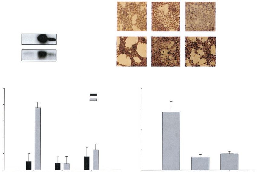
FIG. 3. Induction of growth arrest in EJp53 cells at low multiplicity
of infection with Adp53. (A) Immunoblot analysis of p53 and p21
expression levels in lysates of EJp53 cells cultured in the presence of
TET for 2 days (Control), after p53 induction by TET removal at 2 or
5 days, or at 3 days following infection with a 1:1,000 dilution of the
Adp53 viral stock. (B) Immunostaining with p53 antibody of EJp53
infected with AdLacZ (Control) or Adp53. Cells were photographed
with a Nikon Microphot-FXA microscope (magnification, ⫻200).
(C) PI staining of EJp53 cells after Adp53 infection for 3 days with a
low concentration of virus. Percentages indicate apoptotic cells
FIG. 4. Comparison of the effects of different p53 protein levels on
(sub-G1 fraction) and cells in S phase. (D) ROS levels in EJp53 3 days
arrest and apoptosis in 501T normal human fibroblasts. (A) Morpho-
after infection with AdLacZ (Control) or a low concentration of
logical changes in 501T cells infected with a retrovirus containing a
Adp53, as measured by DCF staining. Grey line indicates mean fluo-
vector (Control) or p53 (Retrop53), 1 week after puromycin selection,
rescent values in control cells.
compared to 48 h after infection with undiluted Adp53. Cells were
photographed with a Nikon Eclipse TE200 microscope (magnification,
⫻100). (B) Percentage of apoptotic cells, as measured by annexin-PI
staining, in 501T cells after infection with Retrop53 (1 week after
The p53 levels and associated ROS increases correlate with selection) or Adp53 (3 days) (grey bars). Control cells (black bars)
the induction of senescence or apoptosis in normal human correspond to infection with a retrovirus containing a vector or
fibroblasts. To investigate whether the responses of normal AdLacZ, respectively. Results reflect mean values of at least two dif-
ferent experiments, and error bars show standard deviations. (C) In-
human fibroblasts to different p53 levels were comparable to crease in intracellular ROS levels in 501T cells after infection with
those seen in cancer cells, we utilized a retrovirus containing Retrop53 or Adp53. Control cells were infected with a retrovirus
p53 (Retrop53) or Adp53. As shown in Fig. 4A and B, fibro- containing a vector or AdLacZ, respectively. Results reflect mean
blasts selected following Retrop53 infection showed growth values of at least two different experiments, and error bars show stan-
arrest, increased cell size, and intact nuclei, which are charac- dard deviations. (D) Immunoblot analysis of p53 expression levels in
lysates of 501T cells after infection with AdLacZ, Adp53 (2 days after
teristic of senescence, in the absence of detectable apoptosis. selection), Retrop53, or a vector containing retrovirus (1 week after
In contrast, Adp53 caused apoptosis in these same fibroblasts selection).
after 3 days. ROS levels induced by Retrop53 increased around
1.5-fold over vector controls, whereas Adp53 caused a greater
than threefold increase in ROS levels (Fig. 4C). These differ-
ent cell responses again correlated with the increased p53 ptosis. To test the role of both Bax and PUMA in Adp53-
protein observed (Fig. 4D). Thus, quantitative differences in induced ROS and apoptosis, we used HCT116 cells with either
p53 expression levels correlated with ROS induction and either of these genes inactivated by somatic gene targeting (59). As
senescence or apoptosis in the same normal cell. shown in Fig. 5B, immunostaining revealed a similarly high
The influence of Bax and PUMA on p53-induced ROS and percentage of cells (⬎50%) expressing p53 in each cell line in
apoptosis. The proapoptotic Bax gene is a Bcl-2 family mem- response to Adp53. Both Bax⫺/⫺ and PUMA⫺/⫺ cells were
ber that has been reported to increase mitochondrial mem- significantly more resistant to apoptosis after infection with
brane permeability (26, 37). It has been shown to be upregu- Adp53 than wild-type parental cells, a result that is consistent
lated by DNA damage and p53 (39), and the requirement for with previous reports (Fig. 5C) (59). Of note, the accumulation
Bax in p53-induced apoptosis has been proposed to be cell of ROS in response to p53 was also markedly reduced in the
context dependent (4, 7, 40). Moreover, EJp53 cells infected absence of either Bax or PUMA (Fig. 5D). These findings
with Adp53 exhibited higher Bax protein levels than observed further correlate with the magnitude of ROS accumulation in
in EJp53 cells after TET removal (Fig. 5A). PUMA is a BH3- response to p53 in determining apoptosis and implicate Bax
only protein that binds to Bcl-2 and Bcl-XL and is directly and PUMA as important effectors of p53-induced ROS, sug-
regulated by p53 (41, 57). When overexpressed, PUMA causes gesting a mitochondrial role in generating ROS after p53 up-
cytochrome c release from the mitochondria and induces apo- regulation.VOL. 23, 2003 ROS INFLUENCES p53-INDUCED CELL FATE DECISIONS 8581
Downloaded from http://mcb.asm.org/ on January 14, 2021 by guest
FIG. 5. Comparison of ROS levels and apoptosis in Adp53-infected Bax- and PUMA-null cells. (A) Immunoblot analysis of p53 and Bax
expression levels in lysates of EJp53 cells after infection with a 1:10 dilution of Adp53 (2 days) or TET removal for 2 days. (B) Immunostaining
with p53 antibody of HCT116 cells (wild type, Bax⫺/⫺, or PUMA⫺/⫺) infected with AdLacZ (Control) or undiluted Adp53. Cells were
photographed with a Nikon Microphot-FXA microscope (magnification, ⫻200). (C) Apoptosis levels in wild-type, Bax⫺/⫺, and PUMA⫺/⫺ HCT116
cells infected with AdLacZ or Adp53 for 3 days, as measured by Annexin-PI staining. (D) Relative ROS levels in wild-type, Bax⫺/⫺, and PUMA⫺/⫺
HCT116 cells infected with Adp53, compared to ROS levels in the same cells infected with AdLacZ. Results represent mean values of at three
different experiments, and error bars show standard deviations. wt, wild type.
ROS inhibition partially blocks senescence and apoptosis in without antioxidant activity, had no effect on colony recovery
EJp53 cells. To investigate the contribution of ROS to p53- (data not shown). All of these results implied that ROS accu-
induced senescence and apoptosis, we tested whether the an- mulation was an important mediator of p53-induced senes-
tioxidant N-acetylcysteine (NAC), a reduced GSH provider cence.
and a direct scavenger of ROS (10, 47), was able to protect We next tested the effects of antioxidants on p53-induced
cells from the permanent growth arrest phenotype. Around apoptosis. As shown in Fig. 6D, treatment with NAC did not
100 EJp53 cells were plated, and p53 was induced by TET affect p53 or Bax protein levels induced by Adp53 infection.
removal for up to 4 days. TET was then added back to the Treatment of EJp53 with either 10 mM NAC or 1 mM reduced
medium, and cells were cultured for another 2 weeks for anal- GSH prior to Adp53 infection significantly inhibited the mag-
ysis of colony formation. It has been shown that the longer nitude of the increase in ROS levels compared to the increase
such cells are exposed to p53, the fewer colonies that can be observed in the absence of antioxidants, whereas the inactive
recovered following p53 down-regulation (54). Figure 6A NAC analogue NAA had no effect on ROS accumulation (Fig.
shows that the addition of 10 mM NAC to the culture medium 6E). NAC and GSH treatment also resulted in a statistically
increased the number of cells able to escape p53-induced se- significant increase in surviving cells after 24 h (Fig. 6F). Be-
nescence, suggesting that inhibition of ROS protected them cause at higher concentrations antioxidants had adverse effects
from this outcome. The fraction of cells that scored positive for on cell survival, it was not possible to determine whether fur-
the senescence marker SA--gal was also reduced in EJp53- ther neutralization of ROS could be achieved, and if so,
induced cells cultured in the presence of NAC (Fig. 6B). DCF whether the magnitude of the p53 apoptotic response would be
staining of these cells confirmed that NAC treatment signifi- further reduced.
cantly reduced ROS accumulation (Fig. 6C). NAC did not Exogenous ROS cooperates with physiological levels of in-
induce cell death at the concentration utilized (see Fig. 6F). As duced p53 to convert a senescent response into apoptosis. The
a further control, 10 mM NAA, a structural analogue of NAC above results demonstrated that high, nonphysiological levels8582 MACIP ET AL. MOL. CELL. BIOL.
Downloaded from http://mcb.asm.org/ on January 14, 2021 by guest
FIG. 7. Comparison of the effects of exogenous ROS on p53-in-
duced or uninduced cells. (A) Percentages of live cells in EJp53 24 h
after treatment with the oxidant tBH. Cells cultured in the presence
(black bars) or absence (grey bars) of TET for 24 h were then treated
FIG. 6. Effect of antioxidants on p53-induced senescence and apo- with different concentrations of tBH for 2 h. (B) Percentages of live
ptosis. (A) Representative plates from a colony formation assay, in cells in PCp53 24 h after treatment with tBH. Cells cultured in the
which around 100 EJp53 cells were plated. Cells were maintained in presence (black bars) or absence (grey bars) of TET for 24 h were then
the absence of TET for 0 (Control), 2, 3, or 4 days, and then TET was treated with different concentrations of tBH for 2 h. Results represent
added again to the medium. Cells were cultured for 14 more days mean values of four different experiments, and error bars show stan-
followed by 10% formalin fixation and Giemsa staining. Where indi- dard deviations. (C) Immunoblot analysis of p53 and Bax expression
cated (plus sign), 10 mM NAC was added at the beginning of the levels in lysates of EJp53 cells cultured for 24 h in the presence or
experiment and at each subsequent medium change, until TET was absence of TET, followed by treatment for 2 h with 100 M tBH.
added back to the medium. (B) Percentage of SA--gal-positive cells
in EJp53 control cells cultured in the presence of TET and after 5 days
of p53 induction in the presence or absence of 10 mM NAC. (C) ROS
levels in the same cells were measured by DCF staining. Results rep- of p53 were needed to induce apoptosis in the same cells in
resent mean values of at least two different experiments, and error bars which physiological levels of this tumor suppressor caused per-
show standard deviations. (D) Immunoblot analysis of p53 and p21
expression levels in lysates of EJp53 cells infected with Adp53 in the
manent growth arrest. Moreover, these outcomes could be
presence or absence of 10 mM NAC. (E) ROS levels in EJp53 cells directly correlated with the magnitude of p53-induced ROS.
cultured in the presence of TET (Control) and infected with Adp53 in Thus, we reasoned that exposure of TET-regulated EJp53 cells
the presence or absence of 10 mM NAC, 1 mM GSH or 10 mM NAA. to an exogenous source of ROS might be sufficient to alter
(F) Anenxin-PI staining showing the percentage of live cells in control their fate so as to convert a senescent response to apoptosis.
EJp53 cultured in presence of TET for 2 days, with 10 mM NAC or 1
M GSH added to the media for the same period, and following infec- To test this possibility, we used tBH, an organic hydroperoxide
tion with Adp53 for 3 days in the presence or absence of the same that causes oxidative stress (58). In combination with physio-
concentrations of NAC or GSH. Results represent mean values of logical levels of p53 induced by 24 h of TET removal, tBH was
three different experiments, and error bars show standard deviations. able to induce apoptosis in EJp53 cells that would otherwise
ⴱ, statistical analysis of Adp53 versus Adp53 ⫹ NAC (P ⬍ 0.00001); ⴱⴱ,
statistical analysis of Adp53 versus Adp53 ⫹ GSH (P ⬍ 0.000001).
have undergone the senescence process (Fig. 7A). Moreover,
such cells were significantly more sensitive to tBH-induced cell
death than uninduced cells. Whereas treatment with 50 MVOL. 23, 2003 ROS INFLUENCES p53-INDUCED CELL FATE DECISIONS 8583
tBH resulted in survival of less than 20% of the p53-induced logical expression levels, there was a reproducible two- to five-
EJp53 cells, greater than 500 M was required to cause com- fold increase in ROS accumulation. Moreover, NAC, a free
parable cell death in uninduced EJp53 cells. As shown in Fig. radical scavenger, was able both to ameliorate ROS accumu-
7B, PC3p53 cells were less sensitive than EJp53 cells to tBH- lation and to partially rescue the same cells from the perma-
induced apoptosis. However, induction of p53 to physiological nent growth arrest phenotype. These findings implicate ROS
levels in these TET-regulatable cells also increased signifi- accumulation in determining p53-induced senescence. At the
cantly their sensitivity to the apoptotic outcome. These results high p53 levels capable of inducing an apoptotic response, we
establish that exogenous ROS can cooperate with physiological observed a more rapid appearance and a greater magnitude of
levels of p53 to convert a senescence response to apoptosis. ROS accumulation in the same cells that underwent p53-in-
Oxidative stress is known to induce p53 expression (11), and duced senescence at physiological p53 expression levels.
we and others have shown that p53 induces ROS (45). To test Previous studies have indicated that Adp53 induces ROS
the possibility of the existence of a positive feedback loop associated with decreased levels of cardiolipin, a component of
between ROS and p53, we measured p53 protein levels in the mitochondrial membrane that is sensitive to oxidative dam-
induced and uninduced EJp53 cells after tBH treatment. At age (45). Moreover, overexpression of ferredoxin reductase, a
Downloaded from http://mcb.asm.org/ on January 14, 2021 by guest
sublethal concentrations, tBH did not alter p53 or Bax protein p53-induced gene, resulted in its localization to the mitochon-
levels (Fig. 7C). These results indicate that oxidants do not drial membrane associated with mitochondrial accumulation
change p53 levels or activity with respect to Bax induction in of ROS (23). In our present studies, an Adp53 apoptotic re-
EJp53 cells. sponse could be ameliorated by NAC and GSH but not by
inactive analogues, and ROS inhibition by antioxidants corre-
lated with a decreased magnitude of apoptosis. All these re-
DISCUSSION
sults strongly imply that the level of p53 protein overexpression
The tumor suppressor p53 is an important sensor of cellular and the p53-induced elevation of intracellular ROS impor-
stress conditions, including DNA damage, hypoxia, survival tantly influence the decision between senescence and apoptosis
factor deprivation, mitogenic oncogenes, and telomere short- in a given cell.
ening. Various outcomes can be observed following p53 acti- It has been reported that p53 can induce the expression of
vation in response to such stresses, including reversible growth Bax, a proapoptotic gene product of the Bcl-2 family. Bax has
arrest, permanent growth arrest or apoptosis (27, 32, 34). In been proposed to control mitochondrial membrane permeabil-
fact, it is thought that the need for a cell to escape these cell ity either by forming a channel in the outer mitochondrial
fates in order to become a cancer cell accounts for the high membrane or by regulating the opening and closing of the
frequency at which p53 function is lost in cancers. Thus, elu- permeability transition pore (37). After the disruption of the
cidation of the p53 signaling pathways involved in each of these membrane, mitochondrial proteins including cytochrome c and
different cellular responses has potentially important implica- Smac/DIABLO are released into the cytosol, activating
tions for understanding cellular aging and cancer as well as for caspase-9 and inhibiting antiapoptotic proteins, which leads to
therapeutic approaches aimed at counteracting these patho- activation of the downstream effector caspases of the apoptotic
logical processes. In the present studies, we manipulated p53 cascade (40). The requirement of Bax by p53 to induce apo-
expression levels in different cell types in the absence of other ptosis has been proposed to be context dependent. Previous
stimuli in an effort to identify those p53 functions critically studies have shown that the absence of Bax did not suppress
involved in determining cell fate decisions with respect to se- ␥-irradiation-induced p53-dependent apoptosis in mouse lym-
nescence and apoptosis. phocytes (28). On the other hand, chemotherapy-induced p53-
At physiological levels of expression similar to those induced dependent apoptosis in Bax⫺/⫺ primary mouse embryo fibro-
by DNA damage, p53 expression alone induced a growth arrest blasts was shown to be attenuated, although not completely
phenotype in EJ cells, which under conditions of TET-regu- suppressed (38). In accordance with these findings, we showed
lated expression became permanent by 4 to 5 days despite that the absence of Bax impaired an Adp53-mediated apopto-
subsequent p53 down-regulation. In contrast, a p53 adenovirus tic response. Our results further show that the absence of Bax
vector, which resulted in p53 protein expression levels that significantly inhibited the accumulation of intracellular ROS.
were at least 10- to 20-fold greater than could be observed in Similar results were observed in the absence of PUMA.
DNA-damaged wild-type p53-containing cells, induced rapid PUMA has been reported to be an exclusively mitochondrial
and efficient apoptosis. Our results show that when p53 was protein that binds to members of the Bcl family (57). These
induced in the same cell context and in the absence of other data, together with our results showing increased mitochon-
stimuli, the magnitude of p53 expression alone determined the drial oxidation, indicate important roles of Bax and PUMA in
decision with respect to these cell fates; this result is consistent ROS accumulation in response to p53 upregulation. Since
with previous reports that demonstrate that the intracellular adaptive changes may occur in cells in which there has been a
level of p53 can influence the decision between arrest and knockout expression of any given gene, further studies will be
apoptosis (12). These findings help to explain the differences in needed to fully elucidate the roles of Bax and PUMA in the
cell fates observed in various studies where Adp53 was com- p53-dependent elevation of intracellular ROS.
pared to other p53 expression systems. A major question in cancer therapeutics is the impact in a
It has been previously shown that p53 induces ROS accu- high fraction of tumors of nonfunctional p53 on the ability of
mulation (45), and a number of genes induced by p53 are specific agents to selectively target the tumor as opposed to
associated with the metabolism of ROS (45). We observed that normal cells. Our observations that p53 induces ROS accumu-
in cells undergoing senescence in response to p53 at physio- lation, which plays an important role in both senescence and8584 MACIP ET AL. MOL. CELL. BIOL.
3. Bartkowiak, D., S. Hogner, H. Baust, W. Nothdurft, and E. M. Rottinger.
1999. Comparative analysis of apoptosis in HL60 detected by annexin-V and
fluorescein-diacetate. Cytometry 37:191–196.
4. Bates, S., and K. H. Vousden. 1999. Mechanisms of p53-mediated apoptosis.
Cell Mol. Life Sci. 55:28–37.
5. Bond, J. A., F. S. Wyllie, and D. Wynford-Thomas. 1994. Escape from
senescence in human diploid fibroblasts induced directly by mutant p53.
Oncogene 9:1885–1889.
6. Buckbinder, L., R. Talbott, S. Velasco-Miguel, I. Takenaka, B. Faha, B. R.
Seizinger, and N. Kley. 1995. Induction of the growth inhibitor IGF-binding
protein 3 by p53. Nature 377:646–649.
7. Burns, T. F., and W. S. El-Deiry. 1999. The p53 pathway and apoptosis.
J. Cell Physiol. 181:231–239.
8. Cai, J., and D. P. Jones. 1998. Superoxide in apoptosis: mitochondrial gen-
eration triggered by cytochrome c loss. J. Biol. Chem. 273:11401–11404.
9. Chen, Q., and B. N. Ames. 1994. Senescence-like growth arrest induced by
FIG. 8. The balance between all the oxidants (like p53, certain hydrogen peroxide in human diploid fibroblast F65 cells. Proc. Natl. Acad.
kinds of stress, or chemotherapeutics) and antioxidants present in the Sci. USA 91:4130–4134.
Downloaded from http://mcb.asm.org/ on January 14, 2021 by guest
cell in a given moment can determine, by controlling intracellular ROS 10. Chen, Q., A. Fischer, J. D. Reagan, L. J. Yan, and B. N. Ames. 1995.
levels, whether a cell fate response would be senescence or apoptosis. Oxidative DNA damage and senescence of human diploid fibroblast cells.
Apoptosis is induced only if intracellular oxidation reaches a certain Proc. Natl. Acad. Sci. USA 92:4337–4341.
threshold. 11. Chen, Q. M., J. C. Bartholomew, J. Campisi, M. Acosta, J. D. Reagan, and
B. N. Ames. 1998. Molecular analysis of H2O2-induced senescent-like growth
arrest in normal human fibroblasts: p53 and Rb control G1 arrest but not cell
replication. Biochem. J. 332:43–50.
apoptosis cell fate decisions, led us to ask whether p53 at 12. Chen, X., L. J. Ko, L. Jayaraman, and C. Prives. 1996. p53 levels, functional
domains, and DNA damage determine the extent of the apoptotic response
physiological levels induced by cellular stresses could cooper- of tumor cells. Genes Dev. 10:2438–2451.
ate with an exogenous ROS source to favor an apoptotic out- 13. Coleman, M. L., and M. F. Olson. 2002. Rho GTPase signalling pathways in
come. By the use of TET-regulatable p53-containing tumor the morphological changes associated with apoptosis. Cell Death Differ.
9:493–504.
cells, we observed that this was indeed the case. At physiolog- 14. Dimri, G. P., X. Lee, G. Basile, M. Acosta, G. Scott, C. Roskelley, E. E.
ical p53 protein levels capable of triggering senescence, an Medrano, M. Linskens, I. Rubelj, O. Pereira-Smith, M. Peacocke, and J.
Campisi. 1995. A biomarker that identifies senescent human cells in culture
apoptotic response was observed when the p53-induced in- and in aging skin in vivo. Proc. Natl. Acad. Sci. USA 92:9363–9367.
crease in intracellular ROS levels was complemented by an 15. Dolphin, D., R. Poulson, and O. Avramovic (ed.). 1989. Glutathione: chem-
exogenous ROS source, which itself was not able to induce ical, biochemical, and medical aspects. John Wiley & Sons, New York, N.Y.
16. Dumont, P., J. I. Leu, A. C. Della Pietra III, D. L. George, and M. Murphy.
apoptosis in the absence of p53. 2003. The codon 72 polymorphic variants of p53 have markedly different
These findings suggest the existence of a threshold of cellu- apoptotic potential. Nat. Genet. 33:357–365.
lar oxidation above which the apoptotic program is initiated. 17. Fang, L., M. Igarashi, J. Leung, M. M. Sugrue, S. W. Lee, and S. A.
Aaronson. 1999. p21Waf1/Cip1/Sdi1 induces permanent growth arrest with
This threshold may vary between cell types or as a function of markers of replicative senescence in human tumor cells lacking functional
other physiological factors. However, the balance between all p53. Oncogene 18:2789–2797.
18. Gewirtz, D. A. 1999. A critical evaluation of the mechanisms of action
the ROS inducers and the antioxidants present in the cell at a proposed for the antitumor effects of the anthracycline antibiotics adriamy-
given moment is likely crucial in determining cell fate decisions cin and daunorubicin. Biochem. Pharmacol. 57:727–741.
(Fig. 8). Certain agents used in cancer therapy, such as ␥-irra- 19. Gossen, M., and H. Bujard. 1992. Tight control of gene expression in mam-
malian cells by tetracycline-responsive promoters. Proc. Natl. Acad. Sci.
diation and doxorubicin, have the ability to induce ROS (18). USA 89:5547–5551.
Based on our present findings, those therapeutic agents that 20. Hagen, T. M., D. L. Yowe, J. C. Bartholomew, C. M. Wehr, K. L. Do, J. Y.
cooperate with p53 in ROS generation would likely be more Park, and B. N. Ames. 1997. Mitochondrial decay in hepatocytes from old
rats: membrane potential declines, heterogeneity and oxidants increase.
toxic for wild-type p53-containing normal than p53-negative Proc. Natl. Acad. Sci. USA 94:3064–3069.
tumor cells. These results provide a rationale for identifying 21. Hayflick, L., and P. Moorehead. 1961. The serial cultivation of human dip-
therapeutic agents that do not complement p53 in ROS gen- loid strains. Exp. Cell Res. 25:585–621.
22. He, T. C., S. Zhou, L. T. da Costa, J. Yu, K. W. Kinzler, and B. Vogelstein.
eration, which would ameliorate the cytotoxic side effects of 1998. A simplified system for generating recombinant adenoviruses. Proc.
such chemicals in normal cells. Natl. Acad. Sci. USA 95:2509–2514.
23. Hwang, P. M., F. Bunz, J. Yu, C. Rago, T. A. Chan, M. P. Murphy, G. F.
Kelso, R. A. Smith, K. W. Kinzler, and B. Vogelstein. 2001. Ferredoxin
ACKNOWLEDGMENTS reductase affects p53-dependent, 5-fluorouracil-induced apoptosis in colo-
rectal cancer cells. Nat. Med. 7:1111–1117.
We thank B. Vogelstein (Johns Hopkins University, Baltimore, Md.) 24. Israeli, D., E. Tessler, Y. Haupt, A. Elkeles, S. Wilder, R. Amson, A. Teler-
for generously providing HCT116 Bax⫺/⫺ cells and Adp53, I. George man, and M. Oren. 1997. A novel p53-inducible gene, PAG608, encodes a
from the Mount Sinai Flow Cytometry Core Facility, and L. Goldin nuclear zinc finger protein whose overexpression promotes apoptosis.
and J. Leung for technical assistance. EMBO J. 16:4384–4392.
S. Macip received support from the Ministerio de Educacion y 25. Johnson, T. M., Z. X. Yu, V. J. Ferrans, R. A. Lowenstein, and T. Finkel.
Cultura of Spain and is a recipient of a postdoctoral fellowship from 1996. Reactive oxygen species are downstream mediators of p53-dependent
the Forchheimer foundation. P. Berggren received support from the apoptosis. Proc. Natl. Acad. Sci. USA 93:11848–11852.
Swedish Cancer Society (Cancerfonden 477-BO2-01SAA). This work 26. Johnstone, R. W., A. A. Ruefli, and S. W. Lowe. 2002. Apoptosis: a link
between cancer genetics and chemotherapy. Cell 108:153–164.
was supported in part by National Institutes of Health grants CA80058
27. Kastan, M. B., O. Onyekwere, D. Sidransky, B. Vogelstein, and R. W. Craig.
and CA85214 (to S.A.A.) and CA78356 and CA82211 (to S.W.L.). 1991. Participation of p53 protein in the cellular response to DNA damage.
Cancer Res. 51:6304–6311.
REFERENCES 28. Knudson, C. M., K. S. Tung, W. G. Tourtellotte, G. A. Brown, and S. J.
1. Attardi, L. D., E. E. Reczek, C. Cosmas, E. G. Demicco, M. E. McCurrach, Korsmeyer. 1995. Bax-deficient mice with lymphoid hyperplasia and male
S. W. Lowe, and T. Jacks. 2000. PERP, an apoptosis-associated target of p53, germ cell death. Science 270:96–99.
is a novel member of the PMP-22/gas3 family. Genes Dev. 14:704–718. 29. Kurimasa, A., H. Ouyang, L. J. Dong, S. Wang, X. Li, C. Cordon-Cardo, D. J.
2. Aubry, J. P., A. Blaecke, S. Lecoanet-Henchoz, P. Jeannin, N. Herbault, G. Chen, and G. C. Li. 1999. Catalytic subunit of DNA-dependent protein
Caron, V. Moine, and J. Y. Bonnefoy. 1999. Annexin V used for measuring kinase: impact on lymphocyte development and tumorigenesis. Proc. Natl.
apoptosis in the early events of cellular cytotoxicity. Cytometry 37:197–204. Acad. Sci. USA 96:1403–1408.VOL. 23, 2003 ROS INFLUENCES p53-INDUCED CELL FATE DECISIONS 8585
30. Lee, A. C., B. E. Fenster, H. Ito, K. Takeda, N. S. Bae, T. Hirai, Z. X. Yu, V. J. is a transcriptional target of p53 in DNA damage-induced apoptosis. Cancer
Ferrans, B. H. Howard, and T. Finkel. 1999. Ras proteins induce senescence Res. 61:6660–6664.
by altering the intracellular levels of reactive oxygen species. J. Biol. Chem. 47. Roederer, M., F. J. Staal, P. A. Raju, S. W. Ela, and L. A. Herzenberg. 1990.
274:7936–7940. Cytokine-stimulated human immunodeficiency virus replication is inhibited
31. Lee, S. W., L. Fang, M. Igarashi, T. Ouchi, K. P. Lu, and S. A. Aaronson. by N-acetyl-L-cysteine. Proc. Natl. Acad. Sci. USA 87:4884–4888.
2000. Sustained activation of Ras/Raf/mitogen-activated protein kinase cas- 48. Rogan, E. M., T. M. Bryan, B. Hukku, K. Maclean, A. C. Chang, E. L. Moy,
cade by the tumor suppressor p53. Proc. Natl. Acad. Sci. USA 97:8302–8305. A. Englezou, S. G. Warneford, L. Dalla-Pozza, and R. R. Reddel. 1995.
32. Levine, A. J. 1997. p53, the cellular gatekeeper for growth and division. Cell Alterations in p53 and p16INK4 expression and telomere length during
88:323–331. spontaneous immortalization of Li-Fraumeni syndrome fibroblasts. Mol.
33. Lin, Y., W. Ma, and S. Benchimol. 2000. Pidd, a new death-domain-contain- Cell. Biol. 15:4745–4753.
ing protein, is induced by p53 and promotes apoptosis. Nat. Genet. 26:122– 49. Rowan, S., R. L. Ludwig, Y. Haupt, S. Bates, X. Lu, M. Oren, and K. H.
127. Vousden. 1996. Specific loss of apoptotic but not cell-cycle arrest function in
34. Lowe, S. W., E. M. Schmitt, S. W. Smith, B. A. Osborne, and T. Jacks. 1993. a human tumor derived p53 mutant. EMBO J. 15:827–838.
p53 is required for radiation-induced apoptosis in mouse thymocytes. Nature 50. Royall, J. A., and H. Ischiropoulos. 1993. Evaluation of 2⬘,7⬘-dichlorofluo-
362:847–849. rescin and dihydrorhodamine 123 as fluorescent probes for intracellular
35. Ludwig, R. L., S. Bates, and K. H. Vousden. 1996. Differential activation of H2O2 in cultured endothelial cells. Arch. Biochem. Biophys. 302:348–355.
target cellular promoters by p53 mutants with impaired apoptotic function. 51. Sager, R. 1991. Senescence as a mode of tumor suppression. Environ. Health
Mol. Cell. Biol. 16:4952–4960. Perspect. 93:59–62.
Downloaded from http://mcb.asm.org/ on January 14, 2021 by guest
36. Macip, S., M. Igarashi, L. Fang, A. Chen, Z. Q. Pan, S. W. Lee, and S. A. 52. Serrano, M., A. W. Lin, M. E. McCurrach, D. Beach, and S. W. Lowe. 1997.
Aaronson. 2002. Inhibition of p21-mediated ROS accumulation can rescue Oncogenic ras provokes premature cell senescence associated with accumu-
p21-induced senescence. EMBO J. 21:2180–2188. lation of p53 and p16INK4a. Cell 88:593–602.
37. Martinou, J. C., and D. R. Green. 2001. Breaking the mitochondrial barrier.
53. Sionov, R. V., and Y. Haupt. 1999. The cellular response to p53: the decision
Nat. Rev. Mol. Cell Biol. 2:63–67.
between life and death. Oncogene 18:6145–6157.
38. McCurrach, M. E., T. M. Connor, C. M. Knudson, S. J. Korsmeyer, and
S. W. Lowe. 1997. bax-deficiency promotes drug resistance and oncogenic 54. Sugrue, M. M., D. Y. Shin, S. W. Lee, and S. A. Aaronson. 1997. Wild-type
transformation by attenuating p53-dependent apoptosis. Proc. Natl. Acad. p53 triggers a rapid senescence program in human tumor cells lacking func-
Sci. USA 94:2345–2349. tional p53. Proc. Natl. Acad. Sci. USA 94:9648–9653.
39. Miyashita, T., and J. C. Reed. 1995. Tumor suppressor p53 is a direct 55. von Zglinicki, T., G. Saretzki, W. Docke, and C. Lotze. 1995. Mild hyperoxia
transcriptional activator of the human bax gene. Cell 80:293–299. shortens telomeres and inhibits proliferation of fibroblasts: a model for
40. Moll, U. M., and A. Zaika. 2001. Nuclear and mitochondrial apoptotic senescence? Exp. Cell Res. 220:186–193.
pathways of p53. FEBS Lett. 493:65–69. 56. Wu, G. S., T. F. Burns, E. R. McDonald III, W. Jiang, R. Meng, I. D. Krantz,
41. Nakano, K., and K. H. Vousden. 2001. PUMA, a novel proapoptotic gene, is G. Kao, D. D. Gan, J. Y. Zhou, R. Muschel, S. R. Hamilton, N. B. Spinner,
induced by p53. Mol. Cell 7:683–694. S. Markowitz, G. Wu, and W. S. el-Deiry. 1997. KILLER/DR5 is a DNA
42. Nemoto, S., K. Takeda, Z. X. Yu, V. J. Ferrans, and T. Finkel. 2000. Role for damage-inducible p53-regulated death receptor gene. Nat. Genet. 17:141–
mitochondrial oxidants as regulators of cellular metabolism. Mol. Cell. Biol. 143.
20:7311–7318. 57. Yu, J., L. Zhang, P. M. Hwang, K. W. Kinzler, and B. Vogelstein. 2001.
43. Oda, E., R. Ohki, H. Murasawa, J. Nemoto, T. Shibue, T. Yamashita, T. PUMA induces the rapid apoptosis of colorectal cancer cells. Mol. Cell
Tokino, T. Taniguchi, and N. Tanaka. 2000. Noxa, a BH3-only member of 7:673–682.
the Bcl-2 family and candidate mediator of p53-induced apoptosis. Science 58. Zamzami, N., I. Marzo, S. A. Susin, C. Brenner, N. Larochette, P. Marchetti,
288:1053–1058. J. Reed, R. Kofler, and G. Kroemer. 1998. The thiol crosslinking agent
44. Oda, K., H. Arakawa, T. Tanaka, K. Matsuda, C. Tanikawa, T. Mori, H. diamide overcomes the apoptosis-inhibitory effect of Bcl-2 by enforcing
Nishimori, K. Tamai, T. Tokino, Y. Nakamura, and Y. Taya. 2000. p53AIP1, mitochondrial permeability transition. Oncogene 16:1055–1063.
a potential mediator of p53-dependent apoptosis, and its regulation by Ser- 59. Zhang, L., J. Yu, B. H. Park, K. W. Kinzler, and B. Vogelstein. 2000. Role of
46-phosphorylated p53. Cell 102:849–862. BAX in the apoptotic response to anticancer agents. Science 290:989–992.
45. Polyak, K., Y. Xia, J. L. Zweier, K. W. Kinzler, and B. Vogelstein. 1997. A 60. Zhu, J., S. Zhang, J. Jiang, and X. Chen. 2000. Definition of the p53
model for p53-induced apoptosis. Nature 389:300–305. functional domains necessary for inducing apoptosis. J. Biol. Chem. 275:
46. Robles, A. I., N. A. Bemmels, A. B. Foraker, and C. C. Harris. 2001. APAF-1 39927–39934.You can also read