Critical immunosuppressive effect of MDSC derived exosomes in the tumor microenvironment
←
→
Page content transcription
If your browser does not render page correctly, please read the page content below
ONCOLOGY REPORTS 45: 1171-1181, 2021

              Critical immunosuppressive effect of MDSC‑derived
                    exosomes in the tumor microenvironment
              MOHAMMAD H. RASHID1,2, THAIZ F. BORIN1, ROXAN ARA1, RAZIYE PIRANLIOGLU1,
                 BHAGELU R. ACHYUT3, HASAN KORKAYA4, YUTAO LIU5 and ALI S. ARBAB1

      1
       Laboratory of Tumor Angiogenesis, Georgia Cancer Center, Department of Biochemistry and Molecular Biology,
          Augusta University, Augusta, GA 30912; 2Nanomedicine Research Center, Department of Neurosurgery,
  Cedars‑Sinai Medical Center, Los Angeles, CA 90048; 3Cancer Animal Models Shared Resource, Winship Cancer Institute,
       Emory University, Atlanta, GA 30322; 4Molecular Oncology and Biomarkers Program, Georgia Cancer Center,
         Department of Biochemistry and Molecular Biology, Augusta University; 5Department of Cellular Biology
                 and Anatomy, Medical College of Georgia, Augusta University, Augusta, GA 30912, USA

                                       Received June 30, 2020; Accepted December 9, 2020

                                                     DOI: 10.3892/or.2021.7936

Abstract. Myeloid‑derived suppressor cells (MDSCs) are             MDSCs are also implemented by MDSC‑derived exosomes
an indispensable component of the tumor microenviron-              which would open up a new avenue of MDSC research and
ment (TME). Along with the role of MDSC immunosuppression          MDSC‑targeted therapy.
and antitumor immunity, MDSCs facilitate tumor growth,
differentiation, and metastasis in several ways that are yet to    Introduction
be explored. Like any other cell type, MDSCs also release a
tremendous number of exosomes, or nanovesicles of endo-            Apart from cancer cells, the tumor microenvironment (TME)
somal origin, that participate in intercellular communications     consists of heterogeneous host cells of the immune system,
by dispatching biological macromolecules. There have been          the tumor vasculature and lymphatics, fibroblasts, pericytes,
no investigational studies conducted to characterize the role      and sometimes adipocytes (1). Myeloid‑derived suppressor
of MDSC‑derived exosomes (MDSC exo) in modulating the              cells (MDSCs) are crucial components of the TME that
TME. In this study, we isolated MDSC exo and demonstrated          play a pivotal role in tumor growth, neovascularization, and
that they carry a significant level of proteins that play an       metastasis (2‑4). MDSCs are a group of vastly heterogeneous
indispensable role in tumor growth, invasion, angiogenesis,        immunosuppressive cells derived from immature myeloid
and immunomodulation. We observed a higher yield and more          progenitors that have been linked to poor patient prognosis (5).
substantial immunosuppressive potential of exosomes isolated       Typically, immature myeloid cells traverse to the peripheral
from MDSCs in the primary tumor area than those in the             organs after originating from bone marrow and rapidly
spleen or bone marrow. Our in vitro data suggest that MDSC         mature into macrophages, dendritic cells, or granulocytes
exo are capable of hyper‑activating or exhausting CD8 T‑cells      (neutrophils, eosinophils, and basophils) (6). That said, in the
and induce reactive oxygen species production that elicits         tumor condition, multifarious factors that are present in the
activation‑induced cell death. We confirmed the depletion of       TME prevent the differentiation of these immature myeloid
CD8 T‑cells in vivo by treating mice with MDSC exo. We also        cells and instigate their actuation into an immunosuppressive
observed a reduction in pro‑inflammatory M1‑macrophages            phenotype (7). MDSCs are usually divided into two subpopu-
in the spleen of those animals. Our results indicate that the      lations: gMDSCs (granulocytic, CD11b +Ly6G +Ly6C low),
immunosuppressive and tumor‑promoting functions of                 which are identical to neutrophils, and mMDSCs (monocytic,
                                                                   CD11b+Ly6G‑Ly6Chi), which are consistent with monocytes
                                                                   with respect to morphology and phenotype (8,9).
                                                                       There is growing evidence that MDSCs harness various
                                                                   immune and nonimmune mechanisms to promote tumor
Correspondence to: Dr Ali S. Arbab, Laboratory of Tumor
                                                                   development. MDSCs inhibit adaptive antitumor immunity by
Angiogenesis, Georgia Cancer Center, Department of Biochemistry
and Molecular Biology, Augusta University, 1410 Laney Walker
                                                                   inhibiting T‑cell activation and function (T‑cell receptor down-
Blvd., Room CN 3315, Augusta, GA 30912, USA                        regulation, T‑cell cell cycle inhibition, and immune checkpoint
E‑mail: aarbab@augusta.edu                                         blockade) (9), and by driving and recruiting T regulatory cells.
                                                                   Immunosuppression by MDSCs is also mediated by the genera-
Key words: exosomes, myeloid‑derived suppressor cells, tumor       tion of reactive oxygen species (ROS) (10) and cytokine release
microenvironment, CD8 T cells, activation‑induced cell death       [interleukin (IL)‑10 and tumor growth factor (TGF)‑β] (11,12),
                                                                   in conjunction with arginine depletion (13). They inhibit
                                                                   innate immunity by polarizing macrophages toward a type 2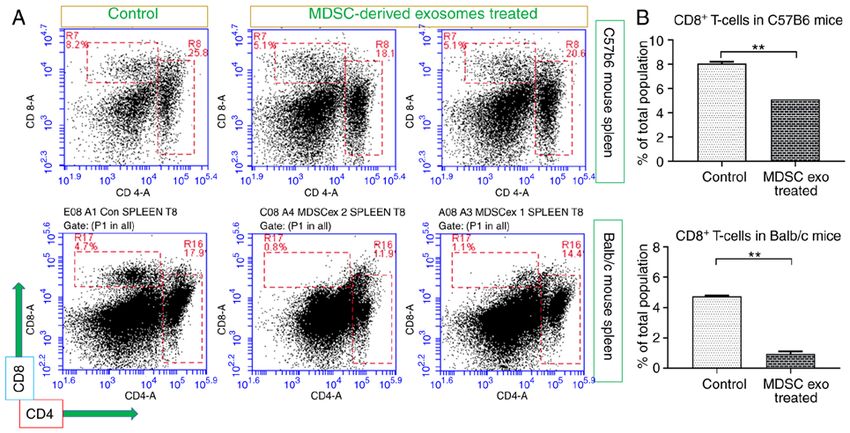
1172                        RASHID et al: IMMUNOSUPPRESSIVE EFFECT OF MDSC-DERIVED EXOSOMES

tumor‑promoting phenotype (M2‑macrophage) (14) and by               Flow cytometry. For the in vivo flow cytometric analysis,
inhibiting natural killer (NK) cell‑mediated cytotoxicity (15).     the collected fresh tissue was dispersed into single cells by
Likewise, MDSCs are efficient recruiters of other immunosup-        filtering through a 70‑µm cell strainer, and spun at 1,200 rpm
pressive cells.                                                     for 15 min. For the in vitro flow cytometric analysis, cells were
    Although the role of MDSCs in tumor growth and                  washed twice with sterile PBS. The pellet was re‑suspended
metastasis is well known, there is a significant knowledge          in 1% BSA/PBS and incubated with LEAF blocker (Stem Cell
gap for understanding the role of MDSC‑derived exosomes             Technologies, cat. #19867) in 100 µl volume for 15 min on ice to
(MDSC exo). During the past decade, there has been a huge           reduce non‑specific staining. The single cells were then labeled
surge of exosome research and publications that are mostly          to detect the immune cell populations using fluorescence conju-
focused on exosomes derived from tumor cells and immune             gated antibodies for CD3 (cat. #100204), CD4 (cat. #100512),
cells. Exosomes are 30‑150 nm lipid bi‑layered extracellular        CD8 (cat. #100732), CD206 (cat. #141708), F4/80 (cat. #123116),
bioactive vesicles of endosomal origin that are secreted by all     CD279 (cat. #135208 and 124312), CD25 (cat. #101910), CD184
cells and are present in various body fluids. Exosomes have         (cat. #146506), CD194 (cat. #131204), CD69 (cat. #104506),
been proposed to act as intercellular communicators as they         CD62L (cat. #104432), CD11b (cat. #101208 and 101228), CD80
can transfer their cargo (proteins, lipids, and nucleic acids)      (cat. #1047220), CD86 (cat. #105028), Gr1 (cat. #108406), Ly6C
to nearby or distant recipient cells. Previously, we observed       (cat. #128012), Ly6G (cat. #127614), and CD45 (cat. #103108).
that MDSC exo carry a significant amount of pro‑tumorigenic         All antibodies were mouse‑specific (BioLegend), and the
factors, and a large percentage of MDSC exo injected intrave-       samples were acquired using the Accuri C6 flow cytometer
nously was found to be distributed in the primary breast tumor      (BD Biosciences). A minimum of 50,000 events were acquired.
and metastatic sites (16). These findings warrant us to further
explore the implication of MDSC exo in immunosuppression            Tumor model. Both 4T1 and AT3 cells expressing the
and tumor progression mechanisms.                                   luciferase gene were orthotopically implanted in syngeneic
    In this study, we characterized the size, yield, and contents   BALB/c and C57BL/J6 mice, respectively (The Jackson
of exosomes collected from different MDSC populations               Laboratory, Bar Harbor, Maine, USA). All mice were
and immature myeloid progenitor cells. We now report that,          between 5‑6 weeks of age and weighed 18‑20 g. Animals
similar to parental MDSCs, exosomes from MDSCs also play            were anesthetized using a mixture of xylazine (20 mg/kg)
a crucial role in inciting the immunosuppressive milieu by way      and ketamine (100 mg/kg) administered intraperitoneally.
of limiting the functions of cytotoxic T cells and pro‑inflam-      Hair was removed from the right half of the abdomen using
matory M1 macrophages in the TME.                                   hair removal ointment, and then the abdomen was cleaned
                                                                    by povidone‑iodine and alcohol. A small incision was made
Materials and methods                                               in the middle of the abdomen, and the skin was separated
                                                                    from the peritoneum using blunt forceps. The separated skin
Ethics statement. All experiments were performed according          was pulled to the right side to expose the mammary fat pad
to the National Institutes of Health (NIH) guidelines and regu-     and either 50,000 4T1 cells or 100,000 AT3 cells in 50 µl
lations. The Institutional Animal Care and Use Committee            Matrigel (Corning Inc.) were injected.
(IACUC) of Augusta University (protocol #2014‑0625)
approved the experimental procedures. All animals were kept         Isolation of MDSCs. MDSCs were isolated from spleens and
under regular barrier conditions at room temperature with           tumors of tumor‑bearing mice 3 weeks after orthotopic tumor
exposure to light for 12 h and dark for 12 h. Food and water        cell implantation. Myeloid progenitor cells were isolated from
were offered ad libitum. All efforts were made to ameliorate        the bone marrow of normal wild‑type mice. We used anti‑mouse
the suffering of animals. CO2 with a displacement rate of           Ly‑6G, and Ly‑6C antibody‑conjugated magnetic beads
30‑70% of the cage volume and delivery rate of two liters/min       (BD Biosciences). The purity of cell populations was >99%.
into the cage followed by a secondary method was used to            In short, the spleen was disrupted in PBS using the plunger of
euthanize animals for tissue collection.                            a 3 ml syringe, and cell aggregates and debris were removed
                                                                    by passing the cell suspension through a sterile 70‑µm mesh
Nanoparticle tracking analysis. Nanoparticle tracking analysis      nylon strainer (Fisherbrand™). Mononuclear cells were sepa-
(NTA) was performed using ZetaView, a second‑generation             rated by lymphocyte separation medium (Corning®) as a white
instrument from Particle Metrix for visualizing and counting        buffy coat layer. Cells were then centrifuged at 1,500 rpm for
individual exosome particles as described previously (16,17).       10 min followed by a washing step with PBS at 1,200 rpm for
This high‑performance integrated instrument is equipped             8 min. Then cells were resuspended at 1x10⁸ cells/ml in PBS
with a cell channel that is integrated into a ‘slide‑in’ cassette   and antibodies conjugated with magnetic beads were added
and a 405‑nm laser. Samples were diluted between 1:100 and          followed by incubation at 4˚C for 30 min. Finally, positive cells
1:2,000 in PBS and injected in the sample chamber with sterile      were collected using a MACS LS column (Miltenyi Biotec)
syringes (BD Discardit II). Ten microliters of EXO suspension       and a MidiMACS™ magnetic stand followed by a wash step
was loaded into the sample chamber and all measurements             with extra PBS. The purity of isolated MDSCs was checked by
were performed at 23˚C and pH 7.4. We used 11 positions with        flow cytometry using Gr1 FITC and CD11b APC antibodies
2 cycles for the measurement mode, and a maximum pixel of           (purchased from BioLegend). Cell viability was checked
200 and minimum of 5 for the analysis parameters. ZetaView          with 7‑AAD which was less than 0.1‑0.2% (dead cells) of the
8.02.31 software and Camera 0.703 µm/px were used for               total population. MDSCs were grown in exosome‑depleted
capturing and analyzing the data.                                   media consisting of RPMI, 2 mM L‑glutamine, 1% MEM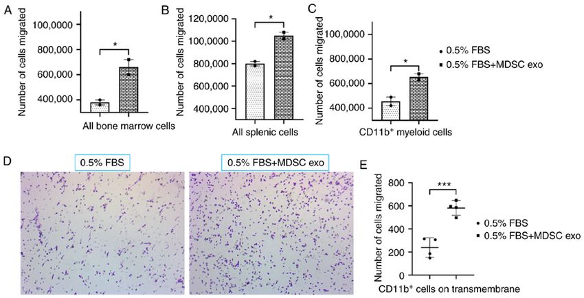
ONCOLOGY REPORTS 45: 1171-1181, 2021                                          1173

non‑essential amino acids, 1 mM sodium pyruvate, and               In vitro scratch assay. Scratch assay was performed to detect
10% FBS, supplemented with 100 ng/ml of GM‑CSF.                    the ability of MDSC‑derived exosomes to increase migration
                                                                   and invasion of tumor cells. 4T1 luciferase positive cells were
Exosome isolation. Exosomes were depleted from the                 seeded in 6‑well plates. After achieving 80‑90% confluency,
complete media by ultracentrifugation for 70 min at                the cells were starved overnight with 0.5% FBS for cell cycle
100,000 x g using an ultracentrifuge (Beckman Coulter) and         synchronization and a measured wound was inflicted at the
SW28 swinging‑bucket rotor. MDSCs (6x10⁶) were grown               center of the culture (from top to bottom). Then, cells were
in a T175 flask for 72 h under normoxic conditions (5% CO2         treated with 50 µl of splenic MDSC‑derived exosomes in
and 20% oxygen) at 37˚C in a humidified incubator. The cell        PBS containing 7.5x10⁸ exosomes for 48 h in 2% FBS media.
culture supernatant was centrifuged at 700 x g for 15 min          Microphotographs were taken every 24 h using an automated
to remove cell debris. To isolate exosomes, we employed a          all‑in‑one microscope (BZ‑X710; Keyence). The wound size
combination of two steps of the size‑based method by passing       was measured using Image J software (NIH) by drawing a
through a 0.20‑µm syringe filter and centrifugation with           rectangular region of interest to quantify the visible area of
100k membrane tube at 3,200 x g for 30 min followed by a           the wound.
single step of ultracentrifugation at 100,000 x g for 70 min [as
described in our previous publication (16)].                       In vivo t reat ment with M DSC‑ derived exosomes.
                                                                   MDSC‑derived exosomes were injected intravenously
Protein quantification. Isolated exosomes resuspended in           (100 µl containing approximately 1.5x10⁹ exosomes) into the
a minimal amount of PBS were lysed by RIPA buffer with             wild‑type Balb/c and C57Bl/6 mice. The animals were treated
protease and phosphatase inhibitor (100:1 dilution). Exosomal      for a week with 3 doses (alternate days) of MDSC‑derived
protein was quantified by Bradford assay using Pierce™ BCA         exosomes. After that, the animals were euthanized and organs
Protein Assay Kit (Thermo Scientific™) and serial dilution         were collected for flow cytometric analysis.
of BSA standard (Thermo Scientific™; Thermo Fisher
Scientific, Inc.).                                                 Isolation of T cells. Both CD4+ and CD8+ cells were isolated
                                                                   from normal mouse splenocytes by immune‑magnetic nega-
Protein array. Proteins were extracted from tumor cells            tive selection kit (Stemcell Technology; catalog #19852 and
and their corresponding exosomes in both untreated and             19853, respectively). In short, harvested spleens from normal
treated conditions to evaluate the expression profiles of          mice were disrupted in cold PBS containing 2% FBS. Clumps
44 factors in duplicate by mouse cytokine antibody array           and debris were removed by passing the cell suspension
(AAM‑CYT‑1000‑8; RayBiotech, Inc.). Protein sample                 through a 70‑µm mesh nylon strainer. The single‑cell suspen-
(500 µg) was loaded onto the membrane according to the             sion was centrifuged at 300 x g for 10 min and resuspended at
manufacturer's instructions, and the chemiluminescent              1x10⁸ nucleated cells/ml. Rat serum was added to the sample
reaction was detected using a LAS‑3000 imaging system              (50 µl/ml) followed by the addition of isolation cocktail
(Fuji Film, Japan). All signals (expression intensity) emitted     (50 µl/ml). After mixing, the sample mix was incubated at
from the membrane were normalized to the average of                room temperature for 10 min. RapidSpheres™ (75 µl/ml) were
6 positive control spots of the corresponding membrane             added to the sample mix and incubated for 3 min. The tube was
using ImageJ software version 1.53c [National Institutes of        placed in EASYSEP™ MAGNETS (catalog #18001; Stemcell
Health (NIH)].                                                     Technologies) for 3 min. The enriched cell suspension was
                                                                   collected by decanting into a new tube. Cells were seeded
In vitro migration assay. A Transwell assay was performed          in 24‑well plates bound with purified anti‑mouse CD3e
to evaluate the chemotaxis property of the MDSC‑derived            (5 µl/ml) in T‑cell media that consists of RPMI, 10% FBS,
exosomes. We used 24 Transwell plates with 8‑µm inserts            1% MEAM, 2.5% HEPES, 1% penicillin‑streptomycin, 0.5%
in polyethylene terephthalate track‑etched membranes               β‑mercaptoethanol, and purified anti‑mouse CD28 (5 µl/ml).
(Corning, Inc.). We collected bone marrow cells and splenic
mononuclear cells using Ficoll gradient centrifugation, and        Quantification of ROS generation by CD8+ T‑cells. ROS
myeloid cells from bone marrow using CD11b + magnetic              production from CD8+ T‑cells following MDSC‑derived
beads from normal Balb/c mice. A total of 1.5x10⁶ cells/insert     exosomes treatment in vitro was estimated by labeling the CD8+
in serum‑free media were added into the upper compartment          T‑cells using CM‑H2DCFDA (Invitrogen™, C6827; Thermo
of the chamber. Inserts were placed in 12‑well plates with         Fisher Scientific, Inc.). In short CD8+ T‑cells were isolated
DMEM containing 0.5% FBS in the presence or absence of             according to the above‑mentioned method. After the final
exosomes (20 µl containing approximately 3x10⁸ exosomes)           wash step of the isolation procedure, cells were resuspended
isolated from MDSCs. After incubating overnight, we                in 1 ml PBS. DCFDA solution at a working concentration of
collected suspended immune cells (migrated) from the media         10 µM/ml was added followed by incubation in the dark at
of the bottom chamber and loosely adherent immune cells on         37˚C for 30 min. The cells were washed with an extra PBS
the surface of the bottom chamber using gentle cell scraping.      to remove the unbound dye and resuspended with appropriate
Then we centrifuged and resuspended the cells in PBS and           T‑cell media. A total of 100,000 cells were seeded per well
counted the cells with a hemocytometer. Insert membranes           of 96 well‑plate. MDSC‑derived exosomes were added in the
were washed, fixed, and stained with 0.05% crystal violet to       treatment group and the same volume of PBS was added in the
detect the migrated/invaded cells. The counting was made           control group. Hydrogen peroxide (H2O2) was used as a posi-
with an inverted microscope (Nikon Eclipse E200).                  tive control for ROS production. Following 4 h of incubation,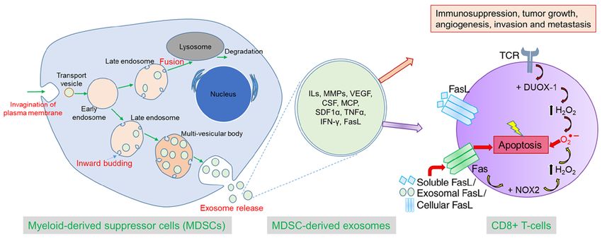
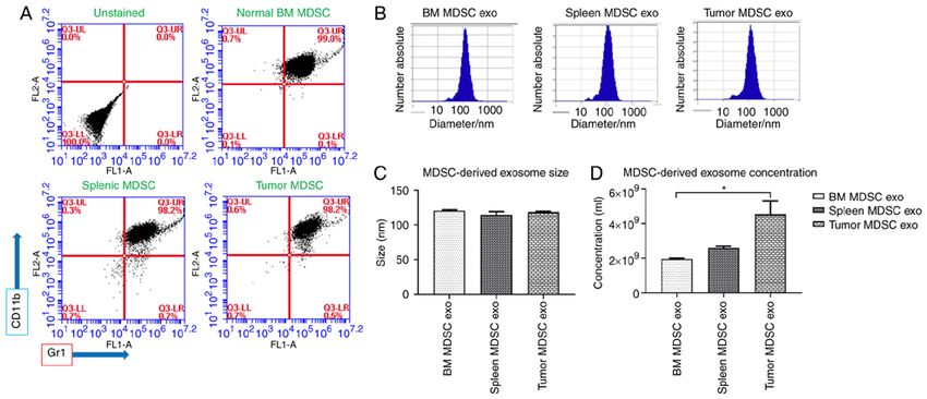
1174                             RASHID et al: IMMUNOSUPPRESSIVE EFFECT OF MDSC-DERIVED EXOSOMES

Figure 1. Isolation of MDSC‑derived exosomes (exo) from different sources. (A) Flow cytometric analysis of isolated MDSCs from normal bone marrow (BM), spleen
of tumor‑bearing mice, and tumors, showing that more than 98% of cells were positive for CD11b and Gr1. (B and C) Nanoparticle tracking analysis (NTA) showing
no significant differences in the size distribution of exosomes isolated from MDSCs of normal bone marrow (BM), the spleen of tumor‑bearing mice, and tumors.
(D) NTA analysis showing exosome concentration per ml. Quantitative data are expressed in mean ± SEM. *P
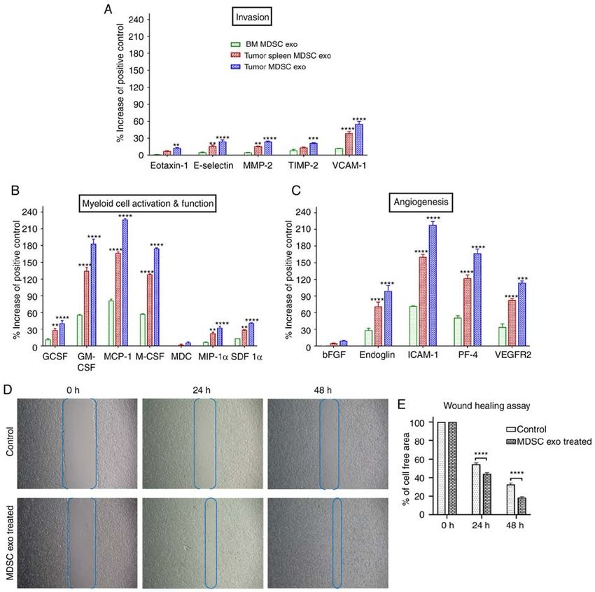
ONCOLOGY REPORTS 45: 1171-1181, 2021                                                             1175

Figure 2. The expression level of cytokines in MDSC‑derived exosomes (exo) that are involved in tumor invasion, angiogenesis, and myeloid cell activation
and function. In vitro quantification of the level of cytokines associated with (A) tumor invasion, (B) myeloid cell activation and function, and (C) angiogen-
esis, detected in the membrane‑based array in protein samples collected from exosomes isolated from MDSCs of normal bone marrow (BM), the spleen of
tumor‑bearing mice and tumors. Quantitative data are expressed as mean ± SEM. **P
1176                            RASHID et al: IMMUNOSUPPRESSIVE EFFECT OF MDSC-DERIVED EXOSOMES

Figure 3. Role of MDSC‑derived exosomes (exo) in immune cell migration. Isolated mouse myeloid cells, bone marrow cells, and splenic cells were seeded
on the top chamber of the Transwell, and splenic MDSC‑derived exosomes were added in the bottom chamber with 0.5% FBS. After 24 h, migrated (A) bone
marrow cells, (B) splenic cells, and (C) myeloid cells in the bottom chamber were counted with a hemocytometer. In addition, (D) attached myeloid cells on
the Transwell membrane were visualized under a light microscope, and (E) quantified. Quantitative data are expressed as mean ± SEM. *P
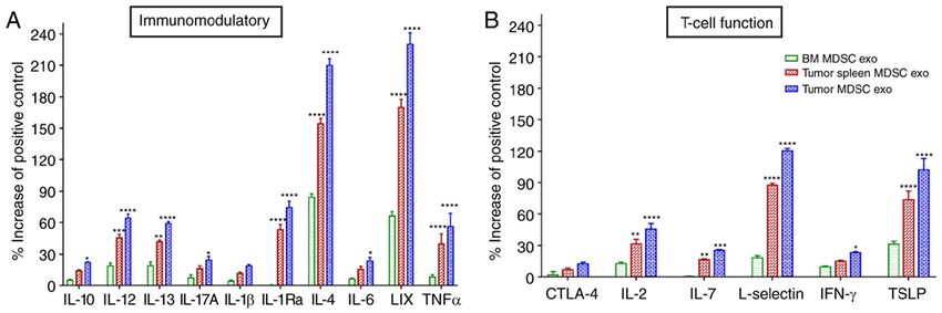
ONCOLOGY REPORTS 45: 1171-1181, 2021                                                          1177

Figure 4. Expression levels of cytokines in MDSC‑derived exosomes (exo) that are involved in T‑cell function and immunomodulation. In vitro quantification
of the level of cytokines associated with (A) immunomodulation and (B) T‑cell function, detected in the membrane‑based array in protein samples collected
from the exosomes isolated from MDSCs of normal bone marrow (BM), the spleen of tumor‑bearing mice and tumors. Quantitative data are expressed as
mean ± SEM. *P
1178                          RASHID et al: IMMUNOSUPPRESSIVE EFFECT OF MDSC-DERIVED EXOSOMES

Figure 6. In vivo depletion of M1‑macrophages, and increased number of M2‑macrophages and gMDSCs by MDSC‑derived exosome treatment. Normal
Balb/c mice were treated with and without (control) splenic MDSC‑derived exosomes for 1 week. Quantification of cells from the bone marrow (BM),
lungs, and spleen showing (A) decreased number of M1‑macrophages and increased M2‑macrophages in the spleen and (B) decreased number of monocytic
mMDSCs and increased number of granulocytic gMDSCs in the spleen following treatment with MDSC‑derived exosomes. Quantitative data are expressed
as mean ± SEM. *P
ONCOLOGY REPORTS 45: 1171-1181, 2021                                                          1179

Figure 7. Effect of MDSC‑derived exosomes on CD4 and CD8‑positive T‑cells in vitro. (A and B) Isolated CD4 and CD8‑positive T‑cells were co‑cultured
with or without splenic MDSC‑derived exosome (exo) treatment for 24 h followed by flow cytometric analysis of the cells. (C) Effect of splenic MDSC‑derived
exosomes on ROS production by CD8+ T‑cells was determined by CM‑H2DCFDA‑labeled CD8+ T‑cells treated with MDSC‑derived exosomes. (D) Effect of
splenic MDSC‑derived exosomes on CD8+ T‑cell proliferation by a cell proliferation assay using WST‑1 reagent. (E and F) Membrane‑based protein array
was used to determine the (E) level of FasL in exosomes from different MDSC populations and (F) expression levels of cytokines in CD8 T‑cells. Quantitative
data are expressed as mean ± SEM. *P
1180                            RASHID et al: IMMUNOSUPPRESSIVE EFFECT OF MDSC-DERIVED EXOSOMES

Figure 8. Schematic diagram showing the process of biogenesis of exosomes from MDSCs and the role of MDSC‑derived exosomes in tumor progression and
immunosuppression by AICD. Exosomes secreted from the MDSCs contain pro‑tumorigenic factors from the parent cells and can play a crucial role in immu-
nosuppression, tumor growth, angiogenesis, invasion, and metastasis by dispensing their contents into the other TME cells or distant cells. MDSC‑derived
exosomes can activate CD8+ T‑cells, and TCR triggering causes activation of DUOX‑1 that leads to H2O2 production and eventually generation of ROS in
mitochondria. Prolonged TCR stimulation triggers overexpression of both Fas (receptor) and FasL (ligand), which culminates in fratricide (from direct cell
contact) or autocrine suicide (interaction of soluble FasL with Fas).

molecules FasL and Bcl‑2 (44). We detected a significantly                     Availability of data and materials
large amount of ROS production from the CD8+ T‑cells that
were treated with MDSC exo. Therefore, we hypothesized                         The datasets used during the present study are available from
that MDSC exo precipitate CD8+ T‑cell apoptosis by AICD                        the corresponding author upon reasonable request.
through hyper‑activation or repeated stimulation, which in
turn results in increased levels of ROS production and activa-                 Authors' contributions
tion of the Fas/FasL (CD95/CD95L) pathway. According to
our data, we believe that ROS are involved in the reduction                    MHR conceived the hypothesis, designed and performed the
of CD8+ T‑cells and there is a possibility that ROS inhibi-                    experiments, and conducted the data collection, data analysis,
tion such as treatment with N‑acetylcysteine might prevent                     and interpretation, and wrote the manuscript. TFB conducted
this reduction. Although we did not look into the lymph                        acquisition of the in vitro data and edited the manuscript. RA
node T‑cell distribution, it will be interesting to see cellular               performed the animal experiments and treated the animals. RP
distribution changes following MDSC exo treatment in future                    conducted the data interpretation and edited the manuscript. BRA
studies.                                                                       helped with planning the in vitro T‑cell experiments. HK guided
    In summary, we comprehensively demonstrated that                           MDSC collection and types, and helped with implantation of
MDSC‑derived exosomes inherit pro‑tumorigenic factors and                      breast cancers. YL provided laboratory facilities for NTA, data
functionally resemble parental cells in immunosuppression,                     interpretation, and editing of the manuscript. ASA supervised
tumor growth, angiogenesis, invasion, and metastasis. In                       the findings of this work, aided in interpreting the results, and
addition, MDSC‑derived exosomes are capable of increasing                      provided the funds and critical revision of the manuscript.
ROS production and inciting the Fas/FasL pathway in
CD8+ T‑cells, which precipitates AICD (Fig. 8). This novel                     Ethics approval and consent to participate
concept would open up a new avenue of MDSC research and
MDSC‑targeted therapy.                                                         All experiments were performed according to the National
                                                                               Institutes of Health (NIH) guidelines and regulations. The
Acknowledgements                                                               Institutional Animal Care and Use Committee (IACUC)
                                                                               of Augusta University (Augusta, GA, USA) (protocol
The authors thank Dr Rhea‑Beth Markowitz, Director, Office                     #2014‑0625) approved the experimental procedures.
of Grant Development, Georgia Cancer Center for help with
the English language editing of the manuscript.                                Patient consent for publication

Funding                                                                        Not applicable.

This study was supported by the Georgia Cancer Center                          Competing interests
Startup Fund and Intramural Grant Program at Augusta
University (Augusta, GA, USA) to ASA.                                          The authors have declared that no competing interest exists.
ONCOLOGY REPORTS 45: 1171-1181, 2021 1181
References 22. Yang L, Huang J, Ren X, Gorska AE, Chytil A, Aakre M,
Carbone DP, Matrisian LM, Richmond A, Lin PC and Moses HL:
Abrogation of TGF beta signaling in mammary carcinomas
1. Balkwill FR, Capasso M and Hagemann T: The tumor microen- recruits Gr‑1+CD11b+ myeloid cells that promote metastasis.
vironment at a glance. J Cell Sci 125: 5591‑5596, 2012. Cancer Cell 13: 23‑35, 2008.
2. Dysthe M and Parihar R: Myeloid‑derived suppressor cells in 23. Pollard JW: Tumour‑educated macrophages promote tumour
the tumor microenvironment. In: Tumor Microenvironment: progression and metastasis. Nat Rev Cancer 4: 71‑78, 2004.
Hematopoietic Cells‑Part A. Birbrair A (ed). Springer 24. De Palma M, Venneri MA, Galli R, Sergi Sergi L, Politi LS,
International Publishing, Cham, pp117‑140, 2020. Sampaolesi M and Naldini L: Tie2 identifies a hematopoietic
3. Achyut BR and Arbab AS: Myeloid derived suppressor cells: lineage of proangiogenic monocytes required for tumor vessel
Fuel the fire. Biochem Physiol 3: e123‑e123, 2014. formation and a mesenchymal population of pericyte progeni-
4. Arbab AS, Rashid MH, Angara K, Borin TF, Lin PC, Jain M and tors. Cancer Cell 8: 211‑226, 2005.
Achyut BR: Major challenges and potential microenvironment‑ 25. Conejo‑Garcia JR, Buckanovich RJ, Benencia F, Courreges MC,
targeted therapies in glioblastoma. Int J Mol Sci 18: 2732, 2017. Rubin SC, Carroll RG and Coukos G: Vascular leukocytes
5. Safarzadeh E, Hashemzadeh S, Duijf PHG, Mansoori B, contribute to tumor vascularization. Blood 105: 679‑681, 2005.
Khaze V, Mohammadi A, Kazemi T, Yousefi M, Asadi M, 26. Nozawa H, Chiu C and Hanahan D: Infiltrating neutrophils mediate
Mohammadi H, et al: Circulating myeloid‑derived suppressor the initial angiogenic switch in a mouse model of multistage carci-
cells: An independent prognostic factor in patients with breast nogenesis. Proc Natl Acad Sci USA 103: 12493‑12498, 2006.
cancer. J Cell Physiol 234: 3515‑3525, 2019. 27. Ohl K and Tenbrock K: Reactive oxygen species as regulators of
6. Trac, N.T. and E.J. Chung: Peptide‑based targeting of immuno MDSC‑mediated immune suppression. Front Immunol 9: 2499,
suppressive cells in cancer. Bioactive Materials 5: 92‑101, 2020. 2018.
7. Lechner MG, Liebertz DJ and Epstein AL: Characterization of 28. Ma J, Xu H and Wang S: Immunosuppressive role of
cytokine‑induced myeloid‑derived suppressor cells from normal myeloid‑derived suppressor cells and therapeutic targeting in
human peripheral blood mononuclear cells. J Immunol 185: lung cancer. J Immunol Res 2018: 6319649, 2018.
2273‑2284, 2010. 29. De Maio A: Extracellular heat shock proteins, cellular export
8. Bronte V, Brandau S, Chen SH, Colombo MP, Frey AB, Greten TF, vesicles, and the stress observation system: A form of communi-
Mandruzzato S, Murray PJ, Ochoa A, Ostrand‑Rosenberg S, et al: cation during injury, infection, and cell damage. It is never known
Recommendations for myeloid‑derived suppressor cell nomen- how far a controversial finding will go! Dedicated to Ferruccio
clature and characterization standards. Nat Commun 7: 12150, Ritossa. Cell Stress Chaperones 16: 235‑249, 2011.
2016. 30. Kucharzewska P and Belting M: Emerging roles of extracellular
9. Yang Z, Guo J, Weng L, Tang W, Jin S and Ma W: Myeloid‑derived vesicles in the adaptive response of tumour cells to microenvi-
suppressor cells‑new and exciting players in lung cancer. ronmental stress. J Extracell Vesicles 2, 2013.
J Hematol Oncol 13: 10, 2020. 31. McAndrews KM and Kalluri R: Mechanisms associated with
10. Corzo CA, Cotter MJ, Cheng P, Cheng F, Kusmartsev S, biogenesis of exosomes in cancer. Mol Cancer 18: 52, 2019.
Sotomayor E, Padhya T, McCaffrey TV, McCaffrey JC and 32. Haverkamp JM, Crist SA, Elzey BD, Cimen C and Ratliff TL:
Gabrilovich DI: Mechanism regulating reactive oxygen species in In vivo suppressive function of myeloid‑derived suppressor cells is
tumor‑induced myeloid‑derived suppressor cells. J Immunol 182: limited to the inflammatory site. Eur J Immunol 41: 749‑759, 2011.
5693‑5701, 2009. 33. Zhao F, Obermann S, von Wasielewski R, Haile L, Manns MP,
11. Bruno A, Mortara L, Baci D, Noonan DM and Albini A: Myeloid Korangy F and Greten TF: Increase in frequency of
derived suppressor cells interactions with natural killer cells myeloid‑derived suppressor cells in mice with spontaneous
and pro‑angiogenic activities: Roles in tumor progression. Front pancreatic carcinoma. Immunology 128: 141‑149, 2009.
Immunol 10: 771, 2019. 34. Sevko A and Umansky V: Myeloid‑derived suppressor cells
12. Dai J, El Gazzar M, Li GY, Moorman JP and Yao ZQ: interact with tumors in terms of myelopoiesis, tumorigenesis and
Myeloid‑derived suppressor cells: Paradoxical roles in infection immunosuppression: Thick as thieves. J Cancer 4: 3‑11, 2013.
and immunity. J Innate Immun 7: 116‑126, 2015. 35. Veglia F, Perego M and Gabrilovich D: Myeloid‑derived
13. Geiger R, Rieckmann JC, Wolf T, Basso C, Feng Y, Fuhrer T, suppressor cells coming of age. Nat Immunol 19: 108‑119, 2018.
Kogadeeva M, Picotti P, Meissner F, Mann M, et al: L‑arginine 36. Ouzounova M, Lee E, Piranlioglu R, El Andaloussi A, Kolhe R,
modulates T cell metabolism and enhances survival and Demirci MF, Marasco D, Asm I, Chadli A, Hassan KA, et al:
anti‑tumor activity. Cell 167: 829‑842.e13, 2016. Monocytic and granulocytic myeloid derived suppressor cells
14. Sin ha P, Clements V K, Bunt SK, A lbelda SM a nd differentially regulate spatiotemporal tumour plasticity during
Ostrand‑Rosenberg S: Cross‑talk between myeloid‑derived metastatic cascade. Nat Commun 8: 14979, 2017.
suppressor cells and macrophages subverts tumor immunity 37. Gao F, Liang B, Reddy ST, Farias‑Eisner R and Su X: Role of
toward a type 2 response. J Immunol 179: 977‑983, 2007. inflammation‑associated microenvironment in tumorigenesis
15. Ostrand‑Rosenberg S and Fenselau C: Myeloid‑derived and metastasis. Curr Cancer Drug Targets 14: 30‑45, 2014.
suppressor cells: Immune‑suppressive cells that impair antitumor 38. Mou W, Xu Y, Ye Y, Chen S, Li X, Gong K, Liu Y, Chen Y, Li X,
immunity and are sculpted by their environment. J Immunol 200: Tian Y, et al: Expression of Sox2 in breast cancer cells promotes
422‑431, 2018. the recruitment of M2 macrophages to tumor microenvironment.
16. Rashid MH, Borin TF, Ara R, Angara K, Cai J, Achyut BR, Liu Y Cancer Lett 358: 115‑123, 2015.
and Arbab AS: Differential in vivo biodistribution of 131I‑labeled 39. Dijkgraaf EM, Heusinkveld M, Tummers B, Vogelpoel LT,
exosomes from diverse cellular origins and its implication for Goedemans R, Jha V, Nortier JW, Welters MJ, Kroep JR and
theranostic application. Nanomedicine 21: 102072, 2019. van der Burg SH: Chemotherapy alters monocyte differentiation
17. Rashid MH, Borin TF, Ara R, Alptekin A, Liu Y and Arbab AS: to favor generation of cancer‑supporting M2 macrophages in the
Generation of novel diagnostic and therapeutic exosomes to tumor microenvironment. Cancer Res 73: 2480‑2492, 2013.
detect and deplete protumorigenic M2 macrophages. Adv Ther 40. Allavena P, Sica A, Solinas G, Porta C and Mantovani A: The
(Weinh) 3: 1900209, 2020. inflammatory micro‑environment in tumor progression: the role
18. Du R, Lu KV, Petritsch C, Liu P, Ganss R, Passegué E, Song H, of tumor‑associated macrophages. Crit Rev Oncol Hematol 66:
Vandenberg S, Johnson RS, Werb Z and Bergers G: HIF1alpha 1‑9, 2008.
induces the recruitment of bone marrow‑derived vascular modu- 41. Biswas SK And Mantovani A: Macrophage plasticity and
latory cells to regulate tumor angiogenesis and invasion. Cancer interaction with lymphocyte subsets: Cancer as a paradigm. Nat
Cell 13: 206‑220, 2008. Immunol 11: 889‑896, 2010.
19. Hiratsuka S, Watanabe A, Aburatani H and Maru Y: Tumour‑ 42. Sikora E: Activation‑induced and damage‑induced cell death in
mediated upregulation of chemoattractants and recruitment of aging human T cells. Mech Ageing Dev 151: 85‑92, 2015.
myeloid cells predetermines lung metastasis. Nat Cell Biol 8: 43. Andersen MH, Schrama D, Thor Straten P and Becker JC:
1369‑1375, 2006. Cytotoxic T Cells. J Invest Dermatol 126: 32‑41, 2006.
20. Pawelek JM and Chakraborty AK: Fusion of tumour cells 44. Hildeman DA, Mitchell T, Kappler J and Marrack P: T cell apop-
with bone marrow‑derived cells: A unifying explanation for tosis and reactive oxygen species. J Clin Invest 111: 575‑581, 2003.
metastasis. Nat Rev Cancer 8: 377‑386, 2008.
21. Umansky V, Blattner C, Gebhardt C and Utikal J: The role of This work is licensed under a Creative Commons
myeloid‑derived suppressor cells (MDSC) in cancer progression. Attribution-NonCommercial-NoDerivatives 4.0
Vaccines (Basel) 4: 36, 2016. International (CC BY-NC-ND 4.0) License.You can also read