Histone H3 phosphorylation is required for the initiation, but not maintenance, of mammalian chromosome condensation
←
→
Page content transcription
If your browser does not render page correctly, please read the page content below
Journal of Cell Science 111, 3497-3506 (1998)                                                                                  3497
Printed in Great Britain © The Company of Biologists Limited 1998
JCS7316

Histone H3 phosphorylation is required for the initiation, but not
maintenance, of mammalian chromosome condensation

Aaron Van Hooser1, David W. Goodrich2, C. David Allis3, B. R. Brinkley1 and Michael A. Mancini1,*
1Department       of Cell Biology, Baylor College of Medicine, One Baylor Plaza, Houston, TX 77030, USA
2Department       of Tumor Biology, The University of Texas M.D. Anderson Cancer Center, Houston, TX 77030, USA
3Department       of Biology, University of Rochester, Rochester, NY 14627, USA
*Author for correspondence (e-mail: mancini@bcm.tmc.edu)

Accepted 1 October; published on WWW 12 November 1998

SUMMARY
The temporal and spatial patterns of histone H3                     absence of mitotic chromosome morphology, indicating
phosphorylation implicate a specific role for this                  that the phosphorylation of H3 is not sufficient for complete
modification in mammalian chromosome condensation.                  condensation. Mild hypotonic treatment of cells arrested in
Cells arrest in late G2 when H3 phosphorylation is                  mitosis results in the dephosphorylation of H3 without a
competitively inhibited by microinjecting excess substrate          cytological loss of chromosome compaction. Hypotonic-
at mid-S-phase, suggesting a requirement for activity of the        treated cells, however, complete mitosis only when H3 is
kinase that phosphorylates H3 during the initiation of              phosphorylated. These observations suggest that H3
chromosome condensation and entry into mitosis. Basal               phosphorylation is required for cell cycle progression and
levels of phosphorylated H3 increase primarily in late-             specifically for the changes in chromatin structure incurred
replicating/early-condensing heterochromatin both during            during chromosome condensation.
G2 and when premature chromosome condensation is
induced. The prematurely condensed state induced by
okadaic acid treatment during S-phase culminates with H3            Key words: Centromere, Heterochromatin, Histone, Mitosis, Okadaic
phosphorylation throughout the chromatin, but in an                 acid, Phosphorylation

INTRODUCTION                                                        of chromatin to condensation factors, transcription factors, and
                                                                    proteases activated during apoptotic cell death (Roth and Allis,
The relaxed chromatin of interphase is condensed during cell        1992; Hendzel et al., 1997; Waring et al., 1997). Additionally,
division to allow its segregation on the spindle apparatus.         the amino-termini of core histones might be involved in
Cytologically, condensation initiates after S-phase and reaches     protein-protein interactions and the recruitment of trans-acting
its maximum during early mitosis (Adlakha and Rao, 1986). It        factors to specific regions of chromatin (Hendzel et al., 1997).
is generally thought that the condensation of chromatin occurs         Site-specific phosphorylation of histone H3 at serine 10
through its folding into loops, compaction of the loops, and        (Ser10) initiates during G2, becomes maximal during
coiling of the axis upon which the loops are anchored               metaphase, and diminishes during late anaphase and early
(reviewed by Manuelidis and Chen, 1990; Koshland and                telophase (Gurley et al., 1978; Paulson and Taylor, 1982;
Strunnikov, 1996). Additionally, tangles are resolved that arise    Hendzel et al., 1997). In Tetrahymena, H3 phosphorylation
during DNA replication and by simple diffusion (Sundin and          occurs only in the mitotic micronucleus; H3 is not
Varshavsky, 1981; Weaver et al., 1985).                             phosphorylated in the macronucleus, which divides
   Several histones are phosphorylated with chromosome              amitotically without obvious chromosome condensation (Allis
condensation, but a causal role for these modifications in          and Gorovsky, 1981). When mammalian interphase cells are
chromatin modeling has not been clearly established (reviewed       fused with mitotic cells, premature chromosome condensation
by Bradbury, 1992). Histone phosphorylations have been              (PCC) is accompanied by significantly increased levels of H3
implicated in both the initiation and maintenance of mitotic        phosphorylation (Johnson and Rao, 1970; Hanks et al., 1983).
chromosome condensation (Bradbury et al., 1973; Marks et al.,       Similarly, rapid and stable increases in H3 phosphorylation at
1973; Gurley et al., 1975, 1978), as well as in the silencing of    Ser10 accompany PCC induced by the protein phosphatase
transcription during mitosis (Paulson and Taylor, 1982). More       inhibitors okadaic acid (OA) and fostriecin (Ajiro et al., 1996;
recently it has been proposed that the phosphorylation of           Guo et al., 1995).
histone amino-terminal tails might reduce their affinity for           In this report, we correlate the initiation of mammalian
DNA and facilitate the movement of nucleosomes and access           chromosome condensation with H3 phosphorylation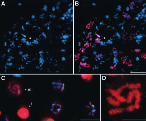
3498 A. Van Hooser and others

specifically in late-replicating/early-condensing chromatin                University of Alabama at Birmingham and has been characterized
during normal division cycles and when premature                           previously (Moroi et al., 1980; Valdivia and Brinkley, 1985). CREST,
condensation is induced. Activity of the kinase that                       anti-phosphorylated H3 (Upstate Biotechnology, Lake Placid, NY),
phosphorylates H3 is required for entry into mitosis, but is not           and anti-acetylated H3 primary antisera were diluted 1:1,000 in TBS-
required for maintaining the condensation of chromosomes in                T and incubated on preparations for 1 hour at 37°C. The samples were
                                                                           then rinsed in TBS-T and blocked again in 5% milk. Fluorophore-
hypotonically-swollen cells. This modification, then, is                   labeled goat anti-mouse, anti-rabbit, and anti-human IgG (H+L)
associated specifically with cell cycle progression and the                secondary antibodies (Jackson Immunoresearch Laboratories, Inc.,
dynamics of chromatin modeling in mammalian cells, but may                 West Grove, PA) were diluted in TBS-T and incubated with samples
not be required for maintaining the condensed state.                       for 45 minutes at 37°C. The preparations were then rinsed in TBS-T,
                                                                           and DNA was counterstained with 0.1 µg/ml 4′,6-diamidino-2-
                                                                           phenylindole (DAPI) in TBS-T. Coverslips were mounted onto glass
                                                                           slides using Vectashield anti-fade medium (Vector Laboratories, Inc.,
MATERIALS AND METHODS                                                      Burlingame, CA). Figs 1I,J and 7 are composite images obtained with
                                                                           a Deltavision, deconvolution-based optical workstation (Applied
Cell culture and synchronization                                           Precision, Issaquah, WA). Z-series stacks of multiple focal planes
CHO-K1 (Chinese hamster ovary), HeLa, and Indian muntjac                   were deconvolved by a constrained iterative algorithm, giving rise to
(Muntiacus muntjac vaginalis) (IM) cells were cultured in Opti-MEM         high-resolution optical sections used in rendering 3-D volumes. Fig.
I (Gibco Laboratories, Grand Island, NY), supplemented with 4%             8 was acquired using a Molecular Dynamics (Sunnyvale, CA) Multi
FBS and 1% penicillin/streptomycin. Cultures were maintained in a          Probe 2001 laser scanning confocal microscope with a KrAr dual-line
humidified 37°C incubator with a 5% CO2 atmosphere. Cell cultures          laser and digitally processed using Imagespace software. All other
were synchronized in G0 by growing them to confluency in serum-            images were collected using a Zeiss Axiophot fluorescence
depleted medium, replating to a density of 1×10⁶ cells/28 mm2, and         microscope with a Hamamatsu high-resolution/high-sensitivity three
incubating in serum-free medium for 66 hours at 37°C, at which time        chip CCD video camera and digitally processed using Adobe
they lacked rounded mitotic cells when examined under the phase            Photoshop.
contrast microscope (Rao and Engleberg, 1966; Ashihara and
Baserga, 1979). The cells were then incubated in serum-containing          Microinjections
medium for 4 hours to allow their entry into G1 before adding 2 mM         Unmodified H3 peptide was synthesized corresponding to the
hydroxyurea (HU; Sigma Chemical Co., St Louis, MO) to inhibit              histone H3 amino-terminal amino acids 1-20 (A1RTKQTAR-
DNA synthesis and arrest cells at the G1/S-phase boundary.                 KSTGGKAPRKQL20C). Phosphorylated H3 peptide was
Synchronized cultures were released after 16 to 20 hours of block by       synthesized such that it contained a single phosphorylated serine
washing twice with PBS (0.14 M NaCl, 2.5 mM KCl, 8 mM                      residue at position 10 (A1RTKQTARKpSTGGKAPRKQL20C). All
Na2HPO4, 1.6 mM KH2PO4, pH 7.4) at 37°C and adding fresh                   peptides contained an artificial cysteine residue at position 21 for
medium with serum. Replicating DNA was labeled with the thymidine          coupling purposes. Peptides were diluted in injection buffer (20 mM
analog 5-bromo-2′-deoxyuridine (BrdU; Sigma Chemical Co.), added           Tris, 50 mM KCl, pH 7.4) with 1:5 biotinylated anti-goat IgG (H+L)
to the medium of cell cultures to a final concentration of 20 µM.          included as a marker, detected by avidin-TXRD (Molecular Probes,
Incorporation was detected using mouse monoclonal anti-BrdU                Inc., Eugene, OR), and injected into the cytoplasm using a gas-driven
antibody with nuclease (Amersham Life Science, Inc., Piscataway,           microinjector (Eppendorf). Results from repeated measurements were
NJ) and the immunofluorescence procedure given below. PCC was              statistically analyzed using Student’s t-test. Statistical significance
induced in CHO, HeLa, and IM cells with 0.25 to 0.5 µM OA (Gibco           was determined with a probability of less than 0.05.
BRL, Gaithersburg, MD) for 1.25 to 5 hours. Cells that became
detached from the substratum with prolonged treatment were
cytospun onto coverslips prior to immunostaining, as described below.
Mitotic cell preparations were obtained by exposing asynchronous           RESULTS
cultures to the microtubule-depolymerizing drug colcemid (Gibco
Laboratories) at 0.1 µg/ml for 4 (HeLa) to 16 (IM) hours. Hypotonic        Late-replicating/early-condensing chromatin is the
treatment consisted of a 10 minute incubation of cells at 37°C in 75       first to undergo H3 phosphorylation during G2
mM KCl containing multiple protease inhibitors (0.1 mM                     Late-replicating chromatin condenses maximally during G2
phenylmethylsulfonyl fluoride (PMSF) and 1 µg/ml each of aprotinin,        and early prophase, followed by the condensation of early-
leupeptin, antipain, and pepstatin).
                                                                           replicating chromatin through late metaphase (Kuroiwa, 1971;
Immunofluorescence microscopy                                              Francke and Oliver, 1978; Yunis, 1980; Drouin et al., 1991).
Cells were grown on poly-amino acid-coated glass coverslips, or,           To determine if late-replicating/early-condensing chromatin is
alternatively, mitotic cells were selectively detached by mitotic shake-   the first to undergo histone H3 phosphorylation, cells
off, pelleted at 1,000 g for 5 minutes, and cytocentrifuged (1,000 rpm,    synchronized in late S-phase of the cell cycle were labeled by
2 minutes) onto coverslips using a Cytospin 3 (Shandon Instruments,        BrdU incorporation and in the subsequent G2 phase with an
Inc., Pittsburgh, PA). Cells were extracted with 0.5% Triton X-100 in      antiserum specific for the phosphorylated amino terminus
PEM (80 mM K-Pipes, pH 6.8, 5 mM EGTA, pH 7.0, 2 mM MgCl2)                 (Ser10) of H3 (Hendzel et al., 1997). CHO cultures become
for 2 minutes at 4°C. Samples were fixed in 4% formaldehyde                enriched for cells in late S-phase approximately 5 hours after
(Polysciences, Inc., Warrington, PA) in PEM for 20 minutes at 4°C          release from HU-block at the G1/S boundary (O’Keefe et al.,
and permeabilized with 0.5% Triton X-100 in PEM for 30 minutes at
RT. Non-specific binding of antibodies was blocked overnight at 4°C
                                                                           1992). At this time, replicating DNA was labeled with BrdU
with 5% milk in TBS-T (20 mM Tris-HCl, pH 7.6, 137 mM NaCl,                for 6 hours, approximately the time it takes CHO cells to reach
0.1% Tween-20). Auto-antiserum from an individual with CREST               late G2 from late S-phase. Immunofluorescence revealed
(calcinosis, Raynaud’s phenomenon, esophageal dysmotility,                 extensive colocalization of anti-BrdU with anti-phosphorylated
sclerodactyly, and telangiectasia) variant scleroderma (designated SH)     H3 (anti-PH3) antibodies, indicating that H3 is phosphorylated
was obtained from the Comprehensive Arthritis Center at the                primarily in late-replicating chromatin during G2 (Fig. 1A-D).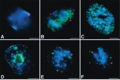
H3 phosphorylation and chromosome condensation 3499
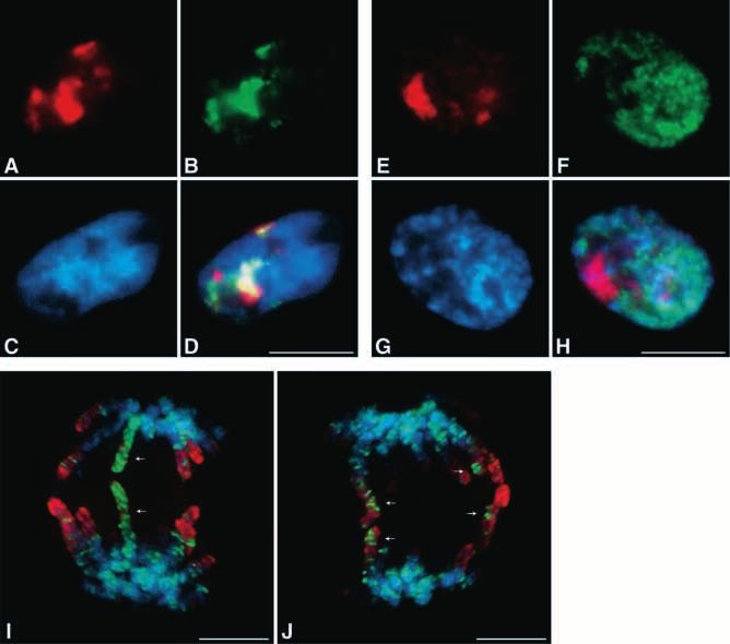
Fig. 1. H3 phosphorylation initiates primarily
in late-replicating chromatin during G2.
(A-D) CHO cultures were labeled in late S-
phase with BrdU, then harvested in the
subsequent G2 and immunostained with anti-
BrdU (green) and anti-PH3 (red) antibodies.
DNA was counterstained with DAPI (blue).
Bar, 10 µm. (E-H) Early-replicating chromatin
was labeled with BrdU prior to harvesting in
G2 and immunostaining as above. Bar, 10 µm.
(I,J) CHO cells were labeled in late S-phase
with BrdU (green), then harvested in the
subsequent mitosis and immunostained as
above. A z-series of deconvolved optical
sections was rotated 180° around the y-axis
and projected as two composite views. Sister
chromatids separating at anaphase were
observed to have mirrored banding patterns of
late-replicating chromatin (arrows). The
dephosphorylation of H3 has begun by late
anaphase, resulting in intermediate levels of
anti-PH3 staining (red). Bar, 5 µm.
Colocalization was not observed between BrdU incorporated that of histone H3 Ser10 (Bialojan and Takai, 1988; Haystead
during the first 5 hours of S-phase, immediately following et al., 1989; Cohen et al., 1990; Mahadevan et al., 1991; Ajiro
release from HU-block, and anti-PH3 staining 6 hours later in et al., 1996). When PCC is induced in IM cells with 0.25 µM
the subsequent G2 (Fig. 1E-H). To additionally determine the OA, the G2 pattern of pericentric H3 phosphorylation is
specificity of BrdU labeling in the above experiments, we observed in most cells within 90 minutes (Fig. 2B). The
labeled late replicating chromatin and harvested the cells in prekinetochores, however, appear as open arrays of CREST-
mitosis, 12 hours after release from HU-block. Sister stained subunits, indicating that they have not reached late S-
chromatids were observed to have identical banding patterns, phase/early G2 and are not fully replicated (Brenner et al.,
indicative of the semi-conservative incorporation of BrdU 1981; He and Brinkley, 1996). H3 is similarly phosphorylated
during late S-phase (Fig. 1I,J). in pericentric heterochromatin when PCC is induced in CHO
and HeLa cells with OA (data not shown). We also examined
H3 phosphorylation initiates in pericentric H3 phosphorylation in cells that were induced to enter mitosis
heterochromatin when premature chromosome prematurely from the G1/S boundary by caffeine treatment
condensation is induced (Schlegel and Pardee, 1986; Brinkley et al., 1988). Prior to
The centromere is a principal site for the initiation of entering mitosis, these cells pass through a G2-like state, in
chromosome condensation in mammalian cells (He and which H3 phosphorylation accumulates primarily in
Brinkley, 1996). H3 phosphorylation concurrently increases in pericentric heterochromatin (data not shown).
pericentric heterochromatin during G2 and advances outward
into the chromosome arms (Hendzel et al., 1997). As shown in
Fig. 2A, chromatic regions surrounding prekinetochores are
the exclusive sites of H3 phosphorylation during G2 in IM
cells, since the number of initiation sites exactly matches the
diploid chromosome number (2N=7么). The centromeres-
prekinetochores appear as doublets, stained with CREST auto-
antiserum, indicating that the cell has reached G2 (Brenner et
al., 1981). To determine if the initiation of H3 phosphorylation
follows the same pattern when PCC is induced, asynchronous Fig. 2. H3 phosphorylation is first detected in pericentric
cell cultures were treated with OA and immunostained with heterochromatin when premature chromosome condensation is
CREST and anti-PH3 antisera. Treatment of mammalian cells induced with OA. (A) IM cells were immunostained with CREST
with OA, which is a specific inhibitor of the serine/threonine (green) and anti-PH3 (red) antisera. DNA was counterstained with
protein phosphatases 1 and 2A, results in PCC and a rapid and DAPI (blue). Bar, 5 µm. (B) IM cells were treated with 0.25 µM OA
stable increase in cellular phosphorylation levels, including for 1.25 hours to induce PCC and immunostained as above. Bar, 5 µm.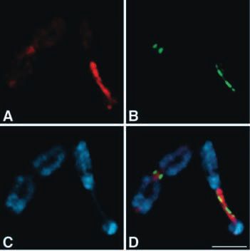
3500 A. Van Hooser and others

H3 phosphorylation is not sufficient for                               et al., 1992; Gliksman et al., 1992; Vandré and Wills, 1992).
chromosome condensation                                                To correlate the extent of H3 phosphorylation with OA-
The effects of OA treatment vary among cell type and cell              induced PCC, we exposed asynchronous cell populations to
cycle stage. Exposure of most mammalian interphase cells to            0.5 µM OA for 2.5 hours. Under these conditions, over 70%
the drug induces a mitosis-like state, characterized by PCC,           of HeLa and IM cells appeared rounded up with compact
increased cdc2/H1 kinase activity, dispersion of nuclear               nuclei characteristic of S-phase PCC. The nuclei of these
lamins, and the appearance of mitotic asters (Haystead et al.,         cells displayed high levels of H3 phosphorylation throughout
1989; Picard et al., 1989; Rime and Ozen, 1990; Yamashita              the chromatin (Fig. 3A,B), particularly at the nuclear
et al., 1990; Zheng et al., 1991). Morphologically normal              periphery, resembling prophase cells in untreated cultures
mitotic chromosome condensation with OA treatment,                     (Hendzel et al., 1997). Rudimentary chromosome
however, is only observed in a small fraction of                       organization developed in less than 10% of the
unsynchronized cells and has been most clearly demonstrated            unsynchronized cells treated under these conditions,
using mouse cells arrested at G2 (Guo et al., 1995; Ajiro et           indicating that H3 phosphorylation throughout interphase
al., 1996). Similarly, OA-treatment of mitotic cells results in        chromatin is not sufficient for mitotic chromosome
the overcondensation of chromosomes, as well as disruption             condensation. The number of cells demonstrating mitotic
of the mitotic spindle and metaphase plate formation (Ghosh            chromosome organization was reduced to less than 1% when
                                                                       cultures were arrested in early S-phase prior to treatment with
                                                                       OA for 2.5 to 5 hours (Fig. 3C-F). CHO cells synchronized
                                                                       in early S-phase demonstrated PCC and high levels of H3
                                                                       phosphorylation only after 5 hours of treatment with 0.5 µM
                                                                       OA (data not shown). Similarly, mitotic chromosome
                                                                       morphology was observed in less than 1% of the cells.
                                                                          In the absence of OA treatment, H3 phosphorylation was
                                                                       undetectable during early interphase in HeLa cells (Fig. 3G,H).
                                                                       Untreated CHO and IM cells, however, display nuclear foci of
                                                                       anti-PH3 immunoreactivity during early interphase (Fig. 4).
                                                                       The small size and number of these nuclear foci make them
                                                                       distinct from the pericentric pattern of phosphorylation
                                                                       observed during G2. To determine the temporal and spatial
                                                                       occurrences of these foci in interphase nuclei, discrete phases
                                                                       of DNA replication were visualized by pulse-labeling with
                                                                       BrdU at progressive time points through S-phase (O’Keefe et
                                                                       al., 1992). Thirty-two per cent of CHO cells blocked at the
                                                                       G1/S boundary had anti-PH3 foci. This number increased upon
                                                                       release and reached a maximum in late S-phase, when the
                                                                       number of cells with foci was near 70%. The foci vary in
                                                                       number from 1 to 5 at each stage and therefore do not appear
                                                                       to be sequentially replicated. They do not correspond
                                                                       exclusively to early-replicating (Fig. 4A-C) or late-replicating
                                                                       regions of chromatin (Fig. 4D-F). All anti-PH3
                                                                       immunoreactivity is abolished when the antiserum is incubated
                                                                       with peptides corresponding to the phosphorylated form of the
                                                                       H3 amino-terminal tail (Hendzel et al., 1997, and data not
                                                                       shown).

                                                                       Activity of the kinase that phosphorylates H3 is
                                                                       required for entry into mitosis
                                                                       The kinase responsible for histone H3 phosphorylation in vivo
                                                                       has not been conclusively identified. It has been shown in a
                                                                       cell-free system that cyclic AMP-dependent protein kinase can
Fig. 3. H3 phosphorylation is not sufficient for chromosome            phosphorylate H3 on Ser10 (Taylor, 1982; Shibata et al., 1990).
condensation. (A,B) Asynchronous HeLa cells were treated with 0.5      Similarly, H3 phosphorylation is linked with elevated cAMP
µM OA for 2.5 hours prior to immunostaining. Rudimentary               levels during gliotoxin-induced apoptosis (Waring et al., 1997).
chromosome morphology was observed in less than 10% of the cells       When H3 phosphorylation is inhibited by the protein kinase
(arrow). Bar, 25 µm. (C,D) HeLa cells were released from HU-block      inhibitor staurosporine, mammalian cells arrest in late G2
into early S-phase and treated with 0.5 µM OA for 2.5 hours prior to
immunostaining. Bar, 25 µm. (E,F) HeLa cells were released from
                                                                       (Crissman et al., 1991; Abe et al., 1991; Th’ng et al., 1994).
HU-block into early S-phase and treated with 0.5 µM OA for 5 hours     However, staurosporine is known to inhibit a wide range of
prior to immunostaining. High degrees of nuclear invagination were     kinases, including p34cdc2, which controls mitotic entry
common (arrowheads), though chromosome morphology was absent.          (Langan et al., 1989; Herbert et al., 1990; Gadbois et al., 1992).
Bar, 25 µm. (G,H) In the absence of OA-treatment, phosphorylated       To more directly define the role of the kinase that
H3 is not observed in HeLa cells during early S-phase. Bar, 25 µm.     phosphorylates H3 in cell cycle progression, we microinjected
H3 phosphorylation and chromosome condensation 3501 proliferating cells with peptide corresponding to the H3 phosphorylation is not required for the structural unmodified amino-terminal 20 amino acids of H3 to compete maintenance of isolated chromosomes for the kinase activity. CHO cells were injected at mid-S-phase, Individual metaphase chromosomes can be microscopically 4 to 5 hours after release from HU-block at the G1/S boundary. identified and examined in much greater detail by incubating Control cells were injected with the phosphorylated (Ser10) cells in water or mild hypotonic solution (75 mM KCl) for 10 form of H3 peptide or with marker only (biotinylated anti-goat minutes (Hsu, 1952). We have found that, though most IgG). Injected cultures were harvested at a time-point when chromosomes are swollen in hypotonic solution without a uninjected cultures become enriched for cells passing or just detectable alteration in their pattern of anti-PH3 staining, a having passed through mitosis, 12 to 13 hours after release significant number of cells (up to 40% in IM preparations and from HU-block. All preparations were masked, and the number 20% in HeLa) have chromosomes within which H3 appears of mitotic cells, identified by DAPI staining, was scored at least completely unphosphorylated (Fig. 6A,B). No difference in the three times. Increasing concentrations of the unmodified H3 level of condensation between phosphorylated and peptide resulted in proportional decreases in mitotic indexes unphosphorylated chromosomes within the same preparation (Fig. 5A). The number of mitotic cells was nearly abolished is observable by DAPI staining, indicating that H3 with the highest concentration of unmodified H3 peptide used. phosphorylation is not required for maintaining high levels of Injection of phosphorylated H3 peptide or marker alone did not chromosome condensation. To determine if the lack of anti- significantly reduce the number of cells passing through PH3 antibody reactivity with hypotonic treatment was due to mitosis when compared to uninjected cells within the same alterations in nucleosome structure, we immunostained the population (Fig. 5A). preparations with an antibody that reacts with the diacetylated To characterize the phenotype of cells inhibited with the (9, 14) H3 tail throughout the mammalian cell cycle (Boggs et H3 competitor peptide, as well as those progressing through al., 1996). The pattern of anti-diacetylated H3 immunostaining mitosis despite injection, we repeated the injections with an was unaltered in all hypotonic-treated cells, indicating that the intermediate concentration (0.42 µg/µl) of unmodified H3 Ser10 epitope was likely still accessible and the distribution of peptide, empirically determined in the previous experiment. H3 unchanged (Fig. 6C,D). All cells arrested in mitosis for Use of an intermediate dose also reduced the possibility that prolonged periods (overnight) with colcemid had we were observing effects of peptide toxicity. The number of chromosomes in which H3 was highly phosphorylated, mitotic, daughter, and binucleate cells was scored indicating that H3 dephosphorylation did not occur with morphologically (Fig. 5B). Control preparations were prolonged mitotic arrest or in the absence of hypotonic injected with approximately five-times the concentration treatment (data not shown). (2.26 µg/µl) of H3 peptide in the phosphorylated form or with marker only. A statistically significant reduction in the Pericentric heterochromatin is resistant to number of mitotic and daughter cells was observed in hypotonic-induced H3 dephosphorylation preparations injected with an intermediate dose of the Intermediate levels of H3 dephosphorylation were observed in unmodified H3 peptide when compared to populations a number (less than 10%) of hypotonically treated IM and injected with the phosphorylated form of H3 peptide or with HeLa mitotic cells. In each case, pericentric regions of marker alone (Fig. 5C). When cell populations injected with chromatin remained highly phosphorylated, whereas the H3 peptide are normalized against populations injected with chromosomal arms were completely dephosphorylated. This marker only, we observed a 38% reduction in the number of retention of H3 phosphorylation is easily observed in the large mitotic cells and a 36% reduction in the number of daughter compound centromeres-kinetochores of IM chromosomes cells with an intermediate dose of competitor peptide (Fig. (Fig. 7) that have been stretched by cytocentrifugation 5D). The number of cells with the focal anti-PH3 staining following brief hypotonic treatment (Zinkowski et al., 1991). pattern characteristic of late G2 was not significantly reduced, The region of phosphorylated chromatin spans the pairing indicating with the above data that arrest occurred just prior domain of the centromere-kinetochore complex, where sister to mitosis. A unique pattern of anti-PH3 staining was chromatids are joined at metaphase, and extends into the observed in 5.5% of the interphase cells injected with chromosomal arms, corresponding to the span of unmodified H3 peptide, characterized by high levels of heterochromatin in these chromosomes (Brinkley et al., 1992). diffuse, predominately nuclear anti-PH3 antibody reactivity Both phosphorylated and dephosphorylated centromeres in the absence of chromosome condensation (Fig. 5E-G). The remain constricted and can be highly elongated, suggesting that percentage of cells with this anti-PH3 staining pattern (5.5%) H3 phosphorylation is not required for maintaining high levels nearly equals the sum of differences between unmodified H3 of chromosome condensation in pericentric heterochromatin and marker only populations in percentages of G2 (1.7- and that stretching occurred by a mechanism other than H3 1.3=0.4%), mitotic (3.9-2.4=1.5%), and daughter cells (9.0- dephosphorylation. Interestingly, the region of chromatin 5.7=3.3%). Anti-PH3 staining was diffuse throughout the immediately subjacent to the CREST-stained centromere- cytoplasm and nucleus of cells injected with the kinetochore lacks anti-PH3 staining (Fig. 7), similar to results phosphorylated form of H3 peptide, with remarkably high obtained by immunogold EM (Hendzel et al., 1997). nuclear staining in 1.3% of the cells (data not shown). Significantly, all mitotic cells identified by DAPI staining had H3 phosphorylation correlates with cell cycle H3 highly phosphorylated despite competitive inhibition progression after hypotonic treatment (Fig. 5E-G), further suggesting that the phosphorylation of Hypotonic swelling of mammalian cells leads to the loss of H3 is essential for entry into mitosis. kinetochore plate morphology, as well as the disruption of
3502 A. Van Hooser and others

                                                                         H3, then released them into tissue culture medium and
                                                                         followed their progress by immunostaining. Congression of
                                                                         IM and HeLa chromosomes to the metaphase plate and entry
                                                                         into anaphase began within 60 minutes of their return to
                                                                         tissue culture medium from hypotonic. Every cell passing
                                                                         through early anaphase had H3 highly phosphorylated,
                                                                         though the chromosomes retained a swollen appearance (Fig.
                                                                         8). No intermediate stages of H3 phosphorylation were
                                                                         observed after 30 minutes of release. If hypotonic-treated
                                                                         cells were returned to tissue culture medium containing
                                                                         colcemid, every mitotic cell was phosphorylated within 30
                                                                         minutes. H3 phosphorylation, then, is correlated with cell
                                                                         viability and progression through mitosis following
                                                                         hypotonic treatment.

Fig. 4. Foci of phosphorylated H3 form in CHO nuclei during early
interphase. G1 (A) and G2 (not shown) phases of the cell cycle were      DISCUSSION
distinguished by a 5-hour BrdU-labeling of DNA replication. The
cells were then immunostained with anti-PH3 (red) and anti-BrdU          The nucleosomal histones are wrapped by DNA as octamers,
(green) antibodies. DNA was counterstained with DAPI (blue). To          consisting of two H2A-H2B dimers and a tetramer of 2 H3 and
visualize discrete phases of DNA replication, cells were arrested at     2 H4 histones (D’Anna and Isenberg, 1974; Moss et al., 1976;
the G1/S boundary with HU, released for 0.5 (B), 2 (C), 5 (D), 7 (E),    Kornberg, 1977). H3 and H4 amino-terminal tails are exposed
or 9 hours (F) and pulse-labeled for 20 minutes with BrdU prior to       from the compact core of the nucleosome and may be involved
immunostaining. Bars, 5 µm.                                              in regulatory interactions with DNA, histone H1, and other
                                                                         proteins (Glotov et al., 1978; Hardison et al., 1977; Mazen et
microtubules, centrioles, and centrosomes (Brinkley et al.,              al., 1987; Roth and Allis, 1992). Interactions of the H3 tail may
1980; Ris and Witt, 1981). However, if the period of                     be modulated by post-translational modifications, including the
hypotonic swelling is brief (under 15 minutes), the damage               acetylation of lysine residues 9, 14, 23, and 27, methylation of
can be reversed by returning the cells to an isotonic tissue             lysines 9 and 27, and serines 10 and 28 may be phosphorylated
culture environment (Brinkley et al., 1980). Under these                 (Dixon et al., 1975; Taylor, 1982; Shibata et al., 1990; Bradbury,
conditions, cell viability is not adversely affected in most             1992). The residues involved in the above interactions are
cell lines. To determine if H3 phosphorylation is required for           largely invariable (Wells and McBride, 1989) and reflect a very
progression through mitosis, we exposed mitotic cells to                 high universality in the mechanisms by which chromatin is
hypotonic conditions, resulting in the dephosphorylation of              modeled. The anti-PH3 antiserum used in these studies reacts

Fig. 5. Delayed entry into mitosis with the competitive
inhibition of the kinase that phosphorylates H3.
(A) Synchronized CHO cells were injected at late S-
phase with increasing concentrations of peptide
corresponding to the unmodified (䉬) or
phosphorylated (䊐) H3 amino terminus. The mitotic
index of each injected population (~125 cells each)
was divided by the mitotic index of uninjected cells on
the same coverslip to derive normalized values on the
y-axis. (B) Injected populations were stained for
coinjected marker antibody, and the number of mitotic
(filled arrow), recently divided daughter (arrowhead),
and binucleate cells (open arrow) was scored. G2 cells
were scored by their characteristic, focal pattern of
anti-PH3 immunostaining (see Figs 1, 2). Bar, 50 µm.
(C) Cells were injected with marker only (n=4), with
0.42 µg/µl unmodified H3 competitor peptide (n=6),
or with 2.26 µg/µl of the phosphorylated form of the
H3 peptide (n=2), where n is the number of separate
experiments consisting of ~200 injected cells each.
(D) Values from C were normalized by dividing the
number of cells injected with unmodified (black bars)
or phosphorylated (hatched bars) H3 peptide by the
number of cells injected with marker only (white bars)
at the same stage within the same preparation. Injected cells were immunostained for coinjected marker antibody (E), anti-PH3 antibody (F),
and DAPI (G). All mitotic chromosomes had H3 phosphorylated (arrow). A number of cells injected with unmodified H3 peptide had high
levels of diffuse, predominately nuclear anti-PH3 staining in the absence of chromosome condensation (arrowhead). Bar, 25 µm.
H3 phosphorylation and chromosome condensation 3503
Fig. 7. Pericentric heterochromatin is resistant to hypotonic-induced
H3 dephosphorylation. IM cells were arrested in mitosis and incubated
in hypotonic solution prior to immunostaining with anti-PH3 (A) and
Fig. 6. H3 phosphorylation is not required for the maintenance of CREST antisera (B). DNA was counterstained with DAPI (C).
condensed chromosomes. (A,B) IM cells were arrested in mitosis (D) Intermediate levels of H3 phosphorylation were observed in some
and incubated in hypotonic solution. Preparations were cells in which pericentric heterochromatin remained highly
immunostained with anti-PH3 antibody (red), and DNA was phosphorylated, whereas chromosome arms were dephosphorylated.
counterstained with DAPI (blue). Unphosphorylated chromosomes Both regions remained highly condensed. Bar, 5 µm.
appeared condensed to the same degree as phosphorylated
chromosomes within the same preparation, as resolved by DAPI
staining (asterisks). Bar, 50 µm. (C) Anti-diacetylated H3 better substrate, free H3 monomers are phosphorylated in vitro
immunoreactivity (red) was observed in all hypotonically swollen by cyclic AMP-dependent protein kinase (Belyavsky et al., 1980;
IM chromosomes (m). Interphase cells (i) stain more intensely Shibata et al., 1990). We therefore expected injected H3 amino-
with the antibody, and low levels of cytoplasmic staining are terminal peptides containing the unmodified Ser10 epitope to
observed regardless of hypotonic treatment. Bar, 25 µm. (D) A compete with, but not necessarily abolish chromatin-associated
higher magnification view of hypotonically-swollen IM H3 phosphorylation. This competition would likely become less
chromosomes immunostained with anti-diacetylated H3 antibody significant with the onset of mitosis and increased kinase activity
(red). Bar, 10 µm.
and/or chromatin-associated substrate accessibility. Injection of
unmodified H3 peptide into cells at mid-S-phase resulted in a
marked reduction of cells entering mitosis when compared to
with the N-terminal phosphorylated form of Ser/Thr 10 in all cells injected with the phosphorylated form of H3 peptide or with
eukaryotes examined, staining with the highest intensity during marker only. Though reduced in number, all injected cells passing
mitotic and, in Tetrahymena, meiotic chromosome through mitosis had H3 highly phosphorylated, suggesting that
condensation (Hendzel et al., 1997; Wei et al., 1998). the modification is essential for entry into mitosis. Since injection
Heterochromatic regions of chromosomes, including of H3 tails in the phosphorylated form did not result in the same
centromeres and telomeres, remain highly condensed reduction of cells entering mitosis, competition for histone
throughout the cell cycle and likely contain elements that direct
chromosome condensation (Koshland and Strunnikov, 1996).
The centromere is a principal site for the initiation of
chromosome condensation at the onset of mitosis in
mammalian cells (He and Brinkley, 1996). Consistent with a
role in the initiation of mammalian cell chromosome
condensation, H3 phosphorylation dramatically increases
during G2 exclusively in the late-replicating/early-condensing
heterochromatin surrounding centromeres. The same pattern of
H3 phosphorylation is observed when PCC is induced by the
phosphatase inhibitor OA and in cells induced to enter mitosis
prematurely. These results provide additional evidence of the Fig. 8. Cells do not pass from metaphase to anaphase with H3
tight coupling between H3 phosphorylation and the initiation of dephosphorylated. (A) Cells arrested in mitosis were incubated
briefly in hypotonic solution to induce the dephosphorylation of H3
chromosome condensation near centromeres-prekinetochores. and released into tissue culture medium. The cells were collected 60
H3 appears highly phosphorylated throughout the chromatin of minutes after release and immunostained with anti-PH3 antibody
OA-treated cells, but mitotic chromosome morphology does not (red) and CREST auto-antiserum (green). All cells entering anaphase
develop. This result indicates that H3 phosphorylation is not after hypotonic treatment had H3 phosphorylated throughout their
sufficient for chromosome condensation. chromosomes, though they retained a swollen appearance relative to
Though chromatin-associated H3 provides a significantly untreated preparations (B). Bar, 10 µm.3504 A. Van Hooser and others acetylase activity did not result in cell cycle arrest under the are particularly susceptible to H3 dephosphorylation and recover conditions of our experiment. It remains possible that the injected poorly from hypotonic treatment relative to other mammalian N-terminal peptides were not suitable substrates for acetylation; cell lines (B.R.B., unpublished observations). or, interestingly, phosphorylated H3 tails are not substrates for Intermediate levels of H3 phosphorylation were observed in acetylation. hypotonic-treated chromosomes of IM and HeLa cells, in which Concomitant with a reduction in the number of cells entering pericentric heterochromatin remained phosphorylated and mitosis in populations injected with unmodified H3 peptide, there chromosome arms were dephosphorylated. Since pericentric was an equivalent accumulation of interphase cells with high heterochromatin is more resistant to hypotonic swelling than levels of anti-PH3 reactivity diffuse throughout uncondensed other chromosomal regions (Brinkley et al., 1980), H3 nuclei. We postulate that this fraction of cells represents the cell phosphorylation may have a role in maintaining the highest cycle stage at which H3 kinase activity is elevated, resulting in levels of chromosome condensation. Similarly, we found that the phosphorylation of competitor peptides in the nucleus. the chromosomes of cells arrested in mitosis with colcemid Together with the fact that the number of cells in late G2 was not were more resistant to hypotonic-induced dephosphorylation of significantly reduced in injected populations, this suggests that H3 than those of unarrested mitotic cells, perhaps related to the the competitive inhibition of H3 phosphorylation prevented varying degrees of hypercondensation observed in colcemid chromosome condensation and entry into mitosis. Our treated preparations (see Rieder and Palazzo, 1992). H3 conclusions support earlier studies, in which the protein kinase phosphorylation, then, correlates with the most condensed inhibitor staurosporine was found to block H3 phosphorylation regions of chromatin during hypotonic treatment, though its and arrest mammalian cells in late G2 (Crissman et al., 1991; Abe removal does not result in decondensation. et al., 1991; Th’ng et al., 1994). However, it remains possible that The periodicity of interaction between DNA and nuclear inhibition of the kinase that phosphorylates H3 prevented the matrix proteins appears to hold interphase chromatin in loops of phosphorylation of other targets essential for entry into mitosis. approximately 60 kb, which may be folded to form the ~200 nm Collectively, the above evidence implicates a role for H3 wide chromatin fiber observed by electron microscopy (Marsden phosphorylation in the initiation of chromosome condensation and Laemmli, 1979; Rattner and Lin, 1985; Fey et al., 1986; and entry into mitosis. This modification, however, may not have Nelson et al., 1986). Euchromatin is compacted from a ~200 nm a critical role in the maintenance of chromosome condensation, diameter interphase fiber to a 700 nm metaphase structure. H3 as chromosomes can be hypotonically swollen with H3 phosphorylation correlates best with the initial events of remaining phosphorylated, and the dephosphorylation of H3 condensation: specifically, compaction of the ~200 nm does not result in decondensation. It is important to note that chromatid fiber (Hendzel et al., 1997). However, hypotonic treatment results in extensive physiological disruption heterochromatin remains at an approximately equivalent level of of cells, albeit reversible in most cases. The primary conclusion compaction (700 nm) throughout the cell cycle (reviewed by that can be drawn from the above data, then, is that H3 Manuelidis and Chen, 1990). The fact that metaphase phosphorylation is not required for maintaining the condensation chromosomes can have H3 hypotonically dephosphorylated of isolated chromosomes. However, this result also confirms the without a cytological loss of chromosome condensation is in vivo observations of Rao and colleagues, who found that the consistent with the fact that heterochromatin is fusion of mammalian mitotic cells with interphase cells results dephosphorylated at the end of mitosis, yet maintains a high in a large reduction of H1 and H3 phosphorylations without a level of compaction. Why then is heterochromatin loss of chromosome condensation (Johnson and Rao, 1970; phosphorylated? One possibility is that H3 phosphorylation in Hanks et al., 1983). It also suggests that the decondensation of pericentric heterochromatin may be required for the release of metaphase chromosomes observed following treatment with the sister chromatid cohesion at the metaphase to anaphase protein kinase inhibitors staurosporine (Th’ng et al., 1994) and transition, a process interrelated with chromosome condensation N-6 dimethylaminopurine (Ajiro et al., 1996) may not have been (Guacci et al., 1997). Additionally H3 phosphorylation may be produced exclusively by H3 dephosphorylation. This is easily required throughout early mitosis for the initiation of conceived, as staurosporine is a potent inhibitor of all kinases chromosome condensation, which continues until late tested in vitro, though its action in vivo may be narrower metaphase (Adlakha and Rao, 1986; Drouin et al., 1991). (Herbert et al., 1990; Th’ng et al., 1994); and N-6 Correlating morphological chromatin data with the dimethylaminopurine is a non-specific kinase inhibitor in vitro, quantification of histone phosphorylations led to the suggestion capable of affecting multiple phases of the cell cycle (Vesely et that H3 phosphorylation occurs during the final organization and al., 1994; Simili et al., 1997). maintenance of chromosomes (Gurley et al., 1978). More Though hypotonic treatment resulted in a large number of recently, however, the phosphorylation of H3 has been observed cells with dephosphorylated chromosomal H3, when these cells to precede detectable chromosome condensation and its were returned to an isotonic tissue culture environment, they dephosphorylation to precede detectable chromosome were not observed to enter anaphase with H3 dephosphorylated. decondensation (Hendzel et al., 1997). Additionally, H3 It is not structural inhibition that prevents cells with phosphorylation may be associated with the relatively relaxed dephosphorylated chromosomes from progressing through state of transcriptionally active chromatin coincident with the mitosis, as these chromosomes appear as compact as those with mitogenic induction of early-response genes, as well as make H3 phosphorylated that undergo anaphase within the same chromatin accessible to nucleases involved in apoptotic pathways preparation. It is both possible that hypotonically-treated cells (Halegoua and Patrick, 1980; Mahadevan et al., 1988, 1991; rephosphorylate chromosomal H3 upon their return to an Barratt et al., 1994; Waring et al., 1997). An attractive hypothesis isotonic environment, or simply that cells with dephosphorylated that reconciles these apparently divergent properties is that the chromosomes are not viable. Suggestive of the latter, IM cells phosphorylation of histone N-terminal tails reduces their affinity
H3 phosphorylation and chromosome condensation 3505

for DNA and facilitates the movement of nucleosomes and                              okadaic acid, on protein phosphatases: specificity and kinetics. Biochem. J.
targeting of factors involved in condensation, transcription, and                    256, 283-290.
apoptosis (Roth and Allis, 1992; Hendzel et al., 1997; Waring et                   Boggs, B. A., Connors, B., Sobel, R. E., Chinault, A. C. and Allis, C. D.
                                                                                     (1996). Reduced levels of histone H3 acetylation on the inactive X
al., 1997). In this way, H3 phosphorylation would work in                            chromosome in human females. Chromosoma 105, 303-309.
conjunction with other mechanisms involved in the modeling of                      Bradbury, E. M. (1992). Reversible histone modifications and the
active chromatin, including the phosphorylation and depletion of                     chromosome cell cycle. BioEssays 14, 9-16.
histone H1 and the enrichment of hyperacetylated histones H3                       Bradbury, E. M., Inglis, R. J., Matthews, H. R. and Sarner, N. (1973).
                                                                                     Phosphorylation of very-lysine-rich histone in Physarum polycephalum.
and H4 (Allegra et al., 1987; Kamakaka and Thomas, 1990; Roth                        Correlation with chromosome condensation. Eur. J. Biochem. 33, 131-139.
and Allis, 1992; Mizzen and Allis, 1998). Our data supports this                   Brenner, S., Pepper, D., Berns, M. W., Tan, E. and Brinkley, B. R. (1981).
role for histone phosphorylation in chromatin modeling, in that                      Kinetochore structure, duplication, and distribution in mammalian cells:
the phosphorylation of H3 appears to be required for dynamic                         analysis by human autoantibodies from scleroderma patients. J. Cell Biol.
changes in chromatin structure, but not for their maintenance.                       91, 95-102.
                                                                                   Brinkley, B. R., Cox, S. M. and Pepper, D. A. (1980). Structure of the mitotic
   The molecular mechanisms by which heterochromatin is kept                         apparatus and chromosomes after hypotonic treatment of mammalian cells
refractory from the dynamic changes of other chromosomal                             in vitro. Cytogenet. Cell Genet. 26, 165-174.
regions remain largely unknown, as do the mechanisms by                            Brinkley, B. R., Zinkowski, R. P., Mollon, W. L., Davis, F. M., Pisegna, M.,
which centromeres-prekinetochores direct the initiation of                           Pershouse, M. and Rao, P. N. (1988). Movement and segregation of
                                                                                     kinetochores experimentally detached from mammalian chromosomes.
chromosome condensation in mammalian cells during G2, and                            Nature 336, 251-254.
why. Seemingly disparate events coalesce at the nuclear                            Brinkley, B. R., Ouspenski, I. and Zinkowski, R. P. (1992). Structure and
envelope. H3 phosphorylation initiates with chromosome                               molecular organization of the centromere-kinetochore complex. Trends Cell
condensation in pericentric heterochromatin, often observed to                       Biol. 2, 15-21.
be at the nuclear periphery (Comings and Okada, 1970; Robbins                      Cohen, P., Holmes, C. F. B. and Tsukitani, Y. (1990). Okadaic acid: a new
                                                                                     probe for the study of cellular regulation. Trends Biochem. Sci. 15, 98-102.
et al., 1970). Similarly, chromosome condensation initiates in                     Comings, D. E. and Okada, T. A. (1970). Condensation of chromosomes onto
foci on the nuclear envelope during the early embryonic                              the nuclear membrane during prophase. Exp. Cell Res. 63, 471-473.
divisions of Drosophila nuclei, though these regions are distinct                  Crissman, H. A., Gadbois, D. M., Tobey, R. A. and Bradbury, E. M. (1991).
from centromeres and telomeres (Hiraoka et al., 1989). The                           Transformed mammalian cells are deficient in kinase-mediated control of
                                                                                     progression through the G1 phase of the cell cycle. Proc. Nat. Acad. Sci.
highest levels of H3 phosphorylation in mammalian prophase                           USA 88, 7580-7584.
nuclei are consistently observed at the periphery of the nucleus.                  D’Anna, J. A. and Isenberg, I. (1974). A histone cross-complexing pattern.
Just as the nuclear lamina is phosphorylated and disassembles,                       Biochemistry 13, 4992-4997.
kinetochore plates first appear. In this way, nuclear structures are               Dixon, G. H., Candido, E. P. M., Honda, B. M., Louie, A. J., MacLeod, A.
central components of the signaling cascades leading to mitosis.                     R. and Sung, M. T. (1975). The structure and function of chromatin. Ciba
                                                                                     Foundn. Symp. 28, 229-256.
                                                                                   Drouin, R., Lemieux, N. and Richer, C.-L. (1991). Chromosome
   The authors thank T. Goepfert, I. Ouspenski, and S. Sazer for                     condensation from prophase to late metaphase: relationship to chromosome
helpful comments regarding the manuscript; and M. G. Mancini, C.                     bands and their replication time. Cytogenet. Cell Genet. 57, 91-99.
P. Schultz, and L. Zhong for contributing their technical expertise.               Fey, E., Krochmalnic, G. and Penman, S. (1986). The nonchromatin
These studies were supported by grants from the NIH to D. W.                         substructures of the nucleus: the ribonucleoprotein (RNP)-containing and
Goodrich (CA70292), to C. D. Allis (GM40922), from the NIH/NCI                       RNP-depleted matrices analyzed by sequential fractionation and resinless
to B. R. Brinkley (CA41424 and CA64255), and a National Scientist                    section electron microscopy. J. Cell Biol. 102, 1654-1665.
Development Grant (9630033N) from the American Heart                               Francke, U. and Oliver, N. (1978). Quantitative analysis of high-resolution
Association to M. A. Mancini. This work is dedicated to T. C. Hsu                    trypsin-giemsa bands on human prometaphase chromosomes. Hum. Genet.
on the occasion of his 82nd birthday.                                                45, 137-165.
                                                                                   Gadbois, D. M., Hamaguchi, J. R., Swank, R. A. and Bradbury, E. M.
                                                                                     (1992). Staurosporine is a potent inhibitor of p34cdc2 and p34cdc2-like
REFERENCES                                                                           kinases. Biochem. Biophys. Res. Commun. 184, 80-85.
                                                                                   Ghosh, S., Paweletz, N. and Schroeter, D. (1992). Effects of okadaic acid on
Abe, K., Yoshida, M., Usui, T., Horinouchi, S. and Beppu, T. (1991). Highly          mitotic HeLa cells. J. Cell Sci. 103, 117-124.
  synchronous culture of fibroblasts from G2 block caused by staurosporine,        Gliksman, N. R., Parsons, S. F. and Salmon, E. D. (1992). Okadaic acid
  a potent inhibitor of protein kinases. Exp. Cell Res. 192, 122-127.                induces interphase to mitotic-like microtubule dynamic instability by
Adlakha, R. C. and Rao, P. N. (1986). Molecular mechanisms of the                    inactivating rescue. J. Cell Biol. 119, 1271-1276.
  chromosome condensation and decondensation cycle in mammalian cells.             Glotov, B. O., Itkes, A. V., Nikolaev, L. G. and Severin, E. S. (1978).
  BioEssays 5, 100-105.                                                              Evidence for close proximity between histones H1 and H3 in chromatin of
Ajiro, K., Yoda, K., Kazuhiko, U. and Nishikawa, Y. (1996). Alteration of            intact nuclei. FEBS Lett. 91, 149-152.
  cell cycle-dependent histone phosphorylations by okadaic acid. J. Biol.          Guacci, V., Koshland, D. and Strunnikov, A. (1997). A direct link between
  Chem. 271, 13197-13201.                                                            sister chromatid cohesion and chromosome condensation revealed through
Allegra, P., Sterner, R., Clayton, D. P. and Allfrey, V. G. (1987). Affinity         the analysis of MCD1 in S. cerevisiae. Cell 91, 47-57.
  chromatographic purification of nucleosomes containing transcriptionally         Guo, X. W., Th’ng, J. P., Swank, R. A., Anderson, H. J., Tudan, C.,
  active DNA sequences. J. Mol. Biol. 196, 379-388.                                  Bradbury, E. M. and Roberge, M. (1995). Chromosome condensation
Allis, C. D. and Gorovsky, M. A. (1981). Histone phosphorylation in macro-           induced by fostriecin does not require p34cdc2 kinase activity and histone
  and micronuclei of Tetrahymena thermophila. Biochemistry 20, 3828-3833.            H1 hyperphosphorylation, but is associated with enhanced histone H2A and
Ashihara, T. and Baserga, R. (1979). Cell synchronization. Meth. Enzymol.            H3 phosphorylation. EMBO J. 14, 976-985.
  58, 248-262.                                                                     Gurley, L. R., Walters, R. A. and Tobey, R. A. (1975). Sequential
Barratt, M. J., Hazzalin, C. A., Cano, E. and Mahadevan, L. C. (1994).               phosphorylation of histone sub-fractions in the Chinese hamster cell cycle.
  Mitogen-stimulated phosphorylation of histone H3 is targeted to a small            J. Biol. Chem. 250, 3936-3944.
  hyperacetylation-sensitive fraction. Proc. Nat. Acad. Sci. USA 91, 4781-4785.    Gurley, L. R., D’anna, J. A., Barhan, S. S., Deaven, L. L. and Tobey, R.
Belyavsky, A. V., Bavykin, S. G., Goguadze, E. G. and Mirzabekov, A. D.              A. (1978). Histone phosphorylation and chromatin structure during mitosis
  (1980). Primary organization of nucleosomes containing all five histones           in Chinese hamster cells. Eur. J. Biochem. 84, 1-15.
  and DNA 175 and 165 base-pairs long. J. Mol. Biol. 139, 519-536.                 Halegoua, S. and Patrick, J. (1980). Nerve growth factor mediates
Bialojan, C. and Takai, A. (1988). Inhibitory effects of a marine sponge toxin,      phosphorylation of specific proteins. Cell 22, 571-581.
3506 A. Van Hooser and others Hanks, S. K., Rodriguez, L. V. and Rao, P. N. (1983). Relationship between Paulson, J. R. and Taylor, S. S. (1982). Phosphorylation of histones 1 and 3 histone phosphorylation and premature chromosome condensation. Exp. and nonhistone high mobility group 14 by an endogenous kinase in HeLa Cell Res. 148, 293-302. metaphase chromosomes. J. Biol. Chem. 257, 6064-6072. Hardison, R. C., Zeitler, D. P., Murphy, J. M. and Chalkley, R. (1977). Picard, A., Capony, J. P., Brautigan, D. L. and Doree, M. (1989). Histone neighbors in nuclei and extended chromatin. Cell 12, 417-427. Involvement of protein phosphatases 1 and 2A in the control of M phase- Haystead, T. A. J., Sim, A. T. R., Carling, D., Honnor, R. C., Tsukitani, promoting factor activity in starfish. J. Cell Biol. 109, 3347-3354. Y., Cohen, P. and Hardie, D. G. (1989). Effects of the tumour promoter Rattner, J. and Lin, C. C. (1985). Radial loops and helical coils coexist in okadaic acid on intracellular protein phosphorylation and metabolism. metaphase chromosomes. Cell 42, 291-296. Nature 337, 78-81. Rao, P. N. and Engleberg, J. (1966). Cell Synchrony. Studies in Biosynthetic He, D. and Brinkley, B. R. (1996). Structure and dynamic organization of Regulation (ed. I. L. Cameron and G. M. Padilla), pp. 332. Academic Press, centromeres-prekinetochores in the nucleus of mammalian cells. J. Cell Sci. New York. 109, 2693-2704. Rieder, C. L. and Palazzo, R. E. (1992). Colcemid and the mitotic cycle. J. Hendzel, M. J., Yi, W., Mancini, M. A., Van Hooser, A., Ranalli, A., Cell Sci. 102, 387-392. Brinkley, B. R., Bazett-Jones, D. P. and Allis, C. D. (1997). Mitosis- Rime, H. and Ozon, R. (1990). Protein phosphatases are involved in the in vivo specific phosphorylation of histone H3 initiates primarily within activation of histone H1 kinase in mouse oocyte. Dev. Biol. 141, 115-122. pericentromeric heterochromatin during G2 and spreads in an ordered Ris, H. and Witt, P. L. (1981). Structure of the mammalian kinetochore. fashion coincident with mitotic chromosome condensation. Chromosoma Chromosoma 82, 153-170. 106, 348-360. Robbins, E., Pederson, T. and Klein, P. (1970). Comparison of mitotic Herbert, J. M., Seban, E. and Maffrand, J. P. (1990). Characterization of phenomena and effects induced by hypertonic solutions in HeLa cells. J. specific binding sites for [3H]-staurosporine on various protein kinases. Cell Biol. 44, 400-416. Biochem. Biophys. Res. Commun. 171, 189-195. Roth, S. Y. and Allis, C. D. (1992). Chromatin condensation: does histone H1 Hiraoka, Y., Minden, J. S., Swedlow, J. R., Sedat, J. W. and Agard, D. A. dephosphorylation play a role? Trends Biol. Sci. 17, 93-98. (1989). Focal points for chromosome condensation and decondensation Schlegel, R. and Pardee, A. B. (1986). Caffeine-induced uncoupling of revealed by three-dimensional in vivo time-lapse microscopy. Nature 342, mitosis from the completion of DNA replication in mammalian cells. 239-296. Science 232, 1264-1266. Hsu, T. C. (1952). Mammalian chromosomes in vitro. I. The karyotype of Shibata, K., Inagaki, M. and Ajiro, K. (1990). Mitosis-specific histone H3 man. J. Hered. 43, 167-172. phosphorylation in vitro in nucleosome structures. Eur. J. Biochem. 192, 87- Johnson, R. T. and Rao, P. N. (1970). Mammalian cell fusion: induction of 93. premature chromosome condensation in interphase nuclei. Nature 226, 717- Simili, M., Pellerano, P., Pigullo, S., Tavosanis, G., Ottaggio, L., de Saint- 722. Georges, L. and Bonatti, S. (1997). 6DMAP inhibition of early cell cycle Kamakaka, R. H. and Thomas, J. O. (1990). Chromatin structure of events and induction of mitotic abnormalities. Mutagenesis 12, 313-319. transcriptionally competent and repressed genes. EMBO J. 9, 3997-4006. Sundin, O. and Varshavsky, A. (1981). Arrest of segregation leads to Kornberg, R. D. (1977). Structure of chromatin. Annu. Rev. Biochem. 46, 931- accumulation of highly intertwined catenated dimers: dissection of the final 954. stages of SV40 DNA replication. Cell 25, 659-669. Koshland, D. and Strunnikov, A. (1996). Mitotic chromosome condensation. Taylor, S. S (1982). The in vitro phosphorylation of chromatin by the catalytic Annu. Rev. Cell Dev. Biol. 12, 305-333. subunit of cAMP-dependent protein kinase. J. Biol. Chem. 257, 6056-6063. Kuroiwa, T. (1971). Asynchronous condensation of chromosomes from early Th’ng, J. P. H., Guo, X.-W., Swank, R. A., Crissman, H. A. and Bradbury, prophase to late prophase as revealed by electron microscopic E. M. (1994). Inhibition of histone phosphorylation by staurosporine leads autoradiography. Exp. Cell Res. 69, 97-105. to chromosome decondensation. J. Biol. Chem. 269, 9568-9573. Langan, T. A., Gautier, J., Lohka, M., Hollingsworth, R., Moreno, S., Valdivia, M. M. and Brinkley, B. R. (1985). Fractionation and initial Nurse, P., Maller, J. and Sclafani, R. A. (1989). Mammalian growth- characterization of the kinetochore from mammalian metaphase associated H1 histone kinase: a homolog of cdc2+/CDC28 protein kinases chromosomes. J. Cell Biol. 101, 1124-1134. controlling mitotic entry in yeast and frog cells. Mol. Cell Biol. 9, 3860-3868. Vandré, D. D. and Wills, V. L. (1992). Inhibition of mitosis by okadaic acid: Mahadevan, L. C., Heath, J. K., Leichtfried, F. E., Cumming, D. V. E., possible involvement of a protein phosphatase 2A in the transition from Hirst, E. M. A. and Foulkes, J. G. (1988). Rapid appearance of novel metaphase to anaphase. J. Cell Sci. 101, 79-91. phosphoproteins in the nuclei of mitogen-stimulated fibroblasts. Oncogene Vesely, J., Havlicek, L., Stroud, M., Blow, J. J., Donella-Deanna, A., Pinna, 2, 249-255. L., Letham, D. S., Kato, J., Detivaud, L., Leclerc, S. and Meuer, L. Mahadevan, L. C., Willis, A. C. and Barratt, M. J. (1991). Rapid histone (1994). Inhibition of cyclin-dependent kinases by purine analogues. Eur. J. H3 phosphorylation in response to growth factors, phorbol esters, okadaic Biochem. 224, 771-786. acid, and protein synthesis inhibitors. Cell 65, 775-783. Waring, P., Khan, T. and Sjaarda, A. (1997). Apoptosis induced by gliotoxin Manuelidis, L. and Chen, T. L. (1990). A unified model of eukaryotic is preceded by phosphorylation of histone H3 and enhanced sensitivity of chromosomes. Cytometry 11, 8-25. chromatin to nuclease digestion. J. Biol. Chem. 272, 17929-17936. Marks, D. B., Paik, W. K. and Borun, T. W. (1973). The relationship of Weaver, D. T., Fields-Berry, S. C. and DePamphilis, M. L. (1985). The histone phosphorylation to deoxyribonucleic acid replication and mitosis termination region for SV40 DNA replication directs the mode of separation during the HeLa S-3 cell cycle. J. Biol. Chem. 248, 5660-5667. for the two sibling molecules. Cell 41, 565-575. Marsden, M. and Laemmli, U. K. (1979). Metaphase chromosome structure: Wei, Y., Mizzen, C. A., Cook, R. G., Gorovsky, M. A. and Allis, C. D. evidence for a radial loop model. Cell 17, 849-858. (1998). Phosphorylation of histone H3 at serine 10 is correlated with Mazen, A., Hacques, M.-F. and Marion, C. (1987). H3 phosphorylation- chromosome condensation during mitosis and meiosis in Tetrahymena. dependent structural changes in chromatin; implications for the role of very Proc. Nat. Acad. Sci. USA 95, 7480-7484. lysine-rich histones. J. Mol. Biol. 194, 741-745. Wells, D. and McBride, C. (1989). A comprehensive compilation and alignment Mizzen, C. A. and Allis, C. D. (1998). Linking histone acetylation to of histones and histone genes. Nucl. Acids Res. (Suppl.) 17, r311-r346. transcriptional regulation. Cell. Mol. Life Sci. 54, 6-20. Yamashita, K., Yasuda, H., Pines, J., Yasumoto, K., Nishitani, H., Moroi, Y., Peebles, C., Fritzler, M., Steigerwald, J. and Tan, E. (1980). Ohtsubo, M., Hunter, T., Sugimura, T. and Nishimoto, T. (1990). Autoantibody to centromere (kinetochore) in Scleroderma sera. Proc. Nat. Okadaic acid, a potent inhibitor of type 1 and type 2A protein phosphatases, Acad. Sci. USA 77, 1627-1631. activates cdc2/H1 kinase and transiently induces a premature mitosis-like Moss, T., Cary, P. D., Crane-Robinson, C. and Bradbury, E. M. (1976). state in BHK21 cells. EMBO J. 9, 4331-4338. Physical studies on the H3/H4 histone tetramer. Biochemistry 15, 2261-2267. Yunis, J. J. (1980). Nomenclature for high-resolution human chromosomes. Nelson, W., Pienta, K. J., Barrack, E. R. and Coffey, D. S. (1986). The role Cancer Genet. Cytogenet. 2, 221-229. of the nuclear matrix in the organization and function of DNA. Annu. Rev. Zheng, B., Woo, C. F. and Kuo, J. F. (1991). Mitotic arrest and enhanced Biophys. Biophys. Chem. 15, 457-475. nuclear protein phosphorylation in human leukemia K562 cells by okadaic O’Keefe, R. T., Henderson, S. C. and Spector, D. L. (1992). Dynamic acid, a potent protein phosphatase inhibitor and tumor promoter. J. Biol. organization of DNA replication in mammalian cell nuclei: spatially and Chem. 266, 10031-10034. temporally defined replication of chromosome-specific α-satellite DNA Zinkowski, R. P., Meyne, J. and Brinkley, B. R. (1991). The centromere- sequences. J. Cell Biol. 116, 1095-1110. kinetochore complex: a repeat subunit model. J. Cell Biol. 113, 1091-1110.
You can also read