TLR2 Regulates Mast Cell IL-6 and IL-13 Production During Listeria monocytogenes Infection
←
→
Page content transcription
If your browser does not render page correctly, please read the page content below
ORIGINAL RESEARCH
                                                                                                                                              published: 14 June 2021
                                                                                                                                     doi: 10.3389/fimmu.2021.650779

                                          TLR2 Regulates Mast Cell IL-6 and
                                          IL-13 Production During Listeria
                                          monocytogenes Infection
                                          Rodolfo Soria-Castro 1, Ángel R. Alfaro-Doblado 1, Gloria Rodrı́guez-López 1,
                         Edited by:       Marcia Campillo-Navarro 2, Yatsiri G. Meneses-Preza 1, Adrian Galán-Salinas 1,
   Juarez Antonio Simões Quaresma,        Violeta Alvarez-Jimenez 3, Juan C. Yam-Puc 4, Rosario Munguı́a-Fuentes 5,
     Evandro Chagas Institute, Brazil     Adriana Domı́nguez-Flores 1, Sergio Estrada-Parra 1, Sonia M. Pérez-Tapia 1,6,
                    Reviewed by:          Alma D. Chávez-Blanco 7* and Rommel Chacón-Salinas 1*
                   Cheorl-Ho Kim,
          Sungkyunkwan University,
                                          1 Departamento de Inmunologı´a, Escuela Nacional de Ciencias Biológicas, Instituto Politécnico Nacional (ENCB-IPN), Mexico
                      South Korea         City, Mexico, 2 Research Coordination, Centro Médico Nacional 20 de Noviembre, Instituto de Seguridad y Servicios
                     Zane Orinska,        Sociales de los Trabajadores del Estado (ISSSTE), Mexico City, Mexico, 3 Unidad de Citometrı´a de Flujo, Lab de Biologı´a
Forschungszentrum Borstel, Germany        Molecular y Bioseguridad Nivel 3, Centro Médico Naval, Secretarı´a de Marina (SEMAR), Mexico City, Mexico, 4 Institute of
                                          Immunology and Immunotherapy, College of Medical and Dental Sciences, University of Birmingham, Birmingham, United
                *Correspondence:
                                          Kingdom, 5 Departamento de Ciencias Básicas, Unidad Profesional Interdisciplinaria en Ingenierı´a y Tecnologı´as Avanzadas,
            Alma D. Chávez-Blanco
                                          Instituto Politécnico Nacional (UPIITA-IPN), Mexico City, Mexico, 6 Unidad de Desarrollo e Investigación en Bioprocesos
         celular_alma@hotmail.com
                                          (UDIBI), Escuela Nacional de Ciencias Biológicas, Instituto Politécnico Nacional (ENCB-IPN), Mexico City, Mexico,
           Rommel Chacón-Salinas         7 Subdirección de Investigación Básica, Instituto Nacional de Cancerologı´a (INCan), México City, Mexico
     rommelchacons@yahoo.com.mx

                   Specialty section:     Listeria monocytogenes (L.m) is efficiently controlled by several cells of the innate
         This article was submitted to    immunity, including the Mast Cell (MC). MC is activated by L.m inducing its
               Microbial Immunology,
               a section of the journal   degranulation, cytokine production and microbicidal mechanisms. TLR2 is required for
              Frontiers in Immunology     the optimal control of L.m infection by different cells of the immune system. However, little
         Received: 08 January 2021        is known about the MC receptors involved in recognizing this bacterium and whether
           Accepted: 24 May 2021
          Published: 14 June 2021
                                          these interactions mediate MC activation. In this study, we analyzed whether TLR2 is
                              Citation:
                                          involved in mediating different MC activation responses during L.m infection. We found
  Soria-Castro R, Alfaro-Doblado ÁR,     that despite MC were infected with L.m, they were able to clear the bacterial load. In
                   Rodrı´guez-López G,   addition, MC degranulated and produced ROS, TNF-a, IL-1b, IL-6, IL-13 and MCP-1 in
                  Campillo-Navarro M,
 Meneses-Preza YG, Galán-Salinas A,      response to bacterial infection. Interestingly, L.m induced the activation of signaling
     Alvarez-Jimenez V, Yam-Puc JC,       proteins: ERK, p38 and NF-kB. When TLR2 was blocked, L.m endocytosis,
                  Munguı´a-Fuentes R,
Domı´nguez-Flores A, Estrada-Parra S,
                                          bactericidal activity, ROS production and mast cell degranulation were not affected.
 Pérez-Tapia SM, Chávez-Blanco AD       Interestingly, only IL-6 and IL-13 production were affected when TLR2 was inhibited in
  and Chacón-Salinas R (2021) TLR2       response to L.m infection. Furthermore, p38 activation depended on TLR2, but not ERK
   Regulates Mast Cell IL-6 and IL-13
            Production During Listeria    or NF-kB activation. These results indicate that TLR2 mediates only some MC activation
            monocytogenes Infection.      pathways during L.m infection, mainly those related to IL-6 and IL-13 production.
          Front. Immunol. 12:650779.
    doi: 10.3389/fimmu.2021.650779        Keywords: mast cell, Listeria monocytogenes, toll like receptor-2, IL-6, IL-13, p38, MAPK, LLO

Frontiers in Immunology | www.frontiersin.org                                     1                                          June 2021 | Volume 12 | Article 650779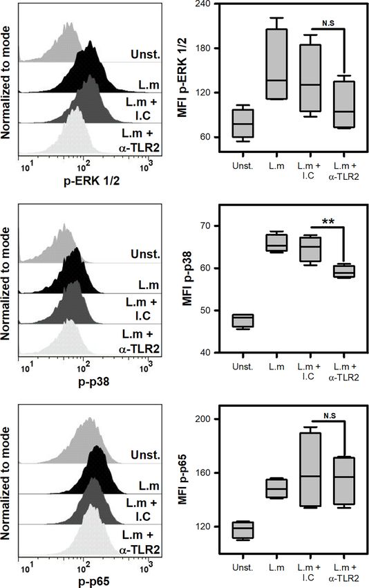
Soria-Castro et al.                                                                   TLR-2 Regulates Mast Cell-Activation by Listeria monocytogenes

INTRODUCTION                                                                Activation of TLR2 promotes diverse cellular functions such
                                                                         as phagocytic activity (23), bactericidal activity (24), ROS
Classically, mast cells (MC) are associated with type I                  production (25), degranulation (26) and cytokine production
hypersensitivity reactions (1). However, growing evidence has            (27). In addition, TLR2-deficient mice are more susceptible to
placed them as initiators of the inflammatory process against             L.m infection than wild type mice, which is consistent with poor
several infectious agents, including bacteria (2). Due to their          control of the bacterial load on target organs (28). In addition,
strategic location in mucosal epithelia, skin, and connective            MyD88-deficient MC produce less IL-6 and MCP-1 in response
tissue, they can respond immediately to the signals derived              to L.m (16). Considering that MC express TLR2 (29), we decided
from mutualistic and pathogenic bacteria, adapting their                 to dissect the activation mechanisms that are regulated by TLR2
response accordingly to maintain host homeostasis (3). In this           during mast cell activation by Listeria monocytogenes infection.
way, MC are provided with at least one member of each of the
Pattern Recognition Receptor (PRR) families. These include
Toll-Like Receptors (TLR), C-type Lectin Receptors (CLR),                MATERIAL AND METHODS
NOD-Like Receptors (NLRs), RIG-Like Receptors (RLRs), and
Scavenger Receptors (4). When MC are activated, they release             Bacteria Culture
many preformed mediators found in their granules, through cell           L.m strain 1778+H 1b (ATCC 43249, USA, Manassas, VA, USA)
degranulation. Furthermore, they can also synthesize de novo             was grown in brain heart infusion broth (BHI, BD-Difco, USA)
molecules such as inflammatory mediators derived from                     for 18 h at 37°C with constant shaking at 112 xg. Bacterial cultures
arachidonic acid, reactive oxygen species (ROS) as well as               were washed with Hanks Balanced Saline Solution (HBSS) (Life
cytokines and chemokines (5). Several studies have shown that            Technologies, USA) and bacterial pellets resuspended in RPMI-
MC are involved in the immune response to pathogenic bacteria,           1640 Glutamax (Life Technologies, USA) supplemented with 40%
including: Pseudomonas aeruginosa (6), Klebsiella pneumoniae             Fetal Bovine Serum (Life Technologies, USA) and frozen at -70 °C
(7), Staphylococcus aureus (8), Mycobacterium tuberculosis (9)           until use. Aliquots of L.m were serially diluted and plated in BHI
and Listeria monocytogenes (L.m) (10), to mention only a few.            agar at 37°C for 18-24 h. Bacterial numbers were determined by
    L.m is a Gram-positive, facultative intracellular bacteria and       counting Colony-Forming Units (CFU).
the causal agent of listeriosis, a foodborne disease with a high
mortality rate (11). L.m presents tropism towards the gravid             Mast Cells
uterus and central nervous system (CNS), contributing to the             Bone Marrow-derived Mast Cell (BMMC) was obtained
most severe clinical manifestations (12). Although, these severe         following the protocol described by (30). Briefly, bone marrow
cases are rare in immunocompetent individuals, they increase in          cells were obtained from femurs and tibias of 6-8-week-old
case of immunosuppression or immunodeficiencies (11). This                C57BL/6 female mice. Cells were maintained in RPMI-1640
implies that host immune response mechanisms are crucial to              supplemented with 10% FBS, 5 mM b-mercaptoethanol (Life
the containment of the bacteria, and its alteration can increase         Technologies, USA) and 2% antibiotic and antimycotic (Sigma,
susceptibility to the infection (13).                                    USA) (complete RPMI 1640 medium) plus 10 ng/mL of
    Cells of the innate immune response play a crucial role in           murine recombinant IL-3 (Peprotech, USA) and 10 ng/mL of
containing L.m infection, notably, macrophages, dendritic cells,         murine recombinant stem cell factor (Peprotech, USA)). Non-
neutrophils, NK cells and MC (14, 15). In experimental murine            adherent cells were transferred to fresh culture medium twice a
listeriosis models, MC have been shown to initiate the effector          week for 6−9 weeks. The purity of BMMC was ≥90% assessed by
response against this bacterium, promoting recruitment of                flow cytometry after staining of CD117 (clone: 2B8, BioLegend,
neutrophils and macrophages to the site of infection (10, 15).           USA; 0.25 mg/mL) and FcϵRI (clone: MAR-1, BioLegend, USA;
Furthermore, MC response to L.m includes intracellular                   0.16 mg/mL) (Supplementary Figure 1A).
infection (16), degranulation (16, 17), ROS production (18)                  Peritoneum-derived mast cells (PMC), cells were obtained
and different cytokines and chemokines (15, 16). However, it is          from peritoneal cavity of mice and cultured in complete RPMI-
unclear which receptors mediates L.m activation by MC.                   1640 medium plus IL-3 (30 ng/mL) and SCF (20 ng/mL). Non-
    TLR2 is a transmembrane type I receptor, which contains a            adherent cells were transferred to fresh culture medium twice a
cytoplasmic TIR domain as well as an extracellular domain with           week for 3−4 weeks. The purity of PMC was ≥90% assessed by
leucine-rich repeats (19). TLR2 ligands include: Diacil-                 flow cytometry (Supplementary Figure 1C).
lipopeptides, lipoarabinomannan, lipoproteins, lipoteichoic                  All experiments followed institutional biosecurity and safety
acid (LTA), peptidoglycan (PGN), porins, phospholipomannan               procedures. All animal experiments were approved by the
and zymosan (20). Once TRL2 binds to its ligand, it is dimerized         Research Ethics Committee of the ENCB, IPN (ZOO-016-2019).
with either TLR1 or TLR6 (21). When this occurs, TIR domain
recruits the adaptive molecules TIRAP and MyD88 that lead to             Toluidine Blue Staining
the activation of the transcription factor NF-kB and the mitogen-        2x10 5 BMMC or 2x10 5 PMC/0.25 mL of RPMI-1640
activated protein kinases (MAPK) that activate the transcription         supplemented with 10% FBS and 5mM b-mercaptoethanol
factor AP-1 (21, 22). Additionally, TLR2 activates the P13K-AKT          (complete medium) were plated in cytospin chambers, and
signaling pathway (22)                                                   then stained with toluidine blue for 10 minutes. Finally, slides

Frontiers in Immunology | www.frontiersin.org                        2                                      June 2021 | Volume 12 | Article 650779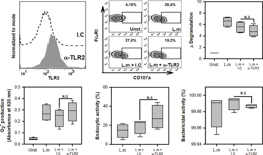
Soria-Castro et al.                                                                  TLR-2 Regulates Mast Cell-Activation by Listeria monocytogenes

were air-dried and mounted with Entellan resin (Merck                   supplemented with ampicillin at 200 ng/mL was added for 1 h.
Millipore, USA) under a coverslip. Images were captured with            Afterwards, cells were washed and then transferred to complete
a digital camera attached to a brightfield microscope (Zeiss             medium supplemented with ampicillin at 200 ng/mL for 0 and
Primo Star, Germany), and analyzed with Micro capture v7.9              24 h. Then, cells were washed with HBSS and then lysed with
software (Supplementary Figure 1B, D).                                  sterile distilled water. Cell suspension were homogenized, and
                                                                        serial decimal dilutions were prepared in saline solution.
Viability Mast Cell Assay                                               Afterwards, 20mL of each dilution were plated on BHI agar at
2.5X10⁵ BMMC/0.25 mL of complete medium were stimulated                 37°C for 48 h, bacterial numbers were determined by
with L.m at different MOI (1:1, 10:1, 100:1) or stimulated with         counting CFU.
LLO (125, 250, 500, 1000 ng/mL) for 24 h. Then cells were
washed with 1 mL of Annexin V binding Buffer 1X (Invitrogen,            Cytokines Quantification
USA) and stained with 1 mg/mL Annexin V (BioLegend, USA)                2x10⁵ BMMC/0.25 mL of complete medium were stimulated with
and 0.5 mg/mL propidium iodide (eBioscience, USA). After                L.m at different MOI or stimulated with Recombinant Listeriolysin-
staining, cell viability was measured by flow cytometry                  O (LLO; RayBiotech, USA) at different concentration (125, 250, 500
(Supplementary Figure 2).                                               and 1000 ng/mL) for 24h. Then, supernatants were collected for the
                                                                        detection of TNF-a, IL-1b, IL-6, MCP-1 (Biolegend, San Diego,
Degranulation Assay                                                     CA., USA) and IL-13 (eBioscience, USA) by ELISA according to
The degranulation assay was carried out as described previously         manufacturer’s instructions.
(31). Briefly, 2x10⁵ BMMC/0.25 mL of complete medium were
incubated with L.m at different MOI for 90 minutes. Then were           Evaluation of Phosphorylated Proteins
washed and stained with anti-CD107a (clone: 1D4B Biolegend,             2x10⁵ BMMC/0.25 mL of complete medium were stimulated
USA; 0.25 mg/mL) and anti-FcϵRI. Staining was measured by               with L.m MOI 100:1 for 15 minutes to evaluate p-ERK 1/2, 30
flow cytometry.                                                          minutes for p-p65 and 60 minutes for p-p38. Then, the cells were
    b-Hexosaminidase release was performed as follows: 2x10⁵            preserved with 250 mL of Fixation buffer (BD-Bioscience, USA)
BMMC/0.25 mL of HEPES-Tyrode Buffer (HBT) (130 mM                       for 10 min/37°C. Subsequently, were washed with 1 mL of Stain
NaCl, 5.5 mM glucose, 2.7 mM KCl, 1.0 mM CaCl2 2 H2O,                   Buffer (BD-Bioscience, USA) and permeabilized with 1mL of
0.1% [wt/vol] Bovine Serum Albumin (BSA), 12 mM HEPES,                  0.5x Perm buffer IV (BD-Bioscience, USA) for 15 min at room
0.45 mM NaH2PO4 1H2O, pH 7.2) were stimulated with L.m at               temperature (RT) and protected from light. Then, the cells were
different MOI or stimulated with PMA (125 nM) plus Ionomicin            washed with 1 mL of Stain Buffer. After blocking with 0.015 mg of
(10 mM) for 90 minutes at 37°C. The supernatants were then              anti-CD16/32 (Mouse BD Fc Block™, clone: 2.4G2. BD-
recovered, and the cell pellet was lysed with 200 mL of 0.2%            Biosciences, USA) cells were stained with antibodies to p-ERK
Triton X-100 in HBT. Both supernatants and cell lysates were            1/2-PE (clone: 20A; 4 mL per tube), p-p65-PE (clone: K10-
incubated with 4-methylumbelliferyl N-acetyl-b-D-                       895.12.50; 4 mL per tube), p-p38-PE (Clone: 36/p38; 4 mL per
glucosaminide (Sigma-Aldrich, USA; 1 mM in 200 mM Na                    tube) or isotype controls (Clone: MOPC-21; Mouse IgG1, k-PE
Citrate Buffer pH 4.5) for 2 h at 37°C. The enzyme reaction             or Clone: MCP-11; Mouse IgG 2b , k-PE). (All from BD-
was stopped by the addition of 100 mL of 200 mM Tris base, pH           Biosciences, USA) for 60 min at RT and protected from light.
10.7. The samples were analyzed in a fluorescence plate reader           Finally, the cells were washed and resuspended in 0.15 mL of
(SpectraMax M, USA) using excitation 356 nm and emission 450            Stain Buffer and analyzed by flow cytometry. The times chosen
nm. The percentage of release of b-Hexosaminidase is calculated         for the detection of each phosphorylated protein were selected
                                        supernatan
using the formula: % Release = ½ (supernatan+cellpellet)   100.       based on kinetic assay (Supplementary Figure 4).

Superoxide Anion(O−2 ) Production                                       Expression of TLR2
2x10⁵ BMMC were cultured in 0.25 mL of RPMI-1640 without                2x10⁵ BMMC/0.1 mL of PBS 1X were marked with anti-TLR2-
phenol red (Life Technologies, USA) and then, stimulated with           biotin (clone: 6C2, eBioscience, USA; 1 mg/0.1 mL) or isotype
L.m at different MOI for 120 minutes. In the last 15 minutes of         control-biotin (clone: eB149/10H5, eBioscience, USA; Rat IgG2b,
incubation 25 mL of a solution of p-nitro blue tetrazolium (NBT)        k-biotin; 1 mg/0.1 mL) and stained with streptavidin-APC (BD-
(Sigma-Aldrich, USA) at 1 mg/mL were added. Then, cells were            Bioscience, USA; 0.02 mg/mL), prior blocking with anti-CD16/
washed with PBS 1X (Life Technologies, USA) and fixed with               32. Staining was measured by flow cytometry.
absolute methanol. The formazan precipitates were dissolved by
adding 54 mL of potassium hydroxide (KOH) 2mM and 46 mL of              TLR2 Blocking Assays
Dimethyl sulfoxide (DMSO) (Sigma-Aldrich, USA). The samples             2x10⁵ BMMC/0.25 mL of complete medium were pre-incubated
were read at a wavelength of 620 nm in a plate reader (Multiskan        30 minutes with 200 ng/mL of anti-TLR2 (clone: C9A12; IgG2a)
EX, Thermo Scientific, USA).                                             or 200 ng/mL of isotype control (clone: T9C6; 1gG2a), both from
                                                                        Invivogen, USA. Afterwards, cells were stimulated with L.m MOI
Bacterial Load Assay                                                    100:1 and the bacterial load, O−2 production, degranulation,
2x10⁵ BMMC/0.25 mL of complete medium were incubated with               cytokines production and evaluation of phosphorylated
L.m at different MOI for 2 h. At the end of incubation, HBSS            proteins assays were performed as indicated above. The

Frontiers in Immunology | www.frontiersin.org                       3                                      June 2021 | Volume 12 | Article 650779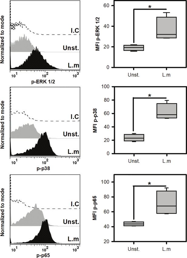
Soria-Castro et al.                                                                  TLR-2 Regulates Mast Cell-Activation by Listeria monocytogenes

bacterial load was analyzed as endocytic activity and bactericidal       reduction of more than 3 logarithms for any MOI tested
activity, according to the following formula: endocytic activity =       (Figure 1D). These results indicate that BMMC degranulate
(CFU at 0 h/MOI added) *100 (%). Bactericidal activity = (100-           and produce ROS with high amounts of L.m and that BMMC are
(CFU at 24 h/CFU at 0 h) *100) (%) (32). To evaluate the                 capable of internalizing and controlling L.m infection.
efficiency of antibody-mediated TLR2 inhibition, 2x10⁵ BMMC/
0.25 mL were pre-incubated with different concentrations of              Listeria monocytogenes Induces Mast Cell
anti-TLR2 (12.5, 25, 50, 100 y 200 ng/mL) for 30 min and then            Cytokine and Chemokine Release
stimulated with the TLR2 agonist: Staphylococcus aureus                  Previous reports have shown that L.m induces the de novo
peptidoglycan (PGN) (Sigma-Aldrich, USA) at 10 mg/mL for                 synthesis of different cytokines and chemokines (16, 33). To
24 h. Then, supernatants were collected for the detection of IL-6        further corroborate the MC response to L.m we determined the
by ELISA (Supplementary Figure 5).                                       production of TNF-a, IL-1b, IL-6, IL-13 and MCP-1. We
                                                                         stimulated BMMC with different MOI of L.m and at 24 h we
Flow Cytometry                                                           determined the levels of each mediator. We found that BMMC
All cell samples stained with fluorochrome-conjugated                     produced TNF-a with MOI 1:1, 10:1 and 100:1 of L.m
antibodies were acquired using FACSAria Fusion (BD                       (Figure 2A). While IL-1b production was only detected in
Biosciences, USA) and analyzed with FlowJo software version              BMMC incubated with MOI 100:1 of L.m (Figure 2B).
6.0 (FlowJo, LLC). Cell debris and doublets were excluded from           Similarly, we found that this bacterium induced IL-6
the analysis.                                                            (Figure 2C) and IL-13 (Figure 2D) only with MOI 100:1 of
                                                                         L.m in BMMC. While MCP-1 was induced with MOI 10:1 and
Statistical Analysis                                                     100:1 of L.m in BMMC (Figure 2E). Together, these results
All statistical analyses were performed with SigmaPlot software          indicate that L.m induces de novo synthesis of cytokines
version 14.0, from Systat Software, Inc., San Jose California USA,       and chemokines.
www.systatsoftware.com. Data normality was assessed by
Kolmogorov-Smirnov with Lilliefors correction. Variables that            Listeria monocytogenes Induces the
followed normal distribution were plotted as mean ± standard             Activation of Signaling Pathways
error mean (s.e.m), represented as bars and analyzed with one            Associated With TLR Activation on
way-analysis of variance (ANOVA) with Student-Newman-                    Mast Cells
Keuls (SNK) post-hoc. While variables that did not follow                Since L.m promoted the activation of BMMC, we decided to
normal distribution, semi-quantitative variables, normalized             evaluate if this was associated with the induction of some
variables or percentages, were plotted as median + range,                intracellular signals that are associated with TLR receptors
represented as boxes and analyzed with the Mann-Whitney                  (22). To this end, we incubated BMMC with L.m (MOI 100:1)
test (comparisons between two groups) or Kruskal-Wallis test             and determined the phosphorylation of some signaling proteins
(comparisons between more than two groups). A value of p <               by flow cytometry. Initially we evaluated the phosphorylation of
0.05 was considered to be significant.                                    ERK 1/2 (Figure 3A) and p38 (Figure 3B), molecules belonging
                                                                         to the MAPK signaling pathway and which have been related to
                                                                         the production of pro-inflammatory cytokines in macrophages
RESULTS                                                                  infected with L.m (34, 35). Interestingly, we found that L.m
                                                                         induced phosphorylation of both signaling proteins in BMMC
Mast Cell Activation Concurs With the                                    (Figures 3A, B). An important transcription factor in the
Control of Listeria monocytogenes                                        production of pro-inflammatory cytokines and chemokines by
Infection                                                                macrophages infected with L.m is NF-kB (34). Therefore, we
Previous studies have noticed that MC can respond to L.m                 determined whether this transcription factor was activated in
infection through degranulation, ROS p ro duction,                       BMMC in response to L.m, by evaluating the phosphorylation of
internalization, and clearance of this bacterium (16–18). To             p65 subunit. Interestingly, we found that L.m induced
corroborate the presence of these MC activation mechanisms               phosphorylation of p65 in BMMC (Figure 3C). Together,
during L.m infection, we incubated BMMC with different MOI               these results indicate that L.m induces the activation in BMMC
of this bacterium to determine degranulation either through the          of cell signaling molecules that are associated with
surface expression of CD107a (Figure 1A) or b-hexosaminidase             TLR activation.
release (Figure 1B), ROS production with detection of O−2
(Figure 1C) and internalization and clearance of L.m by                  TLR2 Is Not Involved in Mast Cell
evaluating intracellular CFU (Figure 1D). We found that                  Degranulation, ROS Release, Endocytosis
degranulation and O−2 production from BMMC occurred using                and Listeria monocytogenes Clearance
L.m MOI 100:1 (Figures 1A–C). Also, we detected viable                   One of the main receptors found in various cells of the innate
bacteria within BMMC at 0-hour post-infection (hpi) with the             immune response that recognizes different components of the
different MOI of L.m tested; however, we recovered small                 cell wall of Gram-positive bacteria, such as L.m, is TLR2 (20).
amounts of this bacterium at 24h, corresponding to a                     The importance of macrophage TLR2 for phagocytosis and ROS

Frontiers in Immunology | www.frontiersin.org                        4                                     June 2021 | Volume 12 | Article 650779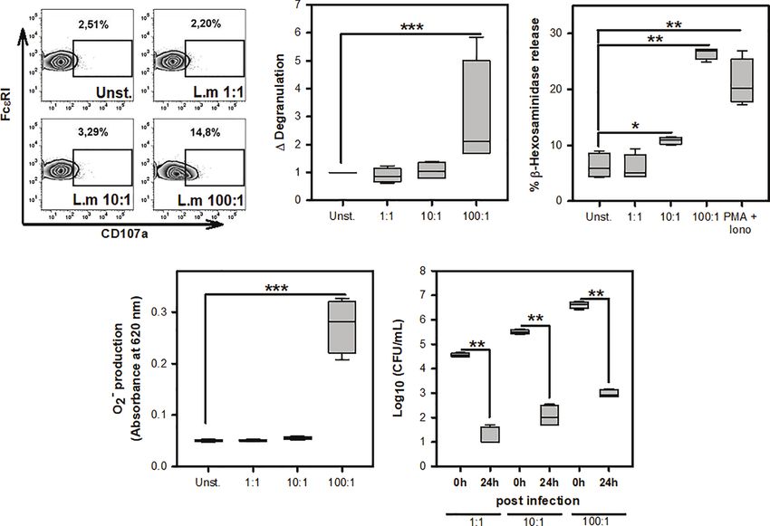
Soria-Castro et al.                                                                                 TLR-2 Regulates Mast Cell-Activation by Listeria monocytogenes

                       A                                                                               B

                                          C                                        D

 FIGURE 1 | Mast cell activation in response to Listeria monocytogenes infection. (A) 2.5X10⁵ BMMC/0.25 mL of complete medium were stimulated with L.m for 90
 minutes at the MOI indicated. BMMC degranulation was determined by flow cytometry. Left panel shows representative zebra-plots. Right panel shows the fold
 change in the percentage of FcϵRI+/CD107a+ cells with respect to unstimulated MC. (sum of 4 independent experiments, n=4 per group; ***p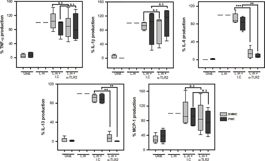
Soria-Castro et al.                                                                                   TLR-2 Regulates Mast Cell-Activation by Listeria monocytogenes

                        A                                        B                                       C

                                            D                                         E

 FIGURE 2 | Listeria monocytogenes induces de novo synthesis of cytokines by mast cells. 2.5x10⁵ BMMC/0.25 mL of complete medium were stimulated with L.m
 for 24 h at the MOI indicated. Mediator levels were evaluated in culture supernatants by ELISA. (A) TNF-a, (B) IL-1b, (C) IL-6, (D) IL-13 and (E) MCP-1. (The figure
 shows the sum of 3 independent experiments, n=3 per group and for each mediator; *p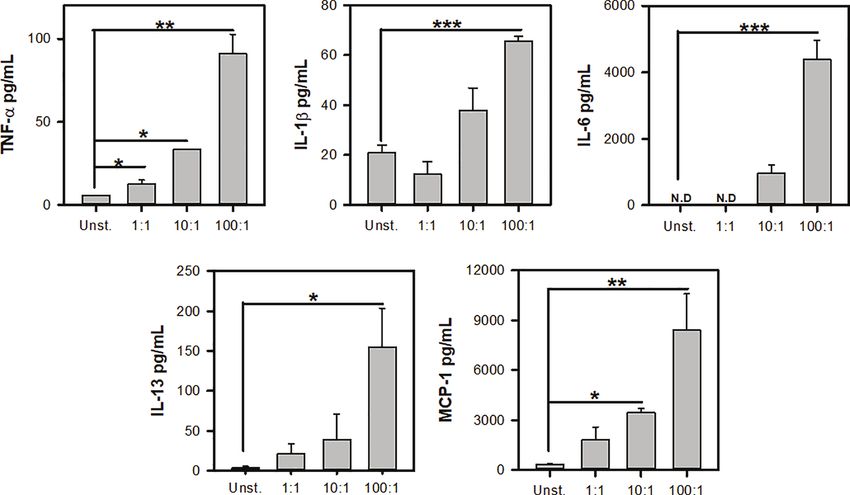
Soria-Castro et al.                                                                                TLR-2 Regulates Mast Cell-Activation by Listeria monocytogenes

                             A

                             B

                             C

 FIGURE 3 | Listeria monocytogenes induce the phosphorylation of cell signaling proteins involved in TLR activation in mast cells. 2.5x10⁵ BMMC/0.25 mL of
 complete medium were stimulated with L.m at MOI 100:1 for different times to determine the phosphorylation of (A) ERK 1/2 at 15 minutes, (B) p38 at 60 minutes
 and (C) p65 at 30 minutes by flow cytometry. The graphs show the median fluorescence intensity (MFI). (Sum of 4 independent experiments, n=4 per group and for
 each phospho-protein. *p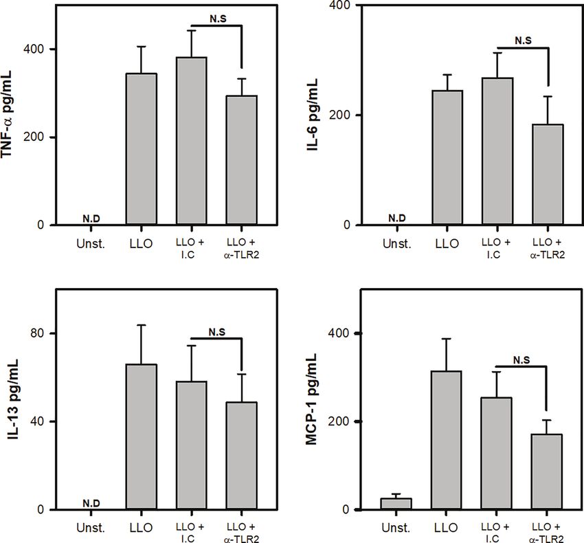
Soria-Castro et al.                                                                                TLR-2 Regulates Mast Cell-Activation by Listeria monocytogenes

                              A                                 B

                          C                                     D                                     E

 FIGURE 4 | Mast cell degranulation, ROS production, endocytosis and clearance of Listeria monocytogenes are TLR2-independent. (A). 2.5x10⁵ BMMC were
 marked with anti-TLR2-biotin (1 mg/0.1 mL) or biotinylated isotype control (I.C) (1 mg/0.1 mL) and stained with streptavidin-APC. Staining was evaluated by flow
 cytometry. (B) 2.5x10⁵ BMMC/0.25 mL of complete medium were preincubated with anti-TLR2 for 30 minutes and then were stimulated with L.m (MOI 100:1) for
 90 minutes. BMMC degranulation was determined by flow cytometry. Left panel shows representative zebra-plots. Right panel shows the fold change in the
 percentage of FcϵRI+/CD107a+ cells with respect to unstimulated MC (sum of 4 independent experiments, n=4 per group; N.S, Not Significance; Kruskal-Wallis
 test). (C) 2.5x10⁵ BMMC/0.25 mL of complete medium were preincubated with anti-TLR2 for 30 minutes. Afterwards, were stimulated with L.m (MOI 100:1) for
 2 h. Then, the determination was performed by the NBT reduction assay (sum of 4 independent experiments, n=4 per group; N.S, Not Significance; Kruskal-Wallis
 test). (D, E) 2.5x10⁵ BMMC/0.25 mL of complete medium were preincubated with anti-TLR2 for 30 minutes. Afterwards, were stimulated with L.m (MOI 100:1) for
 2 h. Then the O−2 intracellular bacteria were quantified by CFU assay. (D) Graphs show the percentage of endocytic activity (CFU at 0 h/MOI added x 100).
 (E) Graphs show the percentage of bactericidal activity (100-(CFU at 24 h/CFU at 0 h) x 100). (sum of 4 independent experiments, n=4 per group; N.S, Not
 Significance; Kruskal-Wallis test).

suggest that TLR2-independent interactions promote MC                                 MC release a wide variety of cytokines and chemokines in
degranulation against L.m.                                                         response to L.m, including TNF-a, IL-1b, IL-6, IL-13 and MCP-
    ROS production in L.m infected MC is associated with the                       1 (15, 16, 33). Interestingly, TNF-a or TNF-receptor deficient
release of MC extracellular traps (MCET), through a mechanism                      mice are highly susceptible to L.m infection (51–53). This could
dependent on NADPH oxidase, MCET contribute to L.m                                 be associated with the TNF-a exacerbation of intracellular killing
extracellular clearance (18). Moreover, ROS production also                        of L.m by macrophages through ROS and nitric oxide
promotes the intracellular clearance of internalized E. coli in                    production (54, 55). On the other hand, antibody-mediated IL-
MC endosomes (50). Furthermore, TLR2 promotes in L.m                               1 receptor blockade affects neutrophil recruitment and
infected macrophages to the release of mitochondrial ROS                           macrophage activation in L.m-infected mice (56), which
associated with TNF-a, IL-1b and IL-6 synthesis via NF-kB                          coincides with an increase in bacterial load in the spleen and
and MAPK activation (36). However, our findings suggest that                        liver (57). Similarly, mice deficient in MCP-1 are more
ROS release by L.m infected MC is not associated with                              susceptible to L.m infection, which correlates with poor
TLR2- interactions.                                                                recruitment of inflammatory monocytes (58). While IL-6
    Despite L.m endocytosis by macrophages has been shown be                       deficient mice are unable to mobilize neutrophils from the
dependent of PI3K-AKT-Rac1 signaling pathway induced by                            bone marrow into the blood circulation, making them highly
TLR2 (23), we did not find any involvement of TLR2 in L.m                           susceptible to L.m infection (59). Finally, administration of IL-13
endocytosis by MC. One possibility is that L.m entry does not                      to mice infected with L.m favors infection control, which
involve TLR2 in MC. Additionally, we also noticed that MC                          coincides with enhanced NK cells activation and an increase in
TLR2 did not participate in L.m intracellular clearance, contrary                  serum IL-12 concentration (60). Therefore, these mediators
to what is observed in TLR2-deficient MC infected with F.                           produced by MC may contribute significantly to host defense
tularensis (24). This suggests that different signals may                          against L. m infection.
promote the activation of intrinsic microbicidal mechanisms in                        In contrast to previous reports where it has been shown that
MC or that L.m virulence factors may promote the permanence                        TLR2-deficient MC exhibit a reduced production of TNF-a in
of this bacterium within MC (16).                                                  response to S. aureus PGN (29) and S. equi (61), we found TNF-

Frontiers in Immunology | www.frontiersin.org                                  8                                         June 2021 | Volume 12 | Article 650779
Soria-Castro et al. TLR-2 Regulates Mast Cell-Activation by Listeria monocytogenes
A B C
D E
FIGURE 5 | Mast cells TLR2 is required for IL-6 and IL-13 production in response to Listeria monocytogenes. 2.5x10⁵ BMMC or 2.5x10⁵ PMC in 0.25 mL of
complete medium were preincubated with anti-TLR2 for 30 minutes and then were stimulated with L.m (MOI 100:1) for 24 h. Mediators levels were evaluated in
culture supernatants by ELISA. (A) TNF-a, (B) IL-1b, (C) IL-6, (D) IL-13 and (E) MCP-1. Data are expressed as percentage of cytokine production, considering as
100% the production induced for L.m only. (Sum of 6 independent experiments, n=6 per group and for each mediator for BMMC (light grey boxes); sum of 4
independent experiments, n=4 per group and for each mediator for PMC (dark grey boxes); N.D, Not detected; N.S, Not Significance; **pSoria-Castro et al. TLR-2 Regulates Mast Cell-Activation by Listeria monocytogenes
A
B
C
FIGURE 6 | Mast cells TLR2 regulates p38 activation in response to Listeria monocytogenes. 2.5x10⁵ BMMC/0.25 mL of complete medium were preincubated with
anti-TLR2 for 30 minutes and then were stimulated with L.m (MOI 100:1) for different times to determine the phosphorylation of (A) ERK 1/2 at 15 minutes, (B) p38
at 60 minutes and (C) p65 at 30 minutes by flow cytometry. The graphs show the median fluorescence intensity (MFI). (The figure shows the sum of 4 independent
experiments, n=4 per group and for each phospho-protein. **pSoria-Castro et al. TLR-2 Regulates Mast Cell-Activation by Listeria monocytogenes
A B
C D
FIGURE 7 | Cytokine production by mast cells in response to Listeriolysin-O is independent of TLR2. 2.5x10⁵ BMMC/0.25 mL of complete medium were
preincubated with anti-TLR2 for 30 minutes and then were stimulated with 1000 ng/mL of Listeriolysin-O (LLO) for 24 h. Mediators levels were evaluated in culture
supernatants by ELISA. (A) TNF-a, (B) IL-6, (C) IL-13 and (D) MCP-1. (Sum of 4 independent experiments, n=4 per group and for each mediator; N.D, Not
detected; N.S, Not Significance; One Way ANOVA test).
Caco-2 cells, mediated mainly by LLO binding to cholesterol and and other mediators produced by MC are able to direct, amplify
subsequent pore formation. Interestingly, this effect correlates and perpetuate the Th2 immune response in allergy, helminthic
with the ability of L.m to invade and replicate into fibroblasts and infection and in oral immunization models (76–80). Interestingly
macrophages (72). Therefore, we suggest that ERK 1/2 activation type 2 response has been recently associated as a relevant
in L.m-stimulated MC may be LLO-dependent. However, it is mechanism during bacterial infection, in particular during the
unclear whether L.m takes advantage of this signaling pathway in skin infection with S. aureus, by favoring IgE production and
MC to invade and replicate in them beyond promoting the effector mechanisms regulated by MC (81). Our results suggest
induction of proinflammatory cytokines. that MC TLR2 could have a relevant role during the innate
Interestingly, we found that LLO induced the release of IL-6 immune response to bacteria and promote an environment that
and IL-13 by MC, without requiring TLR2 signaling. This shows favors type-2 immune response. However, it is unclear whether
that multiple L.m pathogen-associated molecular patterns this response could be elicited by L.m. Our findings suggest that
(PAMPs) lead to IL-6 and IL-13 production, indicating a vital IL-13 production by MC responds to different L.m-activated
role of these cytokines in the host immune response to L.m, and signals, some independent of TLR2 (as is the case with LLO).
suggests that LLO could be recognized by MC through other This is surprising since previous studies have shown that LLO
receptors, with TLR4 being a potential candidate (47, 73). inhibits Th2-mediated reactions in murine models of allergic
IL-13 together with IL-4, IL-5 and IL-10 belong to the group rhinitis (82). Thus, the functional role of IL-13 produced by MC
of cytokines of the type 2 response profile, since they promote the in the context of L.m infection needs to be further explored.
induction of the humoral immune response by favoring the In conclusion, our results show that L.m induces the
production of antibodies by B cells, diminish the cellular activation of signaling pathways in mast cells, mainly related to
immune response and lead to an anti-inflammatory cytokines synthesis. In addition, TLR2 participates in IL-6 and
environment (74, 75). The type response profile 2 cytokines IL-13 production and p38 activation. While TNF-a, IL-1b,
Frontiers in Immunology | www.frontiersin.org 11 June 2021 | Volume 12 | Article 650779Soria-Castro et al. TLR-2 Regulates Mast Cell-Activation by Listeria monocytogenes
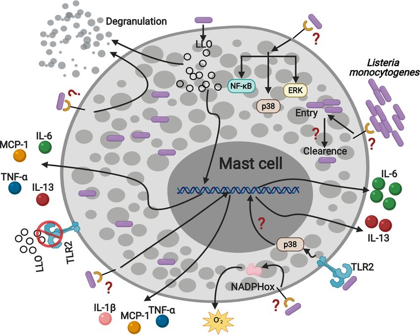
FIGURE 8 | Listeria monocytogenes activates mast cell through TLR2 inducing p38 activation, IL-6 and IL-13 production. MC are infected by L.m and they can
eliminate the intracellular bacterial load. In addition, L.m infection activates several signaling proteins: ERK, p38 and NF-kB, which could drive MC to degranulate,
ROS and cytokines production. Furthermore, the recognition of L.m by TLR2 promotes p38 activation and the production of IL-6 and IL-13. In addition, LLO induced
the production of TNF-a, IL-6, IL-13 and MCP-1 independently of TLR2, indicating that other mechanisms or mast cell receptors are regulating the interaction with
L.m. This figure was created with BioRender.com.
MCP-1 production, ROS release, cell degranulation, the MC-N, VA-J, JY-P, RM-F, and RC-S. Funding: AC-B and
endocytic and bactericidal activity of mast cells, as well as ERK RC-S. All authors contributed to the article and approved the
and NF-kB activation are TLR2-independent mechanisms. submitted version.
Therefore, we demonstrate that mast cell TLR2 plays a crucial
role in regulating the synthesis of IL-6 and IL-13 during Listeria
monocytogenes infection in MC.
FUNDING
This research was supported by SIP, IPN (20210225), and
DATA AVAILABILITY STATEMENT Conacyt Ciencia Bá sica (258738).
The original contributions presented in the study are included in
the article/Supplementary Material. Further inquiries can be
directed to the corresponding authors.
ACKNOWLEDGMENTS
We would like to thank Dr. Jeanet Serafı́n-Ló pez for providing us
with the L.m strain to perform the experiments, Jessica
ETHICS STATEMENT Castañeda-Casimiro for her technical assistance, and Dr.
Maricarmen Godı́nez-Victoria and Dr. Octavio Rodrı́guez-
The animal study was reviewed and approved by Research Ethics Corté s for facilitating the use of the central flow cytometry for
Committee of the ENCB, IPN. the analysis and acquisition of the cell samples. We also want to
thank Cameron Burnett for proofreading the manuscript.
AUTHOR CONTRIBUTIONS
SUPPLEMENTARY MATERIAL
Experimental design and analysis: RS-C, AA-D, GR-L, MC-N,
YM-P, AG-S, VA-J, JY-P, RM-F, AD-F, SE-P, SP-T, AC-B, The Supplementary Material for this article can be found
and RC-S. Experimental performing: RS-C, AA-D, GR-L, online at: https://www.frontiersin.org/articles/10.3389/
YM-P, and AG-S. Manuscript preparation: RS-C, GR-L, fimmu.2021.650779/full#supplementary-material
Frontiers in Immunology | www.frontiersin.org 12 June 2021 | Volume 12 | Article 650779Soria-Castro et al. TLR-2 Regulates Mast Cell-Activation by Listeria monocytogenes
REFERENCES 21. Zhu J, Mohan C. Toll-Like Receptor Signaling Pathways—Therapeutic
Opportunities. Mediators Inflamm (2010) 2010:1–7. doi: 10.1155/2010/781235
1. Galli SJ, Tsai M. Ige and Mast Cells in Allergic Disease. Nat Med (2012) 22. Espinosa-Riquer ZP, Segura-Villalobos D, Ramı́rez-Moreno IG, Pé rez
18:693–704. doi: 10.1038/nm.2755 Rodrı́guez MJ, Lamas M, Gonzalez-Espinosa C. Signal Transduction
2. Abraham SN, St. John AL. Mast Cell-Orchestrated Immunity to Pathogens. Pathways Activated by Innate Immunity in Mast Cells: Translating Sensing of
Nat Rev Immunol (2010) 10:440–52. doi: 10.1038/nri2782 Changes Into Specific Responses. Cells (2020) 9:1–39. doi: 10.3390/cells9112411
3. Garcia-Rodriguez KM, Bahri R, Sattentau C, Roberts IS, Goenka A, 23. Shen Y, Kawamura I, Nomura T, Tsuchiya K, Hara H, Dewamitta SR, et al.
Bulfone-Paus S. Human Mast Cells Exhibit an Individualized Pattern of Toll-Like Receptor 2- and MyD88-dependent Phosphatidylinositol 3-Kinase
Antimicrobial Responses. Immunity Inflamm Dis (2020) 8:198–210. and Rac1 Activation Facilitates the Phagocytosis of Listeria Monocytogenes by
doi: 10.1002/iid3.295 Murine Macrophages. Infect Immun (2010) 78:2857–67. doi: 10.1128/
4. Campillo-Navarro M, Chá vez-Blanco AD, Wong-Baeza I, Serafı́n-Ló pez J, IAI.01138-09
Flores-Mejı́a R, Estrada-Parra S, et al. Mast Cells in Lung Homeostasis: 24. Rodriguez AR, Yu J-J, Guentzel MN, Navara CS, Klose KE, Forsthuber TG,
Beyond Type I Hypersensitivity. Curr Respir Med Rev (2014) 10:115–23. et al. Mast Cell Tlr2 Signaling is Crucial for Effective Killing of Francisella
doi: 10.2174/1573398X10666141024220151 Tularensis. J Immunol (2012) 188:5604–11. doi: 10.4049/jimmunol.1200039
5. da Silva EZM, Jamur MC, Oliver C. Mast Cell Function: A New Vision of an 25. Shishido T, Nozaki N, Takahashi H, Arimoto T, Niizeki T, Koyama Y, et al.
Old Cell. J Histochem Cytochem (2014) 62:698–738. doi: 10.1369/ Central Role of Endogenous Toll-like Receptor-2 Activation in Regulating
0022155414545334 Inflammation, Reactive Oxygen Species Production, and Subsequent
6. Junkins RD, Carrigan SO, Wu Z, Stadnyk AW, Cowley E, Issekutz T, et al. Neointimal Formation After Vascular Injury. Biochem Biophys Res
Mast Cells Protect Against Pseudomonas Aeruginosa-Induced Lung Injury. Commun (2006) 345:1446–53. doi: 10.1016/j.bbrc.2006.05.056
Am J Pathol (2014) 184:2310–21. doi: 10.1016/j.ajpath.2014.05.009 26. Supajatura V, Ushio H, Nakao A, Akira S, Okumura K, Ra C, et al. Differential
7. Sutherland RE, Olsen JS, McKinstry A, Villalta SA, Wolters PJ. Mast Cell IL-6 Responses of Mast Cell Toll-like Receptors 2 and 4 in Allergy and Innate
Improves Survival From Klebsiella Pneumonia and Sepsis by Enhancing Immunity. JClinInvest (2002) 109:1351–9. doi: 10.1172/JCI14704
Neutrophil Killing. J Immunol (Baltimore Md : 1950) (2008) 181:5598–605. 27. Ghosh TK, Mickelson DJ, Fink J, Solberg JC, Inglefield JR, Hook D, et al. Toll-
doi: 10.4049/jimmunol.181.8.5598 Like Receptor (TLR) 2-9 Agonists-Induced Cytokines and Chemokines: I.
8. Lei Z, Zhang D, Lu B, Zhou W, Wang D. Activation of Mast Cells in Skin Comparison With T Cell Receptor-Induced Responses. Cell Immunol (2006)
Abscess Induced by Staphylococcus Aureus (s. Aureus) Infection in Mice. Res 243:48–57. doi: 10.1016/j.cellimm.2006.12.002
Vet Sci (2018) 118:66–71. doi: 10.1016/j.rvsc.2018.01.016 28. Torres D, Barrier M, Bihl F, Quesniaux VJF, Maillet I, Akira S, et al. Toll-Like
9. Garcia-Rodriguez KM, Goenka A, Alonso-Rasgado MT, Herná ndez-Pando R, Receptor 2 Is Required for Optimal Control of Listeria Monocytogenes
Bulfone-Paus S. The Role of Mast Cells in Tuberculosis: Orchestrating Innate Infection. Infection Immun (2004) 72:2131–9. doi: 10.1128/IAI.72.4.2131-
Immune Crosstalk? Front Immunol (2017) 8:1290. doi: 10.3389/ 2139.2004
fimmu.2017.01290 29. Supajatura V, Ushio H, Nakao A, Okumura K, Ra C, Ogawa H. Protective
10. Gekara NO, Weiss S. Mast Cells Initiate Early anti-Listeria Host Defences. Cell Roles of Mast Cells Against Enterobacterial Infection are Mediated by Toll-
Microbiol (2008) 10:225–36. doi: 10.1111/j.1462-5822.2007.01033.x Like Receptor 4. J Immunol (2001) 167:2250–6. doi: 10.4049/
11. Radoshevich L, Cossart P. Listeria Monocytogenes: Towards a Complete jimmunol.167.4.2250
Picture of its Physiology and Pathogenesis. Nat Rev Microbiol (2018) 16:32– 30. Campillo-Navarro M, Leyva-Paredes K, Donis-Maturano L, Rodrı́guez-Ló pez
46. doi: 10.1038/nrmicro.2017.126 GM, Soria-Castro R, Garcı́a-Pé rez BE, et al. Mycobacterium Tuberculosis
12. Vá zquez-Boland JA, Kuhn M, Berche P, Chakraborty T, Domı́ nguez-Bernal Catalase Inhibits the Formation of Mast Cell Extracellular Traps. Front
G, Goebel W, et al. Listeria Pathogenesis and Molecular Virulence Immunol (2018) 9:1161. doi: 10.3389/fimmu.2018.01161
Determinants. Clin Microbiol Rev (2001) 14:584–640. doi: 10.1128/ 31. Rodrı́guez-Ló pez GM, Soria-Castro R, Campillo-Navarro M, Pé rez-Tapia SM,
CMR.14.3.584-640.2001 Flores-Borja F, Wong-Baeza I, et al. The Histone Deacetylase Inhibitor
13. Soria-Castro R, Chá vez-Blanco AD, Garcı́a-Pé rez BE, Wong-Baeza I, Flores- Valproic Acid Attenuates Phospholipase Cg2 and IgE-mediated Mast Cell
Mejı́a R, Flores-Borja F, et al. Valproic Acid Inhibits Interferon-g Production Activation. J Leukocyte Biol (2020) 108:1–8. doi: 10.1002/JLB.3AB0320-547RR
by NK Cells and Increases Susceptibility to Listeria Monocytogenes Infection. 32. Kaneko M, Emoto Y, Emoto M. A Simple, Reproducible, Inexpensive, Yet
Sci Rep (2020) 10:1–14. doi: 10.1038/s41598-020-74836-w Old-Fashioned Method for Determining Phagocytic and Bactericidal
14. D’Orazio SEF. Innate and Adaptive Immune Responses During Listeria Activities of Macrophages. Yonsei Med J (2016) 57:283–90. doi: 10.3349/
Monocytogenes Infection. Microbiol Spectr (2019) 7:1–40. doi: 10.1128/ ymj.2016.57.2.283
microbiolspec.gpp3-0065-2019 33. Gekara NO, Dietrich N, Lyszkiewicz M, Lienenklaus S, Weiss S. Signals
15. Dietrich N, Rohde M, Geffers R, Kroger A, Hauser H, Weiss S, et al. Mast Cells Triggered by a Bacterial Pore-Forming Toxin Contribute to Toll-Like
Elicit Proinflammatory But Not Type I Interferon Responses Upon Activation Receptor Redundancy in Gram-Positive Bacterial Recognition. J Infect Dis
of TLRs by Bacteria. Proc Natl Acad Sci (2010) 107:8748–53. doi: 10.1073/ (2009) 199:124–33. doi: 10.1086/595562
pnas.0912551107 34. Stoiber D, Stockinger S, Steinlein P, Kovarik J, Decker T. Listeria
16. Jobbings CE, Sandig H, Whittingham-Dowd JK, Roberts IS, Bulfone-Paus S. Monocytogenes Modulates Macrophage Cytokine Responses Through Stat
Listeria Monocytogenes Alters Mast Cell Phenotype, Mediator and Serine Phosphorylation and the Induction of Suppressor of Cytokine
Osteopontin Secretion in a Listeriolysin-Dependent Manner. PLoS One Signaling 3. J Immunol (2001) 166:466–72. doi: 10.4049/jimmunol.166.1.466
(2013) 8:1–11. doi: 10.1371/journal.pone.0057102 35. Anand PK, Tait SWG, Lamkanfi M, Amer AO, Nunez G, Pagès G, et al. TLR2
17. Gekara NO, Westphal K, Ma B, Rohde M, Groebe L, Weiss S. The Multiple and RIP2 Pathways Mediate Autophagy of Listeria Monocytogenes Via
Mechanisms of Ca2+ Signalling by Listeriolysin O, the Cholesterol- Extracellular Signal-Regulated Kinase (ERK) Activation. J Biol Chem (2011)
Dependent Cytolysin of Listeria Monocytogenes. Cell Microbiol (2007) 286:42981–91. doi: 10.1074/jbc.M111.310599
9:2008–21. doi: 10.1111/j.1462-5822.2007.00932.x 36. Herb M, Gluschko A, Wiegmann K, Farid A, Wolf A, Utermöhlen O, et al.
18. Campillo-Navarro M, Leyva-Paredes K, Donis-Maturano L, Gonzá lez- Mitochondrial Reactive Oxygen Species Enable Proinflammatory Signaling
Jimé nez M, Paredes-Vivas Y, Cerbulo-Vá zquez A, et al. Listeria Through Disulfide Linkage of NEMO. Sci Signaling (2019) 12:1–15.
Monocytogenes Induces Mast Cell Extracellular Traps. Immunobiology doi: 10.1126/scisignal.aar5926
(2017) 222:432–9. doi: 10.1016/j.imbio.2016.08.006 37. Plum T, Wang X, Rettel M, Krijgsveld J, Feyerabend TB, Rodewald H-R.
19. Krishnan J, Selvarajoo K, Tsuchiya M, Lee G, Choi S. Toll-Like Receptor Human Mast Cell Proteome Reveals Unique Lineage, Putative Functions, and
Signal Transduction. Exp Mol Med (2007) 39:421–38. doi: 10.1038/ Structural Basis for Cell Ablation. Immunity (2020) 52:404–16.e5.
emm.2007.47 doi: 10.1016/j.immuni.2020.01.012
20. Oliveira-Nascimento L, Massari P, Wetzler LM. The Role of TLR2 Ininfection 38. Özören N, Masumoto J, Franchi L, Kanneganti T-D, Body-Malapel M, Ertürk
and Immunity. Front Immunol (2012) 3:79. doi: 10.3389/fimmu.2012.00079 I,̇ et al. Distinct Roles of TLR2 and the Adaptor ASC in IL-1b/Il-18 Secretion
Frontiers in Immunology | www.frontiersin.org 13 June 2021 | Volume 12 | Article 650779Soria-Castro et al. TLR-2 Regulates Mast Cell-Activation by Listeria monocytogenes
in Response to Listeria Monocytogenes. J Immunol (2006) 176:4337–42. 58. Jia T, Serbina NV, Brandl K, Zhong MX, Leiner IM, Charo IF, et al. Additive
doi: 10.4049/jimmunol.176.7.4337 Roles for MCP-1 and MCP-3 in CCR2-Mediated Recruitment of
39. Aubry C, Corr SC, Wienerroither S, Goulard C, Jones R, Jamieson AM, et al. Inflammatory Monocytes During Listeria Monocytogenes Infection.
Both TLR2 and TRIF Contribute to Interferon-b Production During Listeria J Immunol (2008) 180:6846–53. doi: 10.4049/jimmunol.180.10.6846
Infection. PLloS One (2012) 7:1–9. doi: 10.1371/journal.pone.0033299 59. Dalrymple SA, Lucian LA, Slattery R, McNeil T, Aud DM, Fuchino S, et al.
40. Kuhn M, Goebel W. Host Cell Signalling During Listeria Monocytogenes Interleukin-6-deficient Mice are Highly Susceptible to Listeria Monocytogenes
Infection. Trends Microbiol (1998) 6:11–5. doi: 10.1016/S0966-842X(97)01139-6 Infection: Correlation With Inefficient Neutrophilia. Infection Immun (1995)
41. Pengal RA, Ganesan LP, Wei G, Fang H, Ostrowski MC, Tridandapani S. 63:2262–8. doi: 10.1128/IAI.63.6.2262-2268.1995
Lipopolysaccharide-Induced Production of interleukin-10 is Promoted by the 60. Flesch I. Effects of IL-13 on Murine Listeriosis. Int Immunol (1997) 9:467–74.
Serine/Threonine Kinase Akt. Mol Immunol (2006) 43:1557–64. doi: 10.1016/ doi: 10.1093/intimm/9.4.467
j.molimm.2005.09.022 61. Rönnberg E, Guss B, Pejler G. Infection of Mast Cells With Live Streptococci
42. Krämer S, Sellge G, Lorentz A, Krueger D, Schemann M, Feilhauer K, et al. Selective Causes a Toll-Like Receptor 2- and Cell-Cell Contact-Dependent Cytokine
Activation of Human Intestinal Mast Cells by Escherichia Coli Hemolysin. and Chemokine Response. Infect Immun (2010) 78:854–64. doi: 10.1128/
J Immunol (2008) 181:1438–45. doi: 10.4049/jimmunol.181.2.1438 IAI.01004-09
43. Stassen M, Müller C, Richter C, Neudörfl C, Hültner L, Bhakdi S, et al. The 62. Eitel J, Suttorp N, Opitz B. Innate Immune Recognition and Inflammasome
Streptococcal Exotoxin Streptolysin O Activates Mast Cells To Produce Activation in Listeria Monocytogenes Infection. Front Microbiol (2011) 1:149.
Tumor Necrosis Factor Alpha by P38 Mitogen-Activated Protein Kinase- doi: 10.3389/fmicb.2010.00149
and Protein Kinase C-Dependent Pathways. Infection Immun (2003) 71:6171– 63. Hara H, Seregin SS, Yang D, Fukase K, Chamaillard M, Alnemri ES, et al. The
7. doi: 10.1128/IAI.71.11.6171-6177.2003 NLRP6 Inflammasome Recognizes Lipoteichoic Acid and Regulates Gram-
44. Masuda A, Yoshikai Y, Aiba K, Matsuguchi T. Th2 Cytokine Production From Positive Pathogen Infection. Cell (2018) 175:1651–64.e14. doi: 10.1016/
Mast Cells Is Directly Induced by Lipopolysaccharide and Distinctly j.cell.2018.09.047
Regulated by C-Jun N-Terminal Kinase and p38 Pathways. J Immunol 64. McCall-Culbreath KD, Li Z, Zhang Z, Lu LX, Orear L, Zutter MM. Selective,
(2002) 169:3801–10. doi: 10.4049/jimmunol.169.7.3801 a2b1 Integrin-Dependent Secretion of Il-6 by Connective Tissue Mast Cells.
45. Nieto-Patlá n A, Campillo-Navarro M, Rodrı́guez-Corté s O, Muñoz-Cruz S, J Innate Immun (2011) 3:459–70. doi: 10.1159/000324832
Wong-Baeza I, Estrada-Parra S, et al. Recognition of Candida Albicans by 65. Yoshimura T. The Chemokine MCP-1 (CCL2) in the Host Interaction With
Dectin-1 Induces Mast Cell Activation. Immunobiology (2015) 220:1093–100. Cancer: A Foe or Ally? Cell Mol Immunol (2018) 15:335–45. doi: 10.1038/
doi: 10.1016/j.imbio.2015.05.005 cmi.2017.135
46. Vaure C, Liu Y. A Comparative Review of Toll-Like Receptor 4 Expression 66. Siebenlist U, Franzoso G, Brown K. Structure, Regulation and Function
and Functionality in Different Animal Species. Front Immunol (2014) 5:316. of NF-Kappab. Annu Rev Cell Biol (1994) 10:405–55. doi: 10.1146/
doi: 10.3389/fimmu.2014.00316 annurev.cb.10.110194.002201
47. Park JM, Ng VH, Maeda S, Rest RF, Karin M. Anthrolysin O and Other 67. Hershko DD, Robb BW, Luo G, Hasselgren PO. Multiple Transcription
Gram-Positive Cytolysins are Toll-like Receptor 4 Agonists. J Exp Med (2004) Factors Regulating the IL-6 Gene are Activated by cAMP in Cultured Caco-
200:1647–55. doi: 10.1084/jem.20041215 2 Cells. Am J Physiol - Regul Integr Comp Physiol (2002) 283:1140–8.
48. Varadaradjalou S, Thieblemont N, Ben N, Pleau J, Dy M, Arock M. Toll-Like doi: 10.1152/ajpregu.00161.2002
Receptor 2 (TLR2) and TLR-4 Differentially Activate Human Mast Cells. Eur J 68. Lin J, Zhou Z, Huo R, Xiao L, Ouyang G, Wang L, et al. Cyr61 Induces Il-6
Immunol (2003) 2:899–906. doi: 10.1002/eji.200323830 Production by Fibroblast-like Synoviocytes Promoting Th17 Differentiation in
49. Pietrzak A, Wierzbicki M, Wiktorska M, Brzeziń ska-Błaszczyk E. Surface Rheumatoid Arthritis. J Immunol (2012) 188:5776–84. doi: 10.4049/
TLR2 and TLR4 Expression on Mature Rat Mast Cells Can be Affected by jimmunol.1103201
Some Bacterial Components and Proinflammatory Cytokines. Mediators 69. Li J, Lan T, Zhang C, Zeng C, Hou J, Yang Z, et al. Reciprocal Activation
Inflamm (2011) 2011:1–11. doi: 10.1155/2011/427473 Between IL-6/STAT3 and NOX4/Akt Signalings Promotes Proliferation and
50. Malaviya R, Ross EA, MacGregor JI, Ikeda T, Little JR, Jakschik BA, et al. Mast Survival of non-Small Cell Lung Cancer Cells. Oncotarget (2015) 6:1031–48.
Cell Phagocytosis of FimH-expressing Enterobacteria. J Immunol (Baltimore doi: 10.18632/oncotarget.2671
Md : 1950) (1994) 152:1907–14. 70. Carmody RJ, Chen YH. Nuclear factor-kappaB: Activation and Regulation
51. Fontan E. Early Translocation of Acid-Adapted Listeria Monocytogenes During Toll-Like Receptor Signaling. Cell Mol Immunol (2007) 4:31–41.
During Enteric Infection in TNF/Lta–/– Mice. FEMS Microbiol Lett (2001) 71. Drube S, Kraft F, Dudeck J, Müller A-L, Weber F, Göpfert C, et al. Mk2/3 Are
205:179–83. doi: 10.1016/S0378-1097(01)00450-5 Pivotal for IL-33–Induced and Mast Cell–Dependent Leukocyte Recruitment
52. Rothe J, Lesslauer W, Lötscher H, Lang Y, Koebel P, Köntgen F, et al. Mice and the Resulting Skin Inflammation. J Immunol (2016) 197:3662–8.
Lacking the Tumour Necrosis Factor Receptor 1 are Resistant to TNF- doi: 10.4049/jimmunol.1600658
mediated Toxicity But Highly Susceptible to Infection by Listeria 72. Cheng C, Sun J, Yu H, Ma T, Guan C, Zeng H, et al. Listeriolysin O Pore-
Monocytogenes. Nature (1993) 364:798–802. doi: 10.1038/364798a0 Forming Activity is Required for ERK1/2 Phosphorylation During Listeria
53. Endres R, Luz A, Schulze H, Neubauer H, Fütterer A, Holland SM, et al. Monocytogenes Infection. Front Immunol (2020) 11:1146. doi: 10.3389/
Listeriosis in p47phox–/– and TRp55–/– Mice: Protection Despite Absence of fimmu.2020.01146
ROI and Susceptibility Despite Presence of RNI. Immunity (1997) 7:419–32. 73. Herná ndez-Flores KG, Vivanco-Cid H. Biological Effects of Listeriolysin O:
doi: 10.1016/S1074-7613(00)80363-5 Implications for Vaccination. BioMed Res Int (2015) 2015:1–9. doi: 10.1155/
54. Müller M, Althaus R, Fröhlich D, Frei K, Eugster H-P. Reduced Antilisterial 2015/360741
Activity of TNF-deficient Bone Marrow-Derived Macrophages is Due to Impaired 74. Lucey DR, Clerici M, Shearer GM. Type 1, and Type 2 Cytokine Dysregulation
Superoxide Production. Eur J Immunol (1999) 29:3089–97. doi: 10.1002/(SICI) in Human Infectious, Neoplastic, and Inflammatory Diseases. Clin Microbiol
1521-4141(199910)29:103.0.CO;2-D Rev (1996) 9:532–62. doi: 10.1128/cmr.9.4.532
55. Leenen PJM, Canono BP, Drevets DA, Voerman JS, Campbell PA. TNF- 75. Scheller J, Chalaris A, Schmidt-Arras D, Rose-John S. The Pro- and Anti-
Alpha and IFN-gamma Stimulate a Macrophage Precursor Cell Line to Kill Inflammatory Properties of the Cytokine Interleukin-6. Biochim Biophys Acta
Listeria Monocytogenes in a Nitric Oxide-Independent Manner. J Immunol - Mol Cell Res (2011) 1813:878–88. doi: 10.1016/j.bbamcr.2011.01.034
(Baltimore Md : 1950) (1994) 153:5141–7. 76. Kitawaki T, Kadowaki N, Sugimoto N, Kambe N, Hori T, Miyachi Y, et al.
56. Rogers HW, Tripp CS, Schreiber RD, Unanue ER. Endogenous IL-1 is IgE-activated Mast Cells in Combination With Pro-Inflammatory Factors
Required for Neutrophil Recruitment and Macrophage Activation During Induce Th2-promoting Dendritic Cells. Int Immunol (2006) 18:1789–99.
Murine Listeriosis. J Immunol (Baltimore Md : 1950) (1994) 153:2093–101. doi: 10.1093/intimm/dxl113
57. Havell EA, Moldawer LL, Helfgott D, Kilian PL, Sehgal PB. Type I IL-1 77. Liu Z-Q, Song J-P, Liu X, Jiang J, Chen X, Yang L, et al. Mast Cell-Derived
Receptor Blockade Exacerbates Murine Listeriosis. J Immunol (Baltimore Md : Serine Proteinase Regulates T Helper 2 Polarization. Sci Rep (2014) 4:1–7.
1950) (1992) 148:1486–92. doi: 10.1038/srep04649
Frontiers in Immunology | www.frontiersin.org 14 June 2021 | Volume 12 | Article 650779Soria-Castro et al. TLR-2 Regulates Mast Cell-Activation by Listeria monocytogenes
78. Ryan NM, Oghumu S. Role of Mast Cells in the Generation of a T-helper Type Ovalbumin in Mice. Clin Exp Immunol (2006) 144:475–84. doi: 10.1111/
2 Dominated Anti-Helminthic Immune Response. Biosci Rep (2019) 39:1–11. j.1365-2249.2006.03092.x
doi: 10.1042/BSR20181771
79. Kool M, Hammad H, Lambrecht BN. Cellular Networks Controlling Th2 Polarization Conflict of Interest: The authors declare that the research was conducted in the
in Allergy and Immunity. F1000 Biol Rep (2012) 4:1–12. doi: 10.3410/B4-6 absence of any commercial or financial relationships that could be construed as a
80. Aoki I, Itoh S, Yokota S, Tanaka SI, Ishii N, Okuda K, et al. Contribution of potential conflict of interest.
Mast Cells to the T Helper 2 Response Induced by Simultaneous
Subcutaneous and Oral Immunization. Immunology (1999) 98:519–24. Copyright © 2021 Soria-Castro, Alfaro-Doblado, Rodrı ́guez-López, Campillo-Navarro,
doi: 10.1046/j.1365-2567.1999.00878.x Meneses-Preza, Galán-Salinas, Alvarez-Jimenez, Yam-Puc, Munguı ́a-Fuentes,
81. Starkl P, Watzenboeck ML, Popov LM, Zahalka S, Hladik A, Lakovits K, et al. Domı ́nguez-Flores, Estrada-Parra, Pérez-Tapia, Chávez-Blanco and Chacón-Salinas.
Ige Effector Mechanisms, in Concert With Mast Cells, Contribute to Acquired This is an open-access article distributed under the terms of the Creative Commons
Host Defense Against Staphylococcus Aureus. Immunity (2020) 53:1–12. Attribution License (CC BY). The use, distribution or reproduction in other forums is
doi: 10.1016/j.immuni.2020.08.002 permitted, provided the original author(s) and the copyright owner(s) are credited and
82. Yamamoto K, Kawamura I, Tominaga T, Nomura T, Ito J, Mitsuyama M. that the original publication in this journal is cited, in accordance with accepted
Listeriolysin O Derived From Listeria Monocytogenes Inhibits the academic practice. No use, distribution or reproduction is permitted which does not
Effector Phase of an Experimental Allergic Rhinitis Induced by comply with these terms.
Frontiers in Immunology | www.frontiersin.org 15 June 2021 | Volume 12 | Article 650779You can also read