C-Abl Kinase Is Required for Satellite Cell Function Through Pax7 Regulation - Frontiers
←
→
Page content transcription
If your browser does not render page correctly, please read the page content below
ORIGINAL RESEARCH
                                                                                                                                             published: 11 March 2021
                                                                                                                                       doi: 10.3389/fcell.2021.606403

                                            c-Abl Kinase Is Required for Satellite
                                            Cell Function Through Pax7
                                            Regulation
                                            Fabián Montecino 1 , Natalia González 1 , Natasha Blanco 1 , Manuel J. Ramírez 1 ,
                                            Adrián González-Martín 2 , Alejandra R. Alvarez 2 and Hugo Olguín 1*
                                            1
                                             Laboratory of Tissue Repair and Adult Stem Cells, Department of Molecular and Cell Biology, Faculty of Biological
                                            Sciences, Pontificia Universidad Católica de Chile, Santiago, Chile, 2 CARE-UC, Department of Molecular and Cell Biology,
                                            Faculty of Biological Sciences, Pontificia Universidad Católica de Chile, Santiago, Chile

                                            Satellite cells (SCs) are tissue-specific stem cells responsible for adult skeletal muscle
                                            regeneration and maintenance. SCs function is critically dependent on two families of
                                            transcription factors: the paired box (Pax) involved in specification and maintenance
                                            and the Muscle Regulatory Factors (MRFs), which orchestrate myogenic commitment
                            Edited by:
                                            and differentiation. In turn, signaling events triggered by extrinsic and intrinsic stimuli
                         D. Cornelison,     control their function via post-translational modifications, including ubiquitination and
  University of Missouri, United States     phosphorylation. In this context, the Abelson non-receptor tyrosine kinase (c-Abl)
                        Reviewed by:        mediates the activation of the p38 α/β MAPK pathway, promoting myogenesis. c-Abl
                    Alessandro Magli,
  University of Minnesota Twin Cities,      also regulates the activity of the transcription factor MyoD during DNA-damage stress
                         United States      response, pausing differentiation. However, it is not clear if c-Abl modulates other key
                   Julia Von Maltzahn,
      Leibniz Institute on Aging, Fritz
                                            transcription factors controlling SC function. This work aims to determine the role of
    Lipmann Institute (FLI), Germany        c-Abl in SCs myogenic capacity via loss of function approaches in vitro and in vivo. Here
                  *Correspondence:          we show that c-Abl inhibition or deletion results in a down-regulation of Pax7 mRNA
                         Hugo Olguín        and protein levels, accompanied by decreased Pax7 transcriptional activity, without a
                   holguin@bio.puc.cl
                                            significant effect on MRF expression. Additionally, we provide data indicating that Pax7
                    Specialty section:      is directly phosphorylated by c-Abl. Finally, SC-specific c-Abl ablation impairs muscle
         This article was submitted to
                                            regeneration upon acute injury. Our results indicate that c-Abl regulates myogenic
                  Stem Cell Research,
                a section of the journal    progression in activated SCs by controlling Pax7 function and expression.
  Frontiers in Cell and Developmental
                                            Keywords: c-Abl, pax7, satellite cells, muscle stem cells, MRFs, skeletal muscle regeneration, muscle
                                Biology
                                            differentiation
      Received: 14 September 2020
       Accepted: 08 February 2021
         Published: 11 March 2021
                                           INTRODUCTION
                              Citation:
            Montecino F, González N,       Skeletal muscle has a remarkable capacity to regenerate in response to injury or pathologic
              Blanco N, Ramírez MJ,        conditions (Olguín and Pisconti, 2012). This capability rests fundamentally in a group of tissue-
   González-Martín A, Alvarez AR and
                                           specific stem cells called Satellite Cells (SCs). These cells are typically maintained in a quiescent
     Olguín H (2021) c-Abl Kinase Is
   Required for Satellite Cell Function
                                           state (G0 ) localized at the myofiber periphery, between the sarcolemma and the basal lamina (Muir
           Through Pax7 Regulation.        et al., 1965; Hawke and Garry, 2001). In response to acute muscle damage, SCs are activated and
      Front. Cell Dev. Biol. 9:606403.     re-enter the cell cycle. After a robust proliferation period, muscle progenitors can fuse to form a
     doi: 10.3389/fcell.2021.606403        new myofiber or an injured muscle fiber to regenerate tissue function (Partridge, 2002).

Frontiers in Cell and Developmental Biology | www.frontiersin.org                  1                                           March 2021 | Volume 9 | Article 606403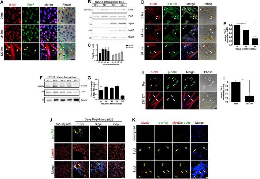
Montecino et al.                                                                                           c-Abl Regulates Pax7 During Myogenesis

    Among other critical regulators, the transcription factor Pax7        MATERIALS AND METHODS
controls the myogenic specification, and survival of SCs. In fact,
in animal models where Pax7 is deleted, the number of SCs                 Cell Culture
rapidly decay after birth, via apoptosis and early differentiation        Adult primary myoblasts and isolated myofibers were obtained
(Relaix et al., 2006; Buckingham and Relaix, 2007; von Maltzahn           as described (Cornelison et al., 2004) and maintained in
et al., 2013). On the other hand, Pax7 overexpression induces a           proliferation medium, F-12C (Life technologies, United States)
quiescent-like state in muscle progenitors, linking its function          supplemented with 15% Horse Serum (HS) (Hyclone,
to SC self-renewal (Olguin and Olwin, 2004). Moreover, high               United States) and 500 pM of FGF-2 at 37◦ C, 6% O2 and
Pax7 levels inhibit MyoD function and myoblast differentiation            5% CO2 . When required, the cells were induced to differentiate
(Olguin et al., 2007).                                                    by replacing the proliferation medium with differentiation
    The Muscle Regulatory Factor (MRF) MyoD, is considered                medium, F-12C, supplemented with 15% HS. For in vitro
the master gene of myogenesis since its ectopic expression in             recombination experiments, primary myoblasts were treated
non-myogenic cells results in the activation of the entire muscle         with 10 µM Tamoxifen T-5648 (Sigma-Aldrich, United States)
differentiation program (Tapscott et al., 1988; Weintraub et al.,         dissolved in ethanol 100% once every 24 h for 2 days.
1989). Pax7 and MyoD co-expression are fundamental in the                    C2C12 myoblasts were maintained in proliferation medium,
first hours after SC activation allowing lineage commitment               DMEM (Thermo Scientific, United States) supplemented
and proliferation while avoiding precocious differentiation               with 10% Fetal Bovine Serum (FBS) (Biological Industries,
(Olguín and Pisconti, 2012). MyoD will eventually induce the              United States) at 37◦ C and 5% CO2 . For differentiation
expression of the MRF Myogenin, which directs myogenic                    experiments, proliferation medium was replaced by
progression to terminal differentiation (Hollenberg et al.,               differentiation medium, DMEM, supplemented with 5%
1993). Different mechanisms are involved in the post-                     HS. Cells with two or more nuclei were considered myotubes.
translational regulation of Pax7 and the MRFs, including                  For c-Abl inhibition assays, C2C12 cells were treated with
the ubiquitin-proteasome system, caspase-mediated proteolysis,            10 µM Imatinib Mesylate (Sigma-Aldrich, United States) for the
and phosphorylation, among other mechanisms (Olguín,                      indicated time in every experiment. For c-Abl activation, cells
2011; Bustos et al., 2015; Dick et al., 2015; González et al.,            were treated with DPH (Sigma-Aldrich, United States) for the
2016; de la Vega et al., 2020). In this context, the Abelson              indicated time and concentrations. DMSO (Sigma-Aldrich) was
non-receptor tyrosine kinase 1 (c-Abl) regulates MyoD activity            used as a vehicle.
during DNA damage response (Simonatto et al., 2013). In
that condition, c-Abl phosphorylates MyoD at the N-terminal
domain (Tyr30), blocking MyoD-dependent gene expression,                  Immunofluorescence Staining
and pausing myogenesis until DNA is repaired (Puri et al., 2002;          Initially, C2C12 cells were plated and maintained as described
Innocenzi et al., 2011).                                                  above. Primary myoblasts were seeded onto 0.66% gelatin-coated
    Early reports described different c-Abl expression levels             glass-slides and maintained as described previously. When
in proliferative (high) and differentiated (low) muscle cells             specified, cells were fixed with 4% paraformaldehyde (PFA)
(Claycomb and Lanson, 1987). Recent work showed that c-Abl                for 10 min, then permeabilized with PBS 0.5% Triton X-100
regulates differentiation in the muscle-derived cell line C2C12           (Sigma-Aldrich, United States) for 5 min and blocked with PBS
by activating the p38 α/β MAPK pathway. c-Abl knock-down                  3% BSA (Sigma-Aldrich, United States) for 60 min and subjected
experiments in C2C12 cells resulted in decreased differentiation,         to standard immunofluorescence (IF) staining (Bustos et al.,
reducing myoblasts fusion, and Myosin Heavy Chain (MyHC)                  2015). The following primary antibodies and dilutions were used:
expression (Bae et al., 2009). Besides, c-Abl exhibits differential       mouse monoclonal anti-Pax7 1:1, anti-Myogenin (F5D) 1:2,
subcellular localization in myoblasts (nuclear) and myotubes              and anti-MyHC (MF20) 1:1 (Hybridoma conditioned medium,
(cytoplasmic), suggesting that c-Abl could exert different                Developmental Studies Hybridoma Bank, United States); rabbit
functions during myogenesis, depending on differentiation                 polyclonal anti-c-Abl (K-12), 1:200 (Santa Cruz Biotechnology,
status and its subcellular localization (Puri et al., 2002;               United States); rabbit polyclonal anti-c-Abl, 1:250 (Cell Signaling,
di Bari et al., 2006).                                                    United States); mouse monoclonal anti-c-Abl (ABL-148), 1:250
    Despite these observations, the role of c-Abl in the                  (Sigma-Aldrich, United States); rabbit polyclonal anti-phospho-
regulation of Pax7 and MRFs expression and function in adult              c-Abl (Tyr412), 1:250 (Sigma-Aldrich, United States); rabbit
muscle progenitors is unclear. Here we describe that c-Abl                polyclonal anti-phospho-c-Abl (Tyr412), 1:100 (Millipore,
is required for myoblasts differentiation and proper muscle               United States); rat monoclonal anti-MyoD (5F11), 1:100
regeneration upon injury. We found that inhibition or deletion            (Merck, United States); goat polyclonal anti-Myogenin (N-20),
of c-Abl results in decreased Pax7 expression and transcriptional         1:500 (Santa Cruz Biotechnology, United States); and chicken
activity in activated SCs, which finally converges in poor                anti-Syndecan-4, 1:500 (Cornelison et al., 2004). Secondary
differentiation and impaired muscle regeneration. Furthermore,            antibodies and dilutions were: donkey anti-mouse IgG Alexa
we show that c-Abl interacts with and phosphorylates Pax7                 555; donkey anti-rabbit IgG Alexa 555; donkey anti-rabbit
protein. Together, these results suggest that c-Abl regulates             IgG Alexa 488; donkey anti-mouse IgG Alexa 488; donkey
SC function during muscle regeneration by controlling Pax7                anti-rat IgG Alexa 555; donkey anti-goat IgG Alexa 555; donkey
expression and activity.                                                  anti-rat IgG Alexa 488; and goat anti-chicken IgY Alexa 555,

Frontiers in Cell and Developmental Biology | www.frontiersin.org     2                                      March 2021 | Volume 9 | Article 606403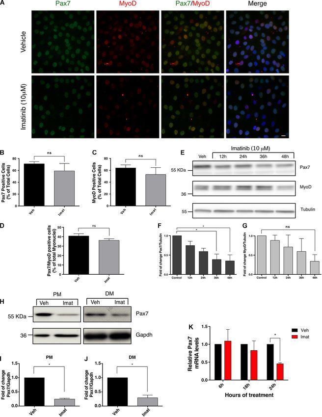
Montecino et al.                                                                                         c-Abl Regulates Pax7 During Myogenesis

1:500 (Life technologies, United States); 1 µg/mL of Hoechst              United States). Secondary antibodies and dilutions were donkey
33342 (Sigma-Aldrich, United States) was added to nuclei                  anti-rabbit IgG Alexa 488; donkey anti-mouse IgG Alexa 647;
counterstaining. Fluoromount (Sigma-Aldrich, United States)               goat anti-chicken IgY Alexa 555; donkey anti-rat IgG Alexa
was used for mounting. Fluorescence was evaluated using a                 647; donkey anti-goat IgG Alexa 647; and donkey anti-rat
Motic BA410 Elite Trinocular microscope equipped with a                   IgG Alexa 555 at 1:500 (Thermo Scientific, United States);
refrigerated Moti-cam Pro 252B camera. Images were acquired               1 µg/mL of Hoechst 33342 was added to nuclei counterstaining.
using the MOTIC IMAGES PLUS 3.0 software. Image analysis                  Fluoromount (Sigma-Aldrich, United States) was used for
were performed using ImageJ software (Schneider et al., 2012).            mounting. For Hematoxylin/Eosin (H&E) staining, sections
                                                                          were fixed with 4% PFA, rinsed with distilled water, and stained
Mice Strains and in vivo                                                  with hematoxylin for 5 min. Then, sections were washed with tap
                                                                          water and stained with eosin for 1 min. Sections were dehydrated
Tamoxifen-Induced Recombination and
                                                                          on an ascending ethanol concentration battery and mounted
Muscle Injury                                                             with Entellan (Sigma-Aldrich, United States).
For conditional c-Abl deletion in SCs, we generated C57BL/6J
Abl1flox/flox -Pax7creERT2 transgenic mouse line (from JAX
stock #013224 and #017763, Jackson Laboratories) (Murphy                  Western Blotting and
et al., 2011). To induce recombination, mice were injected                Co-immunoprecipitation
via intraperitoneal with 1 mg of Tamoxifen T-5648 (Sigma-                 Cells and tissue were lysed in modified RIPA buffer (50 mM
Aldrich, United States) dissolved in sesame oil with 5% Ethanol           Tris-HCl pH 7.4, 150 mM NaCl, 1% IGEPAL), supplemented
(Sigma-Aldrich, United States) per day, for 5 consecutive days.           with Protease Inhibitor Cocktail Set III (Merck, United States)
Seventy two hours after the last tamoxifen dose, muscle injury            and phosphatase inhibitors (1 mM sodium orthovanadate,
was performed as described previously (Cornelison et al.,                 5 mM sodium fluoride, 1 mM β-glycerophosphate, and 1 mM
2004). Briefly, Tibialis anterior (TA) muscles from 12 weeks              pyrophosphate). 10–30 µg of total protein was loaded into
old male mice were injected with 50 µL of 1.2% of barium                  10% SDS-Polyacrylamide gel electrophoresis (PAGE) gels and
chloride (BaCl2 ) diluted in saline (0.9% NaCl); contralateral            transferred to polyvinylidene fluoride (PVDF) membranes.
TAs were injected with 50 µL of saline as control. TA muscles             Western blotting (WB) was performed with the following
were extracted at 7-, 15-, and 30-days post-injury (dpi) and              primary antibodies and dilutions: mouse monoclonal anti-
processed for histological analyses, IF, or lysis. For in vitro SCs       Pax7, anti-MyHC (MF20) and anti-Myogenin (F5D), 1:5
tracing Abl1flox/flox -Pax7creERT2 were bred with B6.129(Cg)-             (Hybridoma conditioned medium, Developmental Studies
Gt(ROSA)26SorTM4(ACTB−tdTomato,−EGFP)Luo/J          (ROSAmT/mG )          Hybridoma Bank, United States); mouse monoclonal
reporter mice, obtained from Jackson Laboratories (JAX stock              anti-c-Abl (24-11), 1:1000 (Santa Cruz Biotechnology,
#007576). This model allows us to identify recombinant cell               United States); rabbit polyclonal anti-c-Abl, 1:2000 (Cell
populations by the expression of cell membrane-localized EGFP             Signaling, United States); mouse monoclonal anti-c-Abl
(mGFP), in contrast with the expression of membrane-tdTomato              (ABL-148), 1:2000 (Sigma-Aldrich, United States); rabbit
(mTomato) in all non-recombinant cells. Only Pax7 expressing              polyclonal anti-phospho-Abl (Tyr412), 1:1000 (Millipore,
cells going under recombination.                                          United States); rat monoclonal anti-MyoD (5F11), 1:1000
   All animal procedures were performed according to National             (Merck, United States); mouse monoclonal anti-myc-
Commission for Science and Technology (CONICYT) guidelines                tag (9B11), 1:5000 (Cell Signaling, United States); mouse
and approved by the School of Biological Sciences and                     monoclonal anti-α-Tubulin (B-5-1-2), 1:20000 (Sigma-Aldrich,
the Pontificia Universidad Católica de Chile Bioethics and                United States); mouse monoclonal anti-Gapdh (6C5), 1:10000
Biosecurity Committee (protocol ID 160929002).                            (Millipore, United States); and mouse monoclonal anti-
                                                                          phosphotyrosine (4G10), 1:2500 (Millipore, United States).
Muscle Tissue Staining                                                    Secondary antibodies used were HRP conjugated: anti-mouse
Tibialis anterior (TA) muscles from 8–12 weeks old male                   IgG (Jackson ImmunoResearch, United States), anti-rabbit IgG
C57BL/6J mice were dissected, and snap-frozen on liquid                   and anti-rat IgG (Thermo Scientific, United States), at 1:5000.
nitrogen chilled isopentane (Sigma-Aldrich, United States),               HRP activity was detected using the Westar ECL Substrates
transverse cryosectioned (7 µm) and subjected to IF. Antigen              (Cyanagen, Italy).
retrieval was performed before Pax7 IF as described previously               For immunoprecipitation (IP), Dynabeads Protein G
(Hussaini et al., 2013). Following primary antibodies and                 (Thermo Scientific, United States) was used according to
dilutions were used: chicken anti-laminin, 1:2000 (Sigma-                 the manufacturer’s instructions. Briefly, 30 µL of Dynabeads
Aldrich, United States), rat monoclonal anti-MyoD (5F11), 1:500           Protein G was incubated with 200 µL of mouse monoclonal
(Merck, United States); rabbit polyclonal anti-phospho-Abl                anti-Pax7 (Hybridoma conditioned medium, Developmental
(Tyr412), 1:100 (Millipore, United States); mouse monoclonal              Studies Hybridoma Bank, United States) or normal mouse
anti-Pax7, 1:1 (Hybridoma conditioned medium, Developmental               IgG (Santa Cruz Biotechnology, United States) at 1:200; for
Studies Hybridoma Bank, United States); rabbit monoclonal anti-           1 h 30 min at room temperature. Then, total protein was
ki67 (SP6), 1:100 (Abcam, United Kingdom); and goat polyclonal            equalized (∼500 µL at 1 mg/mL, 10% was loaded as input)
anti-Myogenin (N-20), 1:250 (Santa Cruz Biotechnology,                    and then incubated with Dynabeads-antibody complexes for

Frontiers in Cell and Developmental Biology | www.frontiersin.org     3                                    March 2021 | Volume 9 | Article 606403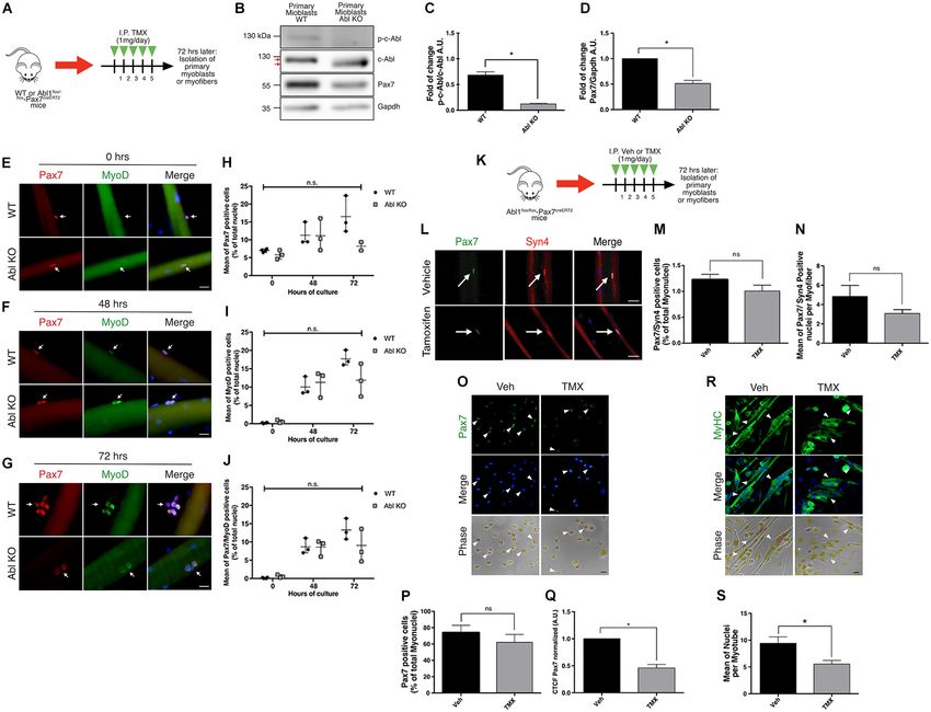
Montecino et al.                                                                                          c-Abl Regulates Pax7 During Myogenesis

2 h at room temperature. Proteins were eluted by resuspending             Reporter Gene Assay
Dynabeads in 30 µL 2X SDS-PAGE loading buffer and                         To evaluate Pax7 transcriptional activity when c-Abl is inhibited,
boiled for 5 min.                                                         C2C12 cells were transfected with the Pax3/7 specific reporter
                                                                          gene 6xPRS9-Luc (Olguin et al., 2007) and pcDNA3-myc-
In vitro Phosphorylation Assay                                            Pax7WT. CMV-LacZ vector for constitutive β-galactosidase
Purified GST-Pax7 Full length (5 µg) and GST-MyoD (1 µg)                  expression was co-transfected in order to normalize luciferase
were incubated with or without 10 ng of recombinant c-Abl His-            activity. Pax7-FKHR was used as positive control for reporter
Tag (ENZO life sciences, United States) in kinase buffer (MOPS            activation, as described previously (Bennicelli et al., 1999;
25 mM pH 7.2, 12.5 mM β-glycerophosphate, 25 mM MgCl2 ,                   Olguin et al., 2007) and CMV empty vector as a negative
325 µM EGTA, 2 mM EDTA, 250 µM DTT) supplemented                          control. Twenty four hours post-transfection, cells were treated
with 100 µM ATP (New England Biolabs, United States) for                  with DMSO as vehicle or Imatinib 10 µM for 18 h.
60 min at 30◦ C. Two hundred and fifty µM of Imatinib                     Finally, whole-cell lysates were collected, and luciferase and
Mesylate (Sigma-Aldrich, United States) or Lambda Protein                 β-galactosidase activities were determined using the Dual-
Phosphatase (New England Biolabs, United States) were added               Light System (Applied Biosystems, United States) following
as indicated. Reactions were loaded into SDS-PAGE gel                     the manufacturer’s instructions. Luminescence was measured in
and analyzed by WB.                                                       TECAN Infinite M200 PRO microplate reader.

Pax7 Phosphorylation in C2C12 Cells                                       Statistics
Experiment                                                                All quantitative data are presented as mean ± SEM unless
C2C12 cells were treated with vehicle, 10 µM Imatinib or DPH,             indicated. The number of independent experiments is
for 24 h and 10 µM pervanadate was added to the medium 1 h                specified in figure legends. Statistical analysis was performed
prior to lysis, except in non-treated cells (N/T). Pervanadate acts       with GraphPad Prism software, using the corresponding
as an inhibitor of protein tyrosine phosphatases (PTPs), allowing         statistical test to determine significance. A P-value < 0.05 was
the accumulation of phospho-tyrosine proteins (Huyer et al.,              considered significant.
1997). Pervanadate was prepared using sodium orthovanadate
and hydrogen peroxide as previously described (Gray et al., 2014).
Pax7 was immunoprecipitated under denaturing conditions in                RESULTS
order to avoid pull-down of its interacting proteins. For this,
denaturing lysis of C2C12 cells was performed using modified              c-Abl Expression and Activation in
RIPA buffer supplemented with 1% sodium deoxycholate                      Muscle Progenitors
and 0.1% sodium dodecyl sulfate (SDS), plus protease and                  It has been described that c-Abl shuttles between the nucleus
phosphatase inhibitors mentioned before. IP was performed as              and cytoplasm in myoblasts and myotubes (Puri et al., 2002).
previously described. In order to achieve greater separation of           We corroborated these findings, and additionally, we observed
immunoprecipitated Pax7 proteins, a 8% SDS-Polyacrylamide gel             a correlation between c-Abl localization and Pax7 levels in
was used in electrophoresis. WB against phospho-tyrosine was              C2C12 cells. High Pax7 expression was accompanied by nuclear
executed in order to detect phospho-tyrosine Pax7 that were               c-Abl localization (Figure 1A, 0 and 24 h). As expected,
immunoprecipitated.                                                       Pax7 expression was not detected in myotubes, where c-Abl
                                                                          was also observed in a cytoplasmic localization (Figure 1A,
Real-Time PCR                                                             120 h and Supplementary Figure 1A). This result suggests
C2C12 cells treated with vehicle or Imatinib 10 µM for 6,                 a correlation between Pax7 expression and c-Abl localization
18, and 24 h, were lysed in TRIzol reagent according to                   during the differentiation of C2C12 cells. Western blot analyses
the manufacturer’s instructions (Invitrogen, United States)               showed that c-Abl expression remains unchanged after 48 h
and total RNA was quantified using NanoQuant plate of                     in differentiating culture conditions (Figures 1B,C). Expression
TECAN Infinite M200 PRO microplate reader; 500 ng                         of the differentiation markers Myogenin and Myosin heavy
of total RNA was retro-transcribed with M-MLV-RT                          chain (MyHC), can be detected from 24 and 36 h, respectively
enzyme (Promega, United States) and Random Hexamer                        (Figure 1B). As described previously (Bustos et al., 2015), Pax7
Primer (Thermo Scientific, United States), according to the               protein remains detectable during this period (Figures 1B,C),
manufacturer’s instructions. qPCR reactions were performed                likely to non-differentiating cells.
with SYBR Green PCR master mix (Thermo Scientific,                           We evaluated c-Abl expression and activation status in
United States), following the manufacturer’s instructions on              C2C12 cells maintained in proliferation (Figure 1D, 0 h)
QuantStudio 3 Real-Time PCR System (Applied Biosystems,                   and differentiation culture conditions (Figure 1D, 24 and
United States) with the following oligonucleotides: Pax7                  96 h). As previously described (Brasher and Van Etten, 2000),
Fwd: 50 -CACCCCTTTCAAAGACCAAA-30 , Pax7 Rev: 50 -                         c-Abl activation was determined by detecting phosphorylation
TGCTTGAAGTTCCTGCTCCT-30 ; and Gapdh as housekeeping                       at tyrosine 412 (phospho-Tyr412). Active c-Abl was detected
gene, Gapdh Fwd: 50 -AGGTCGGTGTGAACGGATTTG-30 ;                           mostly in myoblasts, while the level of phosphorylated c-Abl
Gapdh Rev: 50 -TGTAGACCATGTAGTTGAGGTCA-30 .                               was significantly lower in differentiated, multinucleated cells

Frontiers in Cell and Developmental Biology | www.frontiersin.org     4                                     March 2021 | Volume 9 | Article 606403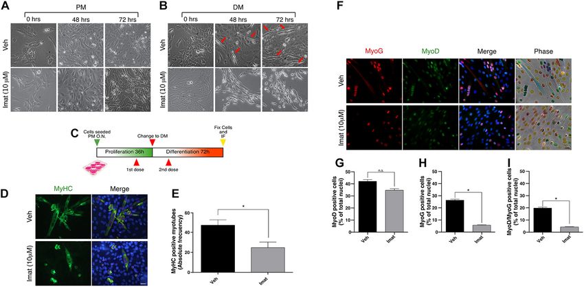
Montecino et al.                                                                                                                   c-Abl Regulates Pax7 During Myogenesis

  FIGURE 1 | c-Abl is expressed in myoblasts and activated during muscle regeneration. (A) Pax7 and c-Abl nuclear expression are correlated. C2C12 cells were
  differentiated the indicated time and then fixed. IF was performed against c-Abl (red), Pax7 (green), and nuclei were stained with Hoechst 33342 (blue). Arrows show
  myotubes without Pax7 expression, while arrowheads show mononucleated cells expressing Pax7 and c-Abl. Scale bar: 10 µm. (B) Expression of Pax7 and c-Abl
  in early myoblast differentiation. C2C12 cells were maintained in differentiation conditions for the indicated times and lysed. WB against Pax7, c-Abl, Myogenin
  (MyoG), MyHC and Gapdh was performed. (C) Graph shows quantification of Pax7 and c-Abl protein levels from B (n = 4). A.U., arbitrary units. (D) Expression and
  phosphorylation of c-Abl in C2C12. Cells were induced to differentiate, and IF for c-Abl (red) and phospho-tyr412 c-Abl (p-c-Abl, green) was performed at the
  indicated times. Nuclei were stained with Hoechst 33342 (blue). Arrows show myotubes and arrowheads myoblasts. Scale bar: 10 µm. (E) Quantification of (D).
  Graph shows quantification of Corrected Total Cell Fluorescence (CTCF) for p-c-Abl normalized to 0 h (n = 3, *P-value < 0.05, Kruskal–Wallis test, ns, not significant)
  A.U., arbitrary units. (F) Expression of p-c-Abl and c-Abl in myoblast differentiation. C2C12 cells were maintained in differentiation conditions for the indicated times
  and lysed. WB against p-c-Abl, c-Abl, and Gapdh was performed. (G) Graph shows quantification of Pax7 and c-Abl protein levels from F (n = 2). A.U., arbitrary
  units. (H) Expression and phosphorylation of c-Abl in primary myoblasts. Cells were maintained in proliferation conditions (Prol) or 7 days in differentiation conditions
  (Diff. D7), followed by IF for c-Abl (red) and p-c-Abl (green). Nuclei were stained with Hoechst 33342 (blue). Arrows show myotubes and arrowheads myoblasts.
  Scale bar: 10 µm. (I) Quantification of (H). Graph shows quantification of CTCF for p-c-Abl normalized to proliferation (n = 3, *P-value < 0.05, Wilcoxon signed-rank
  test). A.U., arbitrary units. (J) c-Abl is transiently phosphorylated in early muscle regeneration. Sections of muscles were obtained at 0 (non-injured), 1, 3, and 7 dpi,
  fixed and IF for p-c-Abl (green) and laminin (red) was performed. Nuclei were stained with Hoechst 33342 (blue). Yellow arrows show p-c-Abl in cells associated with
  laminin. Scale bar: 50 µm. (K) Myogenic cells are positive for p-c-Abl. Muscle cryosections were obtained at 0 (non-injured), 1, and 3 dpi, fixed and IF for p-c-Abl
  (green), and MyoD (red) was performed. Nuclei were stained with Hoechst 33342 (blue). Yellow arrows show MyoD positive cells with expression of p-c-Abl. Scale
  bar: 50 µm.

(Figures 1D,E and Supplementary Figure 1B). Western blot                                  cells (Figure 1I), similar to the distribution observed in C2C12
analyses of whole-cell extracts obtained from C2C12 cells, also                           cells. Together, these results indicate that c-Abl kinase activity
indicate a reduction in phosphorylated c-Abl levels after 96 hrs in                       is differentially regulated in proliferating and differentiating
differentiation conditions (Figures 1F,G).                                                muscle progenitors.
   c-Abl expression and activation status were also analyzed by IF
in primary myoblasts, isolated from adult mice and maintained                             c-Abl Is Activated During Early Muscle
in proliferation and differentiation culture conditions for 7 days.                       Regeneration
Total c-Abl and phospho-c-Abl were detected in both conditions                            To determine c-Abl expression and activation dynamics during
(Figure 1H), although phospho-c-Abl signal in multinucleated                              muscle regeneration, acute injury was performed, by barium
cells was significantly lower in comparison with proliferating                            chloride (BaCl2 ) intramuscular injection, in the Tibialis Anterior

Frontiers in Cell and Developmental Biology | www.frontiersin.org                     5                                              March 2021 | Volume 9 | Article 606403
Montecino et al.                                                                                                                c-Abl Regulates Pax7 During Myogenesis

(TA) muscle from adult mice. Muscles were isolated at 1-, 3-,                           revealed no significant changes in cell morphology between
and 7-days post-injury (dpi) in order to analyze different stages                       treated and untreated cells maintained in PM (Figure 2A).
of early regeneration by IF. The levels of c-Abl phosphorylation                        However, Imatinib treatment resulted in a significant reduction
were determined as described before. No signal for phospho-                             in cell fusion and myotube formation both at 48 and 72 h in
Tyr412 was detected in uninjured muscles, neither in cells nor                          DM, compared to vehicle (Figure 2B). These observations agree
fibers (Figure 1J, non-injured panel). Phospho-c-Abl signal was                         with previous reports, which showed that c-Abl loss of function
detected in myogenic cells early after injury (Figure 1J, 1 and                         impairs myoblasts differentiation (Bae et al., 2009).
3 dpi panels), defined by co-expression of MyoD (Figure 1K,                                 Additionally, we expose the cells to vehicle or Imatinib using
1 and 3 dpi panels). Phospho-c-Abl was also detected in non-                            a two-dose regimen treatment: first during the proliferation
myogenic cells, likely corresponding to infiltrating inflammatory                       phase, followed by a second dose during the differentiation
cells (Figure 1K, 1 and 3 dpi panels). Interestingly, c-Abl                             phase (Figure 2C). Cells were then fixed, and MyHC expression
activation appears to be transient, since phospho-c-Abl was not                         was analyzed by IF (Figure 2D). We observed a decrease in
detected at 7 dpi (Figure 1J and Supplementary Figure 2A).                              the number of MyHC-positive myotubes in cells treated twice
These results indicate that c-Abl Tyr412 phosphorylation is also                        with Imatinib (∼50%) compared to the vehicle (Figure 2E,
differentially regulated in myogenic progenitors in vivo and may                        P-value < 0.05). We then studied the effect of c-Abl inhibition on
play a role in muscle regeneration.                                                     the expression of early differentiation markers such as MyoD and
                                                                                        Myogenin (Figure 2F). We detected no significant differences
                                                                                        on MyoD expression between vehicle and Imatinib treated
c-Abl Inhibition Prevents Differentiation                                               cells (Figure 2G). Interestingly, the percentage of Myogenin
in C2C12 Cells                                                                          expressing cells decreased by ∼5 fold upon Imatinib treatment
c-Abl expression, localization, and activation patterns suggest                         (Figure 2H; P-value > 0.05). The percentage of MyoD/Myogenin
distinct roles in muscle progenitors during their myogenic                              expressing cells was similarly affected by Imatinib treatment
progression. Therefore, we tested the effects of c-Abl inhibition                       (Figure 2I; P-value > 0.05).
in C2C12 myoblasts. For this, we used Imatinib Mesylate                                     Taken together these results indicate that c-Abl kinase activity
which acts as a competitive inhibitor for c-Abl kinase activity                         is required for C2C12 cells differentiation. Since c-Abl inhibition
(Schindler, 2000). Cells were maintained in proliferation (PM)                          did not affect MyoD expression, we hypothesized that c-Abl
or differentiation (DM) culture conditions for 72 h; in the                             activity could regulate myogenesis up-stream of Myogenin
presence of Imatinib or vehicle. Phase-contrast microscopy                              induction in muscle progenitors.

  FIGURE 2 | c-Abl inhibition impairs C2C12 cells differentiation. (A,B) c-Abl inhibition affects C2C12 cells with “myotube like” phenotype. C2C12 cells were
  maintained in proliferation medium (PM) or differentiation medium (DM) for 72 h in the presence of 10 µM of Imatinib (Imat) or vehicle (Veh) (added fresh every 24 h).
  Micrographs were obtained every 24 h to evaluate phenotypic changes in cells. Red arrows indicate C2C12 cells with “myotube like” phenotype. (C) Experimental
  strategy to evaluate C2C12 cells differentiation. Cells were seeded in PM overnight (O.N.) and then treated with vehicle or Imatinib for 24 h (1st dose). Next, PM was
  replaced by DM, and cells were treated again (2nd dose). After 72 h, cells were fixed, and IF for MyoD, Myogenin (MyoG) and MyHC was performed. Nuclei were
  stained with Hoechst 33342 (blue). (D) Long c-Abl inhibition impairs C2C12 cell differentiation. Cells treated twice with Imatinib show a decrease in the number of
  differentiated myotubes (cells with two or more myonuclei), in comparison with the vehicle. Scale bar: 10 µm. (E) Quantification of the number of MyHC positive
  myotubes from (D). Veh mean: 47.0 ± 5.5 cells; Imat mean: 24.66 ± 5.6 (n = 3, *P-value < 0.05, unpaired t-student test. (F) c-Abl inhibition does not change MyoD
  but Myogenin expression in cells. Plots (G,H) show the percentage of MyoD and Myogenin positive cells treated as in (F), respectively. (n = 3, *P-value < 0.05,
  Nested t-test, ns, not significant). Plot (I) shows the mean of MyoD/Myogenin double positive nuclei per myotube (n = 3, *P-value < 0.05, Nested t-test).

Frontiers in Cell and Developmental Biology | www.frontiersin.org                   6                                            March 2021 | Volume 9 | Article 606403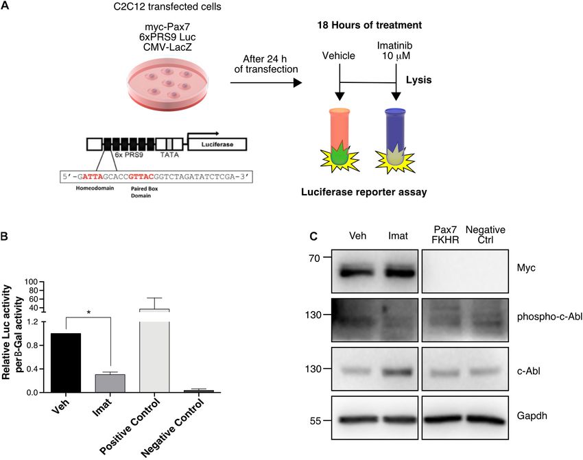
Montecino et al.                                                                                           c-Abl Regulates Pax7 During Myogenesis

c-Abl Inhibition Results in Decreased                                     “Materials and Methods”). First, we tested c-Abl and Pax7
Pax7 Levels                                                               interaction in C2C12 cells treated with vehicle or Imatinib for
                                                                          24 h, followed by Pax7 IP and c-Abl detection by Western blot.
To better understand the molecular basis of the differentiation
                                                                          Under these conditions, there was an enrichment in c-Abl co-IP,
impairment upon c-Abl inhibition, we evaluated the expression
                                                                          compared to control IP with a non-related antibody (Figure 4A,
of Pax7 and MyoD proteins, which may reflect changes
                                                                          left panel). This enrichment was detected both in cells treated
in the differentiation potential of myogenic progenitors. We
                                                                          with vehicle or Imatinib, despite the decrease in Pax7 levels,
performed IF to determine the percentage of cells expressing
                                                                          suggesting that c-Abl could interact with Pax7 independently of
Pax7 and MyoD upon c-Abl inhibition (Figure 3A). C2C12
                                                                          its activation status.
cells maintained in proliferation conditions were treated with
                                                                              Considering the kinase nature of c-Abl, we performed in vitro
vehicle or Imatinib for 48 h. We observed a small, but consistent,
                                                                          phosphorylation assays as described previously (González et al.,
change in the percentage of cells expressing Pax7 after the
                                                                          2016), using recombinant c-Abl His-Tag and GST-Pax7, followed
treatment with Imatinib (59.16 ± 12.62%) in comparison with
                                                                          by Western blot to detect phosphorylated tyrosine residues (see
the vehicle (71.11 ± 3.77%), which did not reach statistical
                                                                          section “Materials and Methods”). We observed two distinct
significance (Figure 3B). A similar trend was observed for
                                                                          bands, just under 100 kDa, coinciding with GST-Pax7 molecular
MyoD expressing cells, with an average of 64.05 ± 5.16%
                                                                          weight (Figure 4B, red arrows). Importantly, these bands were
and 52.97 ± 11.70% for vehicle and Imatinib, respectively
                                                                          only detected when GST-Pax7, c-Abl, and ATP were present
(Figure 3C). The percentage of double positive cells for Pax7 and
                                                                          in the reaction. Moreover, these phospho-proteins were not
MyoD remain unchanged upon Imatinib treatment (Figure 3D).
                                                                          detected when Imatinib was added or when either ATP or c-Abl
Additionally, Imatinib treatment in myofiber associated SCs
                                                                          were omitted from the reaction mix (Figure 4B). Phosphatase
for 48 h, did not alter the number of MyoD positive
                                                                          treatment prior to Western blot had the same effect, further
cells (Supplementary Figure 3). Although the percentage of
                                                                          supporting the phosphoprotein nature of the bands identified
Pax7, MyoD, or Pax7/MyoD expressing cells did not change
                                                                          above. Since MyoD has been described as a c-Abl substrate
significantly, we detected a consistent decrease in fluorescent
                                                                          (Puri et al., 2002; Innocenzi et al., 2011), we used GST-MyoD
signal upon Imatinib treatment. Therefore, Pax7 and MyoD
                                                                          as a positive control for the phosphorylation assay (Figure 4C).
protein levels were evaluated by Western blot (Figure 3E).
                                                                          GST-only was used as an additional negative control, and no
We observed a significant time-depend decay of Pax7 protein
                                                                          phospho-bands were observed nearby Pax7 molecular weight
levels but not in MyoD, reaching a larger decrease at 48 h of
                                                                          (Supplementary Figure 5).
treatment (Figures 3F,G). Moreover, Imatinib treatment induced
                                                                              To determine whether c-Abl phosphorylates Pax7 in a cellular
a significant Pax7 decrease (∼4-fold reduction compared to
                                                                          context, we performed a denaturing IP of Pax7 in C2C12
vehicle) in both proliferating and differentiating conditions
                                                                          cells treated with vehicle, Imatinib or DPH for 24 h. Western
(Figures 3H–J). Accordingly, treatment of C2C12 cells with the
                                                                          blot was performed to analyze the presence of phosphorylated
c-Abl activator DPH (Yang et al., 2011), resulted in a robust
                                                                          tyrosine residues in Pax7. Given the rapid turnover of tyrosine
increase in Pax7 but not in MyoD or Myogenin protein levels
                                                                          phosphorylation in the cell environment, we inhibited PTPs using
(Supplementary Figure 4). These results strongly suggest that
                                                                          pervanadate 1 h prior to lysis (Figure 4D). Pervanadate treatment
Pax7 expression is regulated by c-Abl activity.
                                                                          allows accumulation of phospho-tyrosine proteins compared to
   To determine whether Pax7 down-regulation induced by
                                                                          non-treated cells (Figure 4E, right panel, Veh versus N/T). Under
Imatinib is due to transcriptional changes, we performed
                                                                          these conditions, a phospho-tyrosine signal was detected in Pax7
quantitative Real-Time Polymerase Chain Reaction (qRT-PCR).
                                                                          IP from vehicle-treated extracts, which notably increased upon
C2C12 cells were treated with vehicle or Imatinib for 6, 18,
                                                                          c-Abl activation (DPH treatment). Almost no signal was observed
or 24 h, followed by lysis and total RNA isolation. As shown
                                                                          in Imatinib-treated extracts, or when pervanadate treatment was
in Figure 3K, no significant changes in Pax7 mRNA levels
                                                                          omitted (Figure 4E, left panel, red arrow).
were detected at 6 or 18 h of Imatinib treatment compared
                                                                              Together, these results indicate that Pax7 and c-Abl physically
to vehicle. However, a significant decrease was observed at
                                                                          interact in muscle progenitors and suggest that Pax7 is a c-Abl
24 h. This Pax7 transcript drop precedes the decrease in the
                                                                          kinase substrate in myogenic cells.
protein levels analyzed above, which is noted from 36 h after
Imatinib treatment (Figure 3E). These results indicate that c-Abl
kinase activity regulates Pax7 expression, affecting the outcome          c-Abl Inhibition Impairs Pax7
of muscle differentiation.
                                                                          Transcriptional Activity
                                                                          Considering that c-Abl could regulate Pax7 via direct
c-Abl Interacts With Pax7 in Myogenic                                     phosphorylation and that Imatinib affects Pax7 expression,
Cells                                                                     we explored the possibility that c-Abl regulates Pax7 activity as a
Considering (i) the effects of c-Abl inhibition on Pax7 expression,       transcription factor. For this, we performed a luciferase reporter
and (ii) c-Abl nuclear localization in Pax7-expressing cells,             assay using the Pax3/7 specific reporter gene 6xPRS9-Luc
we hypothesized that c-Abl and Pax7 physically interact in                (Olguin et al., 2007). C2C12 cells, maintained in proliferation
muscle progenitors. With this in mind, we performed co-                   culture conditions, were co-transfected with the 6xPRS9-Luc
immunoprecipitation (co-IP) assays on C2C12 cells (see section            and CMV-LacZ (transfection control) vectors, plus or minus

Frontiers in Cell and Developmental Biology | www.frontiersin.org     7                                     March 2021 | Volume 9 | Article 606403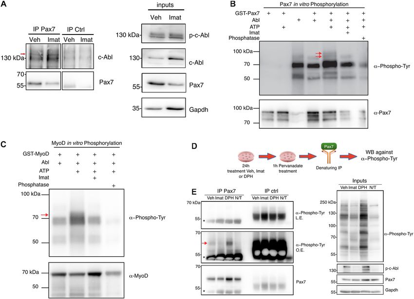
Montecino et al.                                                                                                                  c-Abl Regulates Pax7 During Myogenesis

  FIGURE 3 | c-Abl inhibition decreases Pax7 protein and mRNA levels. (A) Imatinib treatment does not decrease the number of Pax7 or MyoD positive cells. C2C12
  cells were maintained in PM and treated with vehicle (Veh) or 10 µM Imatinib (Imat) for 48 h. Then cells were fixed, and IF for Pax7 (green) and MyoD (red) was
  performed. Nuclei were stained with Hoechst 33342 (blue). Scale bar: 10 µm. (B) Quantification of Pax7 positive cells from (A). Veh mean: 71.11 ± 3.77%; Imat
  mean: 59.16 ± 12.62%. (C) Quantification of MyoD positive cells from (A). Veh mean: 64.05 ± 5.16; Imat mean: 52.97 ± 11.70%. (D) Quantification of Pax7/MyoD
  double positive cells from (A). Veh mean: 40.79 ± 2.38%; Imat mean: 36.06 ± 1.75% (n = 4, P-value > 0.05, Mann–Whitney test, ns, not significant). (E) Imatinib
  induces a time-dependent drop of Pax7 protein. C2C12 cells were maintained in PM as specified and treated with 10 µM Imatinib every 24 h. Cells were lysed, and
  WB was performed to determine levels of Pax7 and MyoD. Tubulin was used to normalize protein levels. Plots (F,G) show Pax7 and MyoD fold of change from (E)
  respectively (n = 3, *P-value < 0.05, ANOVA test, ns: not significant). (H) c-Abl inhibition downregulates Pax7 protein levels in C2C12 cells. Cells were maintained in
  PM or DM for 48 h in the presence of vehicle or Imatinib (10 µM). Then cells were lysed, and WB against Pax7 was performed. Gapdh was used to normalize protein
  levels. Plots (I,J) show a significant decrease in Pax7 protein levels in cells treated with Imatinib versus vehicle (n = 4, *P-value < 0.05, Wilcoxon signed-rank test).
  (K) Pax7 mRNA levels decrease upon c-Abl inhibition. C2C12 cells in PM were treated with vehicle or Imatinib (10 µM) as indicated. A significant drop in Pax7
  mRNA levels is observed at 24 h in Imatinib treated cells. Veh mean: 1.0 ± 0.0; Imat mean: 0.457 ± 0.03 (n = 3, *P-value < 0.05, multiple t-test).

Frontiers in Cell and Developmental Biology | www.frontiersin.org                    8                                              March 2021 | Volume 9 | Article 606403
Montecino et al. c-Abl Regulates Pax7 During Myogenesis
FIGURE 4 | c-Abl interacts and phosphorylates Pax7. (A) c-Abl co-immunoprecipitates with Pax7 in C2C12 cells. Cells maintained in PM were treated with vehicle
(Veh) or 10 µM Imatinib (Imat) for 24 h, followed by lysis and immunoprecipitation (IP) with anti-normal mouse IgG (IP ctrl) or anti-Pax7 (IP Pax7). c-Abl and Pax7
were detected by Western blot (Left panels). Red arrow shows the expected c-abl relative migration. Right panels show inputs for p-c-Abl, c-Abl, Pax7, and Gapdh
(n = 3). (B) Pax7 is phosphorylated by c-Abl. Recombinant c-Abl His-Tag and GST-Pax7 were incubated in kinase buffer as indicated, followed by WB for
Phospho-Tyr and Pax7. Red arrows show Pax7 phosphorylated in tyrosine residues (n = 3). As positive control, GST-MyoD was incubated with c-Abl, as is shown in
(C). Red arrow shows MyoD phosphorylated in tyrosine residues (n = 3). (D,E) Pax7 is phosphorylated by c-Abl in C2C12 cells. The figure shows the following
protocol to determine Pax7 phosphorylation in C2C12. Left panels show Pax7 IP followed by WB against Phospho-Tyr and Pax7 (L.E., less exposed membrane;
O.E., overexposed membrane). Red arrow shows Pax7 phosphorylated in tyrosine residues and asterisk indicates IgG. Right panels show inputs for Phospho-Tyr,
p-c-Abl, Pax7, and Gapdh (n = 3).
myc-Pax7 or Pax7-FKHR (positive control) expression vectors. Recombination was induced by five daily TMX injections
After 24 h, transfected cells were treated with vehicle or Imatinib as described previously (Eon et al., 2008; Reinert et al., 2012).
for 18 h prior to lysis (Figure 5A). We observed a significant After a resting period of 72 h, hind limb muscles were
decrease (>2-fold) in Pax7-induced reporter activity upon c-Abl dissected in order to isolate primary myoblasts or SCs associated
inhibition (Figure 5B). As expected, Pax7-FKHR transfected cells with single myofibers (Figures 6A,K). First, we tested the
showed a transcriptional activity ∼40-fold higher than control effects of c-Abl deletion in primary myoblasts maintained in
and >100-fold higher than cells treated with Imatinib. Myc-Pax7 proliferation culture conditions for 48 h after isolation. Western
expression was corroborated by Western blot (Figure 5C). blot analyses revealed the expression of a lower molecular
Moreover, we observed decreased c-Abl phosphorylation, as weight band reactive to the anti-c-Abl antibody (Figure 6B).
expected upon Imatinib treatment (Figure 5C). In the context of As described by Moresco et al. (2005), a similar protein
our previous results, these findings are consistent with the idea band can be detected with variable expression levels in a
that c-Abl regulates Pax7 activity and expression. tissue-specific manner. In this transgenic mouse model (same
used in our study), Cre-mediated recombination results in
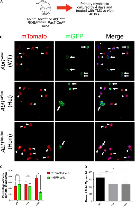
Deletion of c-Abl Impairs Primary the deletion of exon 5 of the abl1 gene, affecting the kinase
Myoblasts Function ex vivo domain. As expected, the same group described that this
To explore the functional consequences of c-Abl dependent recombinant c-Abl has no kinase activity. We confirmed this
regulation of Pax7, we used a c-Abl loss of function by Western blot (Figures 6B,C) and by IF (Supplementary
approach in vivo. Specifically, we generated the Abl1flox/flox - Figure 6). Noteworthy, detection of the c-Abl-kinase-null protein
Pax7creERT2 mouse model, which allows SC-specific c-Abl varied when using different anti-c-Abl antibodies commercially
deletion, upon tamoxifen (TMX) administration (see section available (Supplementary Figure 6), which may partially explain
“Materials and Methods”). differences in expression levels among different tissues and cell
Frontiers in Cell and Developmental Biology | www.frontiersin.org 9 March 2021 | Volume 9 | Article 606403Montecino et al. c-Abl Regulates Pax7 During Myogenesis FIGURE 5 | c-Abl regulates Pax7 transcriptional activity. (A) Protocol for Reporter Assay. C2C12 cells were transfected with the indicated plasmids for 24 h. After that, treatment with DMSO as vehicle or Imatinib (10 µM) was performed for 18 h. Finally, whole-cell lysates were collected, and luciferase and β-galactosidase activities were determined. (B) c-Abl inhibition decreases Pax7 transcriptional activity. Cells treated with Imatinib (mean: 0.305 ± 0.04) exhibits a significant decrease in Pax7 transcriptional activity in comparison with cells treated with vehicle (mean 1.00 ± 0.0), (n = 4, *P-value < 0.05, Wilcoxon signed-rank test). (C) WB from reporter assay cells. We observed a decrease of c-Abl phosphorylation in cells treated with Imatinib corroborating the effectiveness of the treatment. types. Consistent with our previous results, myoblasts expressing differentiation and suggest that c-Abl activity regulates Pax7 the cAbl-kinase-null protein (Abl KO) exhibited significantly expression and myogenic potential in SCs. lower levels of Pax7 protein compared to controls (Figure 6D). To follow the fate of Abl KO myoblasts in culture, we used IF analyses of SCs associated with isolated myofibers indicate a ROSAmT/mG reporter mouse in the Abl1flox/flox -Pax7creERT2 no significant differences in the expression of MyoD, from 0 to genetic background, allowing for lineage tracing in vivo and 72 h (Figures 6E–J). Interestingly, Pax7 expression appears to in vitro (see section “Materials and Methods”). To avoid decrease at 72 h in Abl KO SCs, however, differences did not reach differences in the starting number of cells, primary myoblasts statistical significance due to the high variability in the numbers were isolated from uninjured muscles, and treated with TMX of cells associated with myofibers in each case (Figures 6G,H). for 48 h to induce recombination in vitro (Figure 7A). Next, we analyzed the number of SCs associated with myofibers After additional 96 h, cells were fixed, and the expression of by IF, immediately after isolation. Using Pax7 and Syndecan- mTomato (non-recombined cells) or mGFP (recombined cells) 4 as independent SC markers, we detected no significant was analyzed by direct fluorescence (Figure 7B). We observed changes in the total number of SC upon TMX administration a similar proportion of cells expressing mTomato and mGFP in compared to the vehicle (Figures 6L–N). The reduction of myoblast cultures from WT and Abl1wt/flox mice (Figure 7C). Pax7 expression was corroborated by IF, in primary myoblasts However, we detected a significant difference in mTomato maintained in proliferation culture conditions (Figures 6O–Q). (74.38 ± 5.06%) versus mGFP (25.61 ± 5.06%) expressing cell Moreover, Abl KO myoblasts exhibited a reduced differentiation populations in Abl1flox/flox myoblast cultures. Noteworthy, the capacity, evaluated by MyHC expression and by the formation of average total number of nuclei was not significantly different multinucleated myotubes with reduced nuclei number, compared among conditions (Figure 7D), suggesting that proliferation to control myotubes (Figures 6R,S). Together, these results and/or survival were specifically affected in mGFP(+) cells (i.e., are consistent with the effect of c-Abl inhibition on C2C12 Abl KO myoblasts). Frontiers in Cell and Developmental Biology | www.frontiersin.org 10 March 2021 | Volume 9 | Article 606403
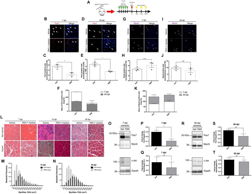
Montecino et al.                                                                                                                c-Abl Regulates Pax7 During Myogenesis

  FIGURE 6 | Specific deletion of c-Abl in SCs impairs myogenesis. (A) In vivo recombination protocol. Abl1WT/WT -Pax7creERT2 (WT) or Abl1flox/flox -Pax7creERT2 (Abl
  KO) mice were injected intraperitoneally (I.P.) with Tamoxifen (TMX, 1 mg daily) for 5 consecutive days. Then, after 72 h, myofibers or primary myoblasts were
  isolated. (B) Decreased expression of phospho-tyr412 c-Abl (p-c-Abl) and Pax7 in Abl KO myoblasts. Primary myoblasts were maintained in PM for 48 h after
  isolation. Then, WB was performed using indicated antibodies. (C) Quantification of normalized p-c-Abl protein levels from (B). A.U., arbitrary units (n = 3,
  *P-value < 0.05, Wilcoxon signed-rank test). (D) Quantification of normalized Pax7 protein levels from (B). A.U., arbitrary units (n = 4, *P-value < 0.05, Wilcoxon
  signed-rank test). (E–G) Expression of Pax7 and MyoD in fiber associated myoblasts from WT and Abl KO mice, at 0, 48, and 72 h of culture. Myofibers were
  isolated, and IF against Pax7 (red) and MyoD (green) was performed. Nuclei were stained with Hoechst 33342 (blue). Arrows indicate satellite cells associated with
  the myofiber. Scale bar: 10 µm. Plots (H–J) show the percentage of Pax7, MyoD and Pax7/MyoD double positive cells per total nuclei, respectively (n = 3,
  P-value > 0.05, Multiple t-Student test, ns, not significant). (K) In vivo recombination protocol for following analyses. Abl1flox/flox -Pax7creERT2 mice were injected
  intraperitoneally (I.P.) with vehicle (Veh) or Tamoxifen (TMX, 1 mg daily) for five consecutive days. Then, after 72 h, myofibers or primary myoblasts were isolated.
  (L) Percentage of Pax7 positive cells do not change in recently isolated myofibers from vehicle or Tamoxifen treated mice. Myofibers were isolated, and IF against
  Pax7 (green) and Syndecan-4 (Syn4, red) was performed. Nuclei were stained with Hoechst 33342 (blue). Arrows indicate satellite cells associated with the myofiber.
  Scale bar: 10 µm. (M) Quantification of Pax7/Syn4 positive cells from (L) (n = 4). Veh mean: 1.22 ± 0.10%; TMX mean: 1.008 ± 0.11% (P-value > 0.05,
  Mann–Whitney test, ns, not significant). (N) Quantification of Pax7/Syn4 positive cells per myofiber from (L) (n = 4). (P-value > 0.05, Mann–Whitney test, ns, not
  significant). (O) IF of proliferative primary myoblasts from mice treated with vehicle or Tamoxifen, using anti Pax7 (green) and nuclei were stained with Hoechst 33342
  (blue). Scale bar: 10 µm. (P,Q) Graphs show quantification of Pax7 positive nuclei and Pax7 Corrected Total Cell Fluorescence (CTCF) from (O). (n = 4,
  *P-value < 0.05, Wilcoxon signed-rank test). A.U., arbitrary units. (R) IF of differentiated (7 days) primary myoblasts from mice treated with vehicle or Tamoxifen,
  using anti MyHC (green) and nuclei were stained with Hoechst 33342 (blue). Arrowheads show representative myotubes. Scale bar: 10 µm. (S) Quantification of
  nuclei per myotube from (R) (Veh n = 4 and TMX n = 3, *P-value < 0.05, Mann–Whitney test).

Muscle Regeneration Is Disturbed in                                                      of 1.2% barium chloride (BaCl2 ) (Cornelison et al., 2004) in
SC-Abl KO Mice                                                                           Abl1flox/flox -Pax7creERT2 mice, previously treated with TMX (SC-
Based on our previous results, we hypothesized that SC function                          Abl KO) or vehicle as described. Contralateral muscles were
would be altered in vivo upon Abl1 deletion, affecting the                               injected with saline solution (NaCl 0.9%) as internal control.
outcome of muscle regeneration. To test this concept, muscle                             TA muscles were collected at different days post injury (dpi)
damage was induced by intramuscular injection (TA muscles)                               for downstream analyses (Figure 8A). Correlating with previous

Frontiers in Cell and Developmental Biology | www.frontiersin.org                   11                                            March 2021 | Volume 9 | Article 606403
Montecino et al.                                                                                                                 c-Abl Regulates Pax7 During Myogenesis

  FIGURE 7 | GFP positive primary myoblasts population decrease in c-Abl Knockout cells recombined in vitro. (A) Protocol to induce Abl1 recombination in vitro.
  Primary myoblasts were isolated from Abl1WT/WT (WT), Abl1wt/flox (Het), or Abl1flox/flox (Hom) ROSAmT/mG -Pax7creERT2 mice and maintained 4 days in PM. During
  this time, cells were treated with vehicle or Tamoxifen (TMX, 10 µM) for 48 h. After that, cells were fixed, and direct fluorescence was detected. Nuclei were stained
  with Hoechst 33342 (blue). (B) Arrowheads show mTomato positive cells and arrows show mGFP positive cells. The asterisk shows non-myogenic cells. Scale bar:
  50 µm. (C) Quantification of the percentage of mTomato positive and mGFP positive myoblasts from (B) (n = 3, *P-value < 0.05, multiple t-Student test, ns, not
  significant). (D) Total number of myoblasts do not change in different mice. Mean of total myoblasts was determined for every mouse from (B). WT mean:
  324.5 ± 39.50; Het mean: 239.7 ± 25.06; Hom mean: 234.7 ± 28.30 (n = 3, P-value > 0.05, ANOVA test, ns, not significant).

findings, IF analyses showed a significant reduction in the                              expected to increase after activation (during early phases of
number of Pax7(+) cells per field at 7 and 30 dpi in SC-Abl                              regeneration), and later decline reaching the numbers observed
KO mice, compared to injured muscles from vehicle-treated                                during quiescence. This change in the Pax7(+) population
animals (Figures 8B–E). The number of Pax7(+) cells are                                  is observed in injured muscles treated with vehicle, but is

Frontiers in Cell and Developmental Biology | www.frontiersin.org                   12                                            March 2021 | Volume 9 | Article 606403
Montecino et al.                                                                                                                  c-Abl Regulates Pax7 During Myogenesis

  FIGURE 8 | Specific deletion of c-Abl in SCs impairs muscle regeneration. (A) Protocol for muscle regeneration in Abl1flox/flox -Pax7creERT2 . Mice were injected
  intraperitoneally (I.P.) with vehicle (Veh) or Tamoxifen (TMX, 1 mg daily) for five consecutive days. Then, after 72 h, muscle injury was induced by intramuscular (I.M.)
  injection of BaCl2 in the tibialis anterior (TA) muscle. Contralateral muscles were injected with saline solution as control. Next, muscles were isolated at 7-, 15-, and
  30-days post-injury (dpi). (B,D) Pax7 positive cells decrease in SC-Abl KO mice during muscle regeneration. IF against Pax7 (red) was performed in 7 dpi (B) and
  30 dpi (D) muscle sections. Nuclei were stained with Hoechst 33342 (blue). Arrows show Pax7 positive cells. Scale bar: 10 µm. Plots (C,E) show quantification for
  Pax7 positive cells normalized per 100 nuclei for 7 and 30 dpi respectively (n = 3, *P-value < 0.05, Mann–Whitney test). (F) Graph shows the mean of total Pax7
  positive cells by mouse. (G,I) Myogenin (MyoG) positive cells in SC-Abl KO mice during muscle regeneration. IF against MyoG (magenta) was performed in 7 dpi (G)
  and 30 dpi (I) muscle sections. Nuclei were stained with Hoechst 33342 (blue). Arrows show MyoG positive cells. Scale bar: 10 µm. Plots (H,J) show quantification
  for MyoG positive cells normalized per 100 nuclei for 7 and 30 dpi respectively (n = 3, P-value > 0.05, Mann–Whitney test, ns: not significant). (K) Graph shows the
  mean of total MyoG positive cells by mouse. (L) Decrease in cross-sectional area (CSA) of regenerative myofibers from c-Abl KO mice. Sections from TA muscles
  were stained with Hematoxylin-Eosin stain. Upper panels: non-injured contralateral muscles; Lower panels: injured TA. Scale bar: 50 µm. (M) Distribution of CSA of
  regenerative myofibers at 15 dpi. (N) Distribution of CSA of regenerative myofibers at 30 dpi (n = 3, *P-value < 0.05, Two-way ANOVA test). (O,R) Pax7 protein
  levels decrease during muscle regeneration in c-Abl KO mice. 7 and 30 dpi TA were lysed, and WB using anti-Pax7, anti-Myogenin (MyoG), or anti-c-Abl was
  performed. Gapdh was used to normalize protein levels. (P,Q) Quantification of normalized Pax7 and MyoG protein levels from 7 dpi extracts (O). A.U., arbitrary units
  (n = 3, *P-value < 0.05, Mann–Whitney test, ns: not significant). (S,T) Quantification of normalized Pax7 and MyoG protein levels from 30 dpi extracts (R). A.U.,
  arbitrary units (n = 3, P-value > 0.05, Mann–Whitney test, ns, not significant).

absent in SC-Abl KO muscles (Figure 8F), suggesting an                                       Histological analysis using H&E staining revealed the
impaired expansion of the progenitor population. This idea is                             expected changes in tissue architecture during regeneration
supported in part by the reduction in the number of MyoD(+)                               (Figure 8L) and an overall reduction in fiber size in SC-Abl
cells in SC-Abl KO muscles at 7 dpi, compared to control                                  KO animals compared to control, quantified at 15 and
(Supplementary Figure 7). No significant changes were observed                            30 dpi (Figures 8M,N). Myofiber cross-sectional area (CSA)
in the number of Myogenin(+) cells between SC-Abl KO                                      measurements revealed a significant difference in the distribution
and control muscles, both at early (7 dpi) or late (30 dpi)                               of fiber size, consistent with the accumulation of small caliber
regeneration (Figures 8G–K).                                                              myofibers in SC-Abl KO regenerating muscles.

Frontiers in Cell and Developmental Biology | www.frontiersin.org                    13                                             March 2021 | Volume 9 | Article 606403
Montecino et al.                                                                                             c-Abl Regulates Pax7 During Myogenesis

   To test if proliferation was affected in SC-Abl KO mice,                 activated muscle progenitors. Specifically, our results show that
we performed IF analysis of ki67 expression in 7 dpi sections.              c-Abl phosphorylated at tyrosine 412 is present in myogenic cells
Interestingly, although not significant, we observed a decrease             during the first 3 days of muscle regeneration but not after 7 days
in the percentage of ki67 positive nuclei in TMX treated                    neither in non-injured muscle. We also determined that c-Abl
animals compared to vehicle (Supplementary Figure 8). This                  inhibition impairs differentiation of C2C12 cells, decreasing the
observation agrees with our previous results in primary                     number of multinucleated cells expressing MyHC. These results
myoblasts and suggests that could be a proliferation defect in              are similar to those obtained by Bae et al. (2009) in which using a
c-Abl KO myoblasts.                                                         c-Abl siRNA in C2C12 cells, they noted a decrease in myotube
   Finally, we performed Western blots to analyze Pax7 protein              formation. In our study, we observed a reduction in the total
levels from whole muscle extracts at 7 and 30 dpi. In accordance            number in Myogenin expressing cells upon c-Abl inhibition,
with IF analyses, we observed a significant decrease in Pax7                however the number of MyoD/Myogenin positive cells remained
protein levels at 7 dpi in SC-Abl KO muscles (Figures 8O,P).                unchanged. These observations support the idea that c-Abl could
Although not statistically significant, a decrease in Myogenin              regulate myogenesis upstream the induction of Myogenin.
protein levels were also observed in regenerating SC-Abl KO                     Previously, it has been described that c-Abl regulates
mice (Figures 8O,Q). As expected from the previous analysis,                MyoD activity by phosphorylation, resulting in a decrease in
decreased Pax7 and Myogenin protein levels still are observed at            transcriptional activity and myogenesis arrest in conditions
30 dpi in SC-Abl KO muscles, however, these differences are not             where cells suffer DNA damage (Puri et al., 2002; Innocenzi et al.,
statistically different from control values (Figures 8R–T).                 2011; Simonatto et al., 2013). These studies showed that c-Abl
   To further confirm c-Abl-Pax7 interaction in vivo, we                    can regulate MyoD activity, however the effect on MyoD protein
performed co-IP from whole muscle extracts obtained at 30 dpi               levels were not explored. Since MyoD binding to DNA prevents
(see section “Materials and Methods”). Surprisingly, we detected            its degradation (Abu Hatoum et al., 1998), we hypothesized
that c-Abl co-immunoprecipitate with Pax7 in samples from                   that c-Abl-dependent regulation of MyoD activity could directly
injured and non-injured muscles, but c-Abl signal is lower for              or indirectly affect MyoD protein levels. Therefore, we decided
TMX-treated animals (Supplementary Figure 9). Noteworthy,                   to analyze the expression of myogenic transcription factors in
we did not detect the c-Abl-kinase-null protein from whole-                 cells treated with a c-Abl inhibitor. Contrary to our first idea,
muscle extracts, which is consistent with the tissue-specific               MyoD protein levels did not significantly decrease when c-Abl
expression variability reported previously (Moresco et al., 2005).          was inhibited. Unexpectedly, we observed a consistent decrease
   Together, our in vivo studies indicate that c-Abl deletion               in Pax7 detection in IF studies. This effect was detected as a
impairs satellite cell function during regeneration. Consistent             decrease in the levels of Pax7 signal per cell, rather than a
with our previous data, these results also suggest that                     decrease in the number of Pax7(+) cells; later corroborated
c-Abl could regulate myogenesis via the control of Pax7                     by Western blot analysis. Conversely, using the c-Abl activator
function and expression.                                                    DPH (Yang et al., 2011), we observed a significant increase in
                                                                            Pax7 protein levels, both by IF and Western blot. Noteworthy,
                                                                            we observed a reduction in Pax7 mRNA levels that preceded
DISCUSSION                                                                  decreased protein levels, suggesting a role for c-Abl in the
                                                                            regulation of pax7 transcription. Previously, it has been shown
The present study uncovers a new target for c-Abl activity                  that TNF/p38α promotes PRC2 recruitment to repress Pax7 gene
during myogenesis. Our results suggest that c-Abl controls Pax7             expression in differentiating myoblasts (Palacios et al., 2010).
expression and activity in adult muscle progenitors, potentially            This could be related to c-Abl-dependent activation of p38α/β
by directly phosphorylating Pax7 protein. Importantly, c-Abl                MAPK during differentiation (Bae et al., 2009), but does not
loss-of-function impairs muscle differentiation without a                   explain diminished Pax7 expression upon c-Abl inhibition. On
significant change in MyoD or Myogenin expression, indicating               the other hand, it has been described that c-Abl phosphorylates
that c-Abl could regulate myogenesis by promoting Pax7                      Emerin (Tifft et al., 2009), an inner nuclear membrane protein
expression and function.                                                    involved in several processes including Pax7 loci localization
    By using C2C12 cells, primary myoblasts and a muscle                    at nuclear lamina during differentiation, which leads to its
injury-and-regeneration model in mice, we provide a more                    transcriptional repression (Demmerle et al., 2013). Future studies
detailed characterization of c-Abl expression pattern during                could determine if c-Abl inhibition affects Pax7 expression by an
muscle differentiation. Our findings indicate that: (i) levels              Emerin-dependent mechanism.
of c-Abl phosphorylated at tyrosine 412 increase in activated                   Exploring the functional interaction between c-Abl and Pax7,
muscle progenitors during early muscle regeneration; (ii) levels            we show that both proteins can be co-immunoprecipitated from
of phospho-c-Abl decreased in differentiated cells; and (iii) c-Abl         cell and whole muscle extracts. Moreover, we provide evidence
is localized to the cytoplasm in Pax7(–) cells.                             for the first time that Pax7 is phosphorylated by c-Abl on tyrosine
    It is known that c-Abl shuttles between nucleus and cytoplasm           residue(s) using an in vitro phosphorylation assay. Preliminary
during differentiation, and also that c-Abl activity is higher in           bioinformatic analysis of motifs in Pax7 protein performed on
proliferative versus differentiated cells (di Bari et al., 2006; Bae        Group-based Prediction System (GPS) 5.0 (Wang et al., 2020)
et al., 2009). Our observations highlight a correlation between             and Scansite 4.0 (Obenauer et al., 2003) suggests at least one
c-Abl activity and localization with the myogenic progression of            residue with a high probability of being phosphorylated by

Frontiers in Cell and Developmental Biology | www.frontiersin.org      14                                     March 2021 | Volume 9 | Article 606403
You can also read


