STING activation in alveolar macrophages and group 2 innate lymphoid cells suppresses IL-33-driven type 2 immunopathology - JCI Insight
←
→
Page content transcription
If your browser does not render page correctly, please read the page content below
STING activation in alveolar macrophages and group 2 innate
lymphoid cells suppresses IL-33–driven type 2
immunopathology
Li She, … , Yong Liu, Xiao-Dong Li
JCI Insight. 2021;6(3):e143509. https://doi.org/10.1172/jci.insight.143509.

 Research Article   Immunology     Inflammation

2′3′-cGAMP is known as a nonclassical second messenger and small immune modulator that possesses potent antitumor
and antiviral activities via inducing the stimulator of IFN genes–mediated (STING-mediated) signaling pathway. However,
its function in regulating type 2 immune responses remains unknown. Therefore, we sought to determine a role of STING
activation by 2′3′-cGAMP in type 2 inflammatory reactions in multiple mouse models of eosinophilic asthma. We
discovered that 2′3′-cGAMP administration strongly attenuated type 2 lung immunopathology and airway hyperreactivity
induced by IL-33 and a fungal allergen, Aspergillus flavus. Mechanistically, upon the respiratory delivery, 2′3′-cGAMP was
mainly internalized by alveolar macrophages, in which it activated the STING/IFN regulatory factor 3/type I IFN signaling
axis to induce the production of inhibitory factors containing IFN-α, which blocked the IL-33–mediated activation of group
2 innate lymphoid (ILC2) cells in vivo. We further demonstrated that 2′3′-cGAMP directly suppressed the proliferation and
function of both human and mouse ILC2 cells in vitro. Taken together, our findings suggest that STING activation by 2′3′-
cGAMP in alveolar macrophages and ILC2 cells can negatively regulate type 2 immune responses, implying that the
respiratory delivery of 2′3′-cGAMP might be further developed as an alternative strategy for treating type 2
immunopathologic diseases such as eosinophilic asthma.

Find the latest version:
https://jci.me/143509/pdf
RESEARCH ARTICLE

                                         STING activation in alveolar macrophages
                                         and group 2 innate lymphoid cells
                                         suppresses IL-33–driven type 2
                                         immunopathology
                                         Li She,1,2 Gema D. Barrera,1 Liping Yan,1 Hamad H. Alanazi,1 Edward G. Brooks,3 Peter H. Dube,1
                                         Yilun Sun,1 Hong Zan,1 Daniel P. Chupp,1 Nu Zhang,1 Xin Zhang,2,4,5,6 Yong Liu,2,4,5,6 and Xiao-Dong Li1
                                         Department of Microbiology, Immunology and Molecular Genetics, Long School of Medicine, University of Texas Health
                                         1

                                         San Antonio, San Antonio, Texas, USA. 2Department of Otolaryngology-Head and Neck Surgery, Xiangya Hospital, Central
                                         South University, Changsha, China. 3Division of Immunology and Infectious Disease, Long School of Medicine, University
                                         of Texas Health San Antonio, San Antonio, Texas, USA. 4Clinical Research Center for Pharyngolaryngeal Diseases and Voice
                                         Disorders, 5Otolaryngology Major Disease Research Key Laboratory of Hunan Province, and 6National Clinical Research
                                         Center for Geriatric Disorders, Xiangya Hospital, Central South University, Changsha, China.

                                                 2′3′-cGAMP is known as a nonclassical second messenger and small immune modulator that
                                                 possesses potent antitumor and antiviral activities via inducing the stimulator of IFN genes–
                                                 mediated (STING-mediated) signaling pathway. However, its function in regulating type 2 immune
                                                 responses remains unknown. Therefore, we sought to determine a role of STING activation by
                                                 2′3′-cGAMP in type 2 inflammatory reactions in multiple mouse models of eosinophilic asthma. We
                                                 discovered that 2′3′-cGAMP administration strongly attenuated type 2 lung immunopathology and
                                                 airway hyperreactivity induced by IL-33 and a fungal allergen, Aspergillus flavus. Mechanistically,
                                                 upon the respiratory delivery, 2′3′-cGAMP was mainly internalized by alveolar macrophages,
                                                 in which it activated the STING/IFN regulatory factor 3/type I IFN signaling axis to induce the
                                                 production of inhibitory factors containing IFN-α, which blocked the IL-33–mediated activation of
                                                 group 2 innate lymphoid (ILC2) cells in vivo. We further demonstrated that 2′3′-cGAMP directly
                                                 suppressed the proliferation and function of both human and mouse ILC2 cells in vitro. Taken
                                                 together, our findings suggest that STING activation by 2′3′-cGAMP in alveolar macrophages
                                                 and ILC2 cells can negatively regulate type 2 immune responses, implying that the respiratory
                                                 delivery of 2′3′-cGAMP might be further developed as an alternative strategy for treating type 2
                                                 immunopathologic diseases such as eosinophilic asthma.

                                         Introduction
                                         Asthma is a chronic condition of airway inflammation characterized by the recurrent episodes of airway
                                         obstruction and wheezing. The prevalence and incidence of asthma are very high and still rising in the
Conflict of interest: The authors have   industrialized world. Clinically, asthma is a very complex and heterogenous disease comprising different
declared that no conflict of interest    subtypes. Eosinophilic asthma is the most common form featured by airway eosinophilia, elevated levels of
exists.                                  serum IgE, and type 2 cytokines IL-4, IL-5, and IL-13. Since the primary cause and underlying mechanism
Copyright: © 2021, She et al. This is    of the disease remain poorly understood, current treatments can only provide a long-term control of symp-
an open access article published under   toms, but fail to cure or prevent the disease (1–4).
the terms of the Creative Commons            In the past few years, it has become increasingly appreciated that group 2 innate lymphoid (ILC2) cells
Attribution 4.0 International License.   play a critical role in the initiation and orchestration of eosinophilic inflammation (5). Innate lymphoid cells
Submitted: August 21, 2020               lack rearranged antigen receptors expressed on B and T cells and are the innate counterparts of T lympho-
Accepted: December 23, 2020              cytes (5–8). By reacting promptly to environmental signals, or type 2 inducer cytokines such as IL-33, activat-
Published: February 8, 2021              ed ILC2 cells are capable of producing massive amounts of the type 2 cytokines IL-5 and IL-13 (9–12), which
                                         promote the development of eosinophilia, airway remodeling, and mucus hypersecretion, respectively. Addi-
Reference information: JCI Insight.
2021;6(3):e143509.                       tionally, ILC2 cell–derived IL-13 can potentiate memory T helper 2 cell responses by licensing dendritic cells
https://doi.org/10.1172/jci.             (13). Notably, IL-33 is a new member of the IL-1 superfamily of cytokines and induces the production of type
insight.143509.                          2 cytokines in ILC2 and Th2 cells to promote the pathogenesis of many eosinophilic diseases such as asthma

                                                                                                                                                                  1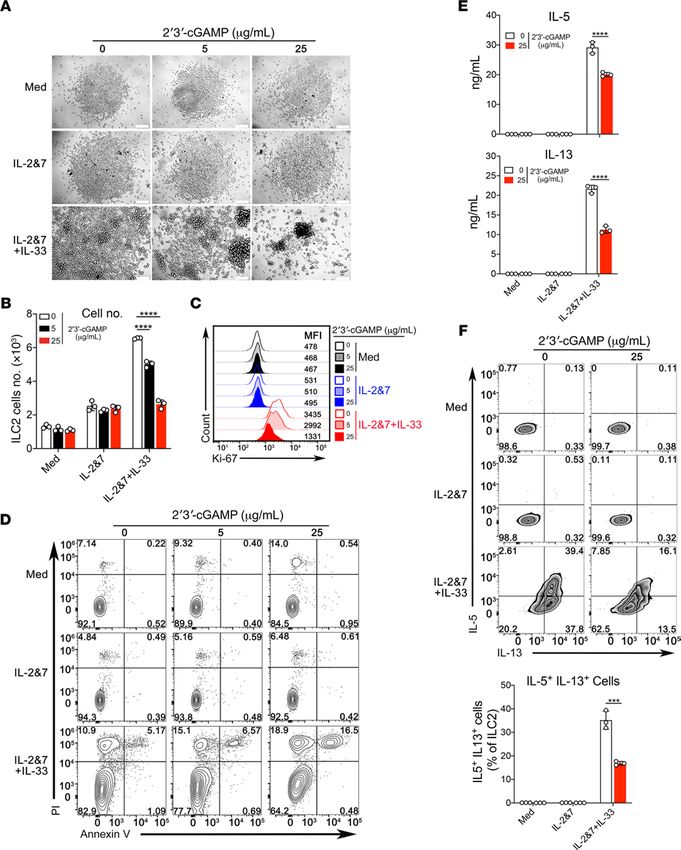
RESEARCH ARTICLE

                                 (9–12). Owing to their enormous capacity in instigating and amplifying type 2 inflammatory responses, acti-
                                 vation of ILC2 cells must be tightly regulated. Indeed, emerging evidence suggests that diversified receptors
                                 expressed on the surface of ILC2 cells can either enhance or repress their activation and proliferation in
                                 response to various signals such as alarmin cytokines, hormones, regulatory cytokines, neuropeptides, and lip-
                                 ids (14). Recent studies have also shown that microbial ligands, including unmethylated CpG-DNA, can acti-
                                 vate the corresponding innate immune responses to attenuate eosinophilic inflammation via inhibiting ILC2
                                 cell function in mouse models of asthma (15–17). However, it is unclear whether other innate immune stimuli
                                 such as 2′3′-cGAMP can regulate type 2 immunity through targeting ILC2 cells. 2′3′-cGAMP was discovered
                                 as a nonclassical second messenger synthesized by the novel DNA sensor cyclic GMP-AMP synthase (cGAS)
                                 in response to invasion of cytosolic DNA when mammalian cells are infected by DNA viruses and intracellu-
                                 lar bacteria (18–20). 2′3′-cGAMP exclusively binds and robustly activates stimulator of IFN genes (STING),
                                 which subsequently recruits the kinase TBK1 to trigger a signaling cascade leading to the production of type
                                 I IFN (IFN-I). Currently, 2′3′-cGAMP or its derivatives are being intensively investigated in a number of
                                 clinical trials for enhancing the efficacy of anti-PD-1–based cancer immunotherapies against multiple tumor
                                 types, including advanced solid tumors and lymphomas (21–23). In addition, 2′3′-cGAMP can be used as an
                                 immune adjuvant to enhance antigen-specific humoral and cellular immunities in a vaccine setting (19).
                                      In this study, we sought to determine an immune modulatory function of 2′3′-cGAMP in regulating
                                 type 2 inflammation in multiple mouse models of eosinophilic asthma. We demonstrate that 2′3′-cGAMP
                                 triggers the robust production of IFN-I in mouse lungs and strongly suppresses both IL-33 and an envi-
                                 ronmental allergen Aspergillus flavus–induced type 2 lung inflammation and airway hyperreactivity (AHR).
                                 Mechanistically, the STING/IFN-I signaling mediates the inhibitory effects of 2′3′-cGAMP and seems to
                                 act directly on ILC2 cells to inhibit their proliferation and function in the cytokine production. Collectively,
                                 our results identify an innate immune-driven mechanism for the 2′3′-cGAMP/STING/IFN-I signaling in
                                 regulating ILC2 cell function and show the potential development of this new mammalian cyclic dinucleo-
                                 tide for the prevention and treatment of eosinophilic asthma.

                                 Results
                                 2′3′-cGAMP inhibits IL-33–induced type 2 immunopathology. It has been recently shown that 2′3′-cGAMP has
                                 a strong immune adjuvant effect to enhance both cellular and humoral immunity (19). However, it remains
                                 undetermined whether 2′3′-cGAMP is capable of modulating overzealous type 2 inflammatory responses
                                 in the context of eosinophilic asthma. To address this issue, we employed a mouse model of acute lung
                                 inflammation induced by a recombinant murine protein IL-33, which is known to robustly activate ILC2
                                 cell–mediated lung inflammation (9, 24). We first determined whether 2′3′-cGAMP could initiate an immune
                                 response in mouse lungs. Mice were administered with increasing doses of 2′3′-cGAMP followed by examin-
                                 ing lung gene expressions by real-time quantitative PCR (RT-qPCR). We found that multiple IFN-stimulated
                                 genes (ISGs) (Mx1, ISG15, IFIT3, and OASL2) were strongly induced to reach a peak level of expression by
                                 2′3′-cGAMP at a dose of 5 μg/mouse. Notably, it appears that at this concentration of 2′3′-cGAMP, the induc-
                                 tion of 2 proinflammatory genes IL-1β and TNF-α were not obvious (Figure 1A). Thus, we chose this dose for
                                 the subsequent in vivo experiments in combination with IL-33 (Figure 1B). We found that 2′3′-cGAMP treat-
                                 ment drastically improved IL-33–induced lung pathology characterized by less infiltration of inflammatory
                                 cells, reduced epithelial cell hyperplasia, and decreased percentage of mucus-producing (PAS+) epithelial cells
                                 (Figure 1C). Functionally, 2′3′-cGAMP treatment resulted in substantially reduced airway resistance and elas-
                                 tance, and increased compliance in response to methacholine (Figure 1D). Consistently, among all analyzed
                                 cell types, eosinophils in both bronchoalveolar lavage fluid (BALF) and lungs were dramatically reduced (Fig-
                                 ure 1, E and F; the FACS gating strategy is shown in Supplemental Figure 1; supplemental material available
                                 online with this article; https://doi.org/10.1172/jci.insight.143509DS1; see complete unedited blots in the
                                 supplemental material). Notably, the number of neutrophils was also significantly reduced when compared
                                 with the IL-33–treated group. Taken together, these results suggest that 2′3′-cGAMP treatment triggered an
                                 immune response that strongly inhibits the development of IL-33–driven lung eosinophilia.
                                      2′3′-cGAMP ameliorates lung eosinophilia and AHR induced by a fungal allergen, A. flavus. To evaluate the ther-
                                 apeutic benefit of 2′3′-cGAMP in the context of environmental allergen-driven type 2 lung inflammation, we
                                 used a clinically relevant model caused by a fungal allergen, A. flavus, that has been recently shown to act through
                                 the IL-33–mediated pathway (25). As with IL-33 described above, we challenged mice with 10 μg extract of A.
                                 flavus after exposing mice to 2′3′-cGAMP. We found that A. flavus–driven type 2 inflammatory markers were

JCI Insight 2021;6(3):e143509 https://doi.org/10.1172/jci.insight.143509                                                                           2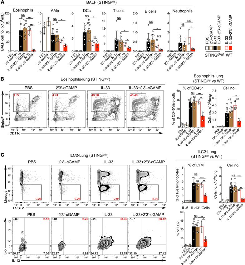
RESEARCH ARTICLE

JCI Insight 2021;6(3):e143509 https://doi.org/10.1172/jci.insight.143509                  3
RESEARCH ARTICLE
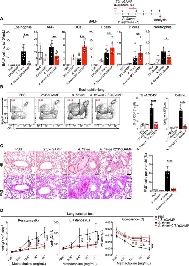
Figure 1. 2′3′-cGAMP inhibits IL-33–induced type 2 lung inflammation. (A) Transcriptional induction of mouse lung gene expressions by 2′3′-cGAMP. Error
bars represent standard error of triplicate assays. Representative data from 1 experiment are shown here. Similar results were obtained from at least 3
experiments. (B) Experimental setup illustrating the animal groups, regimen, and timeline. (C) Lung pathologies were assessed with H&E and PAS staining.
Representative images (scale bars: 100 μm) and the percentage of PAS+ cells are shown here. Original magnification ×200 was used for counting the percent-
age of mucus-producing bronchial epithelial cells (-) (PAS+). (D) Lung functions were examined by Flexivent (Scireq). Airway resistance (R), elastance (E), and
compliance (C) were measured after exposure to increasing doses (6.25–50 mg/mL) of aerosolized methacholine. A P value of less than 0.05 was considered
significant, n = 4–6. Two-way ANOVA followed by Tukey’s multiple comparisons test was conducted. (E) Groups of mice as indicated were treated with PBS,
2′3′-cGAMP, IL-33, or IL-33 + 2′3′-cGAMP. Bronchoalveolar lavage fluid (BALF) was collected and analyzed for differential immune cell types. The result was a
pool of 2 independent experiments. Open circles, n = 3–8 per group. A P value of less than 0.05 was considered significant using unpaired Student’s t test.
(F) Administration of 2′3′-cGAMP decreased the percentage and number of lung eosinophils after exposure to IL-33. Open circles, n = 3–4 per group. A P
value of less than 0.05 was considered significant using unpaired Student’s t test. *P < 0.05, **P < 0.01, ***P < 0.001, ****P < 0.0001.
suppressed, as evidenced by the decrease in eosinophil infiltration of BALF and lung in 2′3′-cGAMP–treated
mice (Figure 2, A and B). In addition, the number of neutrophils was also significantly reduced when com-
pared with the A. flavus–treated group. 2′3′-cGAMP treatment drastically improved A. flavus–induced lung
pathology characterized by infiltration of massive inflammatory cells, mucus overproduction (PAS+ epithelial
cells) and epithelial cell hyperplasia (Figure 2C). Consistent with these observations, 2′3′-cGAMP treatment
improved lung function as evidenced by reduced airway resistance, elastance, and improved compliance (Fig-
ure 2D). Thus, these data indicate that 2′3′-cGAMP treatment can activate a protective response to attenuate
acute type 2 lung inflammation induced by a clinically relevant fungal allergen.
2′3′-cGAMP inhibits ILC2 cell–driven type 2 lung inflammation. Next, we determined whether 2′3′-cGAMP
could affect the function and proliferation of lung ILC2 cells in vivo activated by IL-33 or A. flavus. To easily
track the activated population of ILC2 cells, we performed experiments with IL-13 reporter strain (the hetero-
zygous mouse, IL-13-eGFP+, ref. 26) in addition to WT mice (Figure 3A). The gating strategy for lung ILC2
cells is shown in Supplemental Figure 2. ILC2 cell expansion was significantly suppressed, as determined by
the reduction in total lung ILC2 cell numbers in both strains of mice in the context of IL-33 or A. flavus treat-
ment. Functionally, the percentage of activated ILC2 cells (IL-5 and IL-13 double-positive) was also decreased
upon treatment of 2′3′-cGAMP (Figure 3, B–D). Consistent with previous experiments, eosinophils in the
BALF and lungs were significantly reduced in 2′3′-cGAMP–treated IL-13-eGFP+ mice (Supplemental Figure
3). Further, 2′3′-cGAMP–mediated inhibitory effects on the ILC2 cell proliferation in WT mice was confirmed
by Ki-67 staining (Figure 3E). To further rule out the involvement of adaptive immunity in 2′3′-cGAMP–
induced suppressive effects, Rag1–/– mice that lack mature B and T cells were tested in the context of IL-33 or
A. flavus exposure (Figure 4A). FACS analysis revealed that upon exposures to either IL-33 or A. flavus, Rag1–/–
mice can develop severe type 2 lung inflammation characterized by the increased eosinophils in both BALF
and lungs (Figure 4, B and C); elevated level of mRNA and protein expressions of type 2 effector cytokines
such as IL-5, IL-9, and IL-13 (Figure 4, D and E); percentages of IL-5+ and IL-13+ double-positive cells; and
total numbers of lung ILC2 cells (Supplemental Figure 4). Similar to the 2′3′-cGAMP–treated IL-13-eGFP+ and
WT mice, all above examined parameters of type 2 inflammation in Rag1–/– mice were significantly reduced
by 2′3′-cGAMP (Figure 4, B–E, and Supplemental Figure 4). Taken together, these results strongly suggest that
2′3′-cGAMP activates an innate immune signaling pathway to negatively regulate ILC2 cell–induced eosino-
philic lung inflammation and that suppression is independent of the adaptive immunity in mice.
2′3′-cGAMP inhibits IL-33–, A. flavus–, or house dust mite extract–induced type 2 inflammation via STING/
IFN-I signaling. Next, we investigated whether the inhibitory effect of 2′3′-cGAMP could be dependent on the
STING/IFN-I signaling pathway. As expected, the effect of 2′3′-cGAMP on IL-33–induced type 2 lung inflam-
mation was completely abolished in STING goldenticket/goldenticket (STINGgt/gt) mice (Figure 5, A–C), indi-
cating the in vivo specificity of 2′3′-cGAMP–activated innate immune responses. As responses are known to
be a hallmark of the 2′3′-cGAMP/STING signaling (18–20), we then measured the level of IFN-α protein in
mouse lungs by ELISA. 2′3′-cGAMP treatment, in a dose-dependent manner, triggered a robust production of
IFN-α protein, which was detected from total lung homogenates (Figure 6A). It is very likely that activation of
the 2′3′-cGAMP/STING pathway would generate multiple effector molecules in mouse lungs besides IFN-I.
To demonstrate a possible role of IFN-I signaling, we performed a series of experiments using IFNAR1-de-
ficient mice. The expression of a stimulatory molecule CD40 in bone marrow–derived DCs (BM-DCs) and
alveolar macrophages derived from IFNAR1–/– mice were greatly reduced when stimulated with 2′3′-cGAMP
(Figure 6B). More importantly, in contrast to WT mice shown in Figure 1 and Figure 2, IFNAR1–/– mice treat-
ed with either IL-33 or IL-33 + 2′3′-cGAMP did not show any significant differences in eosinophils and ILC2
JCI Insight 2021;6(3):e143509 https://doi.org/10.1172/jci.insight.143509 4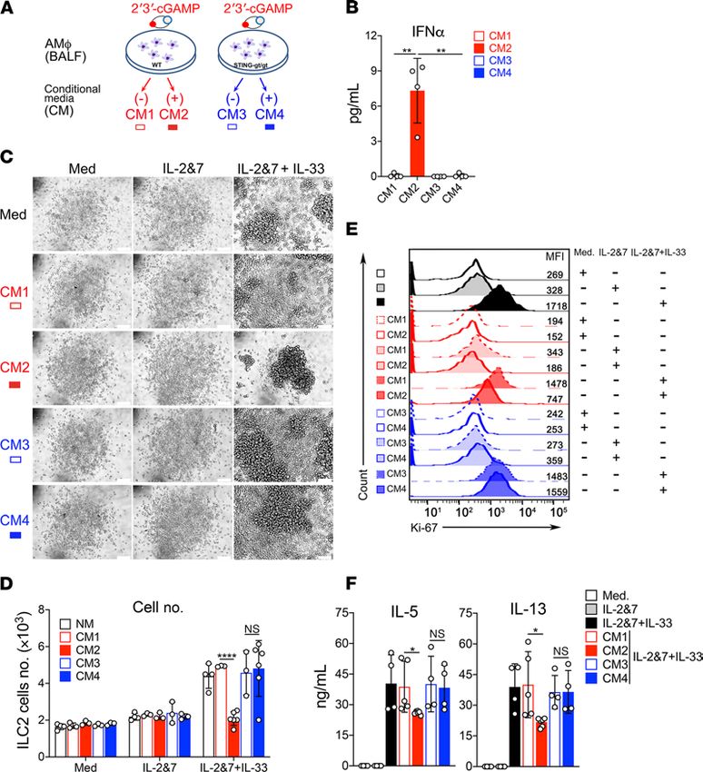
RESEARCH ARTICLE

Figure 2. 2′3′-cGAMP inhibits A. flavus-induced type 2 lung inflammation. (A) Groups of mice as indicated were treated with PBS, 2′3′-cGAMP, A. flavus, or A.
flavus + 2′3′-cGAMP. BALF was collected and analyzed for differential immune cell types. The result was a pool of 2 independent experiments. Open circles, n =
3–7 per group. A P value of less than 0.05 was considered significant using unpaired Student’s t test. *P < 0.05, **P < 0.01, ***P < 0.001. (B) Administration of

JCI Insight 2021;6(3):e143509 https://doi.org/10.1172/jci.insight.143509                                                                                       5
RESEARCH ARTICLE
2′3′-cGAMP decreased the percentage and number of lung eosinophils after exposure to A. flavus. Open circles, n = 3–8 per group. A P value of less than 0.05
was considered significant using unpaired Student’s t test. ****P < 0.0001. (C) Lung pathologies were assessed with H&E and PAS staining. Representative
images (scale bars: 100 μm) and the percentage of PAS+ cells are shown here. Original magnification ×200 was used for counting the percentage of mucus-
producing bronchial epithelial cells (-) (PAS+). ****P < 0.0001. (D) Airway hyperreactivity (AHR) was examined by Flexivent (Scireq). Airway resistance (R) was
measured after exposure to increasing doses (6.25–50 mg/mL) of aerosolized methacholine. Open circles, n = 3–5 per group. A P value of less than 0.05 was
considered significant. Statistics performed by 2-way ANOVA followed by Tukey’s multiple comparisons test. **P < 0.01, ***P < 0.001, ****P < 0.0001.
cells (number and percentage) in BALF or lungs (Figure 6, C–E). We also explored the possibility whether the
eosinophilia and activation of ILC2 cells could be inhibited by 2′3′-cGAMP codelivered or delivered afterward
with A. flavus in Rag1–/– mice (Supplemental Figure 5) or WT mice (Supplemental Figure 6). In addition, we
examined whether 2′3′-cGAMP treatment can affect the established type 2 lung inflammation induced by
house dust mite extract (HDM), a more physiologically relevant aeroallergen (Supplemental Figure 7A). As
shown in Supplemental Figure 7, B and D, 2′3′-cGAMP treatment significantly inhibited HDM-induced lung
eosinophilia and activation of ILC2 cells. In all of these treatments, 2′3′-cGAMP was effective in suppressing
type 2 immunopathology. Taken together, these results suggest that the STING/IFN-I signaling axis mediates
the effector function of 2′3′-cGAMP in vivo to negatively regulate type 2 lung inflammation induced by IL-33
and natural allergens likely via suppressing the activation of ILC2 cells.
Activation of alveolar macrophages by 2′3′-cGAMP leads to the production of ILC2 cell inhibitory factors. To identify
lung cell types that are responsible for taking up 2′3′-cGAMP, we performed a flow cytometric analysis of lung
cells (FACS gating strategy, Figure 7A). Mice were treated with fluorescently labeled 2′3′-cGAMP (cGAMP-
Fluo), and lungs were collected after 16 hours. Lung tissues were then digested, and cells were stained with vari-
ous cell type–specific surface makers. Interestingly, cGAMP-Fluo was mainly detected in alveolar macrophages
and, to a lesser extent, monocyte-derived DCs (MoDCs), but not in other examined lung immune cells such
as conventional DCs, interstitial macrophages, monocytes, T cells, B cells, and nonimmune cells (CD45– cells,
Figure 7B). FACS analysis further revealed that the phosphorylated form of IFN regulatory factor 3 (IRF3) in
alveolar macrophages appeared within 1–2 hours after the 2′3′-cGAMP treatment (Figure 7C). STING is indis-
pensable for the in vivo activity of 2′3′-cGAMP because the phosphorylated form of IRF3 was only detected in
the lung homogenates of WT, but not in STINGgt/gt mice (Figure 7D). To determine whether 2′3′-cGAMP–stim-
ulated alveolar macrophages could produce IFN or ILC2 cell regulatory factors, we collected conditioned media
from the cultured alveolar macrophages as illustrated in Figure 8A. As expected, IFN-α was only detected in the
conditioned media (CM) of alveolar macrophages from WT, but not STING deficient mice by ELISA (Figure
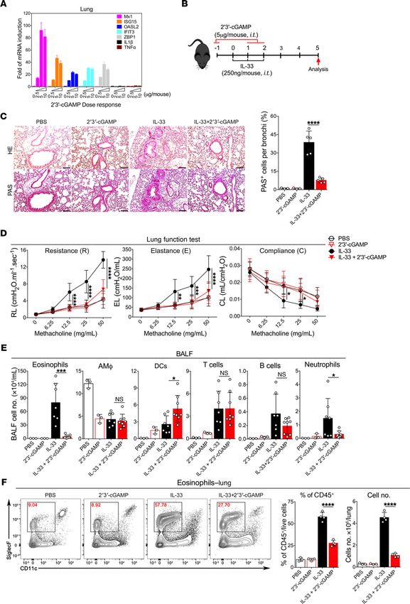
8B). Moreover, although it had no effects on the nonactivated ILC2 cells under the conditions of media or IL-2 +
IL-7, the CM from 2′3′-cGAMP–treated WT alveolar macrophages significantly impaired the growth and func-
tion of IL-33–activated ILC2 cells measured by the cell number and proliferation marker Ki-67 (Figure 8, C–E)
and the cytokine levels of IL-5 and IL-13 (Figure 8F). To further demonstrate an important role of alveolar mac-
rophages in mediating the effects of 2′3′-cGAMP in vivo, we have performed IL-33 treatments in 2 mouse lines
lacking alveolar macrophages (Supplemental Figures 8A and 9A), Csf2–/– and clodronate liposome-treated WT
mice, in which type 2 inflammatory responses were poorly activated (Supplemental Figures 8 and 9). Nonethe-
less, it appears that the 2′3′-cGAMP administration could further reduce the numbers of eosinophils and ILC2
cells when compared with the mouse lines lacking alveolar macrophages treated with the IL-33 alone, implying
that 2′3′-cGAMP might act on other cell types such as ILC2 in vivo. Overall, these results indicate that alveolar
macrophages have the ability to take up extracellular 2′3′-cGAMP and turn on the STING/IRF3 pathway that
may lead to the production of ILC2 inhibitory factors such as IFN-I.
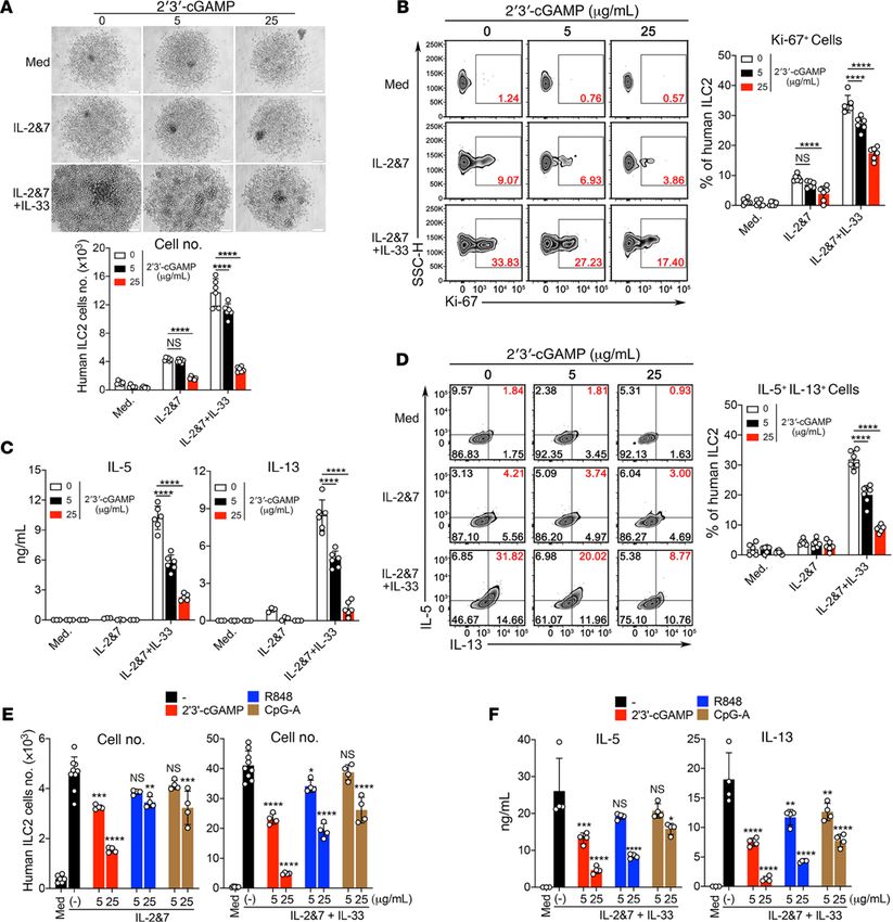
2′3′-cGAMP directly suppresses the proliferation and cytokine production of human and mouse ILC2 cells in vitro.
Because we have shown above that 2′3′-cGAMP treatment could act on alveolar macrophages to negatively
regulate ILC2 cell–driven type 2 inflammation, next we wanted to assess its potential effect on ILC2 cells in
vitro. The purified mouse and human ILC2 cells (Supplemental Figure 10) were cultured under conditions of
media alone, IL-2 + IL-7, or IL-2 + IL-7 + IL-33 and stimulated with 2′3′-cGAMP. Indeed, the purified mouse
and human ILC2 cells were able to take up 2′3′-cGAMP and activate the STING/IFN-I pathway (Supple-
mental Figure 11). Further, in a concentration-dependent manner, 2′3′-cGAMP strongly suppressed the pro-
liferation of both mouse and human ILC2 cells demonstrated by the cell density, the cell number, and the level
of Ki-67 (Figure 9, A–C; and Figure 10, A and B). We found that this suppression by 2′3′-cGAMP was partly
attributed to the induction of cell death (Figure 9D). Moreover, 2′3′-cGAMP strongly suppressed the produc-
tion of IL-5 and IL-13 by mouse and human ILC2 cells activated by IL-33 (Figure 9E and Figure 10C). Intra-
JCI Insight 2021;6(3):e143509 https://doi.org/10.1172/jci.insight.143509 6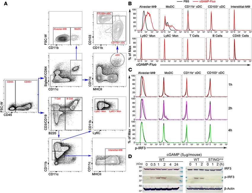
RESEARCH ARTICLE

                                                                           Figure 3. 2′3′-cGAMP inhibits ILC2 cell activation
                                                                           and proliferation induced by IL-33 and A. flavus
                                                                           in WT or IL-13 reporter mice. (A) Experimental
                                                                           protocol showing the animal groups (IL-13-eGFP+
                                                                           and WT), the corresponding treatment regimen
                                                                           and timeline. (B) The heterozygous IL-13-eGFP+
                                                                           mice were treated with PBS, 2′3′-cGAMP, IL-33, or
                                                                           IL-33 + 2′3′-cGAMP. Lung single cell suspensions
                                                                           were prepared and the number of ILC2 cells in
                                                                           lungs were analyzed. In addition, lung cells were
                                                                           stimulated with phorbol 12-myristate 13-acetate
                                                                           in cultures as described in the Methods. The
                                                                           percentage of IL-5+IL-13+ double-positive ILC2
                                                                           cells in lungs was analyzed (n = 2–6 per group
                                                                           as indicated with open circles). (C) Similar to B,
                                                                           instead, WT mice were used (n = 3–4 per group
                                                                           as indicated with open circles). (D) Similar to C,
                                                                           instead of IL-33, WT mice were treated with A.
                                                                           flavus (n = 3–8 per group as indicated with open
                                                                           circles). For A–D, a P value of less than 0.05 was
                                                                           considered significant using unpaired Student’s
                                                                           t test. **P < 0.01, ***P < 0.001, ****P < 0.0001.
                                                                           (E) WT mice were treated with PBS, 2′3′-cGAMP,
                                                                           IL-33, or IL-33 + 2′3′-cGAMP. The lung ILC2 cells
                                                                           were analyzed with Ki-67 staining and isotype
                                                                           antibody. The result is representative of 2 inde-
                                                                           pendent experiments.

JCI Insight 2021;6(3):e143509 https://doi.org/10.1172/jci.insight.143509                                                    7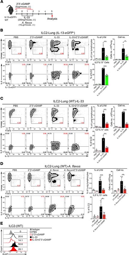
RESEARCH ARTICLE
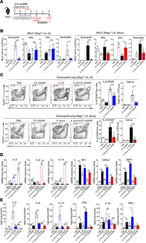
Figure 4. 2′3′-cGAMP inhibits type 2 lung
inflammation induced by IL-33 and A. flavus
in Rag1–/– mice. (A) Experimental protocol
showing the animal groups of Rag1–/– mice
and the corresponding treatment regi-
men and timeline. (B) Administration of
2′3′-cGAMP into Rag1–/– mice decreased the
number of airway eosinophils. BALF analysis
of Rag1–/– mice were treated with 2 kinds of
experimental regimens, PBS, 2′3′-cGAMP,
IL-33, IL-33 +2 ′3′-cGAMP (left panel) or PBS,
2′3′-cGAMP, A. flavus, A. flavus + 2′3′-cGAMP
(right panel). (C) Similar to B, administration
of 2′3′-cGAMP into Rag1–/– mice decreased
the percentage and number of lung eosin-
ophils after exposure to IL-33 (top panel) or
A. flavus (bottom panel) (n = 2–5 per group
as indicated with open circles, a P value of
less than 0.05 was considered significant,
unpaired Student’s t test. **P < 0.01, ***P
< 0.001, ****P < 0.0001). (D) Similar to B,
instead, lung samples were collected on day
3 for RNA extraction, then RT-qPCR analysis
of the selected type 2 effector cytokines and
IFN-stimulated genes (ISGs) as indicated
were performed. Error bars represent stan-
dard error of triplicate assays. Representa-
tive data from 1 experiment are shown here.
Similar results were obtained from at least 3
experiments. (E) Similar to B, instead, lung
samples were collected and homogenized
on day 3 for protein extractions, which were
used for measuring the level of the selected
cytokine as indicated by ELISA. For D and
E, n = 3–4 per group as indicated with open
circles. A P value of less than 0.05 was con-
sidered significant using unpaired Student’s
t test. *P < 0.05, **P < 0.01, ***P < 0.001,
****P < 0.0001.
cellular cytokine staining also demonstrated that 2′3′-cGAMP also suppressed the cytokine expression inside
the ILC2 cells (Figure 9F and Figure 10D). Moreover, 2′3′-cGAMP at a lower concentration (5 μg/mL),
when compared with 2 other TLR agonists (R848, a TLR7 agonist, and CpG-A, a TLR9 agonist), appears
to be more potent in directly suppressing both the growth and production of type 2 effector cytokines of
human ILC2 cells (Figure 10, E and F). Notably, at a higher concentration (25 μg/mL) all 3 agents had inhib-
itory effects on human ILC2 cells. Collectively, these results indicate that 2′3′-cGAMP can directly suppress
JCI Insight 2021;6(3):e143509 https://doi.org/10.1172/jci.insight.143509 8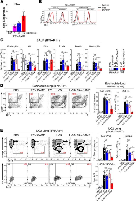
RESEARCH ARTICLE

Figure 5. The inhibitory effect of 2′3′-cGAMP is abolished in STING-deficient mice. Groups of STINGgt/gt and WT mice were treated with PBS, 2′3′-cGAMP,
IL-33, or IL-33 + 2′3′-cGAMP as indicated. BALF was collected and analyzed for differential immune cell types. (A) In contrast to WT mice, administration
of 2′3′-cGAMP into STINGgt/gt mice did not significantly change number of airway eosinophils after exposure to IL-33. (B) Similar to A, administration of
2′3′-cGAMP in STINGgt/gt mice did not significantly change the percentage and number of lung eosinophils after exposure to IL-33. (C) Similar to A, the number
and percentage of IL-5+IL-13+ double- positive ILC2 cells in lungs of STINGgt/gt mice were analyzed (n = 3–5 per group as indicated with open circles, a P value
greater than or equal to 0.05 was not considered significant [NS], Student’s unpaired t test. *P < 0.05, **P < 0.01, ***P < 0.001, ****P < 0.0001).

                                    the activation of ILC2 cell function induced by IL-33. Taken together, our data indicate that IFN-I signal-
                                    ing mediates the effector function of 2′3′-cGAMP to negatively regulate allergen-induced type 2 immune
                                    responses. Based on these findings, as depicted in Figure 11, we propose a working model for 2′3′-cGAMP in
                                    modulating ILC2 cell–mediated type 2 immunity. After the respiratory delivery, 2′3′-cGAMP is first taken up

JCI Insight 2021;6(3):e143509 https://doi.org/10.1172/jci.insight.143509                                                                                           9
RESEARCH ARTICLE
Figure 6. The inhibitory effect of 2′3′-cGAMP is
mediated by the type I IFN signaling pathway.
(A) Induction of IFN-α protein after administra-
tion of 2′3′-cGAMP at 5, 10, and 20 μg/mouse
for 24 hours. The mouse lungs from WT mice
were processed for the ELISA measurement.
(B) BM-DCs and alveolar macrophages derived
from WT and IFNAR1–/– mice were treated with
2′3′-cGAMP. Then, the expression of a costimu-
latory molecule CD40 was analyzed by FACS. (C)
Groups of IFNAR1–/– and WT mice were treated
with PBS, 2′3′-cGAMP, IL-33, or IL-33 + 2′3′-cGAMP
as indicated. BALF was collected and analyzed
for differential immune cell types. In contrast
to WT mice, administration of 2′3′-cGAMP into
IFNAR1–/– mice did not significantly change
number of airway eosinophils after exposure
to IL-33. (D) Administration of 2′3′-cGAMP in
IFNAR1–/– mice did not significantly decrease the
percentage and number of lung eosinophils after
exposure to IL-33. (E) Similar to C, the number
and percentage of IL-5+IL-13+ double-positive ILC2
cells in lungs of IFNAR1–/– mice were analyzed (n
= 3–7 per group as indicated with open circles, a
P value of greater than or equal to 0.05 was not
considered significant [NS], Student’s unpaired t
test, *P < 0.05, **P < 0.01, ***P < 0.001).
by alveolar macrophages, where it activates the STING-mediated pathway to induce the production of IFN-I,
which may in turn act on ILC2 cells to restrain their abilities to proliferate and produce type 2 cytokines. In
addition, 2′3′-cGAMP seems to be able to directly suppress the function of activated ILC2 cells in the context
of exposure to IL-33 and environmental allergens such as A. flavus.
Discussion
In this study, we reveal an important role for 2′3′-cGAMP in negatively regulating type 2 inflammation
induced by IL-33, a fungal allergen, and HDM. We demonstrate that 2′3′-cGAMP administration efficient-
ly protects mice from both IL-33– and a fungal allergen-induced AHR. Mechanistically, 2′3′-cGAMP can
directly act on both alveolar macrophages and ILC2 cells. In alveolar macrophages, 2′3′-cGAMP activates
the STING/IRF3 pathway that leads to a rapid production of ILC2 cell suppressive factors such as IFN-α.
These data provide proof-of-concept evidence to support a therapeutic value of 2′3′-cGAMP in preventing
and mitigating lung inflammation of eosinophilic asthma.
JCI Insight 2021;6(3):e143509 https://doi.org/10.1172/jci.insight.143509 10RESEARCH ARTICLE
Figure 7. 2′3′-cGAMP is mainly taken up by alveolar macrophages in vivo. (A) FACS gating strategy for identification of specific lung cell types by staining
cell surface markers as indicated in the Methods. (B) The fluorescence-labeled 2′3′-cGAMP was mainly detected by FACS analysis in alveolar macrophages,
and to a lesser extent, monocyte-derived DCs (MoDCs), but not in other lung cell types. The result is representative of 3 independent experiments. (C)
FACS analysis shows that the phosphorylated form of IRF3 can be detected in alveolar macrophages at 1–2 hours after the 2′3′-cGAMP treatment. The
result is representative of 3 independent experiments. (D) Western blot analysis of homogenates of mouse lungs shows that 2′3′-cGAMP treatment rapid-
ly activates the STING-mediated pathway within 1–2 hours that results in the phosphorylation of IRF3.
A few recent studies have shown that IFN-I directly inhibits the function and proliferation of ILC2
cells in vitro and in vivo during influenza A infection or treatment with TLR agonists such as CpG-DNA
and R848 (15, 16, 27–30). Interestingly, our results suggest that the inhibitory effects of 2′3′-cGAMP are
likely to be attributed, at least in part, to its ability in triggering production of IFN in mouse lungs, which
generates a Th1-dominant cytokine milieu that is suppressive to ILC2 cells, which may further lead to a
reduced level of Th2-biased adaptive immune response. In a recent phase II clinical trial, administering
inhaled recombinant IFN-α/β was effective in treating steroid-resistant eosinophilic asthma (31). However,
an IFN-based therapy is often associated with a wide array of adverse effects (32, 33). Therefore, instead
of using cytokines directly, agonists that specifically stimulate the innate immune system are becoming
more favorable therapeutics largely attributed to their lower toxicity and better immune response profile.
In this regard, our study presents an important proof of principle for treating eosinophilic asthma through
harnessing 2′3′-cGAMP-STING–mediated innate immune response.
Because of its inherent dual negative charges and the presence of an extracellular enzyme such as ectonu-
cleotide pyrophosphatase/phosphodiesterase (34), an efficient in vivo delivery of 2′3′-cGAMP was usually con-
sidered to be very challenging without proper formulations. However, our results show that when administered
intratracheally at a low dose, 2′3′-cGAMP was capable of inducing innate immune responses comprising IFN-I
JCI Insight 2021;6(3):e143509 https://doi.org/10.1172/jci.insight.143509 11RESEARCH ARTICLE
Figure 8. 2′3′-cGAMP stimulates alveolar macrophages to produce ILC2-inhibitory factors. (A) Diagram showing the
generation of conditioned media (CM) from the ex vivo cultured alveolar macrophage (AMΦ) cells from WT or STINGgt/
gt
mice that were stimulated with or without 2′3′-cGAMP. (B) The level of IFN-α in the CM was measured by ELISA. (C)
Light microscopic images showing the growth of mouse ILC2 cells in the presence of the corresponding CM as indicated.
(D) FACS showing the number of murine ILC2 cells in the presence of the CM. (E) Ki-67 staining of mouse ILC2 cells in
the presence of the CM. (F) ELISA measuring the production of IL-5 and IL-13 by mouse ILC2 cells in the presence of the
CM. Statistics for D were performed using 2-way ANOVA followed by Tukey’s multiple comparisons test by comparing
mouse ILC2 cells treated with CM as indicated. A P value greater than or equal to 0.05 was not considered significant
(NS). *P < 0.05, **P < 0.01, ****P < 0.0001.
that were sufficient to inhibit ILC2 cell activation by IL-33 or a fungal allergen, A. flavus. Given that alveolar
macrophages are the first line of phagocytic defense in lower airways and are approximately 50 times more
numerous than conventional DCs, it was not unexpected that 2′3′-cGAMP was found to be mainly taken up by
alveolar macrophages, and to a lesser extent, MoDCs in mouse lungs. It is possible that the 2′3′-cGAMP–activat-
ed alveolar macrophages can produce chemokines and cytokines, including IFN-I, to create a suppressive local
environment against the development of overzealous type 2 inflammation upon exposures to various aeroaller-
gens. In the context of allergic inflammation, inflamed lung tissue may become leaky and allow immune cells
or cytokines to travel or penetrate across tissue barriers to act on ILC2 cells. However, the molecular mechanism
of how 2′3′-cGAMP enters the cytosol of alveolar macrophages where it engages the downstream STING/
IFN-I signaling pathway remains unknown. In this regard, 2 recent reports suggest that 2 proteins, cGAS and
JCI Insight 2021;6(3):e143509 https://doi.org/10.1172/jci.insight.143509 12RESEARCH ARTICLE Figure 9. 2′3′-cGAMP directly suppresses the proliferation and cytokine production of mouse ILC2 cells. (A) Light microscopic images showing the growth of mouse ILC2 cells in the presence of the increased concentration of 2′3′-cGAMP. (B) FACS showing the number of murine ILC2 cells in the presence of the increased concentration of 2′3′-cGAMP. (C) Ki-67 staining of murine ILC2 cells in the presence of the increased concentration of 2′3′-cGAMP. (D) Cell death of mouse ILC2 cells in the presence of the increased concentration of 2′3′-cGAMP. (E) ELISA measuring the production of IL-5 and IL-13 by mouse ILC2 cells in the presence of the increased concentration of 2′3′-cGAMP. (F) Intracellular staining of IL-5 and IL-13 in mouse ILC2 cells treated with 2′3′-cGAMP (the percentage of the double- positive cells was quantified, lower). Statistics were performed using 2-way ANOVA followed by Tukey’s multiple comparisons test by comparing media-treated mouse ILC2 cells with the individual treatment as indicated. A P value greater than or equal to 0.05 was not considered significant. ***P < 0.001, ****P < 0.0001. JCI Insight 2021;6(3):e143509 https://doi.org/10.1172/jci.insight.143509 13
RESEARCH ARTICLE

                                 human SLC19A1, might be involved in trafficking of extracellular 2′3′-cGAMP into the cytoplasm of cultured
                                 macrophages and monocytes (35, 36). Additionally, in contrast to bacterial cyclic dinucleotides (3′3′-c-di-GMP,
                                 3′3′-c-di-AMP, and 3′3′-cGAMP), which preferentially bind with high affinity and activate 2 new sensors ERAdP
                                 (37) and RECON (37–40), 2′3′-cGAMP is a much more potent and specific ligand for human STING (41, 42).
                                 Collectively, it appears that 2′3′-cGAMP would be a promising candidate for harnessing innate immunity to
                                 treat or prevent many human diseases, such as cancer and eosinophilic asthma.
                                      At present, a number of STING agonists have entered clinical trials for cancer immunotherapy (21–23).
                                 It can also be envisioned that many issues or concerns such as adverse effects may arise and must be further
                                 addressed with independent experimental approaches. At the same time, there will be more exciting oppor-
                                 tunities on the parallel development of these reagents for other applications such as treating eosinophilic
                                 diseases. Supported by this work, the use of 2′3′-cGAMP or other STING agonists as immune modulators
                                 to enhance the efficacy of merits future investigation.
                                      To some extent, our current findings on 2′3′-cGAMP are consistent with a new report showing that a
                                 bacterial cyclic dinucleotide, c-di-GMP, can effectively inhibit IL-33– or Alternaria-induced type 2 inflam-
                                 mation in mice (43). However, it has also recently been reported that in another model of allergic inflam-
                                 mation, the administration of 2′3′-cGAMP had an adjuvant effect that exacerbated the HDM-induced Th2
                                 response (44). This result is contrast to our data presented in Supplemental Figure 7, in which HDM-in-
                                 duced type 2 inflammation was attenuated by 2′3′-cGAMP administration. The exact causes for these
                                 discrepancies are unclear and could be ascribed to many things, such as the dose, timing, and delivery route
                                 for 2′3′-cGAMP. More preclinical experiments, however, are needed to resolve this controversy.
                                      In conclusion, our results highlight the role of an allergen-independent, innate immune-driven effector
                                 function triggered by the 2′3′-cGAMP/STING/IFN-I signaling pathway, which robustly counteracts the rapid-
                                 ly activated ILC2 cells in the context of IL-33 and a fungal allergen-induced acute type 2 inflammation. From
                                 the therapeutic standpoint, our study suggests that further development of a formulated 2′3′-cGAMP for local
                                 delivery into the lungs may serve as an alternative approach for preventing or treating eosinophilic asthma.

                                 Methods
                                 Mice. IL-13-eGFP reporter strain (26) and STINGgt/gt (45) mice have been described previously. WT
                                 C57BL/6J, Csf2–/–, IFNAR1–/–, and Rag1–/– mice were purchased from the Jackson Laboratory. The
                                 IL-13-eGFP+ heterozygous mice were generated by intercrossing with WT C57BL/6J on campus. Mice
                                 were bred and maintained under specific pathogen–free conditions in the animal facility. Age-matched
                                 (8–10 weeks old) female mice were used for the experiments.
                                      Cells and reagents. Bone marrow cells were collected from femurs and tibiae of mice. To obtain BM-DCs,
                                 about 10 million bone marrow cells were cultured in DMEM containing 10% FCS, antibiotics, and Flt3
                                 ligand. After 7 days, mature DCs were harvested and cultured in 96-well plates for experiments. Media were
                                 changed every other day. 2′3′-cGAMP was purchased from InvivoGen.
                                      ELISA to detect cytokines in mouse lungs and cell culture supernatants. For measuring cytokines in mouse
                                 lungs after 2′3′-cGAMP stimulation, the harvested lungs were washed once with cold PBS, transferred into
                                 2 mL tubes, rapidly frozen into liquid-N2, and stored at −80°C. Later, to prepare lung homogenates, 1 mL
                                 tissue protein extraction reagent (T-PER) (Thermo Fisher Scientific) containing protease inhibitors (Roche)
                                 was added and homogenized by a BeadBeater (BioSpec). The lysates were transferred to a 1.5 mL tube and
                                 spun at 14,000g for 30 minutes at 4°C. Supernatant was collected for the ELISA measurement of cytokines.
                                 IFN-α in supernatant of alveolar macrophages culture and lung homogenates were measured with the ELI-
                                 SA kit (PBL Assay Science). Cytokines such as TNF-α, IL-5, IL-9, IL-13, and IFN-γ in lung homogenates
                                 and type 2 cytokines (IL-5 and IL-13) in supernatants of mouse or human ILC2 cell cultures were analyzed
                                 with ELISA kit (all purchased from Invitrogen), and IL-1β in lung homogenates was detected by the ELI-
                                 SA kit (R&D Systems). All final reactions were developed with TMB substrate (Thermo Fisher Scientific)
                                 and stopped by sulfuric acid (0.16 M), and the OD at 450 nm was measured.
                                      Western blot. Western blot analysis was performed as previously described with some modifications (46).
                                 Briefly, protein extracts were obtained from the lungs of 8-week-old female mice treated with 2′3′-cGAMP (5
                                 μg/mouse) for different time periods. One-half of the lung tissue was put into lysing Matrix D tube (MP Bio-
                                 medicals) and immediately frozen in liquid N2. A total of 1.0 mL extraction buffer (8 M urea, 1% SDS, 0.15
                                 M Tris-HCl, pH 7.5) was added, and samples were homogenized by a BeadBeater (BioSpec). The lysates were
                                 transferred to a 1.5 mL tube and spun at 14,000g for 30 minutes at 4°C. Supernatant was collected, and protein

JCI Insight 2021;6(3):e143509 https://doi.org/10.1172/jci.insight.143509                                                                     14
RESEARCH ARTICLE Figure 10. 2′3′-cGAMP directly suppresses the proliferation and cytokine production of human ILC2 cells. (A) Light microscopic images and FACS showing the growth and number of human ILC2 cells in the presence of the increased concentration of 2′3′-cGAMP. (B) Ki-67 staining of human ILC2 cells in the presence of the increased concentration of 2′3′-cGAMP. (C) ELISA measuring the production of IL-5 and IL-13 by human ILC2 cells in the presence of the increased concentration of 2′3′-cGAMP. (D) Intracellular staining of IL-5 and IL-13 in human ILC2 cells treated with the increased concentration of 2′3′-cGAMP (the quantification of the percentage of the double-positive cells, right). (E) Human ILC2 cells cultured under either IL-2&7 or IL-2&7 + IL-33 were treated with 2′3′-cGAMP, R848, or CpG-A as indicated. FACS showing the growth of human ILC2 cells. (F) Same as E. The production of IL-5 and IL-3 was measured by ELISA. The result is representative of 3 independent experiments. Statistics were performed using 2-way ANOVA followed by Tukey’s multiple comparisons test by comparing media or mock (-)-treated human ILC2 cells to the individual treatment as indicated. A P value of greater than or equal to 0.05 was not considered significant (NS). *P < 0.05, **P < 0.01, ***P < 0.001, ****P < 0.0001. JCI Insight 2021;6(3):e143509 https://doi.org/10.1172/jci.insight.143509 15
RESEARCH ARTICLE

                                 Figure 11. A proposed working model illustrates the potential mechanism through which 2′3′-cGAMP inhibits type 2
                                 immunopathology by targeting human and mouse alveolar macrophages and ILC2 cells. Artwork was initially created
                                 in BioRender, https://app.biorender.com.

                                 concentration was measured by Bradford protein assay (Pierce). A total of 55 μg of each sample was loaded and
                                 separated in 10% SDS/PAGE and transferred to PVDF (MilliporeSigma). Membranes were blocked with 5%
                                 nonfat milk and incubated with rabbit mAbs against IRF3, p-IRF3, and β-actin (Cell Signaling Technology).
                                      RT-qPCR. Reverse transcription and RT-qPCR reactions were carried out using iScript cDNA synthesis
                                 kit and iQ SYBR Green Supermix (Bio-Rad). qPCR was performed on a Bio-Rad CFX384 Touch Real-
                                 Time PCR Detection System using the following primers: mouse primers, forward (5′→3′); reverse (5′→3′):
                                 Rpl19 (AAATCGCCAATGCCAACTC; TCTTCCCTATGCCCATATGC), IL-1β (TCTATACCT-
                                 GTCCTGTGTAATG; GCTTGTGCTCTGCTTGTG), IFIT3 (TGGCCTACATAAAGCACCTAGAT-
                                 GG; CGCAAACTTTTGGCAAACTTGTCT), ISG15 (GAGCTAGAGCCTGCAGCAAT; TTCTGG-
                                 GCAATCTGCTTCTT), Mx1 (TCTGAGGAGAGCCAGACGAT; ACTCTGGTCCCCAATGACAG),
                                 OASL2 (GGATGCCTGGGAGAGAATCG; TCGCCTGCTCTTCGAAACTG), TNF-α (CCTCCCTCT-
                                 CATCAGTTCTATGG; GGCTACAGGCTTGTCACTCG), IL-5 (AGGATGCTTCTGCACTTGAG;
                                 CCTCATCGTCTCATTGCTTG), IL-9 (GAACATCACGTGTCCGTCCT; CGGCTTTTCTGCCTTTG-
                                 CAT), IL-13 (TGAGCAACATCACACAAGACC; AGGCCATGCAATATCCTCTG).
                                      In vivo administration and FACS analysis of BALF and lung. Mice were anesthetized by isoflurane inhala-
                                 tion, followed by 3 times of intratracheal administration with 2′3′-cGAMP (5 μg), rIL-33 (0.25 μg), or A.
                                 flavus protease allergen (10 μg) in 80 μL PBS as shown in Figure 1B. Mice were sacrificed at indicated times,
                                 and the trachea was catheterized and flushed with 1 mL ice-cold PBS-EDTA 3 times. Differential cells in
                                 BALF were labeled with antibodies as indicated, then mixed with counting beads (Spherotech) for further
                                 FACS analysis on a BD Celesta cell analyzer. Flow cytometry data were analyzed using FlowJo software.
                                 The antibodies and reagents for FACS analysis are as follows: SPHERO AccuCount Fluorescent (Sphero-
                                 tech, ACFP-70-5), Anti-Mouse Siglec-F PE (clone E50-2440) (BD Biosciences, 552126), Anti-Mouse CD19
                                 Alexa Fluor 647 (clone 1D3) (BD Biosciences, 557684), Anti-Mouse CD3ε APC (clone 145-2C11) (BioLeg-
                                 end, 100322), Anti-Mouse MHC II APC-Cy7 (clone M5/114.15.2) (BioLegend, 10627), Anti-Mouse CD11c
                                 PE-Cy7 (clone N418) (Tonbo Biosciences, 60-0114-U100), Anti-Mouse CD11b V450 Rat (clone M1/70)
                                 (BD Biosciences, 560456), Anti-Mouse Ly-6G FITC (clone RB6-8C5) (Invitrogen, 11-5931-82), Anti-Mouse
                                 Fixable Viability Dye eFluor 506 (Invitrogen, 65-0866-14), Anti-Mouse CD45 PerCP-Cy5.5 (clone 30-F11)
                                 (BioLegend, 103130), Anti-Mouse CD45 APC-Cy7 (clone 30-F11) (BD Biosciences, 561037), Anti-Mouse
                                 CD103 Alexa Fluor 647 (clone 2ET) (BioLegend, 121410), Anti-Mouse CD64 PE (clone X54-5/7.1) (Bio-
                                 Legend, 139303), Anti-Mouse Ly6C PerCP-Cy5.5 (clone AL-21) (BD Biosciences, 560525), Anti-Mouse
                                 Siglec-F FITC (clone S17007L) (BioLegend, 155503), Anti-Mouse CD64 FITC (clone X54-5/7.1) (BioLeg-
                                 end, 139316), Anti-Mouse CD40 PE (clone 3/23) (BioLegend, 124609), Anti-Mouse Isotype Ctrl PE (clone

JCI Insight 2021;6(3):e143509 https://doi.org/10.1172/jci.insight.143509                                                                       16
RESEARCH ARTICLE
RTK2071) (BioLegend, 400408), Anti-Mouse p-IRF3 (S396) Alexa Fluor 488 (clone D601M) (Cell Signal-
ing Technology, 53539S), Anti-Mouse Isotype Ctrl FITC (clone RTK2758) (BioLegend, 400506).
Identification of lung ILC2 cells. Lung ILC2 cell identification was performed as described previously
(47). Lung tissues were digested in 8 mL RPMI 1640 containing liberase (50 μg/mL) and DNase I (1 μg/
mL) for approximately 40 minutes at 37°C. Cell suspensions were filtered through 70 μm cell strainers and
washed once with RPMI 1640. For ILC2 cell identification, total lung cell suspensions were blocked with
2.4G2 antibodies and stained with lineage cocktail mAbs: CD3ε (clone 145-2C11) (BioLegend, 100304),
CD4 (clone GK1.5) (BioLegend, 100404), CD8α (clone 53-6.7) (Tonbo Biosciences, 30-0081-U500),
CD11c (clone N418) (BioLegend, 117304), FceRIα (clone MAR-1) (BioLegend, 134304), NK1.1 (clone
PK136) (BioLegend, 108704), CD19 (clone 6D5) (BioLegend, 115504), TER119 (clone TER-119) (Bio-
Legend, 116204), CD5 (clone 53-7.3) (BioLegend, 100604), F4/80 (clone BM8.1) (Tonbo Biosciences,
30-4801-U500), Ly6G (clone RB6-8C5) (Tonbo Biosciences, 30-5931-U500), APC-conjugated streptavi-
din (BioLegend, 405207), PE-conjugated T1/ST2 (clone DIH9) (BioLegend, 145304), PerCP-Cy5.5-con-
jugated CD25 (clone PC61) (BioLegend, 102030), V450-conjugated Sca-1 (clone D7) (BD Biosciences,
560653), PE-Cy7-conjugated KLRG1 (clone 2F1/KLRG1) (BioLegend, 138416), APC-Cy7-conjugated
CD45, and Fixable Viability Dye eFluor 506.
Intranuclear and intracellular staining. Intranuclear staining of Ki-67 and transcription factors was per-
formed with the True-Nuclear Transcription Factor Buffer Set (BioLegend) according to the manufacturer’s
instructions. For intracellular cytokine staining, single-cell suspensions from the lungs of mice were pre-
pared with Liberase (50 μg/mL) and DNase I (1 μg/mL). A total of 2 × 10⁶ total live nucleated cells were
stimulated in 200 μL RPMI 1640 media containing 10% FBS, Penicillin/Streptomycin (P+S), 2-mercap-
toethanol (50 μM), brefeldin A (GolgiPlug, BD Biosciences), and phorbol 12-myristate 13-acetate (30 ng/
mL) at 37°C for 3 hours. After surface staining, cells were fixed and permeabilized with BioLegend Cyto-
fix/Perm buffer and further stained intracellularly with anti-mouse IL-5 and IL-13. Dead cells were stained
with Fixable Viability Dye eFluor 506 before fixation and permeabilization and excluded during analysis.
ILC2 cell sorting and culture. Murine lung ILC2 cells were isolated from Rag1–/– mice treated with IL-33
(0.25 μg/mouse) for 3 consecutive days plus 2 days of resting before processing lung tissues for sorting
ILC2 cells with a BD FACSAria cell sorter. The criteria for identifying ILC2 cells are lacking classical
lineage markers (CD3ε, CD4, CD8α, CD11c, FceRIα, NK1.1, CD19, TER119, CD5, F4/80, and Gr-1)
but expressing markers of CD45 and T1/ST2. The purity of sorted ILC2 cells should be greater than 95%.
Sorted ILC2 cells were cultured and expanded in RPMI 1640 media supplemented with 10% FBS, murine
IL-2, and IL-7 (all at 10 ng/mL) in 96-well round plates for 6 days before further experiments.
Human ILC2 cells were isolated from peripheral blood of healthy donors or umbilical cord blood samples
from healthy full-term births in the Department of Obstetrics and Gynecology of University of Texas Health
San Antonio. All human samples were used in compliance with University of Texas Health San Antonio
Institutional Review Board. PBMCs or cord blood mononuclear cells were isolated from diluted umbilical
cord blood (1:2) by density gradient centrifugation using density gradient medium, Histopaque (MilliporeSig-
ma), and SepMate 50 mL tubes (STEMCELL Technologies, refs. 47, 48). Cells were then washed once with
Dulbecco’s PBS-FBS buffer (dPBS, 3% FBS, 1 mM EDTA) and resuspended in dPBS-FBS. Cells were stained
with antibodies against CD45, lineage markers (CD3, CD14, CD16, CD19, CD20, and CD56), and ILC2
cell markers CRTH2, CD127, and CD45. Human ILC2 cells were sorted by the BD FACSAria cell sorter as
CD45+Lin–CRTH2+CD127+ cells. The purity of sorted ILC2 cells was determined to be greater than 95%.
Sorted human ILC2 cells were cultured and expanded in RPMI 1640 media supplemented with 10% FBS,
rh–IL-2, and rh–IL-7 (all at 50 ng/mL) in 96-well round plates for 6 days before further experiments.
Culture and treatment of mouse and human ILC2 cells. Sorted ILC2 cells were cultured with or without
2′3′-cGAMP (5 and 25 μg/mL) in 200 μL complete RPMI 1640 media with or without murine IL-2, IL-7,
and IL-33 (all at 10 ng/mL) in 96-well round plates (2000 cells/well) in a 37°C incubator with 5% CO2.
Three days later, the percentage of IL-5+IL-13+ cells, Ki-67 expression, and apoptosis of ILC2 cells were
analyzed by flow cytometry. Five days later, the number and proliferation of ILC2 cells were analyzed by
flow cytometry, and the supernatant was collected for further detecting of IL-5 and IL-13 by ELISA.
Sorted human ILC2 cells were cultured in the complete RPMI 1640 media (200 μL) with or without
rh–IL-2, rh–IL-7, and rh–IL-33 (all at 50 ng/mL) in 96-well round plates (2000 cells/well) in a 37°C incu-
bator with 5% CO2. The cells were treated with 2′3′-cGAMP, R848, and CpG-A (5 or 25 μg/mL as indi-
cated) for 3 or 5 days. The percentage of IL-5+IL-13+ cells and the expression of Ki-67 of ILC2 cells were
JCI Insight 2021;6(3):e143509 https://doi.org/10.1172/jci.insight.143509 17You can also read