Differentiation potential and mRNA profiles of human dedifferentiated adipose cells and adipose derived stem cells from young donors
←
→
Page content transcription
If your browser does not render page correctly, please read the page content below
Molecular Medicine REPORTS 23: 47, 2021

            Differentiation potential and mRNA profiles of human
              dedifferentiated adipose cells and adipose‑derived
                         stem cells from young donors
                          FANGFEI NIE, HONGSEN BI, CHEN ZHANG and PENGBING DING

                 Department of Plastic Surgery, Peking University Third Hospital, Beijing 100191, P.R. China

                                      Received June 23, 2020; Accepted October 27, 2020

                                                 DOI: 10.3892/mmr.2020.11685

Abstract. Dedifferentiated adipose cells (DAs) and                 were identified using RNAseq. Several of these genes were
adipose‑derived stem cells (ADSCs) are two of the primary          involved in biological functions such as transcription regula‑
types of stem cells derived from adipose tissue, which             tion, protein translation regulation, cytokine interactions and
have been reported to possess similar characteristics, but         energy metabolism regulation. The results of the present study
also exhibit unique phenotypic and functional advantages.          suggested a similar functional potential of DAs and ADSCs
However, several reports have described inconsistent results       from young donors undergoing tumescent liposuction opera‑
regarding their differences in multilineage differentiation        tion in regeneration areas and the balance of the differentiative
function. Moreover, to the best of our knowledge, there are        ability of the same cell populations. These data may provide
no studies assessing their myogenic ability, or the differences    a foundation for further clinical administration of stem cells
in the transcriptome between the two cell types derived from       derived from adipose tissues in therapy.
lipoaspirates via tumescent liposuction from the same donors.
The aim of the present study was to compare the properties         Introduction
and expression profiles of these cell types. Subcutaneous
adipose tissue of three female patients (aged 23‑30 years)         Tumescent liposuction is a commonly used and matured
with a physiological BMI (19.1‑23.9 kg/m 2) were obtained          technique for plastic and aesthetic surgery in body contouring
during tumescent liposuction of the abdomen or the thigh.          and fat grafting (1). Thus, it is relatively easy and safe to obtain
The stromal vascular fraction and mature adipocytes were           a large quantity of abandoned fat tissues in the clinical practice.
obtained via collagenase digestion, and ADSCs and DAs              In heathy young individuals with a medium BMI, due to the
were cultured successively. To determine the differences           improved elasticity of their skin, they are more suited for body
between DAs and ADSCs after 6‑7 passages, cell prolifera‑          shaping and fat filling via liposuction (1). As adipose tissue
tion assays, phenotypic assessment, differentiation assays and     is difficult to preserve for long periods of time, traditionally,
high‑throughput RNA sequencing (seq) were used. Similar            the redundant adipose tissue is discarded (2). However, these
cell morphologies, proliferation dynamics, surface markers         discarded tissues contain a substantial quantity of stem
and transcriptome expression profiles were observed between        cells that may be used for tissue repair and regeneration via
the DAs and ADSCs. Whilst there were notable individual            autologous transplantation, especially when the donors get
differences in the osteogenic, lipogenic, chondrogenic and         older (3). Elderly patients are the primary target population
myogenic abilities of the DAs and ADSCs, it was difficult to       that experience various diseases, and additionally, several
determine their differentiation potential based only on the        studies have reported that the viability and differentiation
cell source. Interestingly, the myogenic ability was relatively    capacities of adipose‑derived stem cells (ADSCs) decrease
stronger in cells with relatively weaker lipogenic ability. Only   significantly with age (3‑6). In the present study, the adipose
186 differentially expressed genes between the two groups          tissue of young healthy individuals undergoing tumescent
                                                                   liposuction was used in order to provide an experimental basis
                                                                   for clinical transformation of stem cells.
                                                                       In adipose tissue, ADSCs and dedifferentiated adipose
                                                                   cells (DAs) are the two major stem cell groups that can be
Correspondence to: Dr Hongsen Bi, Department of Plastic
Surgery, Peking University Third Hospital, 49 North Garden Road,   isolated, cultured and amplified in vitro (7). Since Zuk et al (8)
Haidian, Beijing 100191, P.R. China                                reported their potential in 2001, ADSCs have been widely
E‑mail: bihongsen@bjmu.edu.cn                                      used in experimental and clinical research (9,10). At present,
                                                                   collagenase digestion combined with centrifugation is used
Key words: dedifferentiated adipose cells, adipose‑derived         to separate the stromal vascular fraction (SVF) from adipose
stem cells, differentiation, mRNA expression profiles, tumescent   tissues. SVF consists of multiple cell groups with a complex
liposuction                                                        of cell components, primarily including a certain number of
                                                                   ADSCs, fibroblasts, endothelial cells, vascular smooth muscle
                                                                   cells and macrophages (11). As the endothelial cells in SVF
2                             Nie et al: COMPARISON OF HUMAN DAs AND ADSCs FROM YOUNG DONORS

disappear relatively quickly following subculturing, ADSCs            the present study aimed to provide an experimental basis for
are the main types of cell that can be successfully maintained.       the use of these stem cells in regenerative medicine, and iden‑
Heo et al (12) revealed that ADSCs and bone marrow‑derived            tify the best use case for each type.
mesenchymal stem cells exhibited similar proliferation rates,
clonal formation rates, immunophenotypes and differentia‑             Materials and methods
tion potentials in vitro. Although ADSCs are of mesodermal
origin, they can also be differentiated under the correct             Ethical approval and sample collection. The present study
conditions into cells of ectodermal or endodermal origin (13).        was approved by and performed in accordance with the
Kornicka et al (4) reported that the growth kinetics of ADSCs         guidelines and study protocols of the Peking University
were positively correlated with donor age. The number of both         Third Hospital Medical Science Research Ethics Committee
apoptotic and senescent cells increases with age. While the           (approval no. 2014020). Samples of subcutaneous adipose
osteogenic differentiation potential of ADSCs decreases with          tissue (~20 ml from each person) were collected from
donor age, the adipogenic differentiation potential appears           three females with written informed consent during liposuc‑
to remain constant throughout the entire ageing process (4).          tion surgery performed at Peking University Third Hospital
Compared with seeking efficient biotechnological solutions            between January and May 2018. The procedure of tumescent
that may rejuvenate ADSCs in vitro, isolation and storage of          liposuction was as follows: A large amount of tumescent solu‑
an individual's stem cells from lipoaspirates when the donors         tion containing adrenaline, lidocaine, sodium bicarbonate and
are still young and healthy may be an alternative option.             normal saline was rapidly injected into the subcutaneous fat
    In 1986, Sugihara et al (14) reported that mature adipocytes      tissue of the liposuction site. Adipose tissues were then sucked
(MAs) isolated from fat tissue can be dedifferentiated into           out with tissue fluid into a sterile container using negative
fibroblast‑like cells using an in vitro dedifferentiation strategy,   pressure suction. None of the patients had diabetes or other
known as 'ceiling culture'. DAs have been found to possess            severe systemic illness. The characteristics of the subjects are
similar functions as ADSCs, but also have certain advantages.         presented in Table I.
DAs are highly homogeneous cell populations (high purity),
as well as possess a multilineage potential for differentiation       Isolation and culture of DAs and ADSCs. In total, ~20 ml
into various cell types under appropriate inducing conditions         granular adipose tissue without visible blood vessels and
in vitro and in vivo (15). Kishimoto et al (16) cultured DAs and      connective tissue per person was centrifuged with PBS once
ADSCs from human buccal fat pad, and found that DAs showed            with 250 x g at room temperature (RT) for 5 min. Then, 1.67X
increased osteoblastic differentiation capacity compared with         volume 0.14% collagenase I (w/v in DMEM hyperglycemia
ADSCs. Watson et al (17) also reported an increased ability of        medium) was added to the granular adipose tissue and shaken
DAs to redifferentiate and transdifferentiate into adipocytes         at 37˚C with 120 rpm at RT for 50 min. After digestion, loose
and osteoblasts compared with ADSCs in an obese diabetic              connective tissue was further discarded via filtration through
donor. However, comparisons between these two types of                a 425‑µm mesh (Beijing Solarbio Science & Technology Co.,
cells have not resulted in consistent results. For example,           Ltd.) and centrifugated with 250 x g at RT for 5 min, the
Saler et al (18) reported that DAs responded more favorably           floating uppermost layer (containing MAs), as well as the
to the addition of the adipogenic medium compared with                sedimentation at the bottom of the centrifuge tube (containing
ADSCs. While the osteoblastic differentiation capacity of DAs         SVFs) were collected. ADSCs were extracted from SVFs
and ADSCs seem to be similar, there were small differences            obtained above and amplified through conventional adhesive
in the induction time. Additionally, ADSCs have been reported         culture methods (20) in complete culture medium to obtain
to produce greater amounts of acidic mucopolysaccharides              ADSCs (Cyagen Biosciences, Inc.).
compared with DAs during chondrogenic differentiation;                    Preparation of DAs was based on a modified procedure
however, in this study, donors were aged 60‑70 years old with         based on previous studies (8,15). Briefly, MAs were placed in a
a BMI of 22.5‑26.5 kg/m2 (18). Thus, these inconsistent results       tissue culture (TC) treated dish and covered with a plastic dish
may be associated with the age of the donors, BMI, site and           cover without TC treatment, such that floating fat cells could
isolation and culture methods, amongst other factors (19).            attach to it (Fig. 1). A small quantity of DMEM/F12 medium
    Together, the current body of literature suggests that the        (HyClone; Cytiva) supplemented with 10% FBS (Biological
characteristics and potential of ADSCs and DAs are similar            Industries) was added to the dish. The medium was changed
with small differences. However, to the best of our knowledge,        every 3‑5 days to prevent drying up and a plastic tube was
there are no studies comparing these two cell types when              used to prevent the upper cover drifting into the medium.
obtained from the same young individual following tumescent           After 8‑14 days, multiple DAs could be seen growing on the
liposuction, and in particular with regards to their potential        bottom surface of the TC treated dish. Amplification of DAs
use in tissue engineering and differential gene expression.           was performed using the same culture medium and method
Moreover, which type of cell is more suited for application in        as ADSCs. A total of 6‑7 passages of cells was used as this
transplantation to regenerate fat, bone, cartilage and muscle         resulted in a homogeneous culture of both cell types.
remains unknown. To further improve the understanding of
the characteristics of stem cells from adipose tissue sources,        Cell proliferation assay. A Cell Counting Kit‑8 assay
and to determine the most appropriate use of these tissue             (Dojindo Molecular Technologies, Inc.) was used to assess
resources, three pairs of human DAs and ADSCs derived from            cell proliferation according to the manufacturer's instructions.
the same donors were comprehensively compared to further              Cells were trypsinized and seeded in 96‑well tissue culture
examine the mechanisms of tissue transformation. In addition,         plates (1,500 cells/well). After 4, 24, 48, 72 , 96, 120 or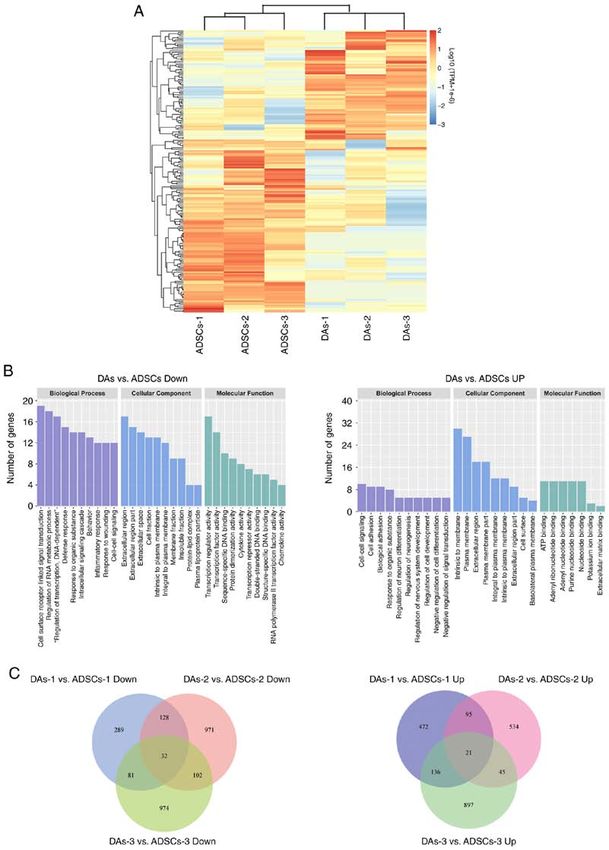
Molecular Medicine REPORTS 23: 47, 2021 3
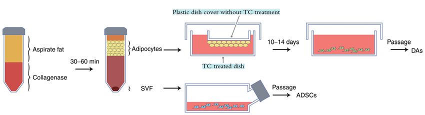
Figure 1. Schematic diagram of the one‑step method used for isolation and culture of ADSCs and DAs from adipose tissues. Following collagenase digestion
and centrifugation the fat cell layer was transferred a TC‑treated dish and a TC untreated dish cover was placed in it, so that the floating fat cells could attach.
After 10‑14 days, spindle cells grew on the bottom dish treated with TC, which were then passaged regularly to acquire the DAs, and ADSCs were obtained via
the conventional culture of SVF. TC, tissue culture; ADSCs, adipose‑derived stem cells; DAs, dedifferentiated adipose cells; SVF, stromal vascular fraction.
Table I. Characteristics of patients enrolled in this study. using medium A for 3 days, the medium was replaced with
medium B for 24 h. After using medium A and B alternately
Patient no. Age, years BMI Biopsy site three times (12 days of culture), cells were stained with Oil
Red O as follows: Cells were fixed with 4% paraformaldehyde
1 30 23.9 Abdomen at RT for 20 min and then dyed with Oil Red O for 15 min at
2 24 20.4 Thigh RT. Induced fat cells contained orange‑red oil droplets. After
3 23 19.1 Thigh washing with distilled water, the cells were observed under a
light microscope with magnification x100. Then, 2 ml isopro‑
panol was added to each well for 1 h at RT. When the Oil Red
O was dissolved in the isopropanol solution, the absorbance
was detected on the spectrophotometer at 500 nm. Then, total
144 h, CCK‑8 solution (10 µl/well) was added to the cells and proteins in each well were extracted with a RIPA lysis buffer
incubated at 37˚C for 1.5 h. The absorbance at 450 nm was (Applygen Technologies Inc.) and quantified using a bicincho‑
measured using a microplate reader. The optical density (OD) ninic acid protein assay kit (CW Biosciences).
values represented the survival/proliferation of cells. For osteogenic differentiation assays, 4.5x10 4 cells
were seeded into 6‑well plates. When the cells reached
Immunophenotypic assessment using flow cytometry. DAs 60% confluence, the medium was replaced with osteogenic
and ADSCs from the same samples were digested when they differentiation medium (Cyagen Biosciences, Inc.). Osteogenic
reached 80% confluence and were resuspended in 90 µl PBS media were changed every 3 days. After 12 days of culture,
(1x10⁵ cells per sample). Then, cell aliquots were incubated Alizarin red S staining was performed. Cells were fixed
with primary antibodies (1:10) in the dark for 15‑20 min at with 70% ethanol at RT for 45 min, and then stained with
RT. Subsequently, the cell suspension was centrifuged at Alizarin red S for 20 min at RT. After washing the floating
300 x g for 5 min at RT, followed by removal of the super‑ color with distilled water, the cells were visualized under a
natant and resuspension of the sediment in 200 µl PBS. The light microscope with magnification of 100 times. Then, 10%
following antibodies were used at 1:20 dilution: Anti‑CD90 cetylpyridinium chloride (w/v in distilled water) was added
(cat. no. E‑AB‑F1167D), anti‑CD44 (cat. no. E‑AB‑F1038D), into each well to wash the dye for 1 h at RT. The OD value
anti‑CD31 (cat. no. E‑AB‑F1050C), anti‑CD34 (cat. was measured at 550 nm, and the protein in each well was
no. E‑AB‑F1143C), anti‑ CD45 (cat. no. E‑AB‑F1137C) and extracted and quantified.
their isotype controls (Elabscience, Inc.), anti‑CD105 (cat. For chondrogenic differentiation, 2.5x10⁵ cells per pellet
no. 12‑1057‑41) and its isotype control (eBioscience, Inc.). in 15‑cm3 conical tubes were used. Cells were maintained at
Positive cells were counted and compared with the signal of 37˚C with 5% CO2 in the chondrogenic differentiation medium
the corresponding immunoglobulin isotypes. Samples were (Cyagen Biosciences, Inc.) for 25 days. The pellets were fixed
analyzed on a BD FACS Aria II (BD Biosciences) and data with 4% paraformaldehyde at RT for 24 h, then underwent
were analyzed using FlowJo version 10.0 (FlowJo, LLC). dehydration, embedding, sectioning at 5 µm, staining with
alcian blue at RT for 30 min and observed under a light micro‑
Multi‑lineage differentiation assay. Differentiation assays scope magnification x40.
for adipocytes, osteoblasts, chondrocytes and skeletal muscle For SkMC differentiation, cells were seeded into
cells (SkMCs) were performed on the three pairs of stem cells. 24‑well plates. When confluence reached 90‑100%, cells were
Assays for adipogenic differentiation potential were incubated with myogenic induction solution (DMEM/F12
performed according to the manufacturer's protocol (Cyagen supplemented with 10% FBS, 5% equine serum (Gibco; Thermo
Biosciences, Inc.). Briefly, 4.5x10⁴ cells were seeded into 6‑well Fisher Scientific, Inc.), 0.1 µmol/l dexamethasone and 50 µmol/l
plates. When the cells reached 80% confluence, the medium hydrocortisone) for 10 days. Then, cells were fixed in 4%
was replaced with adipogenic differentiation medium A. After paraformaldehyde at RT for 15 min, permeabilized in 0.2%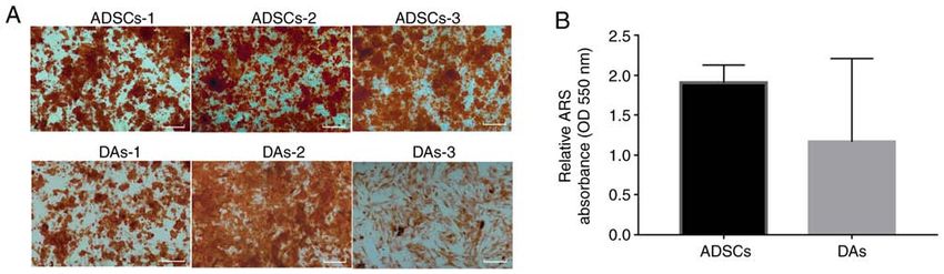
4                           Nie et al: COMPARISON OF HUMAN DAs AND ADSCs FROM YOUNG DONORS

Triton X‑100, blocked with 10% normal goat serum (Beyotime        Table II. Comparison of the surface antigens of DAs and
Institute of Biotechnology) for 20 min at RT. Then incubated      ADSCs from three patients.
with a rabbit anti‑desmin antibody (1:50; cat. no. ab32362;
Abcam) overnight at 4˚C followed by Cy3 goat anti‑rabbit IgG      Antigen	ADSCsa	DAsa
antibody (1:500; Beyotime Institute of Biotechnology). After
staining the nuclei with DAPI for 3 min at RT, the samples were   CD90                     100±0%                    99.97±0.06%
examined using a High Content Imaging system (Operetta            CD44                   99.83±0.06%                 99.73±0.29%
CLS; PerkinElmer, Inc.).                                          CD105                   95.8±3.76%                  93.3±10.50%
                                                                  CD31                   22.63±36.61%                  1.77±1.03%
Comparison of the gene expression profiles between DAs            CD34                   19.37±12.02%                11.02±12.70%
and ADSCs using RNA sequencing (seq). Cell samples were           CD45                   24.94±32.75%                 2.06±2.34%
harvested using trypsin digestion. The total RNA was extracted
using TRIzol® (cat. no. 15596026; Thermo Fisher Scientific,       a
                                                                   Data are presented as the mean percentage of cells expressing each
Inc.) according to the manufacturer's instructions. RNA degra‑    antigen. ADSCs, adipose‑derived stem cells; DAs, dedifferentiated
dation and contamination was monitored on 1% agarose gels.        adipose cells.
RNA purity was checked using the NanoPhotometer ® spec‑
trophotometer (Implen GmbH). RNAseq was performed by
BerryGenomics company. Briefly, sequencing libraries were
generated using NEBNext® Ultra™ RNA Library Prep Kit
for Illumina® (NEB) following manufacturer's recommenda‑          in Fig. 2C. There were notable individual differences in the
tions and index codes were added to attribute sequences to        growth speed of the cells. There was no definite trend in growth
each sample. The library preparations were sequenced on an        speed between the two types of cells from the same individual
Illumina NovaSeq platform and 150 bp paired‑end reads were        source, DAs‑1 grew faster compared with ADSCs‑1, ADSCs‑2
generated. After removing reads containing poly‑N, adapters       grew faster than DAs‑2 while ADSCs‑3 grew at first slower
and low‑quality reads from the raw data, clean high‑quality       and then faster compared with DAs‑3. In general, compared
data obtained were used for the subsequent analysis. EdgeR        with the first and the second pair of cells, ADSCs‑3 and
package (version 3.3.3) was used for the differential expres‑     DAs‑3 proliferated slowly, and the growth curve of ADSCs‑3
sion analysis (21). A |log2(fold change)|>1.00 and P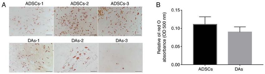
Molecular Medicine REPORTS 23: 47, 2021                                                                   5

Figure 2. Basic characteristics of ADSCs and DAs. (A) Newly inoculated adipocytes, primary DAs, and primary ADSCs are shown successively from left to
right. Scale bar, 200 µm. (B) ADSCs and DAs of 6‑7 passages had similar spindle shapes without lipid droplets. Scale bar, 100 µm. (C) Cell proliferation curves
showed notably individual differences in growth speed, while no definite patterns of growth speed were observed between the two types of cells. *P
6                                Nie et al: COMPARISON OF HUMAN DAs AND ADSCs FROM YOUNG DONORS

Figure 3. Adipogenic capacity of ADSCs and DAs. (A) Morphologies of formed lipid droplets were identified using staining with Oil Red O solution. Scale
bar, 100 µm. (B) Semi‑quantitative analysis using an absorbance wavelength of 500 nm per µg protein. Data are presented as the mean ± SD. ADSCs,
adipose‑derived stem cells; DAs, dedifferentiated adipose cells; OD, optical density.

Figure 4. Osteogenic capacity of ADSCs and DAs. (A) Morphologies of cells stained using ARS. Scale bar, 500 µm. (B) Semi‑quantitative analysis using an
absorbance wavelength of 550 nm per µg protein. Data are presented as the mean ± SD. ADSCs, adipose‑derived stem cells; DAs, dedifferentiated adipose
cells; OD, optical density; ARS, Alizarin red S.

                                                                              dependent’. The most abundant GO‑cellular component was
                                                                              ‘extracellular region’. The most enriched GO‑molecular
                                                                              function was ‘primarily transcription regulator activity’, ‘tran‑
                                                                              scription factor activity’ and ‘sequence specific DNA binding’.
                                                                              ‘Cytokine‑cytokine receptor interactions’ was the most abun‑
                                                                              dant KEGG pathway, with a total of eight genes: C‑C motif
                                                                              chemokine ligand 3 (CCL3), IL6, IL7, C‑C motif chemokine
Figure 5. Chondrogenic differentiation. Morphologies of cells stained using   receptor 1 (CCR1), C‑X‑C motif chemokine ligand 2 (CXCL2),
alcian blue. Scale bar, 1,000 µm. ADSCs, adipose‑derived stem cells; DAs,
                                                                              CX3CL1, CCL5 and TNF superfamily member 18.
dedifferentiated adipose cells.
                                                                                  In the upregulated DEGs in DAs, the most abundant
                                                                              GO‑biological process was primarily ‘cell‑cell signaling’,
                                                                              ‘cell adhesion’ and ‘biological adhesion’. The most enriched
Comparison of gene expression profiles between DAs and                        GO‑cellular component was primarily ‘intrinsic to membrane’,
ADSCs. To further compare the gene expression profiles of                     ‘plasma membrane’ and ‘extracellular region’, while the
DAs and ADSCs, the current study adopted second‑generation                    most enriched GO‑molecular function was primarily ‘ATP
seq technology to detect the three pairs of cells. P1.00 were used as the criteria to screen                  in cancer’, with only six genes: Fibroblast growth factor 18
the DEGs between groups and between each pair of samples.                     (FGF18), WNT16, integrin subunit α 6, MET, hedgehog
Only 186 DEGs were identified between the two groups, with                    interacting protein (HHIP) and MDS1 and EVI1 complex
112 downregulated and 74 upregulated in DAs vs. ADSCs                         locus (MECOM).
(Fig. 7A).                                                                        When comparing the data of each pair of cells, several
    Based on GO and KEGG analysis, the top 10 clusters                        DEGs were identified, but the number of common DEGs
containing the highest number of gene counts were plotted                     across all three cases was low. Venn diagrams (Fig. 7C)
(Fig. 7B). In the downregulated DEGs in DAs vs. ADSCs,                        showing the numbers of downregulated or upregulated DEGs
the most abundant GO‑biological processes were ‘cell surface                  in DAs was 32 (FOSB, CCND2, DUSP1, RGS16, CXCL2,
receptor linked signal transmission’, ‘regulation of RNA                      HES4, FOS, ATF3, ZFP36, DUSP8, GALNT13, RASD1,
metabolic process’ and ‘regulation of transcription, DNA                      ITIH5, F11R, FGD5, FZD3, BCL2A1, CCR1, G0S2, FAM84B,
Molecular Medicine REPORTS 23: 47, 2021 7
Figure 6. Myogenic capacity of ADSCs and DAs. (A) Desmin immunofluorescence staining was used to assess myogenic differentiation. The cytoplasm of the
positive cells were dyed red, and the blue signals represent DAPI‑counterstained nuclei. Scale bar, 200 µm. (B) Semi‑quantitative analysis showing the mean
intensity per well of positive signals in the cytoplasm. (C) Semi‑quantitative analysis showing the mean percentage of positive cells. Data are presented as the
mean ± SD. ADSCs, adipose‑derived stem cells; DAs, dedifferentiated adipose cells.
KLF4, CCDC102B, GADD45G, ALDH8A1, CX3CL1, CCR7, in adipose tissues, and they possess a wide range of therapeutic
USP2, HTR2B, ZNF853, CCL11, TDRD1 and TULP2) and potential in the field of regenerative medicine. However, aging
21 (FGF18, IGF2BP1, SLC38A4, SCN9A, ELOVL2, LYPD1, may attenuate their regenerative potential and metabolic
ALDH1A1, HHIP, NEFM, PLD5, MYH1, RIMS1, IL7R, functions (3‑6,22). The purpose of the present study was to
MECOM, ST6GALNAC3, NOXO1, ADRA2C, NLGN1, provide a reference for future autotransplantation by studying
CAMK4, COL6A6 and GREB1L), respectively. the differentiation and gene expression differences of these
According to the RNAseq results (GEO database, acces‑ two types of cells from the same young donors. In the present
sion number GSE141708), the relative mRNA expression levels study, a modified DA culture method was adopted. ADSCs
of perilipin 1, peroxisome proliferator activated receptor γ, and DAs from the same individual were obtained using a
C/EBPA (CCAAT enhancer binding protein α) and fatty acid one‑step method, and 6‑7 generations of cells were used
binding protein 4 involved in fat metabolism or RUNX family to compare their characteristics. The results indicated that
transcription factor 2 (RUNX2), SP7, activating transcrip‑ there were notable individual differences in the multilineage
tion factor 4 (ATF4) and bone γ‑carboxyglutamate protein differentiation abilities for DAs and ADSCs, and this was not
(BGLAP) involved in osteogenic regulation showed only directly determined by the source of the cells. For the same
individual differences among the three pairs of samples in this individuals, DAs and ADSCs had their own advantages for
experiment, but no significant relationship with adipogenic or different applications, including osteogenic, lipogenic, chon‑
osteogenic differences. The expression of bone morphogenetic drogenic or myogenic repair and regeneration, and future stem
protein 6 (BMP6) in DAs‑1, DAs‑2 and ADSCs‑3 with rela‑ cell autotransplantation should be a personalized treatment.
tively stronger chondrogenic abilities was significantly higher Thus, this study may improve the understanding of the clinical
compared with the corresponding ADSCs‑1, ADSCs‑2 and therapeutic potential of adipose tissues.
DAs‑3. ADSCs are defined as mesenchymal cells within adipose
tissue with multipotent differentiation and self‑renewal
Discussion capacity (23). Since the initial discovery of ADSCs, their
molecular profiles has been the subject of debate. This has
In young adults, adipose tissues can be easily obtained via been primarily due to the use of different ADSC purifica‑
safe and minimally invasive tumescent liposuction. However, tion and culture protocols, as well as the differing use of
as individuals age, the likelihood of suffering from a disease sub‑total vs. whole SVF (24). To date no markers have been
where stem cell therapy is required, such as osteoarthritis, reported to be exclusively expressed in ADSCs (25). In general,
refractory wounds, various tissue defects and other systemic the culture of SVF cells on plastic surfaces yields an adherent
diseases, increases (1,4). ADSCs and DAs are the primary subpopulation of ADSCs. SVF separated from adipose tissues
types of stem cells derived from SVF and MAs, respectively, with collagenase digestion contains a variety of cell types,8 Nie et al: COMPARISON OF HUMAN DAs AND ADSCs FROM YOUNG DONORS Figure 7. Diagrams summarizing the DEGs in ADSCs and DAs. (A) Heatmap of 186 DEGs. The colors in the heat map represent gene expression levels [log10(TPM+1x10 ‑6)]. Red represents higher expression; blue represents lower expression. The brackets represent cluster analysis. (B) GO enrichment bar plots. The clusters with the largest number of DEGs were shown as two histograms (only top 10 if >10 clusters). (C) Venn diagrams summarizing the DEGs in each pair of ADSCs and DAs. DEG, differentially expressed gene; GO, gene ontology; ADSCs, adipose‑derived stem cell; DAs, dedifferentiated adipose cell. including B and T lymphocytes, endothelial cells, fibroblasts, improved the wound healing processes, and found that both macrophages, pericytes and preadipocytes (25). ADSCs are SVF and ADSCs improved the function of endotheliocytes and a relatively homogenous population lacking hematopoietic fibroblasts, regulated gene expression and jointly promoted lineage markers after culturing and expanding (26). In fact, skin healing. However, there were no significant differences in the therapeutic efficacy of ADSCs and SVF are very similar the effect or mechanisms between SVF and ADSCs (20). This when used for autotransplantation in various disease condi‑ may be due to the fact that ADSCs are the primary biological tions, such as orthopedic, inflammatory, degenerative tissue functional cell type in SVF. According to the minimum or organ and autoimmune diseases, in a clinical trial (26). Our criteria for identifying MSCs described by the International previous study revealed that ADSCs and SVF significantly Federation of Adipose Therapeutics and Sciences and the
Molecular Medicine REPORTS 23: 47, 2021                                                9

International Society for Cell Therapy, ADSCs express high          compared with lean individuals, which may be associated with
levels of CD90, CD105 and CD44, whilst remaining negative           the higher activity of MAs, although the specific mechanism
for CD31 (endothelial marker), CD34 (a well‑known stem cell         is yet to be elucidated (30). Due to the limited number of cases
marker for both hematopoietic and endothelial lineages) and         in the present study, this result may also be caused by factors
CD45 (known as a leukocyte common antigen) (27). In the             such as different donor site locations, thus further experiments
present study, all cells expressed high levels of CD90, CD44        are required to clarify this hypothesis.
and CD105, while the intensity and individual differences in            In plastic surgery, several clinical applications have been
the percentage of cells expressing CD31, CD34 and CD45              suggested regarding ADSCs. In addition to promoting fat graft
were notably lower. Moreover, although ADSCs‑1 contained            survival, preclinical and clinical studies have demonstrated
a high proportion of CD31, CD34 and CD45 expressing                 the efficacy of ADSCs in muscle, tendon, bone and cartilage
cells, the expression intensity was too weak, and there was         regeneration (31). Similarly, DAs can transdifferentiate into
no significant difference in the differentiation function           several mature cell types, including adipocytes, chondrocytes,
between the ADSCs‑1 and other cases of cells. These find‑           osteoblasts and skeletal myocytes (32‑34). The present study
ings suggested that ADSCs mixed with a small number of              aimed to establish a more suitable means of seeding cells for
hematopoietic and endothelial lineages did not significantly        different application directions of adipose tissue with rich
affect their differentiation abilities.                             sources by evaluating their differentiation ability into various
    MAs are another abundant cell group present in adipose          lines. Thus, four‑lineage induced differentiation experiments
tissues. When MAs are cultured in vitro, due to the state of        and semi‑quantitative analysis were performed. Although
ischemia and hypoxia, they gradually remove lipid droplets          there were significant differences between each pair of cases,
and changes in their morphology are visible; becoming               there was no significant tendency between the DA and ADSC
spindle cells without lipid droplets and fibroblast‑like (15).      groups overall. Together, on average, the osteogenic and
In the present study, these cells were referred to as DAs. The      lipogenic abilities of ADSCs used in the present study were
mechanism of adipocyte dedifferentiation has not been fully         slightly stronger compared with that of DAs, and the myogenic
elucidated. Jumabay and Bostrom (15) suggested that there are       abilities of DAs were slightly higher compared with that of
two means of dedifferentiation for MAs: By removing lipid           ADSCs.
droplets or asymmetric division to form offspring adipocytes            Although the majority of previous studies have revealed that
and adipose free fibroblast‑like cells. Maurizi et al (28) also     DAs are more favorable than ADSCs for lipogenesis, and our
showed that the dedifferentiation of adipocytes was not due to      unpublished data has suggested that DAs of earlier generations
gradual lipolysis, but instead due to the secretion and excretion   do exhibit significantly higher fat induction ability compared
of lipid droplets. These authors suggested that ceiling culture,    with ADSCs. The results of the present study demonstrated
as a microenvironment stimulus, could induce human adipo‑           that ADSCs and DAs exhibited similar lipogenic abilities,
cytes to reprogram and secrete lipid droplets in large quantities   which may be associated with the higher cell generations used
to obtain new phenotypes adapted to the novel environment. In       in the present study. It was hypothesized that the long‑term
the present study, two simple cell culture materials, non‑TC        growth in the same medium makes the two types of cells
treated and TC treated were used, creating a similar niche of       become homogenous in their differentiation abilities.
hypoxia and low nutrition to the ceiling culture method, and            The semi‑quantitative results of osteogenesis and
thus obtained DAs simply and successfully. Furthermore, it          chondrogenesis also demonstrated the lack of difference
was demonstrated that lipid droplets appeared to be secreted        in the differentiation ability between the two groups.
from the surrounding fibroblast‑like cells.                         Sasahara et al (7) reported a general trend toward decreased
    According to previous studies, comparisons of the               CpG methylation and that increased trimethylation levels of
differentiation abilities between DAs and ADSCs are not             histone H3 at lysine 4 existed in the DAs, compared with the
exactly the same. For instance, it has been reported that the       ADSCs in an epigenetic survey of the promoters of four osteo‑
lipogenic, osteogenic and chondrogenic abilities of DAs are         genic regulatory genes (RUNX2, SP7, ATF4 and BGLAP).
higher compared with those of ADSCs (16,17,29), and that            Moreover, these authors speculated that these genes were more
the osteoblastic differentiation capacity of DAs and ADSCs          likely to be highly expressed in DAs, and that may underlie the
appear to be similar, while the chondrogenic differentiation        improved osteogenic ability of DAs compared with ADSCs (7).
capacity of DAs seems to be weaker (18). It was hypothesized        In the present study, analysis of the differences in the expres‑
that these differences may be associated with the age, BMI,         sion of these four genes in the three pairs of samples and their
source of donors, cell generations, culture medium and              corresponding osteogenic ability yielded inconsistent results,
the detection time points of differentiation induction. To          suggesting that the differences in the osteogenic capacity
the best of our knowledge, no SkM differentiation ability           between cells in each case may be the result of the interac‑
comparisons have been previously reported between DAs               tion of multiple genes. Interestingly, the expression of BMP6
and ADSCs.                                                          in cells with relatively stronger chondrogenic abilities in the
    For the first time, two types of stem cells obtained from       present study was significantly higher compared with that of
liposuction were compared in healthy young women in                 the other cells, suggesting that the basic expression of BMP6
the present study. Interestingly, with the decrease of the          in cells may be associated with chondrogenic differentiation,
donors' BMI, the proliferation activity of the corresponding        which was consistent with several previous studies (35,36).
DAs decreased compared with ADSCs. This tendency was                    In terms of myogenic differentiation, it has been reported
consistent with clinical experience, that is, the survival rate     that ADSCs possess spontaneous myogenic differentiation
of fat transplantation in obese patients is relatively higher       capacity, although the efficiency is very low (37). In the
10 Nie et al: COMPARISON OF HUMAN DAs AND ADSCs FROM YOUNG DONORS
present study, a baseline level of expression of MYH and the same depot. Thus, it is difficult to explain this based only
desmin (DES) in ADSCs and DAs was observed, specific on the sources of the cells or other individual difference. The
to each gene (GEO database accession number GSE141708); results indicated that DAs and ADSCs possessed similar
however, the expression trends were not consistent. It is mRNA expression profiles and differentiation abilities on
worth noting that the skeletal marker MYH1 was one of the average, and DAs and ADSCs may have their own advantages
upregulated genes in DAs common across the three patients, for individuals in different applications in repair and regenera‑
although the expression level was low. Cells with relatively tion.
strong myogenic ability had relatively weak lipogenic ability; There are some limitations in the present study. First,
suggesting a potential opposing effects of genes involved in samples of subcutaneous adipose tissue were collected from
these two processes. It has been reported that microRNAs three healthy females with a medium BMI. The number
directly enhance mitochondrial translation during muscle of patients was small, and therefore the results may not be
differentiation (38). Additionally, differing CpG methylation generalizable. However, the existing data indicated that there
and trimethylation of histone H3 profiles exist in ADSCs were significant individual differences between the two types
and DAs (22). Therefore, a deeper understanding of the of cells. In addition, most studies have reported that obesity and
regulatory mechanisms beyond transcriptomic differences age can significantly affect the differentiation ability of adipose
may assist in elucidating the differences of genes in cell derived stem cells, and whether there is an opposite effect for
differentiation. DAs has only been the subject of relatively few studies (41,42).
The sequencing results of the present study identified a As suggested in the present study, obesity may enhance the
limited number of DEGs between the two groups of cells, proliferation of DAs. However, additional data are required to
similar to previous studies. Perrini et al (39) (GEO accession verify this hypothesis. Only transcriptomic differences were
no. GSE37324) compared genome‑wide mRNA expression studied between the two types of cells, while the mechanistic
profiles of subcutaneous adipose tissue‑derived stem cells processes of the differentiation abilities of adult stem cells
ASCSVF and ASCCeiling from the same fat depot, and reported derived from adipose tissue requires additional study on the
that both principal component analysis and hierarchical cluster role of epigenetic regulatory factors, such as post‑translational
analysis indicated that ASCCeiling and ASCSVF shared a similar histone modifications and non‑coding RNAs. Further study on
pattern of closely related genes. By contrast to the present the differentiation potential and mechanisms involved in the
study, their specimens were obtained from open‑abdominal two types of cells may assist in improved utilizing of adipose
surgery (nine men and six women; average age, 68 years; BMI, tissue for regenerative medicine.
27.06±1.5 kg/m 2), and they used the fourth generation cells. In conclusion, DAs and ADSCs from liposuction of adipose
Another project (GEO no. GSE47869) of microarray analysis tissues in young individuals, can both be easily isolated and
without detailed case information in the GEO database, amplified in vitro, as well as exhibit similar morphologies,
revealed that the global mRNA expression profiles of human proliferation dynamics, surface markers and transcriptome
DAs were very similar to those of ADSCs, but the work has expression profiles. There were notable individual differences
not been published as of yet. GEO2R, an online tool of the in osteogenic, lipogenic, chondrogenic and myogenic abilities,
GEO database (40), was used to analyze the data of GSE37324 and it was difficult to determine whether the differences in
by comparing Sc‑ASCCeiling (DAs) and Sc‑ASCSVF (ADSCs), differentiation potential was due to cell source or other factors.
using a criteria of P1.00 to Thus, DAs and ADSCs from young donors have similar appli‑
screen the DEGs, only 141 named genes were identified, of cation potential in general, while for individuals, DAs and
which 83 were upregulated by ADSCs (no consistent genes ADSCs may have their own advantages based on the specific
with the present study), and 58 were upregulated in DAs (seven repair and regeneration applications.
of which were also upregulated in the DAs in the present study:
BEX1, ENPP1, SCUBE3, HHIP, IL7R, FOXD1 and PODXL; Acknowledgements
however, there were two genes that were downregulated in the
DAs in the present study: EGR1 and IL6, that were upregu‑ Not applicable.
lated in GSE37324). Similarly, using GEO2R to analyze the
data of GSE47869 by comparing ADSCs and DAs with the Funding
same screening standards, only 16 DEGs named genes were
identified, and there were no common DEGs between the This work was supported by the Interdisciplinary Medicine
present study and GSE37324. Seed Fund of Peking University (grant no. BMU2018MX001)
In the DEGs screened in the present study, GO analysis and the National Nature Science Foundation of China (grant
suggested that cytokines may serve a more important role in no. 81501681).
the biological function of ADSCs than they do in DAs, and
that there may be more significant differences in the transcrip‑ Authors' contributions
tion, protein translation regulation and energy metabolism
levels between the two types of cells after multiple passages. FN designed the study, analyzed the data and wrote the
Additionally, although more DEGs can be obtained by manuscript. HB designed the experiments and revised the
comparing the data of each pair of cells, there were only a manuscript. CZ collected the specimen and performed the
few common DEGs identified in the differential genes of each experiments. PD performed the experiments and analyzed
pair of samples, suggesting that several factors may influence the data. All authors read and approved the final manu‑
the gene expression differences of the two types of cells from script.Molecular Medicine REPORTS 23: 47, 2021 11
Availability of data and materials 12. Heo JS, Choi Y, Kim HS and Kim HO: Comparison of molecular
profiles of human mesenchymal stem cells derived from bone
marrow, umbilical cord blood, placenta and adipose tissue. Int J
The RNAseq data have been deposited at the Gene Expression Mol Med 37: 115‑125, 2016.
Omnibus (accession number GSE141708). The other datasets 13. Naderi N, Combellack EJ, Griffin M, Sedaghati T, Javed M,
Findlay M W, Wallace CG, Mosa hebi A, Butler PE,
used and/or analyzed during the present study are available Seifalian AM, et al: The regenerative role of adipose‑derived
from the corresponding author on reasonable request. stem cells (ADSC) in plastic and reconstructive surgery. Int
Wound J 14: 112‑124, 2017.
14. Sugihara H, Yonemitsu N, Miyabara S and Yun K: Primary
Ethics approval and consent to participate cultures of unilocular fat cells: Characteristics of growth in vitro
and changes in differentiation properties. Differentiation 31:
The present study was approved by Peking University Third 42‑49, 1986.
15. Jumabay M and Boström KI: Dedifferentiated fat cells: A
Hospital Medical Science Research Ethics Committee cell source for regenerative medicine. World J Stem Cells 7:
(approval no. 2014020). Samples of subcutaneous adipose 1202‑1214, 2015.
tissue were collected from three females with written informed 16. Kishimoto N, Momota Y, Hashimoto Y, Tatsumi S, Ando K,
Omasa T and Kotani J: The osteoblastic differentiation ability
consent. of human dedifferentiated fat cells is higher than that of adipose
stem cells from the buccal fat pad. Clin Oral Investig 18:
Patient consent for publication 1893‑1901, 2014.
17. Watson JE, Patel NA, Carter G, Moor A, Patel R, Ghansah T,
Mathur A, Murr MM, Bickford P, Gould LJ, et al: Comparison of
Not applicable. markers and functional attributes of human adipose‑derived stem
cells and dedifferentiated adipocyte cells from subcutaneous fat
of an obese diabetic donor. Adv Wound Care (New Rochelle) 3:
Competing interests 219‑228, 2014.
18. Saler M, Caliogna L, Botta L, Benazzo F, Riva F and Gastaldi G:
The authors declare that they have no competing interests. hASC and DFAT, multipotent stem cells for regenerative
medicine: A comparison of their potential differentiation in
vitro. Int J Mol Sci 18: 18, 2017.
References 19. Reumann MK, Linnemann C, Aspera‑Werz RH, Arnold S,
Held M, Seeliger C, Nussler AK and Ehnert S: Donor site
1. Wu S, Coombs DM and Gurunian R: Liposuction: Concepts, location is critical for proliferation, stem cell capacity, and
safety, and techniques in body‑contouring surgery. Cleve Clin J osteogenic differentiation of adipose mesenchymal stem/stromal
Med 87: 367‑375, 2020. cells: Implications for bone tissue engineering. Int J Mol Sci 19:
2. Svalgaard JD, Juul S, Vester‑Glovinski PV, Haastrup EK, 1868, 2018.
Ballesteros OR, Lynggaard CD, Jensen AK, Fischer‑Nielsen A, 20. Bi H, Li H, Zhang C, Mao Y, Nie F, Xing Y, Sha W, Wang X,
Herly M and Munthe‑Fog L: Lipoaspirate storage time and Irwin DM and Tan H: Stromal vascular fraction promotes
temperature: Effects on stromal vascular fraction quality and cell migration of fibroblasts and angiogenesis through regulation of
composition. Cells Tissues Organs 209: 54‑63, 2020. extracellular matrix in the skin wound healing process. Stem
3. Choudhery MS, Badowski M, Muise A, Pierce J and Harris DT: Cell Res Ther 10: 302, 2019.
Donor age negatively impacts adipose tissue‑derived mesen‑ 21. Robinson MD, McCarthy DJ and Smyth GK: edgeR: A biocon‑
chymal stem cell expansion and differentiation. J Transl Med 12: ductor package for differential expression analysis of digital gene
8, 2014. expression data. Bioinformatics 26: 139‑140, 2010.
4. Kornicka K, Marycz K, Tomaszewski KA, Marędziak M and 22. Tansriratanawong K, Tabei I, Ishikawa H, Ohyama A, Toyomura J
Śmieszek A: The effect of age on osteogenic and adipogenic and Sato S: Characterization and comparative DNA methylation
differentiation potential of human adipose derived stromal stem profiling of four adipogenic genes in adipose‑derived stem cells
cells (hASCs) and the impact of stress factors in the course of and dedifferentiated fat cells from aging subjects. Hum Cell 33:
the differentiation process. Oxid Med Cell Longev 2015: 309169, 974‑989, 2020.
2015. 23. Shukla L, Yuan Y, Shayan R, Greening DW and Karnezis T: Fat
5. Nordberg RC, Zhang J, Griffith EH, Frank MW, Starly B and therapeutics: The clinical capacity of adipose‑derived stem cells
Loboa EG: Electrical cell‑substrate impedance spectroscopy can and exosomes for human disease and tissue regeneration. Front
monitor age‑grouped human adipose stem cell variability during Pharmacol 11: 158, 2020.
osteogenic differentiation. Stem Cells Transl Med 6: 502‑511, 2017. 24. Baer PC: Adipose‑derived mesenchymal stromal/stem cells: An
6. Zhu M, Kohan E, Bradley J, Hedrick M, Benhaim P and Zuk P: update on their phenotype in vivo and in vitro. World J Stem
The effect of age on osteogenic, adipogenic and proliferative Cells 6: 256‑265, 2014.
potential of female adipose‑derived stem cells. J Tissue Eng 25. Brooks AES, I m in itoff M, Willia ms E, Da ma n i T,
Regen Med 3: 290‑301, 2009. Jackson‑Patel V, Fan V, James J, Dunbar PR, Feisst V and
7. Sasahara Y, Kubota Y, Kosaka K, Adachi N, Yamaji Y, Nagano H, Sheppard HM: Ex vivo human adipose tissue derived mesen‑
Akita S, Kuroda M, Tanaka T, Bujo H, et al: Adipose‑derived chymal stromal cells (ASC) are a heterogeneous population that
stem cells and ceiling culture‑derived preadipocytes cultured demonstrate rapid culture‑induced changes. Front Pharmacol 10:
from subcutaneous fat tissue differ in their epigenetic character‑ 1695, 2020.
istics and osteogenic potential. Plast Reconstr Surg 144: 644‑655, 26. Kilinc MO, Santidrian A, Minev I, Toth R, Draganov D,
2019. Nguyen D, Lander E, Berman M, Minev B and Szalay AA: The
8. Zuk PA, Zhu M, Ashjian P, De Ugarte DA, Huang JI, Mizuno H, ratio of ADSCs to HSC‑progenitors in adipose tissue derived
Alfonso ZC, Fraser JK, Benhaim P and Hedrick MH: Human SVF may provide the key to predict the outcome of stem‑cell
adipose tissue is a source of multipotent stem cells. Mol Biol therapy. Clin Transl Med 7: 5, 2018.
Cell 13: 4279‑4295, 2002. 27. Bourin P, Bunnell BA, Casteilla L, Dominici M, Katz AJ,
9. Kunze KN, Burnett RA, Wright‑Chisem J, Frank RM and March KL, Redl H, Rubin JP, Yoshimura K and Gimble JM:
Chahla J: Adipose‑derived mesenchymal stem cell treatments Stromal cells from the adipose tissue‑derived stromal vascular
and available formulations. Curr Rev Musculoskelet Med 13: fraction and culture expanded adipose tissue‑derived stromal/stem
264‑280, 2020. cells: A joint statement of the International Federation for
10. Tsubosaka M, Matsumoto T, Sobajima S, Matsushita T, Adipose Therapeutics and Science (IFATS) and the International
Iwaguro H and Kuroda R: The influence of adipose‑derived Society for Cellular Therapy (ISCT). Cytotherapy 15: 641‑648,
stromal vascular fraction cells on the treatment of knee osteoar‑ 2013.
thritis. BMC Musculoskelet Disord 21: 207, 2020. 28. Maurizi G, Poloni A, Mattiucci D, Santi S, Maurizi A, Izzi V,
11. Ramakrishnan VM and Boyd NL: The adipose stromal vascular Giuliani A, Mancini S, Zingaretti MC, Perugini J, et al: Human
fraction as a complex cellular source for tissue engineering appli‑ white adipocytes convert into ‘rainbow’ adipocytes in vitro.
cations. Tissue Eng Part B Rev 24: 289‑299, 2018. J Cell Physiol 232: 2887‑2899, 2017.12 Nie et al: COMPARISON OF HUMAN DAs AND ADSCs FROM YOUNG DONORS
29. Shen JF, Sugawara A, Yamashita J, Ogura H and Sato S: 37. Sung SE, Hwang M, Kim AY, Lee EM, Lee EJ, Hwang SK,
Dedifferentiated fat cells: An alternative source of adult multi‑ Kim SY, Kim HK and Jeong KS: MyoD overexpressed equine
potent cells from the adipose tissues. Int J Oral Sci 3: 117‑124, adipose‑derived stem cells enhanced myogenic differentiation
2011. potential. Cell Transplant 25: 2017‑2026, 2016.
30. Bougaret L, Delort L, Billard H, Lequeux C, Goncalves‑Mendes N, 38. Zhang X, Zuo X, Yang B, Li Z, Xue Y, Zhou Y, Huang J, Zhao X,
Mojallal A, Damour O, Vasson MP and Caldefie‑Chezet F: Zhou J, Yan Y, et al: MicroRNA directly enhances mitochondrial
Supernatants of adipocytes from obese versus normal weight translation during muscle differentiation. Cell 158: 607‑619,
women and breast cancer cells: In vitro impact on angiogenesis. 2014.
J Cell Physiol 232: 1808‑1816, 2017. 39. Perrini S, Ficarella R, Picardi E, Cignarelli A, Barbaro M,
31. Torres‑Torrillas M, Rubio M, Damia E, Cuervo B, Del Romero A, Nigro P, Peschechera A, Palumbo O, Carella M, De
Peláez P, Chicharro D, Miguel L and Sopena JJ: Adipose‑derived Fazio M, et al: Differences in gene expression and cytokine
mesenchymal stem cells: A promising tool in the treatment of release profiles highlight the heterogeneity of distinct subsets
musculoskeletal diseases. Int J Mol Sci 20: 20, 2019. of adipose tissue‑derived stem cells in the subcutaneous and
32. Kazama T, Fujie M, Endo T and Kano K: Mature adipocyte‑derived visceral adipose tissue in humans. PLoS One 8: e57892, 2013.
dedifferentiated fat cells can transdifferentiate into skeletal 40. Barrett T, Wilhite SE, Ledoux P, Evangelista C, Kim IF,
myocytes in vitro. Biochem Biophys Res Commun 377: 780‑785, Tomashevsky M, Marshall KA, Phillippy KH, Sherman PM,
2008. Holko M, et al: NCBI GEO: Archive for functional genomics
33. Matsumoto T, Kano K, Kondo D, Fukuda N, Iribe Y, Tanaka N, data sets‑update. Nucleic Acids Res 41 (D1): D991‑D995, 2013.
Matsubara Y, Sakuma T, Satomi A, Otaki M, et al: Mature 41. Conley SM, Hickson LJ, Kellogg TA, McKenzie T, Heimbach JK,
adipocyte‑derived dedifferentiated fat cells exhibit multilineage Taner T, Tang H, Jordan KL, Saadiq IM, Woollard JR, et al:
potential. J Cell Physiol 215: 210‑222, 2008. Human obesity induces dysfunction and early senescence in
34. Oki Y, Watanabe S, Endo T and Kano K: Mature adipocyte‑derived adipose tissue‑derived mesenchymal stromal/stem cells. Front
dedifferentiated fat cells can trans‑differentiate into osteoblasts Cell Dev Biol 8: 197, 2020.
in vitro and in vivo only by all‑trans retinoic acid. Cell Struct 42. Prieto González EA: Heterogeneity in adipose stem cells. Adv
Funct 33: 211‑222, 2008. Exp Med Biol 1123: 119‑150, 2019.
35. Diekman BO, Estes BT and Guilak F: The effects of BMP6
overexpression on adipose stem cell chondrogenesis: Interactions
with dexamethasone and exogenous growth factors. J Biomed
Mater Res A 93: 994‑1003, 2010. This work is licensed under a Creative Commons
36. Jeon HJ, Yoon KA, An ES, Kang TW, Sim YB, Ahn J, Choi EK, Attribution-NonCommercial-NoDerivatives 4.0
Lee S, Seo KW, Kim YB, et al: Therapeutic effects of human International (CC BY-NC-ND 4.0) License.
umbilical cord blood‑derived mesenchymal stem cells combined
with cartilage acellular matrix mediated via bone morphogenic
protein 6 in a rabbit model of articular cruciate ligament tran‑
section. Stem Cell Rev Rep 16: 596‑611, 2020.You can also read