High-Resolution X-Ray Computed Tomography: A New Workflow for the Analysis of Xylogenesis and Intra-Seasonal Wood Biomass Production
←
→
Page content transcription
If your browser does not render page correctly, please read the page content below
ORIGINAL RESEARCH
                                                                                                                                              published: 06 August 2021
                                                                                                                                         doi: 10.3389/fpls.2021.698640

                                             High-Resolution X-Ray Computed
                                             Tomography: A New Workflow for the
                                             Analysis of Xylogenesis and
                                             Intra-Seasonal Wood Biomass
                                             Production
                                             Romain Lehnebach 1,2*, Matteo Campioli 3 , Jozica Gričar 4 , Peter Prislan 4 , Bertold Mariën 3 ,
                                             Hans Beeckman 5 and Jan Van den Bulcke 1
                                             1
                                               UGCT–UGent-Woodlab, Laboratory of Wood Technology, Department of Environment, Faculty of Bioscience Engineering,
                                             Ghent University, Gent, Belgium, 2 AMAP Laboratory (botany and bio-informatics of plant architecture and vegetation),
                                             Université Montpellier, CIRAD, CNRS, INRAE, IRD, Montpellier, France, 3 Research Group PLECO (Plants and Ecosystems),
                                             Department of Biology, University of Antwerp, Antwerp, Belgium, 4 Department of Yield and Silviculture, Slovenian Forestry
                                             Institute, Ljubljana, Slovenia, 5 Royal Museum for Central Africa, Service of Wood Biology, Tervuren, Belgium

                           Edited by:
                     Fabio Gennaretti,
                                             Understanding tree growth and carbon sequestration are of crucial interest to forecast
       Université du Québec en Abitibi       the feedback of forests to climate change. To have a global understanding of the wood
             Témiscamingue, Canada           formation, it is necessary to develop new methodologies for xylogenesis measurements,
                       Reviewed by:          valid across diverse wood structures and applicable to both angiosperms and
                     Emanuele Ziaco,
           Uppsala University, Sweden        gymnosperms. In this study, the authors present a new workflow to study xylogenesis
                      Valentina Buttò,       using high-resolution X-ray computed tomography (HRXCT), which is generic and
              Université du Québec à
                  Chicoutimi, Canada
                                             offers high potential for automatization. The HXRCT-based approach was benchmarked
                  *Correspondence:
                                             with the current classical approach (microtomy) on three tree species with contrasted
                   Romain Lehnebach          wood anatomy (Pinus nigra, Fagus sylvatica, and Quercus robur). HRXCT proved to
          lehnebach.romain@hotmail.fr        estimate the relevant xylogenesis parameters (timing, duration, and growth rates) across
                                             species with high accuracy. HRXCT showed to be an efficient avenue to investigate
                   Specialty section:
         This article was submitted to       tree xylogenesis for a wide range of wood anatomies, structures, and species. HRXCT
 Technical Advances in Plant Science,        also showed its potential to provide quantification of intra-annual dynamics of biomass
               a section of the journal
            Frontiers in Plant Science       production through high-resolution 3D mapping of wood biomass within the forming
             Received: 21 April 2021
                                             growth ring.
            Accepted: 28 June 2021
                                             Keywords: high-resolution X-ray computed tomography, microtomy, tree growth, xylogenesis, secondary growth
          Published: 06 August 2021
                                             phenology
                                Citation:
  Lehnebach R, Campioli M, Gričar J,
 Prislan P, Mariën B, Beeckman H and         INTRODUCTION
               Van den Bulcke J (2021)
     High-Resolution X-Ray Computed
                                             Forests represent the main CO2 sink of terrestrial ecosystems. Understanding tree growth
 Tomography: A New Workflow for the
             Analysis of Xylogenesis and
                                             and carbon sequestration is of crucial interest to forecast the responses of forests to climate
         Intra-Seasonal Wood Biomass         change and their climate mitigation potential. The forest carbon sink can be assessed
                              Production.    with direct measurement of tree growth (i.e., Hubau et al., 2020) or indirectly through
            Front. Plant Sci. 12:698640.     Dynamic Global Vegetation Models (DGVM). In DGVM, tree growth is often assumed to
        doi: 10.3389/fpls.2021.698640        be limited primarily by photosynthesis (i.e., gross primary production) (Fatichi et al., 2019).

Frontiers in Plant Science | www.frontiersin.org                                    1                                          August 2021 | Volume 12 | Article 698640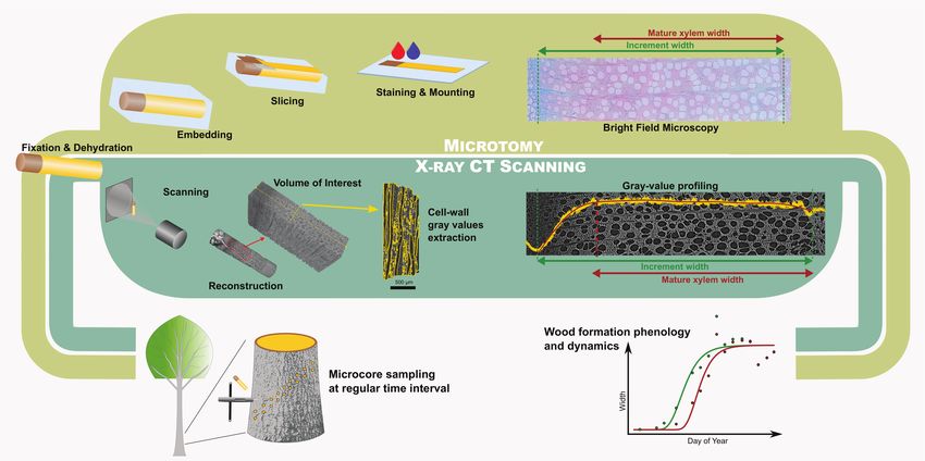
Lehnebach et al.                                                                                   X-Ray Computed Tomography Analysis of Xylogenesis

However, experimental evidence shows that tree growth does                   development (Prislan et al., 2018), intra-seasonal studies of
not directly depend on the photosynthesis and that tree                      secondary growth in angiosperms have been often limited to the
growth is more sensitive to environmental conditions than the                estimation of the timing of qualitative events (e.g., earlywood
photosynthesis (Fatichi et al., 2014). Therefore, more attention             vessel formation, latewood formation). From the biomass
should be devoted to determine the direct constraints on growth              accounting point of view, bypassing cell-level information by
exerted by the environmental conditions at different temporal                characterizing the biomass accumulated during the season at
scales. One way to do this is by studying wood formation                     the tissue level is a promising perspective. In this respect,
(i.e., xylogenesis) that defines seasonal tree growth and carbon             the methodology of Andrianantenaina et al. (2019), based
sequestration (Friend et al., 2019).                                         on an estimation of the apparent density of the forming
    Xylogenesis is driven by the cambium, that is, a layer of                wood, should allow to overcome the issue of structurally
meristematic cells, forming a cylinder along the different axes of           heterogeneous wood and thus to quantify the dynamics of
the plant body. For temperate species, when growing conditions               intra-annual biomass production in angiosperms. The global
become favorable in spring, the cambium cells start to divide and            predominance of angiosperms stresses the importance of
the newly formed cells undergo different structural changes (i.e.,           further methodological development allowing straightforward
differentiation). Xylogenesis can be divided into five sequential            xylogenesis measurements across the diversity of wood structures
processes: (1) cell division and (2) cell enlargement, which                 and species found within the angiosperm group, and across the
drive the growth in size; (3) cell-wall thickening and (4) cell-             different climates and biomes.
wall lignification, which are mainly responsible for the increase                Investigating how tree growth interacts with environmental
in mass and C sequestration (Cuny et al., 2015); and (5)                     factors at a global scale requires the development of a high-
programmed cell death, which allows the woody cell skeleton to               throughput methodology for xylogenesis studies applicable
be used as mechanical support and conduit for water transport.               also to angiosperms. Such methodology should avoid the
The wood formation and its dynamics are strongly influenced by               histological preparation and analysis that are labor intensive,
environmental conditions, such as the temperature, day length,               requiring specific expertise, and several manual steps, such
and water availability (Delpierre et al., 2016; Dox et al., 2021).           as paraffin embedding, slicing, staining, mounting, and
Therefore, climate change is expected to affect the tree growth              observation using bright field microscopy. So far, the labor
and carbon accumulation through the modulation of the timing                 intensity of the manual steps during sample preparation
and duration of xylogenesis.                                                 (requiring skilled technicians) and data collection (observations,
    An appealing method to study the phenology and the seasonal              requiring skilled wood anatomists) has limited the automation
dynamics of wood formation as well as the climate-wood                       of the microtomic approach, pointing out the importance
growth relationships consists in monitoring wood development                 to develop an automated methodology to study seasonal
in small cores (i.e., microcores) collected at regular time                  wood formation.
intervals during the growing season (Rossi et al., 2006). Seasonal               High-resolution X-ray computed tomography (HXRCT) is
observations of anatomical features characterizing the wood                  becoming popular in the analysis and imaging of plant anatomy
formation (e.g., cell morphology, cell developmental stage,                  (Brodersen and Roddy, 2016). Different research fields related
or number of cells in a given stage) are made possible by                    to tree growth and functioning, such as dendrochronology (De
brightfield microscopy after histological slicing and staining.              Mil et al., 2016; Van den Bulcke et al., 2019) and ecophysiology
Most of the xylogenesis studies and the development of the                   (Knipfer et al., 2015; De Baerdemaeker et al., 2019), seized
histological approach (referred hereafter as “microtomy”) have               the opportunity of HXRCT for substantial methodological
been performed on conifers with a homogeneous wood structure                 improvements. However, HXRCT has never been used in the
(Rossi et al., 2009, Deslauriers et al., 2008, Camarero et al., 2010).       demanding field of xylogenesis. Producing three-dimensional
This intensive work on conifers also permitted to refine the                 gray scale images with pixel values (attenuation coefficients)
understanding of the asynchrony between xylem size increase                  directly related to the density of the material, HXRCT represents
and biomass production through a detailed quantification                     a powerful mean to study xylogenesis. More precisely, HXRCT
of cell development kinetic during xylogenesis (Cuny et al.,                 can be used to track cell wall density change occurring during
2015). This approach allowed to develop a clear view on                      the formation and thickening of the secondary cell wall in order
xylogenesis in conifer species (Rathgeber, 2017), while its                  to differentiate the mature wood from the forming wood and
knowledge on angiosperm species remains limited (ref. Delpierre              to assess secondary growth phenology. In addition, HRXCT can
et al., 2016, but see Cufar et al., 2008; Gričar et al., 2018;              quantify the biomass accumulated by trees throughout the course
Prislan et al., 2018; Dox et al., 2020). In fact, the structural             of the season.
heterogeneity of angiosperm wood challenges the acquisition                      The use of HXRCT in the framework of xylogenesis requires
of xylogenesis data from a methodological point of view.                     both high contrast and resolution to discern the forming cells
Indeed, the diversity of cell types encountered in angiosperm                and the cell wall. Obtaining a sufficient contrast between cell
wood (vessels, fibers, and parenchyma) makes cell-counting                   lumens and cell walls with HRXCT requires water removal from
and morphometrics difficult, while limiting the acquisition                  the sample. For mature xylem, water is generally removed by
of information on the xylogenesis dynamics. Therefore, even                  oven-drying the sample prior to scanning (De Mil et al., 2016).
if some authors have been able to quantify the proportion                    However, drying without collapsing and destroying the soft
of growth increment attributed to the different stages of                    tissue, such as cambial and newly forming xylem cells, cannot be

Frontiers in Plant Science | www.frontiersin.org                         2                                    August 2021 | Volume 12 | Article 698640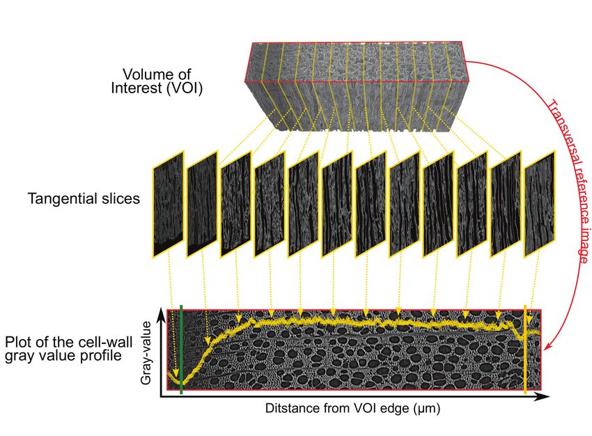
Lehnebach et al.                                                                                X-Ray Computed Tomography Analysis of Xylogenesis

achieved through oven- or air-drying (Stuppy et al., 2003; Leroux         X-Ray CT Workflow
et al., 2009).                                                            Dehydration, Critical Point Drying, and Sample
    Therefore, monitoring of xylogenesis with HXRCT requires              Mounting
the development of an entire workflow including sample                    After dehydration by increasing ethanol concentrations, the
preparation, image acquisition, and an analysis protocol,                 samples for HXRCT were dried to obtain a sufficient contrast
alongside a methodology to follow the cell wall density change            between cell lumens and cell walls. Therefore, a gentle drying
within the forming growth ring.                                           method was required in order to preserve the structure of the soft,
    Here, a new methodology is presented and validated to                 newly forming wood cells. Therefore, critical point drying (CPD),
study xylogenesis using HXRCT based on a new workflow to                  a technique commonly used for the preparation of scanning
monitor cell wall density changes occurring during xylogenesis            electron microscopy samples (Bray, 2000), was used, but has
(Figure 1), applied to three tree species (black pine, common             proven its efficiency in the drying of fresh plant structures also
beech, and pedunculate oak) with contrasted wood anatomy                  for CT imaging (See Supplementary Figure 1 for a comparison
growing in a temperate region. Several relevant parameters,               of an air dried and CPD processed pine microcore) (Leroux et al.,
such as the timing and duration of size growth, mature xylem              2009; Gutiérrez et al., 2018).
production, and overall xylogenesis, as well as rates of size                The samples were critically point dried with liquid CO2 using
growth and mature xylem production, were estimated with                   a CPD device (Baltec CPD 030). Each drying cycle consisted
HXRCT and validated with the classical microtomy approach.                of a mixing-draining procedure (repeated 10 times) allowing
In addition, the gray scale images produced by HRXCT were                 the gradual replacement of the dehydration media (i.e., 100%
used to account for seasonal biomass production dynamics.                 ethanol) by liquid CO2 . Then, the samples were dried by
Concerning the latter aspect, the authors ask whether the intra-          removing the CO2 in a supercritical state (> 31◦ C, > 73 bar).
seasonal stem biomass production and size growth differ between              Six samples were dried at the same time. After drying, the
conifers and angiosperms, specifically among pine, beech, and             samples were mounted on a cylindrical carbon stick (5 cm
oak. While information on intra-seasonal biomass production               x2 mm) for scanning. The mounted samples were stabilized in the
exist for conifers (Cuny et al., 2015; Andrianantenaina et al.,           scanner room for 12 h, at 25◦ C and a relative humidity of 40%, to
2019) to the best of our knowledge, no study has investigated the         avoid sample movement during scanning.
intra-seasonal biomass production in angiosperms.

                                                                          X-Ray CT Scanning and Reconstruction
MATERIALS AND METHODS                                                     Samples were scanned with the Nanowood X-ray CT scanner,
                                                                          developed at the Ghent University Centre for X-ray tomography
Selected Species, Study Sites, and                                        (UGCT; http://www.ugct.ugent.be) (Dierick et al., 2014). The
Sampling                                                                  directional X-ray tube (Microfocus, Hamamatsu) was operated at
To assess the applicability of the HXRCT method on tree                   a tube voltage of 70 kV and a target current of 100 µA. A large (25
species with different functional strategies, three species growing       x 20 cm, 1920 x 1536 px) Varian Si flat detector was used allowing
in Belgium were selected: black pine (Pinus nigra), common                to capture the region of interest (i.e., innermost phloem layers,
beech (Fagus sylvatica), and pedunculate oak (Quercus robur).             cambium, and forming xylem growth ring) in a single field of
With 778 mm.year−1 of precipitation and an average monthly                view. A total of 2001 projections were taken with an exposure
temperature ranging from +3◦ C to 17◦ C (average annual                   time of 700 ms per projection, resulting in 25 min per scan.
temperature of 10.1◦ C), Belgium is characterized by a maritime               Scanned volumes were reconstructed with the Octopus
temperate climate (Campioli et al., 2012). Beech and oak trees            reconstruction software (Vlassenbroeck et al., 2007), including
were sampled in the “park of Brasschaat” (51◦ 12′ N−4◦ 26′ E)             Paganin phase contrast filtering (Paganin et al., 2002). The
while pine trees were sampled in the military domain “Klein               reconstructed volume, with an approximate voxel pitch of
Schietveld” (51◦ 21′ N−4◦ 37′ E) (Dox et al., 2020; Marchand              2.49 µm, consisted of 16-bit tangential images running from the
et al., 2020). The sites are at a distance of 9.5 km in a lowland         bark to the inner side of the xylem.
(18–22 m a.s.l.) region with a similar climate. The three species
present contrasted xylem anatomy: beech and oak are diffuse
and ring porous species respectively, whereas black pine has the          Processing and Analysis of the
typical conifer wood structure made mostly of tracheids. Four             Reconstructed Volume
healthy and (co)dominant trees per species were selected. From            Selection of the Regions of Interest and Gray-Value
the beginning of April to mid-November 2019, two microcores               Extraction
were collected on each tree every two weeks using a Trephor               When sampling, the axis of the Trephor tool can substantially
tool resulting in 18 sampling dates. The sampling was performed           deviate from both the radial direction and the transverse plane of
between 1.3 m and 2 m above the ground following an upward                the stem. Therefore, the reconstructed volume was reoriented to
spiraling pattern with a distance of 4 cm between consecutive             realign the cambium surface parallel to the volume edge in both
sampling points to avoid wound reactions. The microcores were             transverse and longitudinal plane (Supplementary Figure 2)
immediately transferred to an Eppendorf tube filled with 70%              using ImageJ (Rasband, 2012). A volume of interest (VOI)
ethanol solution.                                                         containing the inner layers of the phloem, the cambium, and the

Frontiers in Plant Science | www.frontiersin.org                      3                                    August 2021 | Volume 12 | Article 698640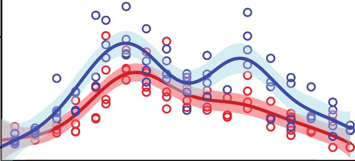
Lehnebach et al.                                                                                            X-Ray Computed Tomography Analysis of Xylogenesis

 FIGURE 1 | Comparison between the microtomy and HXRCT workflow developed in the present study. The flowchart covers the different steps from field sampling to
 data analysis through sample preparation and data extraction.

forming xylem growth ring was selected while avoiding selection                    Within the maturing xylem, the secondary cell wall is formed
of wide rays, resin ducts, or cracks.                                              and lignin is deposited resulting in an increase of the cell wall
   This VOI was then processed with Octopus analysis, formerly                     density from the cambium until the boundary with the mature
distributed by the UGCT spin-off company XRE (now TESCAN-                          xylem zone.
XRE, part of the TESCAN ORSAY HOLDING a.s.) (Brabant                                  Apart from secondary changes occurring many years later,
et al., 2011). For each tangential slice of the VOI, a first                       such as heartwood formation, the structure of the fully mature
thresholding was applied to create a mask covering the area                        cell wall does not change in time (Lehnebach et al., 2019), its
containing the sample. The masks were used to create a first                       density is assumed to vary both between and within species
region of interest (ROI) image stack containing xylem pixels                       in a very narrow range (1.4–1.5 g/cm3). Cell wall density, and
(xylem ROI stack). For each tangential slice of the xylem ROI                      consequently cell wall gray value are therefore assumed to remain
stack, a second thresholding was applied to create a mask                          constant within the mature xylem zone. Consequently, the cell
excluding the pixels associated to cell lumens. The masks were                     wall density profile along the growing ring follows a two-phased
used to create a second region of interest image stack containing                  pattern with (1) an increase from cambium to the end of the
cell wall pixels exclusively (cell wall ROI stack). Both xylem                     maturing xylem zone followed by (2) a constant density toward
and cell wall ROI stacks of each microcore were then processed                     the innermost layers of the mature xylem zone.
with R software (R Development Core Team, 2019), using the                            Identifying the distance from the cambium at which the
packages “magick” (Ooms, 2020) and “imager” (Barthelme, 2019),                     phase transition occurs, allows estimating the width of both
to extract the mean gray value per ROI resulting in two radial                     maturing and mature xylem. An analytical approach is developed
gray-value profiles per microcore, herein after referred to as                     in this study, aiming to quantify the width of both maturing and
“xylem gray profile” and “cell wall gray profile,” respectively.                   mature xylem.

Cell Wall Gray-Value Profiling: Rationale
The gray-value profiling methodology developed here relies on                      Cell Wall Gray-Value Profiling: Implementation
the positive and linear relationship between the gray value of                     For each sample, the cell wall gray profile was plotted over a
HXRCT-derived images and the density of the scanned material.                      transversal reference image, with each data point corresponding
Therefore, cell wall density changes occurring within the forming                  to the average gray value of the corresponding tangential slice
growth ring can be assessed through cell wall gray value changes.                  (Figure 3). We used the smoothing property of a generalized
From a structural point of view, the forming growth ring consists                  additive model (GAM) to analyze the cell wall gray profile of
of a maturing xylem zone (where xylogenesis occurs), and a                         each microcore. GAMs are extremely versatile in fitting non-
mature xylem zone, where xylogenesis already ended (Figure 2).                     linear and non-monotonic data series, and offer the possibility to

Frontiers in Plant Science | www.frontiersin.org                               4                                        August 2021 | Volume 12 | Article 698640
Lehnebach et al.                                                                                                       X-Ray Computed Tomography Analysis of Xylogenesis

 FIGURE 2 | Variation of cell wall gray value or density within the forming growth ring. Following cell wall maturation during xylogenesis, the cell wall density (red line) is
 expected to increase from the cambium (green dotted line) to the most recent mature xylem layer (blue dotted line). Within the mature xylem, the cell wall is fully
 developed and its basic density is assumed to be constant until the growth ring boundary (yellow dotted line) and beyond. The distance from the cambium at which
 the phase transition occurs, allows estimating the width of both mature and maturing xylem.

estimate and analyze their derivative. GAMs are therefore well-                            confidence interval). The median radial position of this part was
suited to analyze cell wall gray profile of diverse tree species. All                      fixed as the cambium position (Figure 4B). The radial position
GAMs were fitted with a Gaussian distribution and identity link                            of the previous growth ring boundary was indicated manually on
function. The goodness of each fit was assessed visually.                                  the reference image (Figure 4A). It allowed to estimate the width
    Consisting of primary-walled cells, the cambium and the                                of the increment, wincr−CT .
enlarging phloem and xylem cells have the lowest cell wall                                     The distance from the cambium for each data point was
density (Figures 2, 3, 4A,B). The cell wall density increases at                           calculated and fitted another GAM over the data spanning from
both sides of the cambium because of phloem and xylem cell                                 the cambium (i.e., distance from the cambium = 0) to the
wall differentiation (Figures 4A,B). We took advantage of this                             previous growth-ring boundary. As for cambium detection, the
feature to define the cambium position semi-automatically by                               95% confidence intervals of the GAM derivatives allowed the
fitting a GAM and analyzing its derivative within a manually                               identification of growth ring parts where the gray values are
defined subset of the sample containing the cambium and the                                constant. The constant part closest to the cambium was selected
newly differentiating phloem and xylem cells (Figure 4B). Within                           as the region where the transition between maturing and mature
this subset, the pointwise 95% confidence intervals of the GAM                             zone occurs (Figure 4C). The radial position of the left edge
derivatives were used to detect the part where the GAM predicted                           of the transition region was considered as the width of the
values were constant (i.e., where the fitted GAM derivative                                maturing xylem, wmaturing−CT . Finally, the width of the mature
values were significantly different from 0, i.e., 0 lying outside the                      zone was computed as, wmat−CT = wincr−CT − wmaturing−CT .

Frontiers in Plant Science | www.frontiersin.org                                       5                                             August 2021 | Volume 12 | Article 698640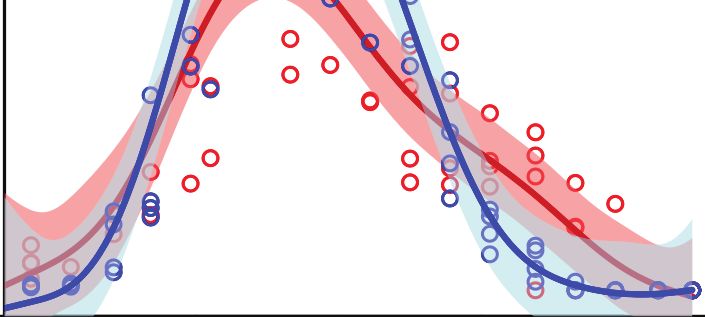
Lehnebach et al.                                                                                                      X-Ray Computed Tomography Analysis of Xylogenesis

 FIGURE 3 | Plot of the cell wall gray profile. The volume of interest (VOI) containing the inner layers of the phloem, the cambium, and the forming xylem growth ring
 (top) is sliced tangentially (middle). Averaging the cell wall gray values of each tangential slices results in a cell wall gray value profile plotted over the transversal
 reference image (bottom). The reference image is in the red frame (top and bottom image), the cambium and previous ring boundary are highlighted by the green and
 yellow lines respectively.

GAM fitting and derivative analysis were performed using the R                            Estimation of Xylogenesis Parameters and
packages “mgcv” (Wood, 2017) and “gratia” (Simpson, 2020). A                              Statistical Analysis
step by step description of the gray-value profiling methodology                          Computation of Xylogenesis Critical Dates and
is available in Supplementary File 1.                                                     Duration, and Growth Rate
                                                                                          Computation of xylogenesis phenological parameters was first
Microtomy Workflow                                                                        performed through a “model-driven” approach, such as the
The microcores for microtomy were dehydrated in graded                                    fitting and derivation of the Gompertz function (e.g., Rossi et al.,
ethanol series, infiltrated, and embedded in paraffin blocks and                          2003). However, a recent methodological breakthrough suggests
finally sliced using a rotary microtome. The thin sections were                           that a “data-driven” approach is able to capture properly the
stained in a water solution of safranin and astra blue, staining                          variation of xylogenesis dynamics (i.e., durations and rates) while
lignin in red and cellulose/hemicellulose in blue, respectively.                          the model-driven approach fails (Cuny et al., 2013). Preliminary
Histological sections were prepared at the Slovenian Forestry                             analysis of the data (not shown) using a Gompertz function
Institute (Ljubljana), following the detailed protocol of Prislan                         produced improper fitting and led us to opt for the GAM data-
et al. (2014). Observation under light microscope allowed to                              driven approach.
distinguish between maturing and mature xylem. For each                                       As the increase of the increment width during the growing
microcore, the width of the cambium, and the width of the                                 season is monotone, a shape constrained additive model (SCAM)
maturing and mature xylem were measured at three positions                                was used (Pya and Wood, 2015) to analyze the phenology of
to consider potential within-sample heterogeneity. The maturing                           wood formation. SCAM was fitted using monotone increasing
zone was measured as the ring part between the cambium and                                P-splines, Gaussian distribution, and identity link function.
the first mature cells. Xylem cells were considered mature when                           Circumferential growth heterogeneity was accounted for using
the entire cell wall was stained red and the lumen was empty of                           the correction formula for cell counting data adapted to widths
any cellular content. In order to be comparable to the HXRCT                              at the individual tree scale:
methodology, the increment width was computed as the sum of
both mature and maturing xylem width and the width of the
cambium divided by two. When the maturing zone was absent
                                                                                                                                    wprev,j
(i.e., for samples collected at the very end of the growing season),                                            wcor,j = wobs,j ∗ 1 P                                     (1)
the mature zone was set equal to the increment width.                                                                             n   wprev,j

Frontiers in Plant Science | www.frontiersin.org                                      6                                            August 2021 | Volume 12 | Article 698640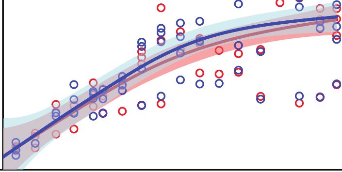
Lehnebach et al.                                                                                                       X-Ray Computed Tomography Analysis of Xylogenesis

       A

       B

       C

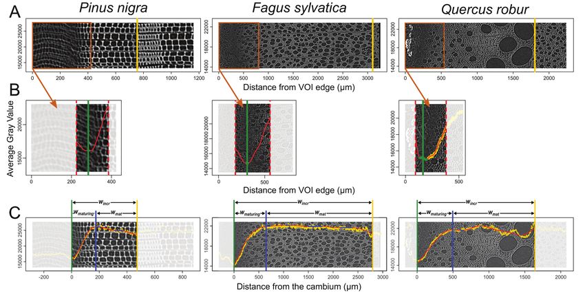
 FIGURE 4 | Implementation of the gray-value profiling. (A) The average gray value of each tangential slice is plotted on the reference cross-section image. At this
 stage, the previous growth ring boundary is identified (yellow vertical line). (B) Within the subset containing the cambium, a GAM fit (red) and its derivative allow
 automatic identification of the cambium position (green vertical line). (C) Within the growth ring, a GAM fit (red) and its derivative allows the detection of a constant
 gray value (blue filled circles). The constant gray value closest to the cambium is set as the boundary between maturing and mature xylem (blue vertical line). This
 procedure allows to estimate maturing xylem width (wmaturing ), mature xylem width (wmature ), and increment width (wincr ). The gray scale is unitless. It varies from 0 to
 65,535 for 16 bits images used here.

where, n is the number of sampling dates, and for each jth sample,                            The same procedure was applied for the width of the mature
wcor,j is the corrected width; wobs,j is the observed width; wprev,j is                    xylem in order to define the date of beginning (tbmat ) and ending
the observed width of the previous growth year (Rossi et al., 2003;                        (temat ), the duration (dmat ) and the average rate (rmat ) of mature
Prislan et al., 2013).                                                                     xylem production. Finally, the duration of the whole xylogenesis
    First, the constrained GAM was fitted over the width of                                was defined as:
the increment to analyze the size growth dynamics at the tree
level (Figure 5). The final increment width was computed as                                                           dxylo = teincr − tbincr                                   (4)
the median of the increment width predicted for the last three
                                                                                           These parameters were estimated for both microtomy and
sampling dates (i.e., the dates for which the increment width
                                                                                           HXRCT datasets.
had already stabilized), in order to account for circumferential
growth heterogeneity. We defined the date of beginning (tbincr )                           Consistency Between HXRCT and Microtomy
and ending (teincr ) of size growth as the day of year for which,                          Measurements
respectively, 5% and 95% (i.e., wbincr and weincr ) of the increment                       The consistency between increment, mature, and maturing xylem
is formed. The duration of size growth was computed as the time                            width measured with HXRCT and microtomy was assessed using
span between both dates:                                                                   major axis regression. The value of both intercept and slope and
                                                                                           their respective 95% confidence intervals (CI) were used to detect
                          dincr = teincr − tbincr                                (2)       significant differences compared to a 1:1 regression. An intercept
                                                                                           differing from 0 (i.e., 0 lies outside the CI of the intercept)
The average daily rate of size growth was calculated as:                                   indicates a fixed bias across the entire range, that is, the HXRCT
                                                                                           provides greater (or lower) values than microtomy. A slope
              rincr = (weincr − wbincr )/(teincr − tbincr )                      (3)       differing from 1 (i.e., 1 lies outside the CI of the slope) indicates
                                                                                           a proportional bias, that is, the HXRCT provided progressively
The differences between the increment width predicted by GAM                               greater (or lower) values with increasing values of the considered
on the consecutive days, spanning from tbincr to teincr , were                             measurement than microtomy. Major axes were fitted with the
calculated to assess the distribution of size growth daily rates.                          “lmodel2” package in R (Legendre, 2018).

Frontiers in Plant Science | www.frontiersin.org                                       7                                             August 2021 | Volume 12 | Article 698640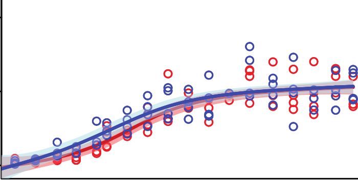
Lehnebach et al.                                                                                                     X-Ray Computed Tomography Analysis of Xylogenesis

                                                                                               was calculated. Being linearly related to the biomass accumulated
                                                                                               in a given volume of the forming xylem ring, this variable
                                                                                               was considered as a proxy of the biomass accumulated at
                                                                                               the trunk base (btrunk ) of the tree from the beginning of the
                                                                                               growing season up to the considered sampling date. As for xylem
                                                                                               widths, we accounted for circumferential growth heterogeneity
                                                                                               by correcting btrunk by the observed width of the previous growth
                                                                                               year at the individual tree scale. We fitted constrained GAM
                                                                                               over btrunk to analyze the biomass production dynamics at the
                                                                                               species level. The biomass production and the size increase
                                                                                               were compared by analyzing the seasonal variation of both
                                                                                               btrunk and the increment width (wincr ). For convenience, both
                                                                                               variables were normalized (i.e., ranging between 0 and 1) using
 FIGURE 5 | Description of the xylogenesis parameters estimated for both                       the median of btrunk and wincr , respectively, predicted for the
 HXRCT and microtomy datasets. Parameters related to size growth (green)                       last three sampling dates. This provided the relative completion
 and mature xylem production (brown) are reported. The date of beginning
                                                                                               of each process (i.e., size growth and biomass production) and
 (tbincr ) and ending (teincr ) of size growth are defined as the day of year for
 which, respectively, 5% and 95% (i.e., wbincr and wcincr ) of the increment is                allowed to compute the time lag between biomass production
 formed. The date of beginning (tbmat ) and ending (temat ) of mature xylem                    and size growth on the course of the growing season. For
 production are defined as the day of year for which, respectively, 5% and 95%                 this purpose, we computed the time lag between the relative
 (i.e., wbmat and wcmat ) of the mature xylem is formed. The duration of size                  completion of a given proportion of the final btrunk and the
 growth (dincr ), mature xylem production (dmat ) and xylogenesis (dxylo ) are
 computed as the time span between teincr and tbincr , temat and tbmat , and
                                                                                               final wincr (e.g., Day of year (DOY) when a given relative
 temat and tbincr respectively. The average daily rate of size growth and mature               completion of the final btrunk is produced - DOY when a given
 xylem production are calculated as, rincr = (weincr − wbincr ) / (teincr − tbincr ) and       relative completion of the final wincr is produced). The time lags
 rmat = (wemat − wbmat ) / (temat − tbmat ).                                                   were computed for relative completion values ranging between
                                                                                               0 and 1 and every 0.01. The differences between both btrunk
                                                                                               and wincr predicted by GAM on the consecutive days were also
                                                                                               calculated. This allowed to assess the seasonal variation of size
Comparison of Parameters Estimated With HXRCT                                                  growth rate and biomass production rate and estimate their
and Microtomy                                                                                  seasonal maximum.
To assess the agreement between critical dates, durations,
and growth rates estimated using HXRCT and microtomy,
the significant differences between the two datasets by case-
based bootstrap resampling were tested. For a given species                                    RESULTS
and for each parameter of interest (Figure 5), the difference                                  Seasonal Growth Pattern
between the median of the four trees based on HXRCT                                            The patterns of seasonal xylem growth, at the species level,
data on one hand, and microtomy data on the other                                              derived from HXRCT and microtomy strongly overlap
hand was computed. The observed median difference was                                          (Figure 6). For both pine and beech, increment and mature
compared to the median reference distribution simulated                                        xylem width increased along the growing season following a
under the null hypothesis (H0 : the parameters extracted from                                  typical sigmoidal curve (Figure 6). For oak, the width of the
HXRCT do not differ from the parameters extracted from                                         increment follows a curvilinear increase while the mature xylem
microtomy). The reference distribution resulted in 10,000                                      width follows a bisigmoidal pattern. Major axis fits did not detect
resampled median differences. A two-tailed test was performed                                  any significant bias in the estimation of increment width by
by computing the achieved significance level (ASL) as the                                      HXRCT compared to microtomy (Supplementary Figure 3;
proportion of absolute values in the reference distribution                                    Supplementary Table 1). Regarding the mature xylem width,
that are equal, or greater, than the observed absolute median                                  no biases were detected for beech and oak, however, a slight
difference. ASL is interpreted in the same way as the p                                        slope departure from 1 for pine (Supplementary Table 1) is
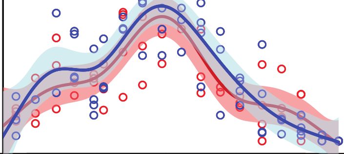
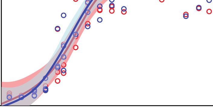
value (P).                                                                                     found. Microtomy and HRXCT described similarly the seasonal
                                                                                               variation of maturing xylem width. For beech, the maturing
Accounting for Biomass Production                                                              xylem width followed a bell-shaped pattern while for oak and
Dynamic Trough Xylem Gray Value                                                                pine the pattern was bimodal, with a first peak in spring and a
The ability of HRXCT to assess biomass production dynamics                                     second peak in July for oak and in September for pine.
throughout the course of the growing season was assessed by                                        Overall, the estimated maturing xylem width by HXRCT is
using the gray-value information. Here, the xylem gray profile                                 significantly lower than for microtomy for pine and beech (see
(i.e., including lumen and cell wall pixels) was preferred to the                              Supplementary Table 1), especially during summer (Figure 6).
cell wall gray profile as it accounts for both the proportions                                 These represent the only cases in our study when HXRCT and
of the xylem layer allocated to cell wall (biomass) and lumens                                 microtomy produce different estimates (see the third paragraph
(void). The sum of the gray values of each xylem gray profile                                  of the discussion section).

Frontiers in Plant Science | www.frontiersin.org                                           8                                    August 2021 | Volume 12 | Article 698640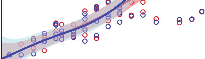
Lehnebach et al.                                                                                            X-Ray Computed Tomography Analysis of Xylogenesis

 FIGURE 6 | Seasonal growth pattern assessed with HXRCT (red) and microtomy (blue). Variations of increment and mature xylem were fitted with constrained GAMs
 (top and middle row), while maturing xylem width was fitted with unconstrained GAMs (bottom row). The shaded band represents the standard error of the fit.

Wood Formation Calendar                                                            while tbincr−CT = DOY 108, the difference is not significant).
Size growth, and therefore xylogenesis, began (tbincr ) at the                     Microtomy and HXRCT gave similar estimates of tbincr (Figure 7;
end of March in oak (one week before the first sampling                            Supplementary Table 2). Size growth was completed in mid-
date, tbincr−micro = DOY 89, while tbincr−CT = DOY 88,                             September for beech (teincr−micro = DOY 256, while teincr−CT =
see Supplementary Table 2), while it started between mid-                          DOY 251), and oak (teincr−micro = DOY 260, while teincr−CT =
and end of April for pine (tbincr−micro = DOY 100, while                           DOY 264) while it lasted until the beginning of October for pine
tbincr−CT = DOY 106) and beech (tbincr−micro = DOY 122,                            (teincr−micro = DOY 277, while teincr−CT = DOY 270). Again,

Frontiers in Plant Science | www.frontiersin.org                               9                                        August 2021 | Volume 12 | Article 698640
Lehnebach et al.                                                                                  X-Ray Computed Tomography Analysis of Xylogenesis

microtomy and HXRCT provided similar estimate of teincr and                 difference between maximum size growth (DOY = 173) and
therefore the duration of size growth (dincr ) was highly similar           biomass production rates (DOY = 177) was four days in pine.
for both methods (dincr−micro = DOY 177, dincr−CT = DOY 162                 This difference was even smaller in beech, that is, two days (DOY
for pine, dincr−micro = DOY 134, dincr−CT = DOY 140 for beech               = 171 vs. DOY = 173). For oak, the relative size growth rate was
and dincr−micro = DOY 163, dincr−CT = DOY 171 for oak, the                  maximal at the end of March (DOY = 89), while the biomass
differences are not significant, see Supplementary Table 2).                production rate peaked at the beginning of June (DOY = 152),
   The mature xylem production began in April for oak                       resulting in a time lag of 63 days.
(tbmat−micro = DOY 105, while tbmat−CT = DOY 104), and from
the end of May to beginning of June for beech (tbmat−micro =
DOY 145, while tbmat−CT = DOY 141) and pine (tbmat−micro                    DISCUSSION
= DOY 152, while tbmat−CT = DOY 154). However, the end of
mature xylem production, and therefore the end of xylogenesis,              This is the first time that xylogenesis dynamics and intra-
occurred by the end of September for beech (temat−micro = DOY               seasonal growth rate parameters are assessed using HRXCT
265, while temat−CT = DOY 264) and oak (temat−micro = DOY                   The development of an entire HRXCT workflow from sample
262, while temat−CT = DOY 262), and by the end of October                   preparation, to reconstructed volume analysis, allowed the
for pine (temat−micro = DOY 304, while temat−CT = DOY 294,                  estimation of relevant xylogenesis parameters (i.e., dates,
the difference is not significant). Overall the duration of mature          durations, and rates) and seasonal time series of increment
xylem production (dmat ) is shorter for beech (dxylo−micro = 142            width. Moreover, the benchmarking of HRXCT with the classical
days, while dxylo−CT = 154 days, the difference is not significant)         microtomy proved the HRXCT approach to produce accurate
than for pine (dxylo−micro = 206 days, while dxylo−CT = 188 days,           estimates of xylogenesis dynamics over species with contrasted
the difference is not significant) and oak (dxylo−micro = 166 days,         xylem anatomies.
while dxylo−CT = 169 days, the difference is not significant).                  The ability of HRXCT to encompass diverse xylem structure
Dates (tbmat , temat ) and durations (dmat ) related to mature xylem        relies on the gray-value profiling methodology. While 2D cell
formation as well as the duration of xylogenesis (dxylo ) were              morphometric analysis is not applicable on various anatomical
similar across methods (Figure 7; Supplementary Table 2).                   structure, the gray-value profiling overcomes this issue by
                                                                            assessing the maturation state from monitoring of density at
Growth Rates                                                                the xylem layer scale. The HRXCT methodology also proved
The estimated size growth rate (rincr ) ranged from 5 to                    to produce accurate estimates of actual ring width along
6 µm.day−1 in pine to 21 to 22 µm.day−1 in beech. The                       the whole growing season ranging from ca. 30 to 5000 µm
mature xylem production rate (rmat ) presented similar values.              (see Supplementary Table 4). Given this result and the high
Overall, estimates of size growth and mature xylem production               resolution of HRXCT (i.e., 2.5 µm/pixel), the authors assume
rate by microtomy and HXRCT are highly similar (Figure 8;                   that HRXCT will succeed in estimating xylogenesis dynamics
Supplementary Table 3). Moreover, the distributions of rincr and            of slow-growing trees. Therefore, the generic and data-driven
rmat estimated with HXRCT and microtomy strongly overlap                    HRXCT method showed to offer a very interesting perspective
(Supplementary Figure 3).                                                   to fill the knowledge gap about secondary growth phenology
                                                                            and intra-annual wood production over a structural and
Seasonal Pattern of Biomass Production                                      taxonomical diversity.
and Size Growth and Their Time Lag                                              It is important to pinpoint that the xylem dimensions
The patterns of biomass production and size growth are very                 estimated with microtomy and HRXCT slightly differ in their
similar at the species level (Figure 9). For both pine and beech,           nature. HRXCT differentiates the mature xylem from the
the biomass increases during the growing season following a                 maturing xylem on the basis of cell wall density differences.
sigmoidal curve, while biomass in oak increases curvilinearly.              Therefore, cells in the very late stages of wood formation, for
However, biomass production lags behind size growth. The time               which the biomass accumulation process is almost accomplished
lag observed in respect of both size and biomass completion                 (i.e., cells at the end of secondary wall formation and lignifying
of the xylem ring is more important for pine (mean = 9 days,                cells) or terminated (i.e., fully lignified cells but still alive)
maximum=15 days) than for beech (mean = 4 days, maximum                     might be considered as mature cells with HRXCT. On the
= 9 days) and oak (mean = 5 days, maximum = 7 days). For pine               other hand, with microtomy such cells are considered as
and beech, the time lag reaches its maximum during the latewood             still maturing since the mature zone consists exclusively of
formation (at the end of the growing season, when 80–90% of                 fully lignified dead cells. Therefore, the slight overestimation
the ring width is formed). In oak, the time lag is maximum from             and underestimation of the mature and the maturing zone,
the end of the earlywood formation up to half of the latewood               respectively, with HRXCT may rely on the different nature
formation (between 40% and 70% of the ring formation).                      of xylem dimensions estimated with HRXCT and microtomy.
   The time lags observed at the peak of both size growth and               However, these differences are not sufficiently important to bias
biomass production rate contrast with the time lag observed                 the estimation of xylogenesis dynamics.
in respect of both size growth and biomass completion. For                      As gray scale information can be directly converted into
both pine and beech, the relative size growth rate, as well as              estimates of biomass, X-ray computed tomography has been
the biomass production rate peaked at the end of June. The                  successfully used in the field of dendrochronology and biomass

Frontiers in Plant Science | www.frontiersin.org                       10                                    August 2021 | Volume 12 | Article 698640
Lehnebach et al. X-Ray Computed Tomography Analysis of Xylogenesis FIGURE 7 | Xylogenesis calendar and duration. Left panels: Estimates of size growth beginning (tbincr ) (i.e., beginning of xylogenesis) and size growth ending (teincr ) (green) as well as mature xylem production beginning (tbmat ) and ending (temat ) (i.e., ending of xylogenesis) (brown) are presented for HXRCT and microtomy. Green and brown lines range from minimum to maximum estimated dates, while second and third quartile are represented by short vertical lines. Right panels: Estimates of xylogenesis duration (dxylo ) (median) for HXRCT (red) and microtomy (blue) are presented. Black lines range from minimum to maximum estimated duration, while second and third quartile are represented by short vertical lines. The significance of the bootstrap resampling test (P) assessing the difference between HXRCT and microtomy is reported. estimation (Bastin et al., 2015; De Mil et al., 2016). A significant accurate than the cell morphometric approach (Cuny et al., 2015), step forward in the quantification of the intra-annual dynamics of it still relies on physical 2D slicing of the sample. Here, it is carbon sequestration, for both conifers and angiosperms, relies showed that HRXCT can also be used in the field of intra-seasonal on the calculation of the apparent density of the forming wood biomass production and carbon sequestration research. In fact, (i.e., the product of cell wall density and the ratio of cell wall the sum of the xylem gray value, which is actually directly related area to xylem area) (Andrianantenaina et al., 2019). Although to density (attenuation coefficients are recorded), and therefore the apparent density approach proved to be more direct and to the biomass accumulated at the trunk base provided a very Frontiers in Plant Science | www.frontiersin.org 11 August 2021 | Volume 12 | Article 698640
Lehnebach et al.                                                                                                                  X-Ray Computed Tomography Analysis of Xylogenesis

                                                                                     Pine
                   | |                                                HRXCT                     ||
                                 ns                                   Microtomy                              ns
                               (P=0.4)                                                                     (P=0.6)
                   |                                                                            |

                                                                                  Beech
                                     |            |                                                                  |        |
                                                         ns                                                                              ns
                                                      (P=0.87)                                                                        (P=0.62)
                                              |                                                                           |       |

                                                                                     Oak
                         | |                                                                         | |
                                      ns                                                                          ns
                                   (P=0.96)                                                                    (P=0.92)
                         |    |                                                                      |    |

             0           10              20            30        40     50      60        0          10              20               30         40        50         60
                                                                      −1                                                                                         −1
                             Size growth rate (r incr , μm.day )                              Mature xylem production rate (r mat , μm.day )

 FIGURE 8 | Size growth (rincr ) and mature xylem (rmat ) production rates (median) estimated with CT (red) and microtomy (blue). Black lines range from minimum to
 maximum estimated duration, while second and third quartile are represented by short vertical lines. The significance of the bootstrap resampling test (P) assessing
 the difference between HXRCT and microtomy is reported.

 FIGURE 9 | Relative completion of size growth and biomass production. The top line presents the relative completion of size growth and biomass production during
 the growing season as well as the time lag between size growth and biomass production (red line). For each species, a reference image and its associated xylem
 gray-value profile (blue line) is shown. The bottom line presents the variation of the relative completion rate, with the vertical lines highlighting the DOY at which the
 maximum rate occurs.

Frontiers in Plant Science | www.frontiersin.org                                     12                                                      August 2021 | Volume 12 | Article 698640
Lehnebach et al. X-Ray Computed Tomography Analysis of Xylogenesis
interesting insight into the capability of HRXCT to assess biomass Therefore, we expect that the new HRXCT workflow
production dynamic throughout the course of the growing presented here will be the foundation for major breakthroughs
season. The observed seasonal variation of the time lag between also in the field of intra-seasonal biomass production and carbon
biomass production and size growth agrees with the anatomical sequestration research. Together with a proper conversion of
signature of the studied species. For both pine and beech, the time gray value into wood density and species-specific allometric
lag reaches its maximum during the latewood formation, when relationships, HXRCT has the clear potential to provide a better
80–90% of the growth ring width is already achieved and thick- quantification of intra-annual dynamics of biomass production
walled tracheids or fibers are produced. In oak, the time lag shows and carbon sequestration through high-resolution 3D mapping
the largest values from the end of earlywood formation to the half of carbon within the forming growth ring and among different
of the completion of the latewood, thus between 40% and 70% of tree species.
the growing ring part consists mainly of thick-walled fibers and HXRCT is a growing and wide spreading technology in the
narrow vessels. Therefore, for all species, the time lag is largest field of tree- and wood sciences. Both forthcoming technological
when the denser part of the growth ring is forming and suggests improvements of scanning devices and protocol optimization
that wall formation and maturation of thick-walled cells require a (batch preparation and actual scanning of samples), as well as
longer period than the formation and maturation of thin-walled increased computer capacity, will offer potential for the further
cells. This pattern already observed in conifers (Cuny et al., 2014) development of a high-throughput analysis of xylogenesis with
seems therefore to apply to angiosperms. HXRCT. Such development will allow to (1) cover a broad
A very short time lag was found (four days) between the range of biomes and climates by increasing drastically the
maximal size growth rate and the maximal biomass production number of sampled individuals and to (2) integrate different tree
rate in pine. This result strongly contrasts with the one-month components (i.e., root, stem, and branches) that may present
time lag generally acknowledged in conifers (Cuny et al., 2015). different phenological patterns and react differently to similar
However, Andrianantenaina et al. (2019) reported a two weeks climatic conditions. Thus, the HRXCT approach, developed
lag between biomass production and size growth in spruce here, offers a promising avenue to investigate seasonal plant-
growing in north-east France. They speculated that this reduced climate growth relationships spanning a wide range of wood
time lag could be accounted for by the occurrence of a long structures, species, biomes, and climatic conditions, and to
summer drought during the measurement period in 2015, generate a substantial amount of data that could trigger a new
resulting in the shortening of the growing season and less generation of vegetations models fully considering the seasonal
investment in cell production. The growing season 2019 (when wood growth dynamics.
the samples were collected) experienced a very intense drought
in late spring-early summer. Also because of the water deficit DATA AVAILABILITY STATEMENT
accumulated during the very dry 2018, the soil in late spring-
early summer of 2019 was dry as never observed before in The raw data supporting the conclusions of this article
Belgium (Mariën et al., 2021). Therefore, wood formation in will be made available by the authors on request, without
black pine might be sensitive to drought as in other conifer undue reservation.
species (Andrianantenaina et al., 2019).
At the same site of our sampling, Dox et al. (2021) found that
AUTHOR CONTRIBUTIONS
the end of wood formation in oak occurs earlier under drought
RL, JVdB, and MC designed the study. BM, MC, and RL collected
conditions while this parameter is rather insensitive to drought
the samples. JG and PP processed the sample for microtomy.
in beech. In 2019, the authors found that wood formation in oak
RL and JG conducted the analysis of the histologic slices. RL
stopped earlier (DOY = 262) than in 2018 (DOY = 282 in Dox
conducted the X-ray computed tomography analysis and the
et al., 2021). As the drought in 2019 was even more pronounced
analysis of the data. RL, JVdB, MC, and HB wrote the first draft
than in 2018 (see above), the results corroborate the drought
of the manuscript. RL, JVdB, MC, and HB wrote the revised
sensitivity of wood formation in oak.
manuscript. BM, JG, and PP made significant contributions to
The time lag in beech (two days) was even lower than for
the manuscript. All authors gave final approval for publication.
pine, while it was much longer in oak (63 days). However, we
note that for oak, the peaks of size growth rate and of biomass FUNDING
production rate are much less pronounced than for the other
species. During earlywood formation, from late March till early This research was funded by the BOF Special Research Fund
June, both size growth rate and biomass production rate were for RL (BOF Starting Grant JVdB, BOFSTG2018000701), the
maximal and rather constant in oak. We believe that the large UGCT Center of Expertise (BOF.EXP.2017.0007), the Research
time lag in oak results from the ring-porous structure of its Foundation Flanders (FWO grant G009720N), and the LEAF-
wood, which implies a fast size growth, due to the enlargement FALL starting ERC grant (714916).
of the vessels, at the beginning of the growing season. Given the
uniqueness of such parameters computed for angiosperms, any ACKNOWLEDGMENTS
comparison with previous studies is not possible.
We believe that further development of HRXCT will open the We acknowledge the institutions that gave permission to conduct
way for more insight into biomass production in angiosperms. research in the study areas: Agency for Nature and Forest
Frontiers in Plant Science | www.frontiersin.org 13 August 2021 | Volume 12 | Article 698640Lehnebach et al. X-Ray Computed Tomography Analysis of Xylogenesis
of the Flemish Government (ANB), the Military Defense of SUPPLEMENTARY MATERIAL
Belgium, and City of Brasschaat. We would like to thank Stijn
Willen for the technical support and Inge Dox and Sebastien The Supplementary Material for this article can be found
Leys for their help in collecting and processing the samples online at: https://www.frontiersin.org/articles/10.3389/fpls.2021.
for microtomy. 698640/full#supplementary-material
REFERENCES Dierick, M., Van Loo, D., Masschaele, B., Bulcke, J., Acker, J., Cnudde, V., et al.
(2014). Recent micro-CT scanner developments at UGCT. Nucl. Instrum.
Andrianantenaina, A. N., Rathgeber, C. B. K., Pérez-de-Lis, G., Cuny, H., and Methods Phys. Res. B. 324, 35–40. doi: 10.1016/j.nimb.2013.10.051
Ruelle, J. (2019). Quantifying intra-annual dynamics of carbon sequestration Dox, I., Gričar, J., Marchand, L. J., Leys, S., Zuccarini, P., Géron, C., et al. (2020).
in the forming wood: a novel histologic approach. Ann. Forest Sci. 76:62. Timeline of autumn phenology in temperate deciduous trees. Tree Physiol. 40,
doi: 10.1007/s13595-019-0846-7 1001–1013. doi: 10.1093/treephys/tpaa058
Barthelme, S. (2019). imager: Image Processing Library Based on ’CImg’. R package Dox, I., Prislan, P., Gričar, J., Mariën, B., Delpierre, N., Flores, O., et al.
version 0.41.42. (2021). Drought elicits contrasting responses on the autumn dynamics of
Bastin, J. F., Fayolle, A., Tarelkin, Y., et al. (2015). Wood specific gravity variations wood formation in late successional deciduous tree species. Tree Physiol. 41,
and biomass of central african tree species: the simple choice of the outer wood. 1171–1185. doi: 10.1093/treephys/tpaa175
PLoS ONE 10:e0142146. doi: 10.1371/journal.pone.0142146 Fatichi, S., Leuzinger, S., and Körner, C. (2014). Moving beyond photosynthesis:
Brabant, L., Vlassenbroeck, J., De Witte, Y., et al. (2011). Three-dimensional from carbon source to sink-driven vegetation modeling. New Phytol. 201,
analysis of high-resolution X-ray computed tomography data with Morpho+. 1086–1095. doi: 10.1111/nph.12614
Microsc. Microanal. 17, 252–263. doi: 10.1017/S1431927610094389 Fatichi, S., Pappas, C., Zscheischler, J., and Leuzinger, S. (2019). Modelling
Bray, D. (2000). “Critical point drying of biological specimens for scanning carbon sources and sinks in terrestrial vegetation. New Phytol 221, 652–668.
electron microscopy,” in Supercritical Fluid Methods and Protocols, eds doi: 10.1111/nph.15451
J. R. Williams, A. A. Clifford (Totowa, NJ: Humana Press), 235–243. Friend, A. D., Eckes-Shephard, A. H., Fonti, P., Rademacher, T. T., Rathgeber, C.
doi: 10.1385/1-59259-030-6:235 B. K., Richardson, A. D., et al. (2019). On the need to consider wood formation
Brodersen, C. R., and Roddy, A. B. (2016). New frontiers in the three-dimensional processes in global vegetation models and a suggested approach. Ann. For. Sci.
visualization of plant structure and function. Am. J. Bot. 103, 184–188. 76:49. doi: 10.1007/s13595-019-0819-x
doi: 10.3732/ajb.1500532 Gričar, J., Zavadlav, S., Jyske, T., Lavri,č, M., Laakso, T., Hafner, P., et al. (2018).
Camarero, J. J., Olano, J. M., and Parras, A. (2010). Plastic bimodal xylogenesis in Effect of soil water availability on intra-annual xylem and phloem formation
conifers from continental Mediterranean climates. New Phytol. 185, 471–480. and non-structural carbohydrate pools in stem of Quercus pubescens. Tree
doi: 10.1111/j.1469-8137.2009.03073.x Physiology 39: 222–233. doi: 10.1093/treephys/tpy101
Campioli, M., Vincke, C., Jonard, M., Kint, V., Demarée, G., and Ponette, Q. Gutiérrez, Y., Ott, D., Töpperwien, M., Salditt, T., and Scherber, C. (2018). X-
(2012). Current status and predicted impact of climate change on forest ray computed tomography and its potential in ecological research: a review
production and biogeochemistry in the temperate oceanic European zone: of studies and optimization of specimen preparation. Ecol. Evol. 8, 7717–7732.
review and prospects for Belgium as a case study. J. For. Res. 17, 1–18. doi: 10.1002/ece3.4149
doi: 10.1007/s10310-011-0255-8 Hubau, W., Lewis, S. L., Phillips, O. L., Affum-Baffoe, K.,Beeckman, H.,
Cufar, K., Prislan, P., de Luis, M., and Gričar, J. (2008). Tree-ring variation, Cuní-Sanchez, A., et al. (2020). Asynchronous carbon sink saturation
wood formation and phenology of beech (Fagus sylvatica) from a in African and Amazonian tropical forests. Nature 579, 80–87.
representative site in Slovenia, SE Central Europe. Trees 22, 749–758. doi: 10.1038/s41586-020-2035-0
doi: 10.1007/s00468-008-0235-6 Knipfer, T., Eustis, A., Brodersen, C., Walker, A. M., and McElrone, A. J. (2015).
Cuny, H. E., Rathgeber, C. B., Kiesse, T. S., Hartmann, F. P., Barbeito, I., Grapevine species from varied native habitats exhibit differences in embolism
and Fournier, M. (2013). Generalized additive models reveal the intrinsic formation/repair associated with leaf gas exchange and root pressure. Plant Cell
complexity of wood formation dynamics. J. Exp. Bot. 64, 1983–1994. Environ. 38, 1503–1513. doi: 10.1111/pce.12497
doi: 10.1093/jxb/ert057 Legendre, P. (2018). lmodel2: Model II Regression. R Package Version 1.7–3.
Cuny, H. E., Rathgeber, C. B. K., Frank, D., Fonti, P., and Fournier, M. (2014). Lehnebach, R., Bossu, J., Va, S., Morel, H., Amusant, N., Nicolini, É., et al. (2019).
Kinetics of tracheid development explain conifer tree-ring structure. New Wood density variations of Legume trees in French Guiana along the shade
Phytol. 203, 1231–1241. doi: 10.1111/nph.12871 tolerance continuum: heartwood effects on radial patterns and Gradients.
Cuny, H. E., Rathgeber, C. B. K., Frank, D., Fonti, P., Mäkinen, H., Prislan, P., et al. Forests 10:80. doi: 10.3390/f10020080
(2015). Woody biomass production lags stem-girth increase by over one month Leroux, O., Leroux, F., Bellefroid, E., Claeys, M., Couvreur, M., Borgonie,
in coniferous forests. Nat. Plants 1:15160. doi: 10.1038/nplants.2015.160 G., et al. (2009). A new preparation method to study fresh plant
De Baerdemaeker, N. J. F., Stock, M., Van den Bulcke, J., De Baets, B., Van structures with X-ray computed tomography. J. Microsc. 233, 1–4.
Hoorebeke, L., and Steppe, K. (2019). X-ray microtomography and linear doi: 10.1111/j.1365-2818.2008.03088.x
discriminant analysis enable detection of embolism-related acoustic emissions. Marchand, L. J., Dox, I., Gricar, J., Prislan, P., Leys, S., Van den Bulcke, J.,
Plant Methods 15:153. doi: 10.1186/s13007-019-0543-4 et al. (2020). Inter-individual variability in spring phenology of temperate
De Mil, T., Vannoppen, A., Beeckman, H., Van Acker, J., and Van den Bulcke, J. deciduous trees depends on species, tree size and previous year autumn
(2016). A field-to-desktop toolchain for X-ray CT densitometry enables tree phenology. Agric. For. Meteorol. 290:108031. doi: 10.1016/j.agrformet.2020.
ring analysis. Ann. Bot. 117, 1187–1196. doi: 10.1093/aob/mcw063 108031
Delpierre, N., Vitasse, Y., Chuine, I., Guillemot, J., Bazot, S., Rutishauser, T., Mariën, B., Dox, I., De Boeck, H. J., Willems, P., Leys, S., Papadimitriou,
et al. (2016). Temperate and boreal forest tree phenology: from organ- D., et al. (2021). Does drought advance the onset of autumn leaf
scale processes to terrestrial ecosystem models. Ann. For. Sci. 73, 5–25. senescence in temperate deciduous forest trees? Biogeosciences 18, 3309–3330.
doi: 10.1007/s13595-015-0477-6 doi: 10.5194/bg-18-3309-2021
Deslauriers, A., Rossi, S., Anfodillo, T., and Saracino, A. (2008). Cambial Ooms, J. (2020). magick: Advanced Graphics and Image-Processing in R. R Package
phenology, wood formation and temperature thresholds in two contrasting Version 2.3.
years at high altitude in southern Italy. Tree Physiol. 28, 863–871. Paganin, D., Mayo, S. C., Gureyev, T. E., Miller, P. R., and Wilkins,
doi: 10.1093/treephys/28.6.863 S. W. (2002). Simultaneous phase and amplitude extraction from a
Frontiers in Plant Science | www.frontiersin.org 14 August 2021 | Volume 12 | Article 698640You can also read


