Cholinergic stimulation with pyridostigmine modulates a heart spleen axis after acute myocardial infarction in spontaneous hypertensive rats
←
→
Page content transcription
If your browser does not render page correctly, please read the page content below
www.nature.com/scientificreports
OPEN Cholinergic stimulation
with pyridostigmine modulates
a heart‑spleen axis after acute
myocardial infarction
in spontaneous hypertensive rats
Robson Luiz Bandoni1, Pamela Nithzi Bricher Choque1, Humberto Dellê1,
Tercio Lemos de Moraes1, Maria Helena Mattos Porter1, Bruno Durante da Silva2,
Gizele Alves Neves1, Maria‑Claudia Irigoyen2, Kátia De Angelis1,3, Valentin A. Pavlov4,
Luis Ulloa5 & Fernanda Marciano Consolim‑Colombo1,2*
The mechanisms regulating immune cells recruitment into the heart during healing after an acute
myocardial infarction (AMI) have major clinical implications. We investigated whether cholinergic
stimulation with pyridostigmine, a cholinesterase inhibitor, modulates heart and spleen immune
responses and cardiac remodeling after AMI in spontaneous hypertensive rats (SHRs). Male adult
SHRs underwent sham surgery or ligation of the left coronary artery and were randomly allocated to
remain untreated or to pyridostigmine treatment (40 mg/kg once a day by gavage). Blood pressure
and heart rate variability were determined, and echocardiography was performed at day six after MI.
The heart and spleen were processed for immunohistochemistry cellular analyses (CD3+ and CD4+
lymphocytes, and CD68+ and CD206+ macrophages), and TNF levels were determined at day seven
after MI. Pyridostigmine treatment increased the parasympathetic tone and T CD4+ lymphocytes in
the myocardium, but lowered M1/M2 macrophage ratio towards an anti-inflammatory profile that
was associated with decreased TNF levels in the heart and spleen. Treatment with this cholinergic
agent improved heart remodeling manifested by lower ventricular diameters and better functional
parameters. In summary, cholinergic stimulation by pyridostigmine enhances the parasympathetic
tone and induces anti-inflammatory responses in the heart and spleen fostering cardiac recovery after
AMI in SHRs.
Acute myocardial infarction (AMI) triggers a sterile inflammatory response characterized by the recruitment
and activation of innate and adaptive immune cells to repair tissue d amage1–3. AMI also elicits systemic inflam-
matory responses that result in organism-wide complications reminiscent of that found in sepsis4–6. In rodents,
AMI triggers hematopoiesis inducing the production of innate immune cells and recruitment of neutrophils
and inflammatory monocytes in the damaged tissue7. These mechanisms regulating monocyte recruitment
from the bone marrow and spleen into the heart during healing have major clinical implications to design novel
therapeutic strategies8, 9. In addition to innate cells, adaptive immune responses also play a fundamental role
in cardiac remodeling after A MI9. More specifically, CD4+ T lymphocytes are required for proper healing and
might prevent chronic remodeling after AMI. Several studies have shown that CD4+ T regulatory cells (Treg)
can modulate inflammation in the myocardium after an ischemic i njury10, 11. Patients with acute coronary syn-
drome show infiltration of CD4+ and CD8+ lymphocytes in MI and non-MI areas, and reduced circulating Treg
cells with compromised modulatory f unction12. These results suggest systemic mechanisms coordinating the
1
Biotechnology Laboratory, Postgraduate Program in Medicine, Universidade Nove de Julho (UNINOVE),
São Paulo, SP, Brazil. 2Hypertension Unit, Heart Institute (INCOR), Medical School of University of São Paulo,
São Paulo, SP, Brazil. 3Departament of Physiology, Federal University of São Paulo (UNIFESP), São Paulo, SP,
Brazil. 4Feinstein Institutes for Medical Research, Northwell Health, Manhasset, NY, USA. 5Department of
Anesthesiology, Duke University Medical Center, Durham, NC, USA. *email: fernanda.consolim@uninove.br
Scientific Reports | (2021) 11:9563 | https://doi.org/10.1038/s41598-021-89104-8 1
Vol.:(0123456789)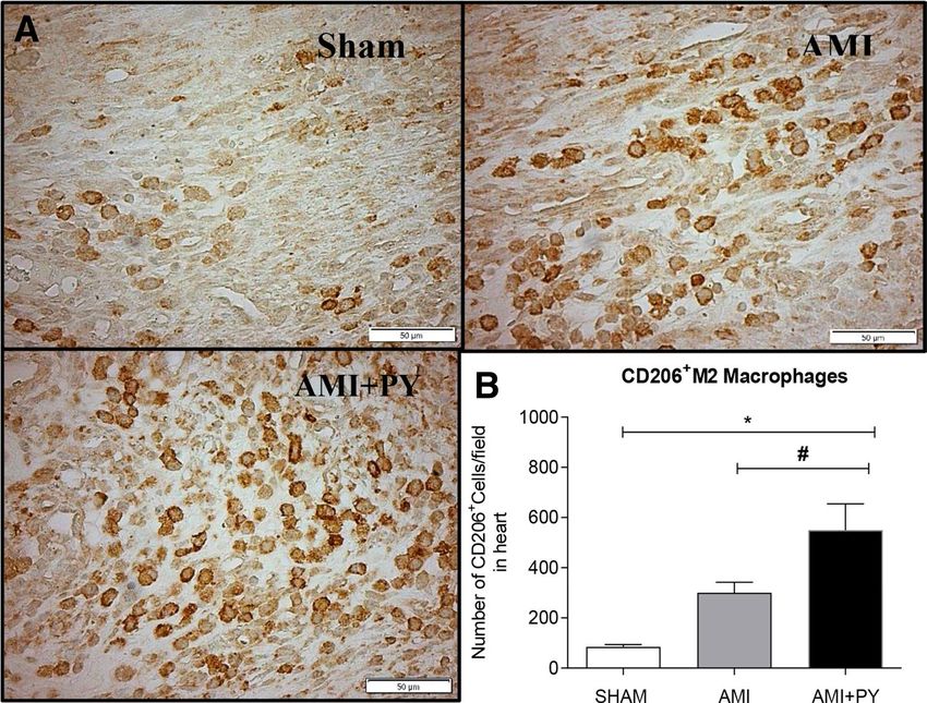
www.nature.com/scientificreports/
Sham (n = 5) AMI (n = 5) AMI + PY (n = 5)
SBP (mmHg) 204 ± 8.2 174 ± 21.6§ 167 ± 13.5*
DBP (mmHg) 144 ± 8.4 126 ± 14.8§ 118 ± 10.9*
MBP (mmHg) 172 ± 8.1 149 ± 17.8§ 142 ± 11.6*
HR (bpm) 376 ± 28.1 392 ± 27.9 369 ± 39.6*
Table 1. Hemodynamic parameters in all three groups. Values expressed as mean ± SEM. §AMI versus
Sham; *AMI + PY versus Sham. Values expressed as mean ± standard deviation. Sham: Control Group; AMI:
Untreated Infarcted Group; AMI + PY: Infarcted Group Treated with Pyridostigmine SBP: Systolic Blood
Pressure; DBP; Diastolic Blood Pressure, MBP = Mean Blood Pressure, HR: Heart rate. *p < 0,05; §p < 0,05.
Statistical significance was determined by one-way ANOVA followed by a multiple comparisons test Tukey’s
was performed using GraphPad Prism version 9.0.1. www.graphpad.com.
autonomic nervous system, bone marrow, and spleen to modulate the immune response from the atherosclerotic
plaque to infarcted m yocardium13.
Recently, autonomic neural regulation of the immune system has attracted investigators’ attention as a novel
therapeutic strategy to control inflammation in multiple clinical settings, including myocardial ischemia–rep-
erfusion injury14 and M I15, 16. The main nerve of the parasympathetic part of the autonomic nervous system—
the vagus nerve modulates the immune function by regulating innate and acquired immune cell-mediated
responses17, 18. Electrical vagus nerve stimulation suppresses aberrant systemic inflammation and decreases serum
TNF levels by inhibiting its production in the spleen19. The vagus nerve interacts with the splenic nerve that
releases norepinephrine in the spleen20, 21. Norepinephrine activates β2-adrenoceptors on splenic T lymphocytes
that contain the enzyme choline acetyltransferase (ChAT), which synthesizes a cetylcholine22–24. Vagus nerve
stimulation causes an increase in splenic acetylcholine levels. In turn, lymphocyte-derived acetylcholine binds to
the α7-nicotinic acetylcholine receptors (α7nAChR) on splenic macrophages to inhibit TNF p roduction19, 24, 25.
On the other hand, sympathetic nerve hyperactivity in the spleen has been linked to chronic immune-mediated
inflammatory diseases26. Moreover, central nervous system angiotensin II infusion and hyperthermia are associ-
ated with enhanced level of efferent splenic sympathetic nerve discharge and splenic pro-inflammatory cytokine
gene expression in r ats27. These findings show the potential of the autonomic nervous system to regulate immune
responses and inflammation; in this regulation the vagus nerve plays a specific role in orchestrating cellular and
cytokine immune responses directed towards promoting tissue healing.
Pyridostigmine is a cholinesterase inhibitor and a cholinergic drug used to treat myasthenia gravis, a chronic
autoimmune, neuromuscular disease that causes muscle weakness28. Pyridostigmine prevents acetylcholine
hydrolysis and enhances parasympathetic modulation in normotensive rats29. We have previously reported that
pyridostigmine treatment prevents deleterious inflammation and oxidative stress in the ischemic myocardium
of these r ats30. Pyridostigmine treatment also increased the proportion of T regulatory cells in peripheral cir-
culation, and decreased activated CD8+ lymphocytes in the spleen. These results suggest that pyridostigmine
modulates the immune cell response by regulating splenic l ymphocytes31. However, the effects of pyridostigmine
in infarcted spontaneous hypertensive rats (SHRs) that exhibit autonomic dysfunction and chronic inflamma-
tion and may represent an animal model that better mimics the clinical profile of patients with AMI remained
unknown. In the present study, we investigated whether pyridostigmine affects splenic lymphocytes and pro-
inflammatory cytokines, and its potential to modulate inflammatory, structural, and functional responses after
myocardial infarction in SHRs.
Results
Pyridostigmine treatment enhances hemodynamic and autonomic parameters. First, we
analyzed the hemodynamic variables as presented in Table 1. Sham animals had very high blood pressure
(204/144 mmHg) characteristic of spontaneous hypertensive SHRs. MI significantly lowered (p < 0.05) systolic
(SBP), diastolic (DBP), and mean (MBP) blood pressure as compared to sham animals (Fig. 1). These results
indicate decreased cardiac output potentially related to reduced left ventricular contraction caused by ischemic
injury. Pyridostigmine treatment affected neither of these hemodynamic parameters. Moreover, all groups had
similar heart rate (p > 0.05). We also analyzed the autonomic tone via heart rate variability (HRV) and baroreflex
sensitivity to determine whether pyridostigmine enhances the parasympathetic modulation (Table 2, Fig. 2).
Root mean square of successive differences (RMSSD) reflects the beat-to-beat variance in heart rate and is the
primary time domain measurement used to estimate vagal mediated changes reflected in HRV. The RMSSD cor-
relates with HF and reflects self-regulatory capacity. Pyridostigmine treatment significantly increased RMSSD
showing its potential to induce cholinergic stimulation (Fig. 2A).
We also performed the spectral analysis of HRV with the three main spectral components distinguished in
short-term recordings: very low frequency (VLF), low frequency (LF), and high frequency (HF). The distribu-
tion of the power and the central frequency of LF and HF are not fixed but may vary in relation to changes
in autonomic modulations of the heart period. We analyzed LF (Fig. 2B) and HF in both absolute ( ms2) and
normalized units (n.u), representing sympathetic and parasympathetic modulation, respectively. Normalization
tends to minimize the effects of LF and HF in total power and emphasizes the balanced behavior (LF/HF) of
the two branches of the autonomic nervous system15, 24, 25). These results showed that AMI decreased HF (nu)
(Fig. 2C), and thereby significantly increased the LF/HF ratio (Fig. 2D) consistent with enhanced sympathetic
Scientific Reports | (2021) 11:9563 | https://doi.org/10.1038/s41598-021-89104-8 2
Vol:.(1234567890)
www.nature.com/scientificreports/
Figure 1. Hemodynamic parameters. (A) SBP; Systolic Blood Pressure. (B) DBP; Diastolic Blood Pressure, and
(C) MBP; Mean Blood Pressure; HR: Heart rate. *p < 0.05 versus Sham; §p < 0.05 versus AMI. Values expressed
as mean ± SEM. Statistical significance was determined by one-way ANOVA followed by a multiple comparisons
test Tukey’s was performed using GraphPad Prism version 9.0.1. www.graphpad.com.
Sham (n = 5) AMI (n = 6) AMI + PY (n = 5)
HRV
RMSSD (ms) 6.2 ± 2.0 5.8 ± 2.2 9.3 ± 2.0 *#
2
PIVAR (ms ) 28.5 ± 13.0 15.8 ± 6.0 63 ± 28.1 *#
LF (nu) 2.6 ± 2.0 2.5 ± 1.0 7.8 ± 4.5 *#
HF (nu) 9.7 ± 3.6 6.0 ± 3.0 23.9 ± 8.7 *#
§
LF/HF 0.3 ± 0.1 0.5 ± 0.2 0.4 ± 0.2
ALPHA-INDEX (ms/mmHg) 0.5 ± 0.3 0.6 ± 0.2 1.1 ± 0.5 *#
Table 2. Heart rate variability (HRV) and Baroreflex Sensitivity (alpha-index) in all groups. Values expressed
as mean ± SEM. §AMI vs Sham; *AMI + PY vs Sham and #AMI + PY vs AMI. HRV: heart rate variability;
RMSSD: square root of the mean of the sum of squares of the differences between successive pulse interval
values; PIVAR: pulse interval variance; LF: Low Frequency nu: Normalized units; HF nu: high frequency
in normalized units; LF/HF: autonomic balance. *P < 0.05; #P < 0.05; §P < 0.05. Statistical significance was
determined by one-way ANOVA followed by a multiple comparisons test Tukey’s was performed using
GraphPad Prism version 9.0.1. www.graphpad.com.
modulation. Conversely, pyridostigmine treatment significantly increased HF (nu) (Fig. 2C), and decreased LF/
HF ratio (Fig. 2D). These results concur in showing pyridostigmine potential to enhance the parasympathetic
vagal tone. Alterations of the baroreceptor-heart rate reflex contribute to reduced parasympathetic activity and
increased sympathetic activity during cardiovascular diseases, including AMI. This baroreflex sensitivity meas-
ured by spectral methods assesses the relationship (in terms of gain) between specific oscillatory components
of the two signals. Spontaneous oscillations in blood pressure elicit an oscillation at the same frequency in RR
interval by the arterial baroreflex activity. BRS is computed as the average value of the transfer function modulus
(i.e., the gain) between systolic pressure and RR interval in the frequency range 0.07–0.14 Hz. The analysis of
BRS can provide prognostic information in experimental and clinical s tudies32. We observed that pyridostigmine
treatment significantly increased BRS (α-index) indicating an improvement of baroreflex sensitivity (Fig. 2E).
Pyridostigmine treatment improves cardiac morphofunctional analyzes. Next, we performed
structural and functional analyses to determine the effects of pyridostigmine on AMI (Table 3). AMI caused an
infarcted and akinetic area consistent with moderate infarction, and significantly increased systolic and left dias-
tolic ventricle (LV) diameters as compared to sham animals. AMI also lowered LV systolic function as inferred
by LV ejection fraction (LVEF) (Fig. 3C) and fractional area change (LV FAC) (Fig. 3D), but increased E/A
ratio (Fig. 3E) (early filling wave/late atrial contraction wave) that was associated with diastolic dysfunction.
Together, these results indicate substantial cardiac remodeling after AMI. Pyridostigmine treatment significantly
improved structural and functional parameters, but not the infarcted and akinetic area (Table 3). Pyridostigmine
treatment significantly decreased systolic (LDV) and left diastolic ventricle (LSV) diameters (Fig. 3A,B, respec-
tively). Although pyridostigmine treatment did not alter LVEF (Fig. 3C), it significantly increased fractional
area change (LV FAC), which is an index of global ventricular function (Fig. 3D), and decreased the E/A ratio
(Fig. 3E). These results show that pyridostigmine improved systolic and diastolic functions and cardiac remod-
eling after AMI in SHRs.
Pyridostigmine treatment increases T helper and M2 macrophages in the infarcted and
peri‑infarcted zones, and inhibition of excessive release of TNF in the spleen and heart tis‑
Scientific Reports | (2021) 11:9563 | https://doi.org/10.1038/s41598-021-89104-8 3
Vol.:(0123456789)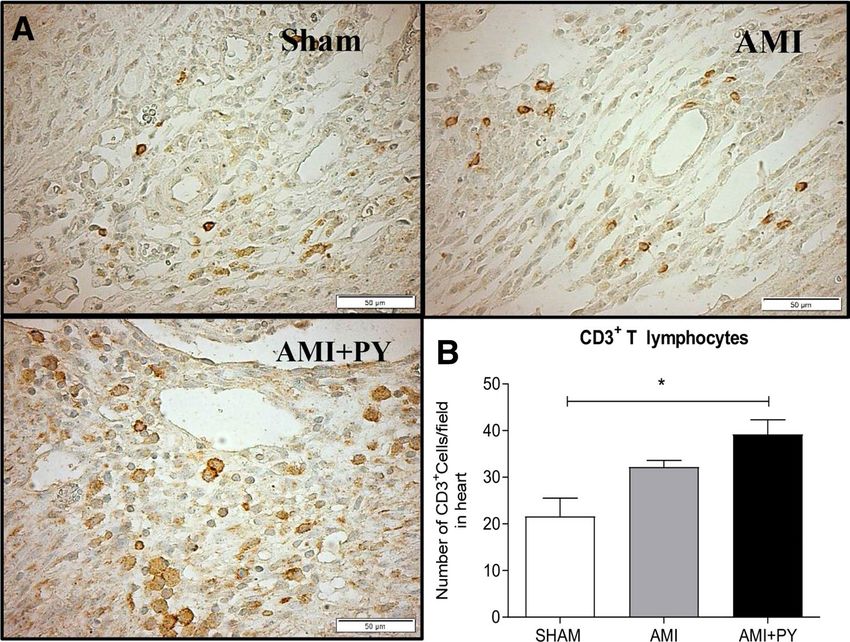
www.nature.com/scientificreports/
Figure 2. Heart rate variability (HRV) and Baroreflex Sensitivity (alpha-index). (A) RMSSD: square root of the
mean of the sum of squares of the differences between successive pulse interval values. (B) LF: Low Frequency
nu: Normalized units. (C) HF nu: high frequency in normalized units and. (D) LF/HF (E) ALPHA-INDEX:
baroreflex sensitivity. *P < 0.05 versus Sham; #P < 0.05 versus AMI and §p < 0.05 versus AMI. Values expressed as
mean ± SEM. Statistical significance was determined by one-way ANOVA followed by a multiple comparisons
test Tukey’s was performed using GraphPad Prism version 9.0.1. www.graphpad.com.
Sham (n = 5) AMI (n = 6) AMI + PY (n = 5)
% infarcted area 0.366 ± 0.03 0.350 ± 0.41
Hipo or acinetic area – 0.172 ± 0.02 0.180 ± 0.06
AO/AE ratio (mm) 0.9 ± 0.1 0.8 ± 0.1 0.9 ± 0.1
LA Diam. (mm) 4.1 ± 0.9 4.4 ± 1.0 3.9 ± 0.6
LSV Diam. (mm2) 5.6 ± 0.8 7.2 ± 1.3 § 6.0 ± 1.2 #
LDV Diam. (mm2) 7.6 ± 0.6 8.6 ± 0.9 § 7.4 ± 1.0 #
LV Mass (g/Kg) 476.9 ± 70.3 524.9 ± 143.5 475.3 ± 171.9
LVEF (%) 49.0 ± 9.8 32.1 ± 17.2 § 36.5 ± 11.1 *
LV FAC (%) 43.8 ± 8.6 27.2 ± 9.0 § 35.9 ± 9.8 #
§
E/A Ratio 1.5 ± 0.4 2.3 ± 1.0 1.6 ± 0.3 #
IVRT (ms) 17.8 ± 4.5 18.3 ± 4.6 20.0 ± 5.2
Table 3. EchoDopplercardiographic parameters in all groups. Values expressed as mean ± SEM. §AMI versus
Sham; AMI + PY vs Sham and #AMI + PY versus AMI. LA: left atrial diameter; AO/LA Ratio: aorta/atrial
diameter ratio; LSV Diam. and LDV Diam.: left systolic and diastolic ventricular diameters, respectively; LSV
Vol. and LDV Vol.: left ventricular systole volume and left ventricular diastolic volume, respectively; LV mass:
left ventricular mass; LVEF: left ventricular ejection fraction; LF FAC: global left ventricular systolic function
estimated by the FAC: fractional area change; E: early filling wave; A: wave of late filling; E/A: transmitral
flow/E/A ratio; IVRT: isovolumetric relaxation time. *P < 0.05; #P < 0.05; §P < 0.05. Statistical significance
was determined by one-way ANOVA followed by a multiple comparisons test Tukey’s was performed using
GraphPad Prism version 9.0.1. www.graphpad.com.
Scientific Reports | (2021) 11:9563 | https://doi.org/10.1038/s41598-021-89104-8 4
Vol:.(1234567890)www.nature.com/scientificreports/
Figure 3. EchoDopplercardiographic parameters. (A) LSV Diam. And (B) LDV Diam.: left systolic and
diastolic ventricular diameters, respectively. (C) LVEF: left ventricular ejection fraction (D) LV FAC: global left
ventricular systolic function estimated by the FAC: fractional area change and (E) E/A: transmitral flow/E/A
ratio. *P < 0.05 versus Sham; #P < 0.05 versus AMI; §P < 0.05 Sham versus AMI. Values expressed as mean ± SEM.
Statistical significance was determined by one-way ANOVA followed by a multiple comparisons test Tukey’s was
performed using GraphPad Prism version 9.0.1. www.graphpad.com.
sue. Sham SRH animal have few immune cells in the myocardium. However, AMI induces a significant
infiltration of immune cells into the infarcted and peri-infarcted zones at day seven. Among these cells, AMI
significantly increased CD68+ (M1) macrophage counts, and pyridostigmine significantly decreased these
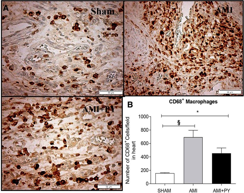
counts (Fig. 4). Likewise, AMI also increased CD206+ (M2) macrophage counts, but pyridostigmine treatment
further increased these cells in the infarcted and peri-infarcted zones (Fig. 5). Therefore, pyridostigmine treat-
ment altered the M1/M2 ratio towards a M2 anti-inflammatory profile. We also detected that sham and AMI
groups have similar counts of T (CD3 +) total lymphocytes. However, pyridostigmine treatment increased the
number of T cells as compared to those in sham group, and these cells were concentrated in the infarcted and
peri-infarcted areas (Fig. 6). Likewise, Sham and AMI animals had similar counts of T helper (CD4 +) lympho-
cytes, and pyridostigmine treatment increased these cells in the infarcted and peri-infarcted areas (Fig. 7). In
agreement with previous studies, we also observed that AMI increased TNFα levels both in heart and spleen,
and PY treatment significantly decreased these levels in both organs (Fig. 8A,B). Thus, AMI failed to affect the
lymphocyte counts, and pyridostigmine significantly increased T helper counts, decreased the M1/M2 ratio, and
decreased TNF levels in the infarcted area but also in the spleen (Fig. 8).
Discussion
The main new findings of this study are that pyridostigmine treatment increases parasympathetic modulation and
baroreflex sensitivity, promotes anti-inflammatory immune cell modulation in the myocardium, and decreases
splenic and cardiac TNF levels after AMI in SHRs. These results also correlate with the potential of pyridostig-
mine to improve cardiac morphofunctional remodeling after AMI in SHRs. Thus, cholinergic stimulation using
pyridostigmine induces cardiac protection after ischemic injury in SHRs.
The bidirectional interplay between the nervous and immune systems has gained great clinical attention to
design novel therapeutic strategies for AMI17, 33. The inflammatory reflex and its efferent arm—the cholinergic
anti-inflammatory pathway have been successfully explored in therapeutic approaches for sepsis, arthritis and
other inflammatory c onditions17, 18. Activation of cholinergic signaling in the inflammatory reflex using cho-
linesterase inhibitors, including the cholinergic drug galantamine suppresses inflammation in preclinical and
clinical settings of numerous disorders characterized by immune and metabolic dysregulation33, 34. This is the
first study investigating the effects of cholinergic stimulation by the cholinesterase inhibitor pyridostigmine on
immune alterations after AMI in SHRs. This is a clinically relevant study, because SHRs have hemodynamic,
neural, and target organ damage reminiscent of that found in essential neurogenic human hypertension. Oka-
moto and Aoki (1963) developed this strain of hypertensive r ats35 that are widely used in the literature to study
natural history, genetic determinants, and pathophysiological changes in arterial hypertension36, 37. SHRs exhibit
immune deficiencies in T and B lymphocytes, decreased T cell proliferative r esponses36, and lower ChAT mRNA
Scientific Reports | (2021) 11:9563 | https://doi.org/10.1038/s41598-021-89104-8 5
Vol.:(0123456789)www.nature.com/scientificreports/
Figure 4. CD68+ cell count (M1 macrophages) (A) Photomicrographs showing the M1 macrophages within
the infarcted zone from the left ventricle (magnification 400×) and (B) bar graphs of C D68+ positive cells in
each group was compared. Fifteen microscopic fields of the infarcted and peri-infarcted zones were analyzed
(5–6 animals per group). *P < 0.05; §P < 0.05 statistically significant. §AMI versus Sham; *AMI + PY vs Sham.
(Immunohistochemistry, diaminobenzidine: scale bar equals 50 µm). Values expressed as mean ± SEM.
Statistical significance was determined by one-way ANOVA followed by a multiple comparisons test Tukey’s was
performed using GraphPad Prism version 9.0.1. www.graphpad.com.
expression in circulating and splenic mononuclear cells as compared to normotensive Wistar rats38. The pos-
sibility that hypertension in SHRs is related to immune anomalies is still under investigation36, 37. Enhanced
innate immune responses and cytokine release from splenocytes have been demonstrated in pre-hypertensive
SHR rats and evoke profound activation of the adaptive immune system contributing to vascular damage and
hypertension37. In addition, SHRs exhibit autonomic dysfunction (increased sympathetic and lower parasym-
pathetic vagal modulation) and chronic inflammation, suggesting that they may represent an animal model that
better mimics the clinical profile of patients with A MI36, 37.
SH animals have a higher sympathovagal ratio; AMI further increased the sympathetic drive and worsened
baroreflex function. Still, pyridostigmine treatment significantly improved the cardiovascular autonomic bal-
ance and baroreflex sensitivity. The baroreflex is a fundamental self-regulatory mechanism of the cardiovascular
system. La R overe32 demonstrated a close correlation between post-MI mortality and baroreflex sensitivity
(BRS). Although the pathological importance of impaired baroreflex function in high post-MI mortality has
already been recognized, no effective long-term pharmacological intervention is a vailable32, which is the direct
consequence of the lack of targeted drugs. These findings suggest a mechanistic basis for restoring baroreflex
sensitivity with pyridostigmine in AMI.
Pyridostigmine is a potent acetylcholinesterase inhibitor and a clinically-approved cholinergic drug for the
treatment of Myasthenia Gravis. Pyridostigmine significantly increases vagal modulation as reported in experi-
mental and human studies. Short-term administration of pyridostigmine increases HRV in healthy h umans39
29
and rats . In patients with heart failure, pyridostigmine also ameliorates the autonomic and hemodynamic
performance during dynamic e xercise40 and reduces ventricular arrhythmia d ensity41. Also, pyridostigmine
enhances cholinergic modulation of the immune cells by preventing vagal-derived acetylcholine d egradation38.
We and others have demonstrated that pyridostigmine treatment started just after coronary artery ligation can
improve autonomic and cardiocirculatory function in normotensive rats analyzed weeks after the A MI15, 16, 42. A
limited number of studies have evaluated the effects of peripheral and centrally-acting cholinesterase inhibitors
on autonomic, hemodynamic, and inflammatory indices in SH a nimals43, 44. Importantly, our findings indicate
that pyridostigmine induces autonomic improvement in infarcted SHRs that is associated with anti-inflammatory
responses in the heart-spleen axis, and cardiac remodeling markers.
Myocardial infarction triggers a sterile inflammatory response characterized by sequential recruitment and
activation of innate and adaptive immune cells to repair the tissue d amage1–3. Macrophages are among the first
Scientific Reports | (2021) 11:9563 | https://doi.org/10.1038/s41598-021-89104-8 6
Vol:.(1234567890)www.nature.com/scientificreports/
Figure 5. CD206+ cell count (M2 macrophages) (A) Photomicrographs showing the M2 macrophages within
the infarcted zone from the left ventricle (magnification 400×) and (B) bar graphs of C D206+ positive cells in
each group was compared. Fifteen microscopic fields of the infarcted and peri-infarcted zones were analyzed
(5–6 animals per group). *P < 0.05; #P < 0.05 statistically significant. *AMI + PY vs Sham; #AMI + PY vs AMI
(Immunohistochemistry, diaminobenzidine: scale bar equals 50 µm). Values expressed as mean ± SEM.
Statistical significance was determined by one-way ANOVA followed by a multiple comparisons test Tukey’s was
performed using GraphPad Prism version 9.0.1. www.graphpad.com.
responders to local damage. Depending on their phenotype, macrophages may be harmful or protective. M1
macrophages express high levels of inflammatory mediators and dominate the first days after AMI, while M2
macrophages express high levels of anti-inflammatory cytokines involved in tissue repair, gradually appear and
remain predominant over five days after the i nfarction1, 3. Pyridostigmine treatment decreased M1 and increased
M2 macrophages, favoring an anti-inflammatory profile with increased T helper (CD4 +) lymphocyte counts
and decreased TNF levels in myocardium and spleen. These results provide new insights into the efficacy of
pyridostigmine in SHRs subjected to MI and substantially extend the information generated by previous studies
using normotensive infarcted a nimals30, 31. As in these previous studies, a better anti-inflammatory, immune cell
profile in the myocardium was associated with decreased levels of pro-inflammatory cytokines.
The sympathetic nervous system enhances macrophage recruitment in the heart after an ischemic event. Sym-
pathetic fibers stimulate the bone marrow to release hematopoietic stem and progenitor cells, as observed in mice
and humans after AMI6, 7, 45. The spleen is also a major source of pro-inflammatory cytokines and immune cells.
Indeed, within the first day after coronary ligation, the spleen releases monocytes from its subcapsular red pulp
into the bloodstream as shown in mice and rats6, 7, 9. As a consequence, bone marrow and splenic leucocyte pro-
duction lead to monocytosis, and these cells are recruited into the ischemic heart8, 9, 13. This increased metabolic
activity of spleen and bone marrow has been shown in patients with acute AMI using 18F-fluorodeoxyglucose
PET image46. Our results show that pyridostigmine decreased sympathovagal balance in infarcted rats, improved
M1/M2 macrophage ratio, and decreased TNF levels in spleen and heart. Thus, cholinergic stimulation may
interfere with local mechanisms preventing macrophage polarization in the ischemic myocardium. The idea of
targeting macrophage and lymphocyte polarization to control inflammation following AMI has been recently
proposed47. In addition to innate cells, lymphocytes also play a fundamental role after AMI10. More specifically,
CD4+ T helper cells are required for proper healing and can attenuate chronic remodeling after AMI. CD4+
T-cells regulate the infiltration of inflammatory monocytes and are required for proper extracellular matrix
formation and angiogenesis during post-MI healing48. Furthermore, CD4+ T cell-derived cytokines modulate
monocyte differentiation and macrophage activity within the m yocardium49.
Our results indicate a link between increased vagal modulation with pyridostigmine and an anti-inflammatory
M1/M2 profile, increased T helper counts, and decreased myocardium TNF levels. Given the detrimental poten-
tial of TNF on the myocardium, TNF local production may play an important role in ventricular dysfunction
and adverse remodeling after i nfarction50. TNF is produced soon after AMI, regulates myocardial apoptosis and
triggers additional cellular inflammatory r esponses50. Anti-TNF treatment was associated with smaller infarct
Scientific Reports | (2021) 11:9563 | https://doi.org/10.1038/s41598-021-89104-8 7
Vol.:(0123456789)www.nature.com/scientificreports/
Figure 6. CD3+ cell count (Total lymphocytes) (A) Photomicrographs showing the Total lymphocytes within
D3+ lymphocytes
the infarcted zone from the left ventricle (magnification 400×) and (B) bar graphs of C
positive cells in each group was compared. Fifteen microscopic fields of the infarcted and peri-infarcted
zones were analyzed (5–6 animals per group). *P < 0.05 statistically significant. *AMI + PY vs Sham.
(Immunohistochemistry, diaminobenzidine: scale bar equals 50 µm). Values expressed as mean ± SEM.
Statistical significance was determined by one-way ANOVA followed by a multiple comparisons test Tukey’s was
performed using GraphPad Prism version 9.0.1. www.graphpad.com.
size and decreased ventricular dysfunction in ischemic-reperfusion and permanent ischemia in animal m odels51
including AMI. Thus, it is plausible that TNF inhibition by pyridostigmine may trigger a positive remodeling
and decrease heart failure in infarcted SHRs.
Targeting cholinergic signaling in the inflammatory reflex provides novel therapeutic opportuni-
ties for diseases characterized with inflammatory and cardiovascular derangements, including myocardial
ischemia–reperfusion injury and AMI damage17. The vagus nerve can act reflexively on the spleen, reducing
systemic inflammation18. Likewise, efferent vagal signals may facilitate the release of lymphocytes from the thy-
mus through nicotinic receptors52. Vagal stimulation can increase acetylcholine release, which activates α7nAChR
on macrophages to decrease pro-inflammatory cytokines in the s pleen19,22,24. A main source of acetylcholine
in the spleen is a subgroup of T lymphocytes expressing β2-adrenoreceptors and ChAT (ChAT-T cells) that
synthesize acetylcholine22. Upon vagal activation, the splenic nerve releases norepinephrine (NE) in the spleen
to stimulate ChAT-T cells to release acetylcholine, which in turn inhibits TNF production in macrophages via
α7nAChR-mediated signaling15, 20, 21, 23.
As both acetylcholine and NE modulate splenic immune cell functions, our study highlights the need to
consider the balance between these two mediators, which probably determines the final immunoregulatory
outcome53. We show the potential of pyridostigmine to reduce cardiac inflammation and improve heart remod-
eling after AMI. As expected, AMI caused significant morphofunctional alterations, detected by echocardiog-
raphy at day 7, which affects circulation and reduces blood pressure. Pyridostigmine treatment significantly
improves systolic and diastolic function, but they were insufficient to restore normal blood pressure as seen in
sham animals. Noninvasive diastolic function measurements with echocardiography correlated with LV cardiac
measurements as determined by catheterization in rat models of myocardial hypertrophy and AMI22; these
noninvasive parameters may predict further ventricular dysfunction5. Ventricular dysfunction and heart failure
are frequent complications of AMI and are associated with poor prognosis. Some studies have shown the ben-
eficial effects of more prolonged pyridostigmine treatment on cardiac function and remodeling in normotensive
infarcted rats15, 54, 55. It can be speculated that this immediate improvement may lead to positive remodeling and
decrease the development of heart failure in infarcted SHRs.
Our study has some limitations. As mentioned above, the specific role of ChAT-positive T cells and possibly
B cells in the effects observed in the spleen is unknown and remains to be evaluated in future studies. In addi-
tion, there is a lack of specific insight into the role of ChAT-containing immune cells in the heart and cardiac
myocytes in mediating local immunomodulatory effects in our model. On a related note, we cannot assess
Scientific Reports | (2021) 11:9563 | https://doi.org/10.1038/s41598-021-89104-8 8
Vol:.(1234567890)www.nature.com/scientificreports/
Figure 7. CD4+ cell count (Lymphocytes T helper) (A). Photomicrographs showing the Lymphocytes T helper
within the infarcted zone from the left ventricle (magnification 400×) and (B) bar graphs of C D4+ positive cells
in each group was compared. Fifteen microscopic fields of the infarcted and peri-infarcted zones were analyzed
(5–6 animals per group). *P < 0,05; #P < 0.05 statistically significant. *AMI + PY versus Sham; #AMI + PY versus
AMI. (Immunohistochemistry, diaminobenzidine: scale bar equals 50 µm). Values expressed as mean ± SEM.
Statistical significance was determined by one-way ANOVA followed by a multiple comparisons test Tukey’s was
performed using GraphPad Prism version 9.0.1. www.graphpad.com.
Figure 8. TNF levels in the heart (A) and the spleen (B) in all groups. #p < 0.05; §P < 0.05 statistically significant.
Values expressed as mean ± SEM. One-way ANOVA. §AMI versus Sham; #AMI + PY versus AMI. Values
expressed as mean ± SEM. Statistical significance was determined by one-way ANOVA followed by a multiple
comparisons test Tukey’s was performed using GraphPad Prism version 9.0.1. www.graphpad.com.
the relative contribution of neural acetylcholine versus non-neuronal acetylcholine in the beneficial immune
and cardiometabolic pyridostigmine effects on the heart. Previously, vagus nerve stimulation has been shown
to suppress cardiac (myocardial) and systemic TNF levels in rodents during e ndotoxemia18, 19. Vagus nerve
stimulation in animals and patients with heart failure also results in improved cardiac function, indicating the
cardioprotective efficacy of vagus nerve cholinergic signaling56–58 Previous studies have indicated that cardiac
myocytes also contain ChAT and synthesize and secrete acetylcholine, which plays a role in myocardium regu-
latory functions59, 60. Acetylcholine released from cardiac myocytes reportedly has various protective effects in
Scientific Reports | (2021) 11:9563 | https://doi.org/10.1038/s41598-021-89104-8 9
Vol.:(0123456789)www.nature.com/scientificreports/
pathological scenarios, including cardiac hypertrophy and failure as indicated in transgenic mouse models61, 62.
Further studies are necessary to evaluate the specific role of non-neuronal cell-derived acetylcholine in addi-
tion to the neural vagus nerve-derived cholinergic output in mediating pyridostigmine effects in the heart. In
addition, further work using cell-type-specific transcriptomic and epigenomic analysis is necessary to reveal the
underlying molecular mechanisms of how pyridostigmine induces the M1-M2 cell type conversion.
In conclusion, cholinergic stimulation with pyridostigmine modulates a heart-spleen axis in the immune
response after myocardial infarction and improves heart remodeling. Our results warrant future studies to
determine mechanisms of these effects and the consequences in heart failure.
Materials and methods
Experimental design. All animal procedures were approved by the Committee on the Ethics of Animal
Experimentation at the University of Nove de Julho (CEUA protocol No. 7612011118). Animal care was per-
formed following the Guide for the Care and Use of Laboratory Animals published by the U.S. National Institutes
of Health. Adult male SHR (2–3 months old, 200–250 g) were housed in collective plastic cages (four animals
per cage) with controlled temperature (23 °C), a 12:12-h light–dark cycle with rat chow provided ad libitum and
unlimited access to water. Animals were randomly assigned to one of three groups, with 10–12 animals in each
group: sham rats (Sham), untreated infarcted rats (AMI), and pyridostigmine-treated infarcted rats (AMI + PY).
All animals were monitored for 7 days. The AMI + PY Group received pyridostigmine bromide (Sigma-Aldrich,
St Louis, MO), as described previously (40 mg/kg once a day, by gavage) started one h after surgery and contin-
ued for seven days after this procedure8, 63. According to a prior study, the dose and period of pyridostigmine
administration chose were appropriate to inhibit approximately 40% of plasma acetylcholinesterase activity29.
All methods were reported in accordance with ARRIVE guidelines.
Myocardial infarction. Rats in the AMI and AMI + PY groups were anesthetized (80 mg/kg ketamine and
12 mg/kg xylazine intraperitoneal injected, I.P.) and underwent induction of AMI by surgical occlusion of the
escribed30, 31. A left thoracotomy performed by dissecting the third intercostal
left coronary artery, as previously d
space and exposing the heart. Then, the left coronary artery was occluded with a single nylon (6.0 mm) suture
1 mm distal to the left atrial appendage. The chest was then sutured. The rats were maintained under ventilation
until recovery. The sham group underwent the same procedure, but AMI was not induced. Infarcted rats were
randomly allocated to receive or not P.Y. The analytical investigators were blind to the treatment.
Arterial catheterization, hemodynamic measurements, and cardiovascular variability analy‑
sis. At day six, rats were anesthetized (80 mg/kg ketamine and 12 mg/kg xylazine, I.P.), and a catheter filled
with 0.06 mL of saline solution was implanted into the femoral a rtery29 for hemodynamic measurements. The
arterial cannula was connected to a strain gauge transducer (Blood Pressure XDCR; Kent Scientific, Torrington,
CT), and arterial pressure (A.P.) signals and pulse interval heart rate (H.R.) were digitally recorded over a 30-min
period in conscious, awake animals using a data acquisition system (WinDaq, 2 kHz; DATAQ, Springfield, OH),
as described29. This basal acquisition was used to evaluate heart rate variability (HRV) and systolic arterial pres-
sure variability (as described below). HRV. For time and frequency domains analysis of cardiovascular auto-
nomic modulation, the time series (three time series of 5 Min for each animal) of pulse interval (PI) and systolic
arterial pressure (SAP) were cubic spline-interpolated (250 Hz) and cubic spline-decimated to be equally spaced
in time after linear trend removal; power spectral density was obtained through the Fast Fourier transformation.
Spectral power for low frequency (LF, 0.20–0.75 Hz) and high frequency (HF, 0.75–4.0 Hz) bands was calculated
by power spectrum density integration within each frequency bandwidth, using a customized routine (Cardi-
oseries). The time domain variables were: root mean square of the successive differences (RMSSD) and total
variance of pulse interval (VAR-PI) for pulse interval (PI); and total variance of systolic arterial pressure (VAR-
SAP) for systolic arterial pressure (SAP). The α-index in the low-frequency band was calculated only when the
magnitude of the squared coherence between the PI and SAP signals exceeded 0.5 (range, 0–1). After coherence
calculation, the α-index was obtained from the square root of the ratio between PI and SAP variability in the two
major low frequency (LF) band29.
Echocardiographic evaluation. Echocardiographic evaluations were performed by a blind observer
under the guidelines of the American Society of Echocardiography. Rats were anesthetized (80 mg/kg ketamine
and 12 mg/kg xylazine intraperitoneal-I.P.), and images were obtained with a 10–14-MHz linear transducer in
a G.E. Vivid 7 Ultra-Definition Clarity Control (G.E. Healthcare, USA). This procedure was performed six days
after AMI or sham surgeries in order to analyze AMI area (hipo or acinetic ventricular areas) and LV ejection
fraction (LVEF%), and to calculate the following parameters: left atrial diameter, ventricular mass (LV mass); left
ventricular end-diameter during systole and diastole (LVSD, LVDD); E wave A wave ratio (E/A); isovolumetric
relaxation time (IVRT); fractional area change (FAC), as described in detail elsewhere as described p reviously15,
63
. Through midtransversal and apical transversal views, AMI size was measured by bi-dimensional echocar-
diogram. In diastole, three measurements of the endocardial perimeter (E.P.) and the length of the infarcted
segment (ISe) were obtained for each view. The AMI size for each ISi was calculated by the equation ISi (%) = ISe/
E.P. × 100. The total infarct size of each animal was calculated as the mean of ISi (%) of the three segments. AMI
was defined as increased echogenicity and change in myocardial systolic movement (hypokinesia, akinesia, or
dyskinesia), following Santos et al.28. Our group15 and o thers63 have demonstrated strong correlations between
the AMI area assessed by echocardiogram and postmortem histological analysis, showing that this is a valid
method to estimate AMI area in rats.
Scientific Reports | (2021) 11:9563 | https://doi.org/10.1038/s41598-021-89104-8 10
Vol:.(1234567890)www.nature.com/scientificreports/
Immunohistochemistry for immune cells. At day seven, 6–7 animals from each group were anesthe-
tized (80 mg/kg ketamine and 12 mg/kg xylazine, I.P.) and perfused with 0.9% NaCl plus 14 mmol/l KCl solution
(IV; with a pressure equal to 13 c mH2O) to arrest the heart in diastole, followed by perfusion of 4% buffered for-
malin for tissue fixation. Harvested hearts were immersed in formalin for 24 h. Transverse slices were processed
and embedded in paraffin. Serial sections of paraffin-embedded tissues (3 μm) were placed on glass slides coated
with 2% 3-aminopropyl-triethyl silane (Sigma-Aldrich) and deparaffinized in xylene, then immersed in alcohol
and incubated with 3% hydrogen peroxide. The sections were immersed in a citrate buffer (pH 6.0; Sigma-
Aldrich) at 95 °C for 20 min for antigen retrieval. Nonspecific signals were blocked using specific antibody
diluents (Antibody Diluent, cat. no. S0809; Dako, Glostrup, Denmark)30. The slides were then incubated with
the following primary antibodies: anti-rat CD4 (1:500, T Lymphocyte marker, rabbit monoclonal—EPR19514,
cat. no. 221775; Abcam, Cambridge, UK), anti-rat CD3 (1:100, T helper Lymphocyte marker, rabbit monoclo-
nal—SP7; cat 16669, Abcam), anti-rat CD68 (1:100, M1 macrophage marker, mouse monoclonal—ED-1 cat. no.
31630, Abcam), and anti-rat CD206 (1:800, M2 macrophage marker cat. no. 64693; Abcam). The samples were
kept overnight at 4 °C in a humidified chamber. The sections were incubated with LSAB + System-HRP reagents
for 30 min (K0690; Dako Co, Denmark). Finally, the sections were incubated in 3,3-diminobenzidine in a chro-
mogen solution (K346811; Dako Co, Denmark) at room temperature for 2–5 min, and then stained with Mayer’s
hematoxylin (Sigma-Aldrich) and covered. For the negative controls, the primary antibodies were replaced with
1% PBS/BSA and nonimmune mouse serum (X501-1, Dako).
Cell counts in the infarcted and peri‑infarcted zones. Fifteen consecutive microscope fields (magni-
fication: 400×) of the infarcted and peri-infarcted zones were photographed (fluorescence Microscope (Olympus
AX70) with a digital camera (Olympus Japan Co, Tokyo, Japan). An investigator blinded to the animal group
samples analyzed the images and manually counted them with the aid of the Image J version 1.48v 17 (free soft-
ware, NIH, Bethesda, Maryland, EUA), using the "cell counter" plug-in31.
Cytokine measurements. A set of 5–6 animals in each group was euthanized by decapitation on day
seven after thoracotomy to collect fresh heart and spleen for TNF-α analyses. Measurement of the TNFα was
performed in samples of the LV and spleen by ELISA using Duo-set available kits for TNFα (BD Pharmingen,
San Jose, CA, USA) as previously describe30. The sensitivity of the assays was 15 pg/mL. The results were normal-
ized by LV or spleen total protein30.
Statistical analysis. All data were represented as means ± the standard error of the mean (SEM). For para-
metric data, the one-way analysis of variance (one-way ANOVA) was performed with Turkey’s multiple com-
parison tests was performed using GraphPad Prism version 9.0.1 for Windows, GraphPad Software, San Diego,
California USA, www.graphpad.com. For nonparametric data, the Kruskal–Wallis test was used. P values less
than 0.05 were considered significant.
Accordance statement
All methods were reported in accordance with ARRIVE guidelines.
Received: 1 September 2020; Accepted: 15 April 2021
References
1. Frangogiannis, N. G. The immune system and cardiac repair. Pharmacol. Res. 58, 88–111 (2008).
2. Kain, V., Prabhu, S. D. & Halade, G. V. Inflammation revisited: inflammation versus resolution of inflammation following myo-
cardial infarction. Basic Res. Cardiol. 109, 444 (2014).
3. Yan, X. et al. Temporal dynamics of cardiac immune cell accumulation following acute myocardial infarction. J. Mol. Cell Cardiol.
62, 24–35 (2013).
4. Anzai, T. Post-infarction inflammation and left ventricular remodeling: a double-edged sword. Circ. J. 77, 580–587 (2013).
5. Alp, E. et al. Incidence, risk factors and mortality of nosocomial pneumonia in intensive care units: a prospective study. Ann. Clin.
Microbiol. Antimicrob. 3, 17 (2004).
6. Ulloa, L. & Deitch, E. A. Neuroimmune perspectives in sepsis. Crit. Care 13, 133 (2009).
7. Swirski, F. K. et al. Identification of splenic reservoir monocytes and their deployment to inflammatory sites. Science 325, 612–616
(2009).
8. Dutta, P. et al. Myocardial infarction accelerates atherosclerosis. Nature 487, 325–329 (2012).
9. Hoyer, F. F. et al. Tissue-specific macrophage responses to remote injury impact the outcome of subsequent local immune chal-
lenge. Immunity 51(899–914), e897 (2019).
10. Hofmann, U. & Frantz, S. Role of T-cells in myocardial infarction. Eur Heart J. 37, 873–879 (2016).
11. Li, J., Tan, J., Martino, M. M. & Lui, K. O. Regulatory T-cells: potential regulator of tissue repair and regeneration. Front. Immunol.
9, 585 (2018).
12. Santos-Zas, I., Lemarie, J., Tedgui, A. & Ait-Oufella, H. Adaptive immune responses contribute to post-ischemic cardiac remod-
eling. Front. Cardiovasc. Med. 5, 198 (2018).
13. Libby, P., Nahrendorf, M. & Swirski, F. K. Leukocytes link local and systemic inflammation in ischemic cardiovascular disease: an
expanded “cardiovascular continuum”. J. Am. Coll. Cardiol. 67, 1091–1103 (2016).
14. Calvillo, L. et al. Vagal stimulation, through its nicotinic action, limits infarct size and the inflammatory response to myocardial
ischemia and reperfusion. J. Cardiovasc. Pharmacol. 58, 500–507 (2011).
15. de La Fuente, R. N. et al. Cholinergic stimulation with pyridostigmine improves autonomic function in infarcted rats. Clin. Exp.
Pharmacol. Physiol. 40, 610–616 (2013).
16. Durand, M. T. et al. Pyridostigmine restores cardiac autonomic balance after small myocardial infarction in mice. PLoS ONE 9,
e10447 (2014).
Scientific Reports | (2021) 11:9563 | https://doi.org/10.1038/s41598-021-89104-8 11
Vol.:(0123456789)www.nature.com/scientificreports/
17. Pavlov, V. A., Chavan, S. S. & Tracey, K. J. Molecular and functional neuroscience in immunity. Annu. Rev. Immunol. 36, 783–812
(2018).
18. Olofsson, P. S., Rosas-Ballina, M., Levine, Y. A. & Tracey, K. J. Rethinking inflammation: neural circuits in the regulation of
immunity. Immunol. Rev. 248, 188–204 (2012).
19. Borovikova, L. V. et al. Vagus nerve stimulation attenuates the systemic inflammatory response to endotoxin. Nature 405, 458–462
(2000).
20. Besedovsky, H. O. & del Rey, A. Immune-neuro-endocrine interactions: facts and hypotheses. Endocr. Rev. 17, 64–102 (1996).
21. Bratton, B. O. et al. Neural regulation of inflammation: no neural connection from the vagus to splenic sympathetic neurons. Exp.
Physiol. 97, 1180–1185 (2012).
22. Rosas-Ballina, M. et al. Acetylcholine-synthesizing T cells relay neural signals in a vagus nerve circuit. Science 334, 98–101 (2011).
23. Vida, G. et al. beta2-Adrenoreceptors of regulatory lymphocytes are essential for vagal neuromodulation of the innate immune
system. FASEB J. 25, 4476–4485 (2011).
24. Olofsson, P. S. et al. alpha7 nicotinic acetylcholine receptor (alpha7nAChR) expression in bone marrow-derived non-T cells is
required for the inflammatory reflex. Mol. Med. 18, 539–543 (2012).
25. Fujii, T. et al. Physiological functions of the cholinergic system in immune cells. J. Pharmacol. Sci. 134, 1–21 (2017).
26. Bellinger, D. L., Lorton, D., Hamill, R. W., Felten, S. Y. & Felten, D. L. Acetylcholinesterase staining and choline acetyltransferase
activity in the young adult rat spleen: lack of evidence for cholinergic innervation. Brain Behav. Immun. 7, 191–204 (1993).
27. Ganta, C. K. et al. Central angiotensin II-enhanced splenic cytokine gene expression is mediated by the sympathetic nervous
system. Am. J. Physiol. Heart Circ. Physiol. 289, H1683-1691 (2005).
28. Breyer-Pfaff, U., Maier, U., Brinkmann, A. M. & Schumm, F. Pyridostigmine kinetics in healthy subjects and patients with myas-
thenia gravis. Clin. Pharmacol. Ther. 37, 495–501 (1985).
29. Soares, P. P., da Nobrega, A. C., Ushizima, M. R. & Irigoyen, M. C. Cholinergic stimulation with pyridostigmine increases heart
rate variability and baroreflex sensitivity in rats. Auton. Neurosci. 113, 24–31 (2004).
30. Bezerra, O. C. et al. Cholinergic stimulation improves oxidative stress and inflammation in experimental myocardial infarction.
Sci. Rep. 7, 13687 (2017).
31. Rocha, J. A. et al. Increase in cholinergic modulation with pyridostigmine induces anti-inflammatory cell recruitment soon after
acute myocardial infarction in rats. Am. J. Physiol. Regul. Integr. Comp. Physiol. 310, R697-706 (2016).
32. La Rovere, M. T., Pinna, G. D. & Raczak, G. Baroreflex sensitivity: measurement and clinical implications. Ann. Noninvasive
Electrocardiol. 13, 191–207 (2008).
33. Pavlov, V. A. & Tracey, K. J. Neural circuitry and immunity. Immunol. Res. 63, 38–57 (2015).
34. Metz, C.N. & Pavlov, V.A. Treating disorders across the lifespan by modulating cholinergic signaling with galantamine. J. Neuro-
chem. (2020). pp 1– 22
35. Okamoto, K. & Aoki, K. Development of a strain of spontaneously hypertensive rats. Jpn. Circ. J. 27, 282–293 (1963).
36. Rodriguez-Iturbe, B., Pons, H. & Johnson, R. J. Role of the immune system in hypertension. Physiol. Rev. 97, 1127–1164 (2017).
37. Harwani, S. C., Chapleau, M. W., Legge, K. L., Ballas, Z. K. & Abboud, F. M. Neurohormonal modulation of the innate immune
system is proinflammatory in the prehypertensive spontaneously hypertensive rat, a genetic model of essential hypertension. Circ.
Res. 111, 1190–1197 (2012).
38. Fujimoto, K., Matsui, M., Fujii, T. & Kawashima, K. Decreased acetylcholine content and choline acetyltransferase mRNA expres-
sion in circulating mononuclear leukocytes and lymphoid organs of the spontaneously hypertensive rat. Life Sci. 69, 1649–1638
(2001).
39. Nobrega, A. C. et al. Enhancement of heart rate variability by cholinergic stimulation with pyridostigmine in healthy subjects.
Clin. Auton. Res. 11, 11–17 (2001).
40. Androne, A. S., Hryniewicz, K., Goldsmith, R., Arwady, A. & Katz, S. D. Acetylcholinesterase inhibition with pyridostigmine
improves heart rate recovery after maximal exercise in patients with chronic heart failure. Heart 89, 854–858 (2003).
41. Behling, A. et al. Cholinergic stimulation with pyridostigmine reduces ventricular arrhythmia and enhances heart rate variability
in heart failure. Am. Heart J. 146, 494–550 (2013).
42. Santos-Almeida, F. M., Girao, H., da Silva, C. A., Salgado, H. C. & Fazan, R. Jr. Cholinergic stimulation with pyridostigmine
protects myocardial infarcted rats against ischemic-induced arrhythmias and preserves connexin43 protein. Am. J. Physiol. Heart
Circ. Physiol. 308, H101-107 (2015).
43. Lataro, R. M., Silva, C. A., Tefe-Silva, C., Prado, C. M. & Salgado, H. C. Acetylcholinesterase inhibition attenuates the development
of hypertension and inflammation in spontaneously hypertensive rats. Am. J. Hypertens. 28, 1201–2120 (2015).
44. Lataro, R. M. et al. Chronic treatment with acetylcholinesterase inhibitors attenuates vascular dysfunction in spontaneously
hypertensive rats. Am. J. Hypertens. 32, 579–587 (2019).
45. Katayama, Y. et al. Signals from the sympathetic nervous system regulate hematopoietic stem cell egress from bone marrow. Cell
124, 407–421 (2006).
46. Kim, E. J., Kim, S., Kang, D. O. & Seo, H. S. Metabolic activity of the spleen and bone marrow in patients with acute myocardial
infarction evaluated by 18f-fluorodeoxyglucose positron emission tomograpic imaging. Circ. Cardiovasc. Imaging 7, 454–460
(2014).
47. Harel-Adar, T. et al. Modulation of cardiac macrophages by phosphatidylserine-presenting liposomes improves infarct repair. Proc.
Natl. Acad. Sci. U. S. A. 108, 1827–1832 (2011).
48. Tang, T. T. et al. Regulatory T cells ameliorate cardiac remodeling after myocardial infarction. Basic Res. Cardiol. 107, 232 (2012).
49. Weirather, J. et al. Foxp3+ CD4+ T cells improve healing after myocardial infarction by modulating monocyte/macrophage dif-
ferentiation. Circ. Res. 115, 55–67 (2014).
50. Nian, M., Lee, P., Khaper, N. & Liu, P. Inflammatory cytokines and postmyocardial infarction remodeling. Circ. Res. 94, 1543–1553
(2004).
51. Hartman, M. H. T., Groot, H. E., Leach, I. M., Karper, J. C. & van der Harst, P. Translational overview of cytokine inhibition in
acute myocardial infarction and chronic heart failure. Trends Cardiovasc. Med. 28, 369–379 (2018).
52. Antonica, A., Magni, F., Mearini, L. & Paolocci, N. Vagal control of lymphocyte release from rat thymus. J. Auton. Nerv. Syst. 48,
187–197 (1994).
53. Ratcliffe, E. M., deSa, D. J., Dixon, M. F. & Stead, R. H. Choline acetyltransferase (ChAT) immunoreactivity in paraffin sections
of normal and diseased intestines. J. Histochem. Cytochem. 46, 1223–1231 (1998).
54. Lu, Y. et al. Pyridostigmine ameliorates cardiac remodeling induced by myocardial infarction via inhibition of the transforming
growth factor-beta1/TGF-beta1-activated kinase pathway. J. Cardiovasc. Pharmacol. 63, 412–420 (2014).
55. Barboza, C. A. et al. Cholinergic stimulation by pyridostigmine bromide before myocardial infarction prevent cardiac and auto-
nomic dysfunction. Sci. Rep. 9, 2481 (2019).
56. Zhang, Y. et al. Chronic vagus nerve stimulation improves autonomic control and attenuates systemic inflammation and heart
failure progression in a canine high-rate pacing model. Circ. Heart Fail. 2, 692–699 (2009).
57. De Ferrari, G. M. et al. Chronic vagus nerve stimulation: a new and promising therapeutic approach for chronic heart failure. Eur.
Heart J. 32, 847–855 (2011).
58. Schwartz, P. J. & De Ferrari, G. M. Vagal stimulation for heart failure: background and first in-man study. Heart Rhythm 6, S76-81
(2009).
Scientific Reports | (2021) 11:9563 | https://doi.org/10.1038/s41598-021-89104-8 12
Vol:.(1234567890)You can also read