SINGLE MRNP ANALYSIS BY SUPER-RESOLUTION MICROSCOPY AND FLUORESCENCE CORRELATION SPECTROSCOPY REVEALS THAT SMALL MRNP GRANULES REPRESENT MRNA ...
←
→
Page content transcription
If your browser does not render page correctly, please read the page content below
bioRxiv preprint first posted online Feb. 22, 2019; doi: http://dx.doi.org/10.1101/558098. The copyright holder for this preprint
 (which was not peer-reviewed) is the author/funder, who has granted bioRxiv a license to display the preprint in perpetuity.
                              It is made available under a CC-BY-NC-ND 4.0 International license.

                                                                                                                            Mateu-Regué et al.

               Single mRNP analysis by super-resolution microscopy and
                  fluorescence correlation spectroscopy reveals that
                   small mRNP granules represent mRNA singletons

                             Àngels Mateu-Regué1, Jan Christiansen2, Frederik Otzen Bagger1,
                                      Christian Hellriegel3 and Finn Cilius Nielsen*1

                                         1
                                         Center for Genomic Medicine, Rigshospitalet,
                             2
                                 Department of Biology, University of Copenhagen, Denmark and
                                        3
                                          Carl Zeiss Microscopy GmbH, Jena, Germany

 1
   Center for Genomic Medicine, Rigshospitalet, 2100 Copenhagen, Denmark
 2
   Department of Biology, Copenhagen Biocenter, 2200 Copenhagen, Denmark
 3
   Carl Zeiss Microscopy GmbH, 07745 Jena, Germany
 *Correspondence: finn.cilius.nielsen@regionh.dk
                                                                                                                                            1
bioRxiv preprint first posted online Feb. 22, 2019; doi: http://dx.doi.org/10.1101/558098. The copyright holder for this preprint
 (which was not peer-reviewed) is the author/funder, who has granted bioRxiv a license to display the preprint in perpetuity.
                              It is made available under a CC-BY-NC-ND 4.0 International license.

                                                                                                                            Mateu-Regué et al.

 Abstract

 Small cytoplasmic mRNP granules are implicated in mRNA transport, translational control and decay.

 Employing Super-resolution Microscopy and Fluorescence Correlation Spectroscopy, we analyzed the

 molecular composition and dynamics of single cytoplasmic YBX1_IMP1 mRNP granules in live cells. Granules

 appeared elongated and branched with patches of IMP1 and YBX1 distributed along mRNA, reflecting the

 attachment of the two RNA-binding proteins in cis. Particles form at the nuclear pore and are spatially

 segregated from translating ribosomes, so the mRNP is a repository for mRNAs awaiting translation.

 Individual mRNPs contain a single mRNA and 5 to 15 molecules of YBX1 and IMP1, which is in agreement with

 the average number of mRNA-binding sites calculated from CLIP analyses. We conclude that small

 cytoplasmic mRNP granules are mRNA singletons, thus depicting the cellular transcriptome. Consequently,

 expression of functionally related mRNAs in RNA regulons is unlikely to result from coordinated assembly.

 Keywords: RNA granules, mRNP granules, IMP1, YBX1, Singleton, Structured Illumination Microscopy,

 Localization Microscopy, Fluorescence Correlation Spectroscopy, mRNA regulons

                                                                                                                                            2
bioRxiv preprint first posted online Feb. 22, 2019; doi: http://dx.doi.org/10.1101/558098. The copyright holder for this preprint
 (which was not peer-reviewed) is the author/funder, who has granted bioRxiv a license to display the preprint in perpetuity.
                              It is made available under a CC-BY-NC-ND 4.0 International license.

                                                                                                                            Mateu-Regué et al.

 Introduction

 Essential steps from the nuclear mRNA processing and export to cytoplasmic localization, translation and

 decay of mRNAs are implicated in fine tuning of gene expression. Regulatory steps are governed by RNA-

 binding proteins, which interact with the mRNA in a sequential manner. Mammalian cells comprise about

 1500 RNA-binding proteins of which nearly half bind to mRNAs (Gerstberger et al., 2014), and the majority is

 widely distributed with a minority exhibiting a particular spatial and temporal expression. Some mRNA-

 binding proteins shuttle between the nucleus and the cytoplasm, whereas others are mainly present in the

 cytoplasm (Shyu and Wilkinson, 2000), including proteins such as FMRP, HuD as well as YBX1 and the family

 of insulin-like growth factor 2 mRNA binding proteins IMP1, 2 and 3 (Darnell and Richter, 2012). Cytoplasmic

 mRNA-binding proteins are found in small membrane-less granules. In contrast to the larger P-bodies and

 stress granules, which mostly embody stress-induced condensations of RBPs and mRNA promoted via

 intrinsic disordered regions in the attached RBPs (Molliex et al., 2015; Reijns et al., 2008), small cytoplasmic

 mRNP granules represent the unperturbed state of cellular mRNA. Due to their significance in dendritic and

 axonal mRNA transport, cytoplasmic mRNP granules are sometimes referred to as neuronal RNP granules

 (Anderson and Kedersha, 2006; Kiebler and Bassell, 2006), but mRNP granules are found in any cell

 throughout the body at all developmental stages. Global biochemical analyses have shown that granules

 contain ribosomal subunits, translation factors, decay enzymes, helicases, scaffold proteins, and RNA-binding

 proteins, but we have limited data about the molecular composition of individual granules, that allow us to

 understand their function and relation to the other granular assemblies.

 Insulin-like growth factor 2 (IGF2) mRNA-binding protein 1 (IMP1, IGF2BP1) belongs to a conserved family of

 heterochronic mRNA-binding proteins (IMP1, IMP2, and IMP3) (Hansen et al., 2004; Nielsen et al., 2001;

 Nielsen et al., 1999; Yaniv and Yisraeli, 2002). Together with the cytoplasmic mRNA-binding protein YBX1,

 IMPs form typical cytoplasmic RNP granules (Eliscovich et al., 2017) (Figure 1). Granules are mobile and

 widespread in the cytoplasm although they exhibit a preponderance for the perinuclear regions and the

                                                                                                                                            3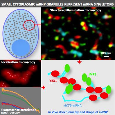
bioRxiv preprint first posted online Feb. 22, 2019; doi: http://dx.doi.org/10.1101/558098. The copyright holder for this preprint
 (which was not peer-reviewed) is the author/funder, who has granted bioRxiv a license to display the preprint in perpetuity.
                              It is made available under a CC-BY-NC-ND 4.0 International license.

                                                                                                                            Mateu-Regué et al.

 lamellipodia in motile cells (Nielsen et al., 1999; Oleynikov and Singer, 2003). In conventional laser scanning

 microscopy they exhibit an optical diameter of about 200-700 nm (Nielsen et al., 2002). Transcriptome-wide

 CLIP analyses have shown that both IMP1 and YBX1 associate with large parts of the transcriptome (Conway

 et al., 2016; Goodarzi et al., 2015). Individual RNAs exhibit numerous IMP1 and YBX1 attachment sites

 distributed along the target mRNA (Nielsen et al., 2004; Runge et al., 2000; Singh et al., 2015), although it is

 unknown whether binding is taking place on the same mRNA molecule. IMP1 and YBX1 are essential for

 normal development; IMP1- and YBX1-deficient mice are both small with imperfect organ development

 including neuronal defects (Hansen et al., 2004; Uchiumi et al., 2006), and at the cellular level both factors

 promote cell growth (Bell et al., 2013; Bommert et al., 2013; Shiota et al., 2008). Moreover, YBX1 and IMP1

 have been implicated in a series of complex biological pathways such as F-actin formation and protein

 secretion (Jønson et al., 2007; Uchiumi et al., 2006), and specifically IMP1 is a participant in the embryonal

 heterochronic network consisting of HMGA2, let-7 and Lin28B mRNAs (Jonson et al., 2014; Nishino et al.,

 2013). Finally, YBX1 plays a role in nodal signaling via sqt RNA localization, processing and translation (Kumari

 et al., 2013).

 To advance our understanding of cytoplasmic mRNP granules, we employed super-resolution and

 fluorescence correlation microscopy of YBX1, IMP1 and associated mRNA. In contrast to the current

 perception based on conventional light microscopy, our data show that granules represent irregular

 branched and elongated structures composed of alternating patches of IMP1 and YBX1 along a common

 mRNA. Formation of the particles requires mRNA, and mRNPs are first observed at the nuclear pore. The

 mRNPs are not connected to actively translating ribosomes, which are located in a separate vicinal

 compartment. Each particle contains a single mRNA and between 5-15 IMP1 and YBX1 molecules, in

 agreement with the average number of binding sites in the target mRNAs. Taken as a whole, we conclude

 that mRNP granules represent singletons and that coordinated expression of functionally related mRNAs is

 unlikely to be due to coordinated assembly.

                                                                                                                                            4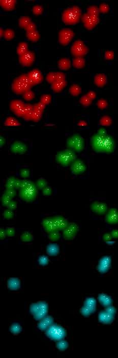
bioRxiv preprint first posted online Feb. 22, 2019; doi: http://dx.doi.org/10.1101/558098. The copyright holder for this preprint
 (which was not peer-reviewed) is the author/funder, who has granted bioRxiv a license to display the preprint in perpetuity.
                              It is made available under a CC-BY-NC-ND 4.0 International license.

                                                                                                                            Mateu-Regué et al.

 Results

 IMP1 and YBX1 coexist in mRNPs in an RNA-dependent manner

 To provide an overview of the subcellular distribution and structure of IMP1 and YBX1 RNPs in vivo, HeLa

 cells were stained with anti-IMP1 and anti-YBX1 antibodies and examined by Structured Illumination

 Microscopy (SIM). IMP1 and YBX1 were cytoplasmic and prominent at the perinuclear region and in the

 lamellipodia of the cells (Figure 2A-C). Granules appeared branched and elongated and ranged from 250 to

 800 nm in size and were composed of alternating patches of IMP1 and YBX1 (Figure 2D-F). In general, YBX1

 was observed along the entire outline of the particle, whereas IMP1 exhibited a preponderance for

 projections and ends. To visualize the associated mRNA, we performed FISH of ACTB mRNA combined with

 immunostaining of YBX1 and IMP1 (Figure 2N-R). ACTB mRNA was hybridized to 48 labelled Quasar® 570

 Dye-labeled probes covering the entire mRNA from the 5´ to the 3´ end. Probe staining was partially masked

 by the attached RNA-binding proteins, in particular when YBX1 was present. To corroborate the putative

 RNA-dependent interaction of YBX1 and IMP1, an immunoprecipitation of 3xFLAG-tagged or GFP-tagged

 IMP1 was carried out (Supplemental Figure 1). YBX1 was enriched in the immunoprecipitate, and treatment

 with RNAse A reduced the amount of immunoprecipitated YBX1, indicating that IMP1 does not interact

 directly with YBX1. In agreement with the observation that the particles contain multiple IMP1 molecules

 binding independently to the mRNA, IMP1 was also reduced by RNAse A (Supplemental Figure 1B). The

 mRNPs were clearly distinct from cytoplasmic P-bodies and stress-granules identified by G3BP and DCP1a,

 respectively (Figure 2G-H and Supplemental Figure 2). The average size of stress-granules were at least an

 order of magnitude larger than IMP1_YBX1 mRNPs, that could be distinguished within the larger bodies.

 YBX1 and IMP1 mRNP is formed in the nuclear pore and awaits translation

 Since the observed size of the mRNP is difficult to reconcile with facilitated diffusion of IMP1_YBX1 mRNP

 particles through the central nuclear pore channel, whose size limit is regarded to be about 40 nm (Hoelz et

 al., 2011), we examined the first appearance of the mRNP. SIM revealed very faint and almost non-existent

                                                                                                                                            5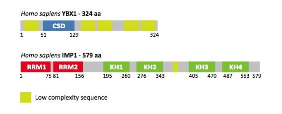
bioRxiv preprint first posted online Feb. 22, 2019; doi: http://dx.doi.org/10.1101/558098. The copyright holder for this preprint
 (which was not peer-reviewed) is the author/funder, who has granted bioRxiv a license to display the preprint in perpetuity.
                              It is made available under a CC-BY-NC-ND 4.0 International license.

                                                                                                                            Mateu-Regué et al.

 YBX1 and IMP1 nuclear staining, and we failed to observe any colocalization between the two factors and

 mRNA. This led us to image the nuclear pore in closer detail. Nuclear pores were visualized by staining of

 Nup153, and both IMP1 and YBX1 mRNPs were found to align with the pore (Figure 3), where mRNPs

 projected towards the cytoplasm. The cytoplasmic distribution and alignment was unaffected by incubation

 with leptomycin (data not shown). We infer that IMP1_YBX1 mRNPs are likely to form at the nuclear pore.

 To define the relation of the mRNP to the translation apparatus, we employed O-propargyl-puromycin (OPP)

 to depict actively translating ribosomes (Liu et al., 2012). After fixation, OPP was conjugated to an Alexa Fluor-

 488 fluorophore by click chemistry. Moreover, IMP1 and YBX1 were stained by immunofluorescence, and

 SIM Z-stacks of the cells were generated. As shown in Figure 4 (Panels A and B) and supplemental Figure 3,

 IMP_YBX1 mRNP did not colocalize with translating ribosomes. However, ribosomes were positioned in close

 proximity to the mRNPs, facilitating a possible handover of mRNA. To substantiate the cytological findings,

 we performed a polysome fractionation analysis confirming that both IMP1 and YBX1 predominantly

 sedimented as free mRNP in monosomal fractions corresponding to 40S - 80S (Figure 4C). Moreover, we

 performed an immunofluorescence staining combining IMP1 and the elongation factor eEF1A or eIF4A3. In

 its GTP-form, eEF1A is responsible for bringing aminoacyl-tRNA to the A-site of the ribosome, whereas eIF4A3

 is a core component in the exon junction complex defining virgin mRNAs (Maquat et al., 2010; Mateyak and

 Kinzy, 2010) (Figure 4, panel D). eEF1A was absent from mRNPs, whereas eIF4A3 colocalized with the mRNP.

 The results are in agreement with a spatial segregation of mRNPs from actively translating ribosomes and

 imply that protein synthesis follows unloading of the mRNA.

 Dynamics of IMP1_YBX1 mRNPs

 To assess the dynamics of cytoplasmic IMP1_YBX1 mRNPs, we employed Fluorescence Correlation

 Spectrosopy (FCS). Wild-type GFP-tagged IMP1 and -YBX1 and a GFP-tagged IMP1 GXXG mutant (GFP-

 IMP1_KH1-4mut), with impaired RNA-binding (Supplemental Figure 4), were expressed in HeLa cells.

 Moreover, GFP was included as a reference. As depicted in Figure 5A, GFP and GFP-IMP1_KH1-4mut

 exhibited faster Lag times than GFP-IMP1 and GFP-YBX1. Whereas GFP best fitted a 1-component model, the

 best fit of the experimental autocorrelation curves of GFP-IMP1_KH1-4mut, GFP-IMP1 and GFP-YBX1 was a

                                                                                                                                            6
bioRxiv preprint first posted online Feb. 22, 2019; doi: http://dx.doi.org/10.1101/558098. The copyright holder for this preprint
(which was not peer-reviewed) is the author/funder, who has granted bioRxiv a license to display the preprint in perpetuity.
It is made available under a CC-BY-NC-ND 4.0 International license.
Mateu-Regué et al.
2-component model. The modelling showed that GFP-IMP1_KH1-4mut could be resolved into two relatively
fast diffusion times of 2.9E-04 s and 1.2E-03 s, respectively, whereas GFP-IMP1 and GFP-YBX1 exhibited a fast
moving fraction with a Lag time of ~10E-03 s and a much slower fraction moving in the range of 10E-01 s
(Figure 5C). Compared to GFP, wild-type IMP1 and YBX1 exhibited fractions diffusing 10 fold and a 1000 fold
slower. In agreement with the pull-down analysis described above, Fluorescence Cross Correlation
Spectroscopy with GFP-IMP1 or GFP-IMP1_KH1-4mut combined with mCherry-YBX1 demonstrated that the
interaction between the two factors in vivo relies on mRNA-binding and not on other factors (Figure 5B). To
further substantiate FCS data, we visualized the mobility of the RNP in the live cell. We employed mEos3.2,
a photoconvertible fluorescent protein (Zhang et al., 2012), and expressed mEos3.2-IMP1. After localized
photoactivation at 405 nm, cellular movements of the photoconverted red mEos3.2-IMP1 was followed by
time lapse microscopy (Supplementary Figure 5). Both a slow and a rapid transport (in the range of seconds)
was observed. Slow particles diffused in all directions and migrated at very slow rate of about 0.1 μm/s. Even
after 1.5 minute the majority of the granular RNP remained in the center of the photoactivation. In contrast,
the rapid diffusion accumulated quickly at a particular distant site 25 μm away within seconds after
photoconversion, corresponding to a speed of at least ~5 μm/s. The results show that assembly of granules
requires RNA-binding, and that IMP1_YBX1 mRNP motility is multidirectional and involves fast and slow
trafficking and regulated anchoring.
Molecular composition of IMP1_YBX1 mRNP
We analysed the number of molecules in IMP1_YBX1 mRNP in vivo by means of Fluorescence Correlation
Spectroscopy (FCS) and Localization Microscopy and compared the results to available transcriptome-wide
IMP1 and YBX1 eCLIP and RIP-seq data. Moreover, the interplay between IMP1 and YBX1 upon binding to
high-affinity RNA targets such as ACTB and C-MYC mRNAs (Leeds et al., 1997; Ross et al., 1997), was examined
in order to provide a framework for an understanding of the stoichiometric data (Figure 6).
Figure 6A shows a schematic representation of ACTB and C-MYC transcripts together with the CLIP-derived
number of cross-linked and immunoprecipitated reads at particular positions (Conway et al., 2016; Goodarzi
7bioRxiv preprint first posted online Feb. 22, 2019; doi: http://dx.doi.org/10.1101/558098. The copyright holder for this preprint
(which was not peer-reviewed) is the author/funder, who has granted bioRxiv a license to display the preprint in perpetuity.
It is made available under a CC-BY-NC-ND 4.0 International license.
Mateu-Regué et al.
et al., 2015). Although IMP1 exhibits a preponderance for the 3´UTR and YBX1 binding is more widespread,
some binding sites are overlapping and the two factors may compete for a number of binding sites. The CLIP
data show that YBX1 and IMP1 exhibit strong binding to ACTB mRNA exon 5 and the ZIP code in the 3´UTR,
respectively, and to the CRD region and the 3’UTR of C-MYC, respectively. Therefore, we performed
electrophoretic mobility-shift assays (EMSA) with these mRNAs, shown in Figure 6B. YBX1 and IMP1
concentration ratios were according to the protein copy number per HeLa cell, as previously characterised
(Singh et al., 2015). YBX1, IMP1 or both proteins were incubated with the radioactively labelled RNA targets.
The mobility-shifts showed that the binding of each protein to the chosen mRNA segments was mutually
exclusive since there was no evidence of a supershift with any of the RNA probes. For C-MYC, we observed
that both YBX1 and IMP1 were able to bind to the CRD and 3’UTR targets. IMP1 exhibited a higher affinity
for CRD than YBX1, since at 1:10 of the YBX1 concentration IMP1 was able to out-compete YBX1. The same
happened with C-MYC 3’UTR, although in this case IMP1 was able to achieve a higher degree of
multimerization. Regarding the ACTB segments, we observed that YBX1 had a very high affinity for exon 5
and showed a high degree of multimerization, in accordance with the CLIP data. IMP1 was essentially unable
to bind to ACTB exon 5 and could not compete with YBX1. Both IMP1 and YBX1 were able to bind to the
3´UTR target, and we observed the same competition pattern as described for the C-MYC transcripts,
although both proteins exhibited lower affinity. Taken together, we infer that IMP1 and YBX1 compete for
shared binding segments – so at a given time IMP1 or YBX1 may not occupy all their possible binding sites.
To determine the number of IMP1, YBX1 and RNA molecules in the individual mRNP, we employed
Localization Microscopy and Fluorescence Correlation Spectroscopy. For Localization Microscopy, HeLa cells
were stained with anti-IMP1 or anti-YBX1 antibodies and Alexa Fluor 568 or 555 secondary antibodies,
respectively, or with a set of ACTB mRNA probes as shown in Figure 2. The number of secondary antibodies
binding a primary antibody was determined by FCS, and this showed that two secondary antibodies bind to
each primary antibody (data not shown). Consequently, events (emitted photons at a particular site) were
divided by two (Figure 7C). To avoid counting the same secondary antibody more than once due to the
presence of multiple fluorophores and to improve positioning accuracy, a grouping of 5.5 pixels was applied.
8bioRxiv preprint first posted online Feb. 22, 2019; doi: http://dx.doi.org/10.1101/558098. The copyright holder for this preprint
(which was not peer-reviewed) is the author/funder, who has granted bioRxiv a license to display the preprint in perpetuity.
It is made available under a CC-BY-NC-ND 4.0 International license.
Mateu-Regué et al.
Panel A shows Localization Microscopy readings with GAUSS distributions and crosses. Clustered photons
(crosses) corresponding to 50 granules were counted (Figure 7A), and the median and range are summarized
in the Table (Figure 7C). The median number of molecules was 7 for YBX1 (range 2-34) and 6 for IMP1 (range
2-15) in the RNP. Moreover, we analyzed the number of ACTB RNA probes in the mRNP granules. As
illustrated in Figure 2, parts of the ACTB mRNA appeared to be masked by attached RBPs, and we counted
on average 11 probes ranging from 3-42 in the mRNP. In no ACTB cluster, we counted more than the maximal
number of 48 probes. Therefore, we infer that there is only a single ACTB transcript in a particle.
To corroborate the findings described above, we extracted information regarding the average number of
molecules in the particles in live cells by FCS. The counts per particle (related to the amplitude of the
fluorescence fluctuations) of FCS measurements in the free GFP and GFP-IMP1_KH1-4mut constructs
(monomers) were compared with the wild type GFP-IMP1 and GFP-YBX1 in HeLa cells. We could measure
differences in the fluctuations during the recorded measurements (Figure 7B), and this was also represented
in the amplitude of the autocorrelation curves. Cytoplasmic readings from 9 different cells showed that the
number of GFP-IMP1 molecules per particle was from 7 to 11, considering GFP-IMP1_KH1-4mut as the
monomer state of the protein, whereas the results for GFP-YBX1 were unexpectedly low compared to what
we observed in the Localization Microscopy analysis. Endogenous YBX1 concentration is 10 times higher than
IMP1, and we suspected that GFP-YBX1 counts per particle were underestimated due to the high levels of
competing endogenous protein. To examine if this was the case, endogenous YBX1 was knocked down using
a siRNA against the 3’UTR. As observed in Figure 7, counts per particle during these conditions increased
from 3-4 to 8-13 YBX1 molecules per particle. Finally, we estimated the number of global IMP1 and YBX1
mRNA binding sites from public CLIP and RIP-seq data (Conway et al., 2016; Goodarzi et al., 2015) in order to
put the Localization Microscopy and FCS data in perspective (Figure 7, Panel C). Raw CLIP-seq data (fastq)
were downloaded from the short read archive (SRA) and aligned to the human genome sequence. Binding
sites were estimated by read-islands. The lower detection level was adjusted to the nearest threshold, where
islands in the mock control samples equalled the total number of transcripts, therefore a cut-off of 7 and 6
for IMP1 and YBX1, respectively, was employed. We estimated that the average number of IMP1 and YBX1
9bioRxiv preprint first posted online Feb. 22, 2019; doi: http://dx.doi.org/10.1101/558098. The copyright holder for this preprint
(which was not peer-reviewed) is the author/funder, who has granted bioRxiv a license to display the preprint in perpetuity.
It is made available under a CC-BY-NC-ND 4.0 International license.
Mateu-Regué et al.
binding sites per mRNA to be 7 and 5, respectively. IMP1 has been described to dimerize to form stable RNA-
binding complexes (Nielsen et al., 2004), corroborated by the electrophoretic mobility-shift analysis in Figure
6, and YBX1 has also been shown to multimerize on a single RNA attachment site (Figure 6) (Skabkin et al.,
2004). Taken as a whole, Localization Microscopy and FCS analyses, which provide the molecular composition
of the individual RNP granules in vivo, are in agreement with the transcriptome-wide CLIP estimates, when
dimerization of IMP1 and a multimerization of YBX1 on mRNA are taken into account. We therefore conclude
that IMP1_YBX1 RNPs represent solitary mRNAs with attached proteins.
10bioRxiv preprint first posted online Feb. 22, 2019; doi: http://dx.doi.org/10.1101/558098. The copyright holder for this preprint
(which was not peer-reviewed) is the author/funder, who has granted bioRxiv a license to display the preprint in perpetuity.
It is made available under a CC-BY-NC-ND 4.0 International license.
Mateu-Regué et al.
Discussion
Cytoplasmic RNP granules have been recognized for several decades, and although we have a relatively deep
understanding of their biochemistry, the molecular composition and cellular distribution are incompletely
understood. This has partly been due to the lack of technologies to characterize molecular complexes in
intact and live cells, so we employed super resolution microscopy and correlation spectroscopy to provide a
deeper insight into the nature of single mRNP granules.
Compared to conventional laser scanning microscopy, where mRNP granules appear spherical with a size of
200-700 nm (Nielsen et al., 2002), structured illumination microscopy (SIM) has a resolution 2-3 times below
that of diffraction-limited instruments, providing a lateral resolution of about 100 nm (Stelzer, 2014). An
average 2 kb mRNA has an outline of about 300 nm - taking secondary structure into account - so it is feasible
to distinguish RBPs on the same mRNA by SIM (Milo et al., 2010) (http://book.bionumbers.org/which-is-
bigger-mrna-or-the-protein-it-codes-for/). IMP1_YBX1 mRNPs were on average 300 nm in size, which is in
agreement with atomic force microscopy of isolated granules (Jønson et al., 2007). YBX1- and IMP1-stainings
were partly overlapping but also alternating along the mRNP. Whereas YBX1 was distributed along the entire
particle, IMP1 had a preponderance for projections and ends. YBX1 is one of the core proteins of mRNPs and
has previously been described to coat the entire mRNA (Singh et al., 2015; Skabkin et al., 2004), whereas
IMP1 preferentially binds to single-stranded CA-rich elements in the 3’UTR and loop regions (Conway et al.,
2016; Hafner et al., 2010). As previously described, the embedded mRNAs were to a large extent masked by
the associated proteins (Buxbaum et al., 2014). In our data, mRNA appeared to be masked particularly in
regions covered by YBX1, which is in accordance with the fact that YBX1 acts as a translational repressor
(Evdokimova et al., 2006). Due to the distinct localization of the core proteins in the mRNP, the complete
structure was only perceived in the composite pictures. If focus had been directed towards one of the factors,
we would have failed to recognize the entire size and shape of the mRNP. Moreover, SIM directly
demonstrates that YBX1 and IMP1 bind in cis, which is of significance for the interpretation of CLIP and
immunoprecipitation analysis that fail to make a distinction between cis and trans.
11bioRxiv preprint first posted online Feb. 22, 2019; doi: http://dx.doi.org/10.1101/558098. The copyright holder for this preprint
(which was not peer-reviewed) is the author/funder, who has granted bioRxiv a license to display the preprint in perpetuity.
It is made available under a CC-BY-NC-ND 4.0 International license.
Mateu-Regué et al.
RNA-binding proteins associate with and dissociate from the mRNA along its journey from the nucleus to the
translating ribosomes (Singh et al., 2015). Based on the presence of nuclear export signals in IMP1 and YBX1,
the general idea has been that the factors enter the nucleus and bind their target mRNAs (Nielsen et al.,
2003; Oleynikov and Singer, 2003). We hardly observed nuclear IMP1 and YBX1 staining, and proper mRNP
complexes composed of IMP1 and YBX1 and mRNA were only identified at the cytoplasmic side of the nuclear
pore. This observation supports a recent study, indicating that Actb mRNA first associates with IMP1 in the
nuclear envelope (Wu et al., 2015). The nuclear pore is flexible and dynamic (Knockenhauer and Schwartz,
2016), and the largest macromolecular complexes that have been shown to pass are viral capsids up to ~40
nm diameter. Our data reflect that the mRNA is brought to the pore by a canonical mRNA export pathway,
before IMP1 and YBX1 are loaded onto the mRNA. In this way, the nuclear pore may represent a crucial
remodeling step of the mRNP (Singh et al., 2015). Whether the nuclear export signals in IMP1 and YBX1 have
a function in localization to the nuclear pore remains to be addressed.
In the cytoplasm, IMP1 and YBX1 protect mRNAs from miRNA-mediated degradation (Jonson et al., 2014)
and YBX1 is also a mRNA stabilizer (Evdokimova et al., 2001), so the mRNP is considered an mRNA
repository/safe house until translation. Although granules incorporate ribosomal subunits (Krichevsky and
Kosik, 2001), we observed no direct association with translating ribosomes, which were closely intertwined
between the mRNPs. The mRNP also contained the exon-junction complex core factor eIF4A3, which is a
hallmark of mature mRNA prior to the pioneer round of translation. Our earlier compositional analysis of
IMP1 RNPs, that identified the remaining exon-junction components together with the nuclear cap-binding
subunit CBP80 and the nuclear poly(A)-binding protein PABPN1, corroborates the pre-translational status of
these cytoplasmic mRNPs (Jønson et al., 2007). In agreement with their large composite nature the majority
of the mRNPs exhibited a very slow diffusion rate compared to the KH domain GXXG loop mutant that failed
to bind RNA. Particles migrated in all directions, and judged from the photoconversion experiments some
were almost immobile, whereas about one-third of the mRNPs exhibited a faster mobility. The fastest
particles were able to migrate several micrometers per second. Intriguingly, they accumulated at a particular
location indicating that they may be subject to a regulated cytoplasmic docking. Transport may involve simple
diffusion but also cytoplasmic streaming and motors, which are not mutually exclusive mechanisms (Lu et al.,
12bioRxiv preprint first posted online Feb. 22, 2019; doi: http://dx.doi.org/10.1101/558098. The copyright holder for this preprint
(which was not peer-reviewed) is the author/funder, who has granted bioRxiv a license to display the preprint in perpetuity.
It is made available under a CC-BY-NC-ND 4.0 International license.
Mateu-Regué et al.
2016; Song et al., 2015; Suzuki et al., 2017). Moreover, the single photoconverted mRNP was fairly stable,
indicating that fluidity is low in contrast to what is observed within liquid droplets (Courchaine et al., 2016).
We determined the molecular composition of the individual granules by Localization Microscopy and FCS.
Both methods roughly led to the same result and showed that particles were composed of 5-15 molecules of
both IMP1 and YBX1. Due to the masking, the Localization Microscopy analysis of ACTB mRNA should
obviously be interpreted with some caution. However, since we never arrived at probe counts exceeding the
total number of applied ACTB probes, we infer that there is only one ACTB mRNA in the particles in agreement
with what we observed by SIM. Moreover, the comparative data from CLIP analysis (Conway et al., 2016;
Goodarzi et al., 2015) show - in agreement with the Localization Microscopy and FCS data - that the average
number of YBX1 and IMP1 mRNA attachment sites at a global level are 6 and 7, respectively. The reason that
FCS and Localization Microscopy provide slightly higher numbers of IMP1 molecules is in agreement with
IMPs binding as dimers and that YBX1 to some extent multimerizes. The results are in line with recent findings
showing that neuronal mRNAs travel singly into dendrites (Batish et al., 2012) and that MAP2, CaMKIIa and
ACTB RNAs localize independently in low copy numbers (Mikl et al., 2011), thus supporting the “sushi-belt
model” (Doyle and Kiebler, 2011). The findings may also be corroborated by a rough estimate. Assuming that
a HeLa cell contains about 200.000 transcripts in a volume of 3000 μm3 there will be 60 mRNAs per μm3
(Shapiro et al., 2013). By simple counting of SIM stacks, we observe 30-50 mRNPs per μm3, which is
compatible with a single transcript in each mRNP, considering that some mRNAs may undergo translation or
reside in the nucleus. RNA-binding proteins have been proposed to coordinate the production of functionally
related proteins by organizing their mRNAs in regulons (Keene, 2007). The finding that small cytoplasmic
mRNP granules represent singletons implies that coordinate expression of functionally related mRNAs in RNA
regulons is unlikely to result from coordinated assembly, but rather results from regulated docking, as
described above, or from selective stabilization of mRNA in the particles.
Small cytoplasmic mRNP granules are distinct from large stress granules and P bodies, that represent dynamic
mRNP assemblies formed in response to stress and mRNA decay, respectively. Based on our findings, we
13bioRxiv preprint first posted online Feb. 22, 2019; doi: http://dx.doi.org/10.1101/558098. The copyright holder for this preprint
(which was not peer-reviewed) is the author/funder, who has granted bioRxiv a license to display the preprint in perpetuity.
It is made available under a CC-BY-NC-ND 4.0 International license.
Mateu-Regué et al.
propose that smaller cytoplasmic granules should be designated mRNP singletons rather than granules to
clearly distinguish them from the larger assemblies. Moreover, this would allude to the fact that stress
granules probably incorporate elements of mRNP singletons.
14bioRxiv preprint first posted online Feb. 22, 2019; doi: http://dx.doi.org/10.1101/558098. The copyright holder for this preprint
(which was not peer-reviewed) is the author/funder, who has granted bioRxiv a license to display the preprint in perpetuity.
It is made available under a CC-BY-NC-ND 4.0 International license.
Mateu-Regué et al.
Acknowledgements
The Danish National Program for Infrastructure is thanked for donating the super resolution microscope and
Lena Bjørn Johansson is thanked for technical assistance.
Author Contributions
FCN and AM designed the study. AM, FCN, JC, FOB, CH designed the experiments. AM, FOB and JC performed
the experiments. AM, FCN, JC, FOB and CH analysed the results. AM, FCN and JC prepared the manuscript.
All authors read and approved the final manuscript.
Declaration of Interests
The authors declare no competing interests.
15bioRxiv preprint first posted online Feb. 22, 2019; doi: http://dx.doi.org/10.1101/558098. The copyright holder for this preprint
(which was not peer-reviewed) is the author/funder, who has granted bioRxiv a license to display the preprint in perpetuity.
It is made available under a CC-BY-NC-ND 4.0 International license.
Mateu-Regué et al.
Methods
Contact for Reagent and Resource Sharing
Further information and requests for resources and reagents should be directed to and will be fulfilled by the
Lead Contact, Finn Cilius Nielsen (finn.cilius.nielsen@regionh.dk).
Experimental model and Subject Details
Cell lines
HeLa cells (ATCC® CCL-2TM) were cultured in phenol red-free Dulbecco’s Modified Eagle Medium (DMEM),
high glucose (4.5 g/L) + GlutaMAX and 1 mM sodium pyruvate (Thermo Fisher Scientific) supplemented with
10% Fetal bovine serum (Tetracycline free, Biowest) and penicillin/streptomycin (Invitrogen). HT1080 cells
(ATCC® CCL-121TM) were cultured in Dulbecco’s Modified Eagle Medium (DMEM), high glucose (4.5 g/L) +
GlutaMAX and 1 mM sodium pyruvate (Thermo Fisher Scientific) supplemented with 10% Fetal bovine serum
(Tetracycline free, Biowest) and penicillin/streptomycin (Invitrogen). TREX HT1080 3xFLAG-IMP1 cells were
cultured in Dulbecco’s Modified Eagle Medium (DMEM), high glucose (4.5 g/L) + GlutaMAX and 1 mM sodium
pyruvate (Thermo Fisher Scientific) supplemented with 10% Fetal bovine serum, 5 ug/mL Blasticidin (Thermo
Fisher Scientific), 100 ug/ml Hygromycin (Thermo Fisher Scientific) and penicillin/streptomycin. All cell lines
were grown at 37°C with 5% CO2 in a humidified incubator
Method Details
Transfections
Vectors
IMP1 was cloned into pEGFP-C2 (Clontech) and pmEos3.2-C1 vector (Addgene). GFP-IMP1_KH1-4mut
construct was obtained by mutating the GXXG loops of the 4 KH domains (Hollingworth et al., 2012) from
GK(E/K/G)G to GELG. YBX1 was cloned into pcDNA3.1+N-EGFP (Genscript) and pmCherry-C1 (Clontech)
16bioRxiv preprint first posted online Feb. 22, 2019; doi: http://dx.doi.org/10.1101/558098. The copyright holder for this preprint
(which was not peer-reviewed) is the author/funder, who has granted bioRxiv a license to display the preprint in perpetuity.
It is made available under a CC-BY-NC-ND 4.0 International license.
Mateu-Regué et al.
vectors, inserting a 25 amino acid flexible linker composed of 5x GlyGlyGlyGlySer between the fluorescent
tag and YBX1 to reduce aggregation.
Plasmid transfections
HeLa cells were plated in 35mm glass-bottom dishes (P35G-1.5-14-C, MatTek) and were transfected using
FuGENE® 6 (Promega). For each transfection, 3 µl of FuGENE® 6 and 1.3 µg of plasmid were added to 100 µl
of OPTI-MEM Medium (Thermo Fisher Scientific) . The mixture was incubated at room temperature for 30
minutes prior to the addition to the coverslips.
siRNA and plasmid co-transfection
HeLa cells were plated in 35mm glass-bottom dishes (P35G-1.5-14-C, MatTek) and were transfected using
Lipofectamine 2000 (Thermo Fisher Scientific). For each transfection, 3 µl of Lipofectamine® 2000, 1 µg of
plasmid and siRNA to a final concentration of 2.5 nM were added to 100 µl of OPTI-MEM Medium (Thermo
Fisher Scientific). The mixture was incubated at room temperature for 30 minutes prior to the addition to the
coverslips.
Western blot analysis
Protein extracts were separated in 10% RunBlue SDS gels and transferred to PVDF membranes (Invitrogen).
After blocking, membranes were incubated overnight with a peptide specific rabbit anti-IMP1 antibody
(Nielsen et al., 1999) an anti-YBX1 antibody (ab12148, Abcam), an anti-GFP antibody (ab1218, Abcam) and a
GAPDH antibody (FL-335, Santa Cruz) in blocking solution at 4°C before they were washed and incubated
with horseradish peroxidase-conjugated anti-rabbit IgG for 1 h at room temperature. Immunoreactive
proteins were detected with SuperSignal chemiluminescence reagents (Thermo Fisher Scientific) according
to the manufacturer's instructions. Blots were scanned using a C-DiGit Blot Scanner (LI-COR Biosciences).
Immunoprecipitation
17bioRxiv preprint first posted online Feb. 22, 2019; doi: http://dx.doi.org/10.1101/558098. The copyright holder for this preprint
(which was not peer-reviewed) is the author/funder, who has granted bioRxiv a license to display the preprint in perpetuity.
It is made available under a CC-BY-NC-ND 4.0 International license.
Mateu-Regué et al.
TREX HT1080 cells stably expressing 3xFLAG-IMP1 were generated as described (Jønson et al., 2007). Briefly,
the expression of 3xFLAG-IMP1 was induced by the addition of 1 μg/ml tetracycline, and cells were harvested
24 hours after the addition of tetracycline in lysis buffer on ice. HT1080 cell line was used as a negative
control. In parallel, HeLa cells were transiently transfected with pEGFP-C1 and pEGFP-IMP1 and cell pellets
were collected 48 hours after transfection. Cell pellets containing 1×10⁷ cells were lysed in lysis buffer
containing 20 mM Tris-HCl (pH 7.5), 1.5 mM MgCl2, 140 mM KCl, 1 mM DTT, 0.5% NP-40 supplemented with
mammalian protease inhibitor cocktail (Sigma). Cell lysates were cleared by centrifugation at 8200 xg for 5
minutes before addition of EZviewTM Red Anti-FLAG Affinity Gel (Sigma) or GFP antibody (ab1218, Abcam)
coupled DynabeadsTM Protein G (Invitrogen). Cleared cell lysates were incubated with the beads for 2 hours
at 4°C with rotation. After that, samples were washed 3x with lysis buffer, split and treated with 20 ug/mL
RNAse A (DNase and protease-free, Thermo Scientific) or RiboLock RNase inhibitor (Thermo Scientific) for 20
minutes at room temperature with rotation. Beads were subsequently washed and proteins were eluted
directly in 2x SDS buffer.
Polysome fractionation analysis
Polysome analysis was performed as described (Nielsen et al., 2002). Briefly, HeLa cells (5×10⁶ cells) were
lysed in 500 μl 20 mM Tris-HCl (pH 8.5), 1.5 mM MgCl2, 140 mM KCl, 0.5 mM DTT, 0.5% NP-40, 200 U of
RNasin (Promega) per ml and 0.1 mM cycloheximide. The lysate was centrifuged at 10,000 xg for 10 min, and
the supernatant was applied to a linear 20 to 47% sucrose gradient in 20 mM Tris-HCl (pH 8.0), 140 mM KCl,
5 mM MgCl2. Centrifugation was carried out at 200,000 g for 2 hours and 15 min in a Beckman SW 41 rotor.
Fractions of 1 ml were collected, followed by protein precipitation with 10% TCA.
Electrophoretic mobility-shift analysis (EMSA)
Electrophoretic mobility-shift analysis was carried out essentially as described previously (Nielsen et al.,
2004). RNA targets were C-MYC CRD (positions 1181-1362 in CDS), C-MYC 3’UTR (positions 1-226 in 3’UTR),
ACTB exon 5, and ACTB 3’UTR (positions 1-233). Tag-less recombinant human IMP1 and IMP1_KH1-4mut
(with the four GXXG signature loops mutated to GELG) were expressed and purified as described earlier
18bioRxiv preprint first posted online Feb. 22, 2019; doi: http://dx.doi.org/10.1101/558098. The copyright holder for this preprint
(which was not peer-reviewed) is the author/funder, who has granted bioRxiv a license to display the preprint in perpetuity.
It is made available under a CC-BY-NC-ND 4.0 International license.
Mateu-Regué et al.
(Nielsen et al., 2004). Recombinant human YBX1 with a C-terminal FLAG-tag was purchased from OriGene
Technologies, Inc.
Immunofluorescence, fluorescence in situ hybridization and structured illumination microscopy (SIM).
HeLa cells were seeded in glass-bottom coverslips (P35G-0.170-14-C, MatTek) and fixed 24 hours after with
3.7% formaldehyde solution in PBS, followed by a permeabilization step with 0.5% Triton X-100 in PBS.
Immunofluorescence of IMP1 and YBX1 was performed using antibodies against IMP-1 (E-20, Santa Cruz) and
YBX1 (ab12148, Abcam). Nup153, eIF4A3 and eEF1a were detected using anti-Nup153 antibody (ab24700,
Abcam), anti-eIF4A3 antibody (ab32485, Abcam) and anti-eEF1A1/2/L3 antibody (ab37969, Abcam).
Coverslips were washed 3x with PBS prior to Alexa Fluor conjugated secondary antibodies (Thermo Fisher
Scientific) incubation for an hour at RT. Samples were washed 3x with PBS and mounted in VECTASHIELD
mounting media (RI = 1.45).
Structured Illumination Microscopy (SIM) was performed using a Zeiss ELYRA PS.1 microscope and channel
correction was applied using a channel alignment file created by imaging 0.1 µm TetraSpeck Microspheres
(Thermo Fisher Scientific). The image sets comprised 5 rotations and processed images were thresholded in
accordance with current standards to remove diffuse background (honeycombs) caused by stray pollutants
or some residual autofluorescence on the SIM reconstructed pictures before contrasting. An example is
provided in Supplemental Figure 6, that also provides a comparison between conventional confocal imaging
and SIM of the mRNA granules and image overlays demonstrating the measurements of the granules.
RNA FISH of ACTB mRNA was performed with ACTB DesignReady Probe Set (LGC Biosearch Technologies)
following the “Sequential Stellaris FISH and Immunofluorescence using Adherent Cells protocol” from LGC
Biosearch Technologies
(https://biosearchassets.blob.core.windows.net/assets/bti_custom_stellaris_immunofluorescence_seq_pr
otocol.pdf). Briefly, immunofluroescence of IMP1 and YBX1 was performed as described above followed by
a fixation step with fixation buffer (3.7% formaldehyde in PBS), Wash Buffer A (LGC Biosearch Technologies)
19bioRxiv preprint first posted online Feb. 22, 2019; doi: http://dx.doi.org/10.1101/558098. The copyright holder for this preprint
(which was not peer-reviewed) is the author/funder, who has granted bioRxiv a license to display the preprint in perpetuity.
It is made available under a CC-BY-NC-ND 4.0 International license.
Mateu-Regué et al.
was incubated for 5 minutes and ACTB probe set was hybridized in for 16 hours at 37°C with Hybridization
buffer (LGC Biosearch Technologies). After hybridization, dishes were incubated with Wash Buffer A for 30
minutes at 37°C followed by addition and incubation of Wash Buffer B (LGC Biosearch Technologies) for 5
minutes. Samples were mounted and imaged using the same protocol described above.
OP-puromycin protein synthesis assay
HeLa cells were seeded in glass-bottom coverslips (Mattek) and Click-iT® Plus OPP Alexa Fluor® 488 Protein
Synthesis Assay Kit was used to localize nascent polypeptides following the manufacturers protocol.
Cycloheximide (50 µg/mL) was added prior to OPP to one sample as a negative control. After the click
reaction, immunofluorescence detection of IMP1 and YBX1 was performed as described previously. Samples
were mounted in Vectashield mounting medium and images were acquired using a Zeiss ELYRA 3.2
microscope (Structured Illumination Microscopy).
Photoconversion of mEos3.2-IMP1 for in vivo protein tracking
HeLa cells were plated in 35mm glass-bottom dishes (P35G-1.5-14-C, MatTek) and transfected with
pmEos3.2-IMP1 with FuGENE 6 as described above in “Plasmid transfection” section. Photoconversion was
performed using a Zeiss LSM780 microscope with a Plan-Apochromat 63x/1.4 Oil objective using a 405 laser
pulse in a selected area in the cytoplasm. Cells were imaged before and after photoconversion and
photoconverted mEos3.2-IMP1 was monitored every 10 seconds and followed throughout the cytoplasm.
Localization Microscopy
HeLa cells were stained with either IMP1 and YBX1 antibodies and Alexa Fluor 568 and Alexa Fluor 555
secondary antibodies, respectively, or with ACTB Quasar-570 probes (LGC Biosearch Technologies). Samples
were mounted in non-hardening Vectashield mounting medium with a refraction index of 1.45 (Olivier et al.,
2013). Localization Microscopy was performed on a Zeiss ELYRA PS.1 microscope using an alpha-Plan-
Apochromat 100x/1.46 objective. Total internal reflection microscopy (TIRF) with a excitation wavelength of
20bioRxiv preprint first posted online Feb. 22, 2019; doi: http://dx.doi.org/10.1101/558098. The copyright holder for this preprint
(which was not peer-reviewed) is the author/funder, who has granted bioRxiv a license to display the preprint in perpetuity.
It is made available under a CC-BY-NC-ND 4.0 International license.
Mateu-Regué et al.
561 nm and appropriate emission filters (BP 570-650) was used in all localization microscopy experiments
and ZEN 2012 software was used to analyse and filter the data obtained in a total of 80.000 frames acquired.
Frames were corrected for drift over a time scale of 36 min 13 sec (Model-based correction), and grouping
was applied in the antibody stained samples in order to compile all the events that came from a single
antibody. Frames corresponding to the bleaching period of the sample were discarded for the final counting.
Fluorescence correlation spectroscopy (FCS) and Fluorescence Cross Correlation Spectroscopy (FCCS)
FCS measurements were performed with a Zeiss LSM 780 confocal microscope. HeLa cells were transfected
with pEGFP-C1 (GFP), GFP-IMP1, GFP-IMP1_KH1-4mut and GFP-YBX1 (-/+ cotransfection with YBX1 3’UTR
siRNA) as described above in the Plasmid transfection and siRNA and plasmid cotransfection sections and
incubated for ∼16 h and 48 hours respectively before measurements were conducted. Argon laser with a
488 excitation wavelength was used making sure that the count rate was linear at each particular laser power
used. Transfected cells were located and FCS measurements were performed in a Zeiss LSM780 confocal
microscope using a C-Apochromat 40x/1.2 W Corr M27 objective. Measurements were recorded in 10 second
intervals during a total time of 60 seconds choosing arbitrary points in the cytoplasm and experimental
autocorrelation curves were obtained. Intervals showing bleaching were discarded for the average. GFP-
IMP1 or GFP-IMP1_KH1-4mut and mCherry-YBX1 were cotransfected, and FCCS measurements were
performed following the same procedure as with FCS.
CLIP-seq analysis
Five public datasets were acquired by use of fastq-dump
(https://trace.ncbi.nlm.nih.gov/Traces/sra/sra.cgi?view=software) using the parameters: “skip-technical”,
“readids”, “read-filter pass”, “dumpbase”, “split-files”, and “clip”. Fastq files were aligned with STAR (version
2.5.2b) (Dobin et al., 2013) to the GRCh38 release 87 genome assembly as provided by ENSEMBL with
corresponding annotation (ftp://ftp.ensembl.org/pub/release-
87/gtf/homo_sapiens/Homo_sapiens.GRCh38.87.gtf.gz), using an overhang of 50 for all samples. Aligned
reads (bam files) were imported into GenomicAlignments (Lawrence et al., 2013) in R, and single end reads
21bioRxiv preprint first posted online Feb. 22, 2019; doi: http://dx.doi.org/10.1101/558098. The copyright holder for this preprint
(which was not peer-reviewed) is the author/funder, who has granted bioRxiv a license to display the preprint in perpetuity.
It is made available under a CC-BY-NC-ND 4.0 International license.
Mateu-Regué et al.
were resized to the estimated fragment length (300bp) (Jothi et al., 2008). A standard peak calling algorithm
could not be applied (e.g. MACS2) because of lack of a paired input sample. Hence, peaks were estimated
from read pile-up (islands), where the lower boundary was estimated to be a cutoff where the mock samples
proximate one island per transcript (closest number). In one case a mock was not available with the sample,
and the lower boundary was estimated from median of mocks from ENCODE. All mock samples analysed
required a cut-off lower than 9 reads in order to proximate one island per transcript. Crosslinked
immunoprecipitation of endogenous YBX1 followed by high-throughput sequencing (CLIP-seq) in human
MDA-parental breast cancer cells, YBX1 were acquired from the short read archive (SRA) with accession
numbers: SRR1662159, SRR1662160, and SRR1662161 (Goodarzi et al., 2015). Enhanced CLIP-seq for
IGF2BP1/IMP1 was acquired from SRA, accession numbers: SRR5112331 and SRR5112330 (Consortium,
2012).
22bioRxiv preprint first posted online Feb. 22, 2019; doi: http://dx.doi.org/10.1101/558098. The copyright holder for this preprint
(which was not peer-reviewed) is the author/funder, who has granted bioRxiv a license to display the preprint in perpetuity.
It is made available under a CC-BY-NC-ND 4.0 International license.
Mateu-Regué et al.
References
Anderson, P., and Kedersha, N. (2006). RNA granules. J Cell Biol 172, 803-808.
Batish, M., van den Bogaard, P., Kramer, F.R., and Tyagi, S. (2012). Neuronal mRNAs travel singly
into dendrites. Proc Natl Acad Sci U S A 109, 4645-4650.
Bell, J.L., Wachter, K., Muhleck, B., Pazaitis, N., Kohn, M., Lederer, M., and Huttelmaier, S. (2013).
Insulin-like growth factor 2 mRNA-binding proteins (IGF2BPs): post-transcriptional drivers of cancer
progression? Cell Mol Life Sci 70, 2657-2675.
Bommert, K.S., Effenberger, M., Leich, E., Kuspert, M., Murphy, D., Langer, C., Moll, R., Janz, S.,
Mottok, A., Weissbach, S., et al. (2013). The feed-forward loop between YB-1 and MYC is essential
for multiple myeloma cell survival. Leukemia 27, 441-450.
Buxbaum, A.R., Wu, B., and Singer, R.H. (2014). Single beta-actin mRNA detection in neurons reveals
a mechanism for regulating its translatability. Science 343, 419-422.
Consortium, E.P. (2012). An integrated encyclopedia of DNA elements in the human genome. Nature
489, 57-74.
Conway, A.E., Van Nostrand, E.L., Pratt, G.A., Aigner, S., Wilbert, M.L., Sundararaman, B., Freese, P.,
Lambert, N.J., Sathe, S., Liang, T.Y., et al. (2016). Enhanced CLIP Uncovers IMP Protein-RNA Targets
in Human Pluripotent Stem Cells Important for Cell Adhesion and Survival. Cell Rep 15, 666-679.
Courchaine, E.M., Lu, A., and Neugebauer, K.M. (2016). Droplet organelles? EMBO J 35, 1603-1612.
Darnell, J.C., and Richter, J.D. (2012). Cytoplasmic RNA-binding proteins and the control of complex
brain function. Cold Spring Harb Perspect Biol 4, a012344.
Dobin, A., Davis, C.A., Schlesinger, F., Drenkow, J., Zaleski, C., Jha, S., Batut, P., Chaisson, M., and
Gingeras, T.R. (2013). STAR: ultrafast universal RNA-seq aligner. Bioinformatics 29, 15-21.
Doyle, M., and Kiebler, M.A. (2011). Mechanisms of dendritic mRNA transport and its role in synaptic
tagging. EMBO J 30, 3540-3552.
Eliscovich, C., Shenoy, S.M., and Singer, R.H. (2017). Imaging mRNA and protein interactions within
neurons. Proc Natl Acad Sci U S A 114, E1875-E1884.
Evdokimova, V., Ovchinnikov, L.P., and Sorensen, P.H. (2006). Y-box binding protein 1: providing a
new angle on translational regulation. Cell Cycle 5, 1143-1147.
Evdokimova, V., Ruzanov, P., Imataka, H., Raught, B., Svitkin, Y., Ovchinnikov, L.P., and Sonenberg,
N. (2001). The major mRNA-associated protein YB-1 is a potent 5' cap-dependent mRNA stabilizer.
EMBO J 20, 5491-5502.
23bioRxiv preprint first posted online Feb. 22, 2019; doi: http://dx.doi.org/10.1101/558098. The copyright holder for this preprint
(which was not peer-reviewed) is the author/funder, who has granted bioRxiv a license to display the preprint in perpetuity.
It is made available under a CC-BY-NC-ND 4.0 International license.
Mateu-Regué et al.
Gerstberger, S., Hafner, M., and Tuschl, T. (2014). A census of human RNA-binding proteins. Nat Rev
Genet 15, 829-845.
Goodarzi, H., Liu, X., Nguyen, H.C., Zhang, S., Fish, L., and Tavazoie, S.F. (2015). Endogenous tRNA-
Derived Fragments Suppress Breast Cancer Progression via YBX1 Displacement. Cell 161, 790-802.
Hafner, M., Landthaler, M., Burger, L., Khorshid, M., Hausser, J., Berninger, P., Rothballer, A.,
Ascano, M., Jr., Jungkamp, A.C., Munschauer, M., et al. (2010). Transcriptome-wide identification of
RNA-binding protein and microRNA target sites by PAR-CLIP. Cell 141, 129-141.
Hansen, T.V.O., Hammer, N.A., Nielsen, J., Madsen, M., Dalbaeck, C., Wewer, U.M., Christiansen, J.,
and Nielsen, F.C. (2004). Dwarfism and Impaired Gut Development in Insulin-Like Growth Factor II
mRNA-Binding Protein 1-Deficient Mice. Molecular and Cellular Biology 24, 4448-4464.
Hoelz, A., Debler, E.W., and Blobel, G. (2011). The structure of the nuclear pore complex. Annu Rev
Biochem 80, 613-643.
Hollingworth, D., Candel, A.M., Nicastro, G., Martin, S.R., Briata, P., Gherzi, R., and Ramos, A. (2012).
KH domains with impaired nucleic acid binding as a tool for functional analysis. Nucleic Acids Res
40, 6873-6886.
Jonson, L., Christiansen, J., Hansen, T.V., Vikesa, J., Yamamoto, Y., and Nielsen, F.C. (2014). IMP3
RNP safe houses prevent miRNA-directed HMGA2 mRNA decay in cancer and development. Cell Rep
7, 539-551.
Jothi, R., Cuddapah, S., Barski, A., Cui, K., and Zhao, K. (2008). Genome-wide identification of in vivo
protein-DNA binding sites from ChIP-Seq data. Nucleic Acids Res 36, 5221-5231.
Jønson, L., Vikesaa, J., Krogh, A., Nielsen, L.K., Hansen, T.v., Borup, R., Johnsen, A.H., Christiansen,
J., and Nielsen, F.C. (2007). Molecular composition of IMP1 ribonucleoprotein granules. MCP 6, 798-
811.
Keene, J.D. (2007). RNA regulons: coordination of post-transcriptional events. Nat Rev Genet 8, 533-
543.
Kiebler, M.A., and Bassell, G.J. (2006). Neuronal RNA granules: movers and makers. Neuron 51, 685-
690.
Knockenhauer, K.E., and Schwartz, T.U. (2016). The Nuclear Pore Complex as a Flexible and Dynamic
Gate. Cell 164, 1162-1171.
Krichevsky, A.M., and Kosik, K.S. (2001). Neuronal RNA granules: a link between RNA localization
and stimulation-dependent translation. Neuron 32, 683-696.
Kumari, P., Gilligan, P.C., Lim, S., Tran, L.D., Winkler, S., Philp, R., and Sampath, K. (2013). An essential
role for maternal control of Nodal signaling. Elife 2, e00683.
24bioRxiv preprint first posted online Feb. 22, 2019; doi: http://dx.doi.org/10.1101/558098. The copyright holder for this preprint
(which was not peer-reviewed) is the author/funder, who has granted bioRxiv a license to display the preprint in perpetuity.
It is made available under a CC-BY-NC-ND 4.0 International license.
Mateu-Regué et al.
Lawrence, M., Huber, W., Pages, H., Aboyoun, P., Carlson, M., Gentleman, R., Morgan, M.T., and
Carey, V.J. (2013). Software for computing and annotating genomic ranges. PLoS Comput Biol 9,
e1003118.
Leeds, P., Kren, B.T., Boylan, J.M., Betz, N.A., Steer, C.J., Gruppuso, P.A., and Ross, J. (1997).
Developmental regulation of CRD-BP, an RNA-binding protein that stabilizes c-myc mRNA in vitro.
Oncogene 14, 1279-1286.
Liu, J., Xu, Y., Stoleru, D., and Salic, A. (2012). Imaging protein synthesis in cells and tissues with an
alkyne analog of puromycin. Proc Natl Acad Sci U S A 109, 413-418.
Lu, W., Winding, M., Lakonishok, M., Wildonger, J., and Gelfand, V.I. (2016). Microtubule-
microtubule sliding by kinesin-1 is essential for normal cytoplasmic streaming in Drosophila oocytes.
Proc Natl Acad Sci U S A 113, E4995-5004.
Maquat, L.E., Tarn, W.Y., and Isken, O. (2010). The pioneer round of translation: features and
functions. Cell 142, 368-374.
Mateyak, M.K., and Kinzy, T.G. (2010). eEF1A: thinking outside the ribosome. J Biol Chem 285,
21209-21213.
Mikl, M., Vendra, G., and Kiebler, M.A. (2011). Independent localization of MAP2, CaMKIIalpha and
beta-actin RNAs in low copy numbers. EMBO Rep 12, 1077-1084.
Milo, R., Jorgensen, P., Moran, U., Weber, G., and Springer, M. (2010). BioNumbers--the database
of key numbers in molecular and cell biology. Nucleic Acids Res 38, D750-753.
Molliex, A., Temirov, J., Lee, J., Coughlin, M., Kanagaraj, A.P., Kim, H.J., Mittag, T., and Taylor, J.P.
(2015). Phase separation by low complexity domains promotes stress granule assembly and drives
pathological fibrillization. Cell 163, 123-133.
Nielsen, F.C., Nielsen, J., and Christiansen, J. (2001). A family of IGF-II mRNA binding proteins (IMP)
involved in RNA trafficking. Scand J Clin Lab Invest Suppl 234, 93-99.
Nielsen, F.C., Nielsen, J., Kristensen, M., Koch, G., and Christiansen, J. (2002). Cytoplasmic trafficking
of IGF-II mRNA-binding protein by conserved KH domains. J Cell Sci 115, 2087-2097.
Nielsen, J., Adolph, S., Rajpert-De Meyts, E., Lykke-Andersen, J., Koch, G., Christiansen, J., and
Nielsen, F. (2003). Nuclear transit of human zipcode-binding protein IMP1. The Biochemical journal
376, 383-391.
Nielsen, J., Christiansen, J., Lykke-Andersen, J., Johnsen, A.H., Wewer, U.M., and Nielsen, F.C. (1999).
A family of insulin-like growth factor II mRNA-binding proteins represses translation in late
development. Mol Cell Biol 19, 1262-1270.
25You can also read