Metabolomics Adaptation of Juvenile Pacific Abalone Haliotis discus hannai to Heat Stress - Nature
←
→
Page content transcription
If your browser does not render page correctly, please read the page content below
www.nature.com/scientificreports

            OPEN           Metabolomics Adaptation of
                           Juvenile Pacific Abalone Haliotis
                           discus hannai to Heat Stress
                           Fei Xu1*, Tingting Gao1,3 & Xiao Liu1,2
                           Temperature fluctuation is a key abiotic factor for the growth and survival of Pacific abalone Haliotis
                           discus hannai, particularly during climate change. However, the physiological mechanism underlying
                           the abalones’ response to heat stress remains unknown. We sought to understand the metabolic
                           adaptation mechanism of Pacific abalone to heat stress for further analyzing its heat tolerance capacity.
                           For two groups experienced different acclimate temperature (10 °C and 30 °C for 62 days), the Pacific
                           abalone juveniles displayed significantly different survival rates under 31 °C acute heat treatment.
                           A total of 1815 and 1314 differential metabolites were identified from the 10 °C and 30 °C acclimate
                           groups respectively, by comparing mass spectrometry data of the samples before and after heat
                           stimulation. Heat stress led to mitochondrial failure, resulting in incomplete oxidative metabolism
                           of amino acids and fatty acids in the mitochondria, and massive accumulation of unstable metabolic
                           intermediates in cells. The 10 °C acclimated group accumulated more harmful substances after heat
                           stimulation, provoking further stress responses and pathophysiological processes. In comparison,
                           the 30 °C acclimated group showed stronger regulation capacity to produce beneficial substances
                           for metabolic homeostasis. The findings provided insight into the heat response of marine animals,
                           especially concerning mitochondrial metabolism.

                           Global warming will trigger more extreme weather events that can potentially be deleterious to marine animals,
                           especially the ones distributed in the intertidal and neritic zones1,2. Organisms inhabiting this range face higher
                           water temperature elevations and more environmental fluctuations during the summer season. Aquaculture ani-
                           mals, which are subjected to fluctuations in natural conditions, are more vulnerable to these physical and chemi-
                           cal stimulations as they are usually stocked intensively at high density and rely on water influx from coastal areas3.
                           Even a slight change in the climate may cause enormous economic losses in mariculture. The analysis of thermal
                           stress on aquaculture animals has thus gained more research interests4–7.
                               Pacific abalone Haliotis discus hannai, a cold water gastropod, is the predominantly farmed abalone species
                           in many Asian countries8. It is naturally distributed in cold water, implying that high temperatures would limit
                           its southern expansion along the Asian coast. In recent years, the dominant aquaculture strategy in China is
                           transferring juvenile abalones to warmer south China during the winter, thereby accelerating the animal’s growth
                           rate. In the meantime, the traditional genetic selection based on growth traits may contribute to a decreased heat
                           stress tolerance of the organism. Consequently, the massive mortality in summer months has become a challenge
                           that has led to an enormous economic loss in abalone and the entire molluscs industry in recent years9. While
                           some overwintering abalones were transferred back to the northern region during spring, some were cultured
                           for a whole year in the southern region where the seawater temperature approaches the heat tolerance limit
                           of the organism in the summer. Both genetic breeding and aquaculture technologies altered the distribution
                           region of the Pacific abalone. However, little is known about the metabolic response of Pacific abalone to thermal
                           challenges and their adaptation mechanism. Thus, having a robust and accurate understanding of the effects of
                           temperature on abalone energetic metabolism is imperative and critical for predicting the future impacts of the
                           rising ocean temperatures on abalone farming.
                               For marine ectothermic organisms, environmental temperature could strongly affect the rate of virtually all
                           metabolic processes, which is mediated mainly through the metabolic rate10,11. The physiological responses to

                           1
                            Key Laboratory of Experimental Marine Biology, Center for Mega-Science, Institute of Oceanology, Chinese
                           Academy of Sciences, Qingdao, 266071, China. 2Fishery College of Zhejiang Ocean University, Key Laboratory of
                           Marine Fishery Equipment and Technology of Zhejiang, Zhoushan, 316022, China. 3University of Chinese Academy
                           of Sciences, Beijing, 10049, China. *email: xufei@qdio.ac.cn

Scientific Reports |   (2020) 10:6353 | https://doi.org/10.1038/s41598-020-63122-4                                                             1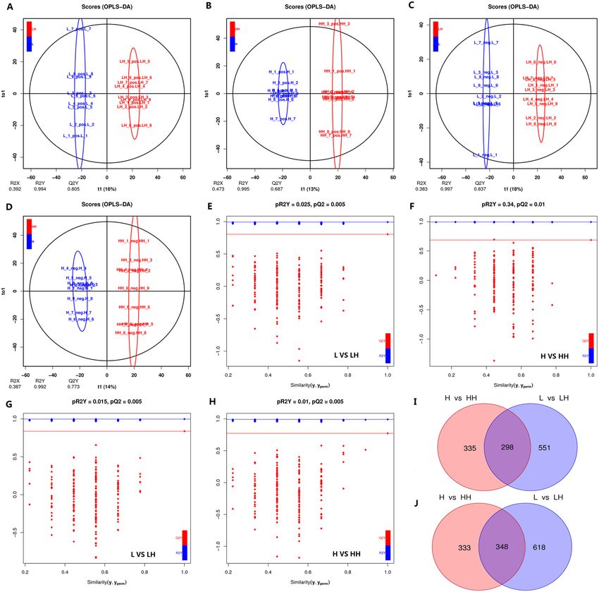
www.nature.com/scientificreports/                                                                  www.nature.com/scientificreports

                           thermal stress initially concentrate upon redeploying pathways to increase energy production12,13 and oxidation
                           resistance14. Many factors may be related to heat tolerance in these organisms, including the rate and range of
                           temperature change, animal size, state of ontogeny15, and heat exposure history16,17. At the same time, hereditary
                           factors also play essential roles in the adaptation to thermal stress. South African abalone has significant genetic
                           diversification18 and should have been a result of adaptive to different water temperatures between west and east
                           coast19. The biochemical reaction rates within abalone tissues increase with temperature rise, which leads to an
                           increase in the energy need to maintain homeostasis. Consequently, a coordinated response of various metabolic
                           pathways and oxidation of multiple energy substances is required20. For instance, under a high-temperature envi-
                           ronment, adenosine triphosphate (ATP) production is elevated in abalones by enhancing oxygen consumption
                           and activating the pathways for anaerobic metabolism12. In addition, the accessorial rate and extent of protein11
                           and lipids21 oxidation are also redeployed for ATP production, for a stable equilibrium between energy output
                           and energy consumption.
                               However, oxidative stress occurs when the adjustment of substrate oxidation fails to fully compensate for the
                           increased demand for metabolism at higher body temperature. The consequent accumulation of reactive oxygen
                           species can directly result in the oxidative damage of macromolecules, including lipids, proteins and nucleic
                           acids22. The side effects of oxidative damage on cells can in part be abolished by cellular antioxidant defence
                           systems or by the expression of heat shock proteins. Whereas the former mechanism requires antioxidases and
                           antioxidants to degrade free radicals to substances with lower toxicity, the latter mechanism entails the repair of
                           misfolded polypeptide chains of damaged proteins or the elimination of irretrievably damaged proteins. Both of
                           these mechanisms have been described in abalones23–25.
                               The hepatopancreas is an organ of the digestive tract of arthropods and molluscs. Mollusc hepatopancreas
                           plays an important role in energy storage, metabolite transformation, enzyme synthesis, and antioxidant pro-
                           tection26, indicating its potential as the ideal target tissue for the study of the metabolism mechanism of heat
                           stress on abalones. We thus investigated the abalone’s hepatopancreas with metabolomics based techniques. By
                           comparing the results of the phenotypic and metabolic differences between animals acclimated under different
                           seawater temperatures, we analyzed the heat tolerance and adaptation mechanisms of the juvenile Pacific abalone.

                           Methods
                           Ambient seawater temperature data collection.                Seawater temperature data were collected for two
                           typical abalone culture regions in northern and southern China. The data for the northern abalone culture region
                           was generated from the Xiaomaidao station of the National Marine Science Data Center (http://www.nmdis.org.
                           cn/). The data for the southern region was from Fuzhou Marine Fisheries Bureau (http://hyj.fuzhou.gov.cn/),
                           which averaged the seawater temperature from three aquaculture sites around Fuzhou city (Huangqi, Tongxin
                           bay, and Minjiang estuary). Monthly average sea surface temperature data of these two regions in the past three
                           years were collected.

                           Animals and acclimatization.              Pacific abalone juveniles (shell length: 26.2 ± 2.3 mm, shell width:
                           16.5 ± 2.5 mm, total wet weight: 2.17 ± 0.56 g) used in this study were purchased from a commercial farm in
                           Qingdao (Shandong, China), and were kept in culture tank at 20 ± 0.3 °C for two-week recovery. The animals
                           were then divided into two groups for a 62-day acclimatization period under 10 °C and 30 °C seawater controlled
                           by a digital temperature controller. The decrease rate of seawater temperature was 1 °C per day, while the increase
                           rate was 1 °C per day till 25 °C and then 0.5 °C per day to 30 °C. During this time, the seawater was aerated, and
                           the salinity was maintained at 31 ± 1. Excess feeding with brown alga Laminaria japonica was given daily. The
                           remaining food and excrement were removed the following day and renewed with clean, sand-filtered seawater
                           at the corresponding temperature. After the 62-day acclimatization, animals from each acclimated group were
                           recovered to 20 °C at a rate of + (-) 2 °C per day, and were then maintained for one week to make the L group
                           (10 °C acclimated) and H group (30 °C acclimated) respectively.

                           Acute heat treatment and sampling.                 The heat tolerance of L and H groups of juvenile abalones were
                           compared by metabolomics assay and survival measurement after exposure to 31 °C seawater. A total of 72 indi-
                           viduals from each group were randomly selected and transferred into 31 °C seawater immediately. After three
                           hours treatment, 27 individuals were randomly sampled from each group to make LH group (31 °C treated L
                           group) and HH group (31 °C treated H group) for metabolomics assay, while the remaining 45 individuals in
                           each group were continually cultured till 48 h for measuring survivals. Hepatopancreas tissues were dissected
                           from four groups of juvenile abalones (L, H, LH, and HH). Within each group, tissues from three abalones were
                           mixed and ground into powder in liquid nitrogen for subsequent metabolite extraction and metabonomic-based
                           analysis. In this way, nine biological replications were produced for each group.

                           Metabolite extraction. 50 mg of sample was taken and placed in an Eppendorf tube, followed by the addi-
                           tion of 1000 μl extraction solvent (Vmethanol:Vacetonitrile:Vwater = 2:2:1, containing internal standard 2 μg/ml). The mix-
                           ture was homogenized in ball mill for 4 min at 45 Hz, then ultrasound treated for 5 min (incubated in ice water).
                           After homogenization for three times, the mixture was incubated for one hour at −20 °C to precipitate proteins.
                           Centrifugation was performed at 12000 rpm for 15 min at 4 °C. A total of 825 μl supernatant was carefully trans-
                           ferred into a fresh Eppendorf tube and dried in a vacuum concentrator without heating. 200 μl extraction solvent
                           (Vacetonitrile:Vwater = 1:1) was then added for reconstitution, followed by vortex 30 s, sonicated for 10 min (4 °C water
                           bath), and centrifuge for 15 min at 12000 rpm (4 °C). 75 μl supernatant was transferred into a fresh 2 ml LC/MS
                           glass vial for UHPLC-QTOF-MS analysis, while another 20 μl was taken from each sample and pooled as QC
                           samples. Six technical replicates were conducted to QC sample with 75 μL each for UHPLC-QTOF-MS analysis.

Scientific Reports |   (2020) 10:6353 | https://doi.org/10.1038/s41598-020-63122-4                                                                  2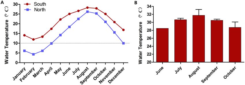
www.nature.com/scientificreports/                                                              www.nature.com/scientificreports

                           Figure 1. Seawater temperature data. (A) The monthly average temperature of south and north sites for the past
                           three years. (B) The maximum temperature of the south site in summer months for the past three years.

                           Total ion chromatogram and correlation analysis of quality control (QC) samples are presented in Supplementary
                           Figs. 1–6

                           Liquid chromatography tandem mass spectrometry analysis. LC-MS/MS analyses were performed
                           according to previous report27. In brief, the UHPLC system (1290, Agilent Technologies) with a UPLC BEH
                           Amide column (1.7 μm 2.1*100 mm, Waters) coupled to TripleTOF 6600 (Q-TOF, AB Sciex) was used. The
                           mobile phase consisted of 25 mM NH4OAc and 25 mM NH4OH in water (pH = 9.75) (A) and acetonitrile (B)
                           with gradient elution as follows: 0 min, 95% B; 7 min, 65% B; 9 min, 40% B; 9.1 min, 95% B; 12 min, 95% B, with
                           solvent delivery at a fixed rate of 0.5 mL/min. The sample injection volume was 3 μL. The triple time of flight
                           (TOF) mass spectrometer was used due to its ability to acquire MS/MS spectra on an information-dependent
                           basis (IDA) during an LC-MS experiment. In this mode, the acquisition software (Analyst TF 1.7, AB Sciex) con-
                           tinuously evaluates the full scan survey MS data as it is collected and triggers the acquisition of MS/MS spectra
                           depending on preselected criteria. In each cycle, 12 precursor ions whose intensities were greater than 100 were
                           chosen for fragmentation at collision energy (CE) of 30 V (15 MS/MS events with product ion accumulation time
                           of 50 msec each). The electrospray ionization (ESI) source conditions were set as follows: Ion source gas 1 as 60
                           Psi, Ion source gas 2 as 60 Psi, Curtain gas as 35 Psi, source temperature 600 °C, Ion Spray Voltage Floating (ISVF)
                           5000 V or −4000 V in positive or negative ion mode, respectively.

                           Data preprocessing and annotation.              According to previous report27, MS raw data files were converted
                           to the mzXML format using ProteoWizard28, and processed by R29 package XCMS (version 3.2)30. The preproc-
                           essing results generated a data matrix that consisted of the retention time, mass-to-charge ratio (m/z) values, and
                           peak intensity. R package CAMERA31 was used for peak annotation after XCMS data processing. In-house MS2
                           database and KEGG32, PubChem33, ChEBI34 and HMDB35 were applied in metabolites identification. The identi-
                           fied metabolites’ function annotations were excerpted from the description in HMDB, PubChem and UniProt36.

                           Identification of differential metabolites. Orthogonal projections to latent structure-discriminant
                           analysis (OPLS-DA) was applied to the analysis of the metabolite content to obtain more reliable relevant dif-
                           ferential metabolites (DEMs) information between experimental groups referring to the method of Thevenot’s
                           report37. The parameters of the evaluation of the OPLS-DA model included R2X, R2Y, and Q2Y. The R2X and R2Y
                           represented the accounted rate of the developed model to the X and Y parameter matrices, respectively, while
                           Q2Y indicated the predictive capability of the model to Y. The model was considered more reliable when R2X,
                           R2Y, and Q2Y were closer to 1, and was considered adequate when Q2Y > 0.5 and excellent when Q2Y > 0.9. The
                           model quality was evaluated by cross validation. A VIP value was given to each metabolite by OPLS-DA model
                           to express the degree of its contribution to the model construction. In other words, the VIP value demonstrates
                           the closeness of the metabolite and heat shock treatment to a linear relationship. DEMs were screened by com-
                           bining fold change (FC), P values of Student’s t-test and VIP value of each metabolite based on OPLS-DA. A set
                           of substances from differential expression analysis was referred to the DEM set, named as “A-B”. Metabolites with
                           concentration higher in sample B than in sample A were designated upregulated DEMs, while those with a lower
                           level in sample B compared with sample A were downregulated DEMs.

                           Results
                           Ambient seawater temperature of different abalone culture regions. Abalones cultured in the
                           northern sea area experienced at least four months low temperature (averagely < 10 °C) annually (Fig. 1A), while
                           that in the southern sea area experienced as many as five months hot temperature (average maximum seawater
                           temperature was higher than 28 °C, Fig. 1B). The maximum water temperature in the Fuzhou aquaculture area
                           was around 30 °C from July to September. In this context, acclimation temperatures were designed as 10 °C and
                           30 °C, to compare the heat response differences of abalones.

                           Heat tolerance comparison. Within the 48 hours of heat treatment at 31 °C, abalone juveniles from L
                           and H groups displayed significantly different survival rates (Fig. 2). All animals from the L group died within

Scientific Reports |   (2020) 10:6353 | https://doi.org/10.1038/s41598-020-63122-4                                                           3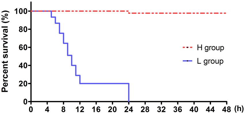
www.nature.com/scientificreports/ www.nature.com/scientificreports
Figure 2. Kaplan-Meyer survival analysis of different Pacific abalone juveniles at 31 °C heat shock. L group,
Pacific abalone juveniles acclimated for 62 days at 10 °C and then cultured for 7 days at 20 °C; H group, Pacific
abalone juveniles acclimated for 62 days at 30 °C and then recovered 7 days at 20 °C.
24 hours, showing high sensitivity to extremely high temperatures. Whereas, only one individual from the H
group died within the 48 hours of observation, indicating tolerance to extremely high temperature.
DEMs identification. OPLS-DA model distinguished group pairs well (L-LH and H-HH) with metab-
olites detected from either positive or negative ion mode (Fig. 3A–D, positive ion mode: R2YL- LH = 0.994,
R2YH-HH = 0.995, Q2YL-LH = 0.805, Q2YH-HH = 0.687; and negative ion mode: R2YL-LH = 0.997, R2YH-HH = 0.992,
Q2YL-LH = 0.837, Q2YH-HH = 0.773). Permutation test confirmed the validation (pQ2Y < 0.05) of OPLS-DA mod-
els (Fig. 3E–H). With P < 0.05 and VIP > 1 as threshold, 1815 (849 in positive ion mode and 966 in negative ion
mode) and 1314 (633 and 681) DEMs were identified in L-LH and H-HH comparison pairs, respectively. Besides,
there were 298 (positive ion mode) and 348 (negative ion mode) overlapped DEMs between the two comparison
pairs (Fig. 3I,J).
Metabolite biomarkers. Within the 1815 and 1314 identified DEMs, annotated substances account for 247
(Supplementary Table S1) and 241 (Supplementary Table S2) in L-LH and H-HH comparison pairs, respectively.
Stricter criteria were then used with FC > 1.5 or 1.5 to identify metabolite biomarkers
(MBs) within annotated DEMs. As a result, 42 substances (34 of which were upregulated and eight downregu-
lated) were obtained in L-LH comparison pair (Supplementary Table S3), while 100 were selected in H-HH set
(92 upregulated and eight downregulated, Supplementary Table S4). There were 28 MBs, which were all upreg-
ulated, overlapped by both comparison pairs (Table 1). Functional annotation on the 28 overlapped substances
indicated the possible dysfunction of some metabolic enzymes either localized in the mitochondria or the cyto-
plasm. The mitochondrion enzymes possibly being influenced were mainly involved in the oxidative catabolism
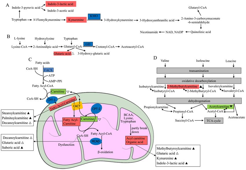
of amino acids and fatty acids (Fig. 4).
Functional annotation of MBs specifically represented in each comparison pair was also conducted. In the
L-LH set, the remaining six upregulated MBs after removing the 28 overlapped MBs were regarded as L-LH
specific (Table 2). Similarly, 64 MBs were obtained as H-HH specific, in which the top 15 was listed in Table 2.
There were significant differences regarding the functions of the two specific MBs sets, suggesting different heat
response at metabolites accumulation level of L and H groups. The six MBs in the L-LH set suggested the occur-
rence of oxidative stress. In comparison, the top 15 MBs in the H-HH specific set consisted of substances benefi-
cial to metabolic homeostasis, including various dipeptides, antioxidants, and neuroprotective substances.
The oxidative metabolism occurring in the mitochondria, the powerhouse of cells, is crucial to heat stress
response and heat tolerance. Figure 4 shows the effect of heat stress on substances related to the metabolism of
amino acids and fatty acids in the mitochondria of hepatopancreas tissues of juvenile abalones.
Discussion
After experiencing two weeks of homogeneous environment culture, juvenile abalones from 30 °C acclima-
tion group exhibited higher survival than 10 °C group, under short-term extreme heat stress (31 °C). The result
highlights the role of acclimation in the heat stress response of abalones. We inferred that high-temperature
acclimation exerts specific effects on metabolic processes in abalones, which allow them to respond quickly in a
physiologically beneficial manner when exposed to heat stress11, predisposing them to survive in a harsh environ-
ment. Based on this hypothesis, a metabolomics study was conducted to further assay the physiological biochem-
istry adaptation mechanism of juvenile abalone to heat stress.
Homeostasis of physiological processes requires the activation of various metabolic pathways to increase
energy supply for animals in response to environmental stress. Temperature changes can alter the biochemi-
cal reaction rate of many enzymatic processes in poikilothermic animals, whereas acute heat stress can induce
prompt adjustment38. In this study, a variety of intermediates produced from incomplete degradation of amino
and fatty acids were identified to be significantly accumulated in the hepatopancreas of thermally treated aba-
lone juveniles (HH-group and LH-group), indicating that excessive ambient temperature leads to dysfunction
of pathways utilizing protein and lipid oxidation as energy supply. This should exacerbate the energy shortage
experienced by the heat-stressed animals. Mitochondria, the powerhouse of cells, is the main site for amino and
fatty acids metabolism. Heat stress affects the normal function of mitochondrial oxidative metabolism39. It causes
accumulation of intermediates, some of which were reported to disturb cell homeostasis and hinder cellular
function, leading to pathophysiological processes such as muscle spasm, muscle weakness40, heart failure, cardiac
arrest, and sudden death41,42.
Scientific Reports | (2020) 10:6353 | https://doi.org/10.1038/s41598-020-63122-4 4www.nature.com/scientificreports/ www.nature.com/scientificreports
Figure 3. OPLS-DA model analysis and DEMs identification. (A–D) are OPLS-DA model analysis results
(A,B) data from positive ion mode; (C,D), negative ion mode) based on LC/MS data of metabolites from Pacific
abalone hepatopancreas samples of control (blue) and heat shock groups (red). Permutation test confirmed the
validation of each model (E–H). (I,J) show Venn diagram analysis results of DEMs identified from different
comparison pairs. L, Pacific abalone juveniles acclimated for 62 days at 10 °C and then cultured for 7 days
at 20 °C; LH, Pacific abalone juveniles from L group treated 3 hours by 31 °C heat shock; H, Pacific abalone
juveniles acclimated 62 days at 30 °C and then recovered after 7 days at 20 °C; HH, Pacific abalone juveniles
from H group treated for 3 hours with 31 °C heat shock. Image was created by R software version 3.5.3 (https://
www.r-project.org/).
As reported in previous studies, abalones experienced elevated ammonia excretion rate and weight loss when
reared under high temperature11,24, suggesting the increased contribution of protein/amino acid oxidation to their
energy supply. The metabolomics-based studies on small abalone H. diversicolor43, sea cucumber Apostichopus
japonicushas38,44 also showed the influence of heat stress on amino acid metabolism. In this study, tissue concen-
trations of two tryptophan metabolic intermediates significantly increased after heat stress (Table 1): L-kynurenine
and indole-3-lactic acid (3-IAA). In humans, the aromatic amino acid L-tryptophan has two main metabolic path-
ways (Fig. 4A). The kynurenine pathway accounts for more than 95% of tryptophan oxidative catabolism, while the
other pathway includes a series of indole compounds45. The dysfunction or absence of the mitochondrial enzyme
kynurenine-3-monooxygenase (EC: 1.14.13.9) results in a significant accumulation of kynurenine46, which is also
associated with human neuromuscular dysfunction47. Kynurenine elevation in heat-treated abalone may reflect the
breakdown of mitochondrial function and possible disorder of normal physiological function of the neuromuscular
Scientific Reports | (2020) 10:6353 | https://doi.org/10.1038/s41598-020-63122-4 5www.nature.com/scientificreports/ www.nature.com/scientificreports
L-LH H-HH
No. Name Function FC VIP FC VIP
Component of the coenzyme NAD, cofactor, antioxidant,
1 Nicotinamide 11.53 2.08 11.21 1.55
neuroprotective agent, anti-inflammatory agent.
2 (-)-Naringenin Inhibitor to cytochrome P450 isoform CYP1A2. 8.74 1.68 6.22 1.77
3 O-Acetyl-L-serine Takes part in cysteine and glutathione metabolism. 5.16 2.2 5.47 1.78
4 Maleamic acid Conjugate acid of a maleamate. 5.94 2.08 5.06 1.74
5 2-Methylbutyroylcarnitine* Intermediate of isoleucine metabolism. 9.55 2.36 4.55 2.25
Metabolite biomarker suggests carnitine palmitoyltransferase
6 Stearoylcarnitine* 4.15 1.97 3.71 2.46
(CPT) II deficiency.
Metabolite from the metabolism of methylxanthines (caffeine,
7 1,3-Dimethyluric acid 3.54 2.21 3.59 2.26
theophylline, and theobromine) mainly catalyzed by CYP1A2.
Chelator of Ca , Mg
2+ 2+
and polymer to create an ion-exchange
8 Iminodiacetic acid 3.6 2.34 3.45 2.38
resin.
9 Imidazoleacetic acid Metabolite product of histamine metabolism. 1.52 1.7 3.45 2.38
A metabolite from the metabolism of methylxanthines (caffeine,
10 1,7-Dimethyluric acid 2.19 1.78 3.33 1.6
theophylline, and theobromine) mainly catalyzed by CYP1A2.
Essential molecule for membranes biosynthesis and repair,
11 CDP-choline 2.72 2.26 3.11 2.12
molecule for neuroprotection.
12 Amrinone Acardiotonic, vasodilator. 2.08 2.04 2.8 2.06
13 DL-Indole-3-lactic acid* Metabolite of tryptophan. 1.93 2.14 2.67 2.36
Metabolite biomarker suggests deficiencies of medium-chain
14 Suberic acid* acyl-CoA dehydrogenase, carnitine-acylcarnitine translocase, or 1.85 1.76 2.34 2.14
malonyl-CoA decarboxylase.
15 alpha-Guanidinoglutaric acid* Derivative of glutaric acid, which can induce seizures. 1.95 2.28 2.33 2.36
16 Creatine Facilitator of ATP biosynthesis. 1.75 2.08 2.19 2.37
An important component of enzymes involved in metabolizing
17 D-Biotin 2.24 2.05 2.19 1.75
fats and carbohydrates. Maintains cellular metabolism.
18 MDMA Nervous stimulant. Facilitates ATP production. 1.96 2.16 2.11 2.24
Non-selective and irreversible monoamine oxidase inhibitor,
19 Phenelzine 1.73 2.14 2.08 2.35
neuroregulator.
Metabolite from the metabolism of methylxanthines (caffeine,
20 7-Methylxanthine 1.54 1.69 2.05 1.95
theophylline, and theobromine) mainly catalyzed by CYP1A2.
Metabolite biomarker suggests aromatic L-amino acid
21 N-Acetyl-L-tyrosine# 1.92 1.72 2.04 1.59
decarboxylase deficiency.
Metabolite could stabilize the chemical balance with side effect of
22 Acamprosate 2.11 1.85 2.03 1.6
irregular heartbeats.
23 Erythrono-1,4-lactone Lactone of tetronic acid. 2.07 2.42 1.79 2.29
Post-transcriptional modifications found in tRNA, snRNA, and
24 5-Methylcytidine 1.86 2.21 1.72 1.95
rRNA.
Metabolite involved in tryptophan metabolism. Breakdown
25 5-Hydroxyindoleacetate# 1.98 1.75 1.72 1.84
product of serotonin.
Inhibitor of chloride channels and T-type calcium channels.
26 Niflumic Acid 2.06 1.85 1.7 1.65
Inhibitor of phospholipase A2 and cyclooxygenase-2.
Intermediate metabolite of tryptophan. Metabolite biomarker
27 L-Kynurenine* 1.81 2.18 1.64 1.73
suggests kynurenine-3-monooxygenase deficiency.
Metabolite biomarker suggests carnitine palmitoyltransferase
28 L-Palmitoylcarnitine* 1.78 1.54 1.62 1.8
(CPT) II deficiency.
Table 1. Functional annotation of overlapped metabolite biomarkers between L-LH and H-HH sets. Note:
HMDB, PubChem, and UniProt were applied in metabolites function annotation. *Indicates metabolites
associated with enzymes in mitochondria; #Indicates metabolites associated with enzymes in cytoplasm.
system. The data regarding the cytotoxicity of 3-IAA are limited. Some studies indicated that this metabolite could
activate p38, c-Jun N-terminal kinases, caspase-8, caspase-9, and caspase-3 to induce cell apoptosis48. Another
amino acid-related metabolic biomarker, glutaric acid, was identified to be increased in the LH group. The mas-
sive accumulation of glutaric acid is attributable to the dysfunction of many mitochondrial enzymes, including
glutaryl-CoA dehydrogenase (EC: 1.3.8.6), electron transfer flavoprotein (ETF), and ETF-ubiquitin oxidoreductase
(ETF-QO) (EC: 1.5.5.1)49. Glutaryl-CoA dehydrogenase catalyzes glutaryl-CoA to generate crotonyl-CoA and CO2
during the mitochondrial oxidative metabolism of lysine, tryptophan, and hydrolysine (Fig. 4B). Due to the dysfunc-
tion of this enzyme, glutaryl-Co A is directed toward glutaric acid production50. Excessive accumulation of glutaric
acids and derivatives can cause a variety of neurometabolic and muscular disturbances40, partially explaining the
poor performance of L group under heat stress.
Massive accumulation of substances related to fatty acid metabolism was also observed in this study. Two carni-
tine linked metabolites were identified in MBs: stearoylcarnitine and L-palmitoylcarnitine (Table 1), accumulation
of which has been recognized to indicate the malfunction or absence of mitochondrial enzyme carnitine palmito-
yltransferase II (CPT-2, EC: 2.3.2.21)51. Carnitine plays an obligate role in long-chain fatty acids transferring from
Scientific Reports | (2020) 10:6353 | https://doi.org/10.1038/s41598-020-63122-4 6www.nature.com/scientificreports/ www.nature.com/scientificreports
Figure 4. Effect of heat stress on metabolites in the mitochondria of juvenile Pacific abalones. ▲ indicates
metabolites significantly increased in both L and H groups after heat stress; ▼indicates metabolites significantly
decreased in both L and H groups after heat stress; △ and ▽ indicate metabolites significantly increased and
decreased, respectively, in only L group after heat stress. FACS, fatty acyl-CoA synthase; CPT-1, 1-type carnitine
palmitoyltransferase; CPT-2, 2-type carnitine palmitoyltransferase; CACT, carnitine acylcarnitine translocase;
MCAD, medium chain acyl-CoA dehydrogenase; BCAA, branched-chain amino acid; GD, glutaryl-CoA
dehydrogenase; KMO: kynurenine monooxygenase. Blue background indicates enzyme protein; orange
background indicates channel protein; red background indicates significantly accumulated metabolites; green
background indicates significantly reduced metabolites.
the cytoplasm to the mitochondrial matrix for subsequent β-oxidation52. The import was mainly conducted by com-
bined reactions of fatty acyl-CoA synthase (FACS, EC: 6.2.1.3), carnitine palmitoyltransferase (CPT-1 and CPT-2)
and carnitine acylcarnitine translocase (CACT) (Fig. 4C)52. CPT-1 (EC: 2.3.1.21) is localized in the outer mitochon-
drial membrane, where it is responsible for exchanging the CoA for carnitine to produce fatty acyl-carnitines (e.g.
stearoylcarnitine and L-palmitoylcarnitine) which then enters the mitochondrial matrix via facilitated diffusion by
CACT. The fatty acyl-carnitine complex is then disrupted by CPT-2, and the fatty acid rebinds to CoA in the mito-
chondria, where the replaced carnitine then diffuses back across the membrane by CACT into the mitochondrial
intermembrane space53. Consequently, CPT-2 dysfunction causes accumulation of long-chain fatty acyl-carnitines
(e.g. stearoylcarnitine and L-palmitoylcarnitine) in cells while reducing the carnitine content51. Significantly dimin-
ished L-carnitine was indeed observed in the L-LH pair (Supplementary Table S1).
Furthermore, carnitine is also involved in the catabolism of branched-chain amino acid (e.g. isoleucine
and valine)52. High level of 2-Methylbutyroylcarnitine (Table 1, Fig. 4D) is indicative of the function loss of the
enzyme short/branched chain specific acyl-CoA dehydrogenase (EC: 1.3.8.-), which is responsible for catalyz-
ing the third metabolic step in the pathway for mitochondrial oxidative catabolism of isoleucine and valine54.
Elevated stearoylcarnitine, L-Palmitoylcarnitine, and 2-Methylbutyroylcarnitine content in the hepatopancreas
of heated abalone juveniles indicated the accumulation of these intermediates in mitochondrial, instead of pro-
ceeding to downstream oxidation. Accumulation of long-chain fatty acyl-carnitines was also observed during cell
apoptosis process55. Accumulated palmitoylcarnitines was reported to be able to activate caspase 3, 7, and 8, and
initiate cell apoptosis55. Furthermore, fatty acids combined with carnitines can also affect redox chemistry, impair
tissue function, and cause myocardial injury through perturbed membrane molecular dynamics, potentially trig-
gering cardiac arrhythmia and cardiac arrest41,42.
The speculation of fatty acid oxidation disorders of abalone under heat stress was further supported by the
observation of the suberic acid accumulation (Table 1)56 and significant accumulation of an intermediate of
medium-chain fatty acid, Decanoyl-L-carnitine, in LH group (Supplementary Table S1). In humans, syndrome
with hypoglycemia, hypoketonemia, cardiomyopathy, neuronal migration abnormalities, and other tissues patho-
logical signature are usually presented during fatty acid oxidation disorders57. Hypoketonemia of abalone under
heat stress was also observed in this study: significantly reduced concentration of acetoacetic acid in both L-LH
and H-HH comparison pairs (Supplementary TableS S1 and S2). We failed to detect the other two main ketone
Scientific Reports | (2020) 10:6353 | https://doi.org/10.1038/s41598-020-63122-4 7www.nature.com/scientificreports/ www.nature.com/scientificreports
Set Name Description FC P VIP
Pyrimidine base, product of CTP dephosphorylation. The CTP
L-LH Cytosine 5.71 8.37E-04 1.84
can transfer a phosphate to convert ADP to ATP.
L-Gulonic gamma-lactone Direct precursor of vitamin C. 2.83 4.71E-04 1.85
Phenoxybenzamine Metabolite causes vasodilatation and increases cardiac output. 2.55 7.38E-03 1.68
Lavandulol Pheromone. 2.06 5.57E-04 2.00
Neurotoxin, mitochondrial toxin, and metabotoxin when at a high
beta-Alanine 1.74 5.48E-05 2.09
level.
Product during metabolism of some amino acids. Act as an
Glutaric acid 1.62 1.66E-03 1.89
acidogen and a metabotoxin when at high levels.
H-HH Pro-Tyr Dipeptide. 5.14 9.23E-04 2.15
Pro-Asn Dipeptide. 2.13 5.52E-04 2.15
Thr-Ala Dipeptide. 2.10 4.08E-04 2.09
D-Glucose-6-phosphate Metabolic hub. 3.20 1.37E-02 1.68
Ammelide Hydrolysis product of melamine. 2.33 7.10E-05 2.17
Metabolite influencing the regulation of gene expression,
S-Methyl-5’-thioadenosine 2.07 6.65E-03 1.83
proliferation, differentiation, and apoptosis.
4-Hydroxycinnamic acid Important source of antioxidants. 2.05 2.54E-05 2.31
trans-2-Hydroxycinnamic acid Important source of antioxidants. 2.02 5.25E-06 2.39
Theobromine Vasodilator, diuretic and heart stimulator. 2.01 6.87E-03 1.70
Metabolite initiating a protective response to stress, modulating a
N-Acetyl-D-glucosamine 2.00 1.91E-03 1.90
cell’s capacity to grow and divide, and regulates gene transcription.
Tiopronin Reducing agent, neuroprotective agent. 1.99 5.42E-05 2.23
Metabolite could be converted to tyrosine, used in the biosynthesis
L-Phenylalanine 1.97 1.10E-02 1.66
of dopamine and norepinephrine neurotransmitters.
Dimethylglycine Byproduct of the metabolism of choline. 1.95 6.11E-03 1.78
Phe-Ala Dipeptide. 1.92 1.74E-02 1.52
Ajmalicine Monoterpenoid indole alkaloid, vasodilator agent. 1.87 4.52E-03 1.8
Table 2. Functional annotation of set specific metabolite biomarkers. Note: HMDB, PubChem, and UniProt
were applied in metabolites function annotation.
bodies possibly because the volatile acetone is undetectable by the LC-MS/MS technology used in this study,
while hydroxybutyric acid usually occurs at low concentration in animal cells. At the same time, neuronal migra-
tion abnormalities of heat-treated abalones, i.e. pathophysiological symptoms of rotating and shaking the shell,
stiff muscles, and loss of adhesion, fainting, and cardiac arrest, were also observed in this study, consistent with
observations in other studies and other Haliotis species58.
Besides the accumulation of more intermediates related to mitochondrial dysfunction in the LH group, LH and
HH groups showed significantly different metabolic responses to heat stress. The LH group specifically accumulated
some MBs harmful to cell homeostasis. Among the six L-LH specific MBs, L-Gulonic gamma-lactone is the sub-
strate of the last step of the biosynthesis of vitamin C59, a powerful reducing agent. Its accumulation should indicate
the deficiency of vitamin C biosynthesis ability under heat stress, if the abalone has any. Beta-Alanine is a neurotoxin,
mitochondrial toxin, and metabotoxin at elevated levels in human nerve tissues60. However, H-HH specific MBs
enriched some substances beneficial to metabolic homeostasis. S-Methyl-5’-thioadenosine is a metabolite that can
influence the regulation of gene expression, proliferation, differentiation, and apoptosis61. Both 4-Hydroxycinnamic
acid and trans-2-hydroxycinnamic acid were important sources of antioxidants62,63. Both theobromine and ajmali-
cine were regarded as vasodilators. N-Acetyl-D-glucosamine is a metabolite that can initiate a protective response
to stress, modulate the cells’ capacity to grow and divide, and regulate gene transcription64. Tiopronin is a reducing
agent and a neuroprotective agent. Juvenile abalones in the HH group showed quicker response to regulate the pro-
duction of metabolic promotion and stabilization, cell injury repair agents, and neuroprotection after heat treatment,
which could explain their adaptation mechanism to heat tolerance. However, more molecular studies on mitochon-
drial activities during heat response are still needed to support our findings.
The mitochondria function as the cellular powerhouse and are responsible for accommodating increased energy
demand during heat stress. Therefore, the structure and function of mitochondria in the Pacific abalone need to be
stabilized to sustain their tolerance to heat stress. The single population is limited to reflect the general heat response
scenario of the Pacific abalone. Environmental metabolomics response in abalones is worthy of further investiga-
tions, as well as molecular studies on mitochondrial activities during heat response in marine animals.
Conclusion
We studied the metabolic mechanism of heat tolerance in juvenile Pacific abalones. While comparing different
groups with different acclimation history, we found that substances involved in the oxidation decomposition of
amino acids and fatty acids were accumulated, which indicated dysfunction of mitochondrial enzymes under heat
stress. Abnormal accumulation of these metabolites aggravates stress response and leads to pathophysiological
processes, especially when the organisms face increased energy demand under conditions of increased ambient
temperature. Individuals with high-temperature acclimation history produced more beneficial substances for
Scientific Reports | (2020) 10:6353 | https://doi.org/10.1038/s41598-020-63122-4 8www.nature.com/scientificreports/ www.nature.com/scientificreports
metabolic homeostasis. The findings revealed the critical role of mitochondria in heat response of animals and
provided insight into the heat adaptation of marine molluscs.
Data availability
All raw data files are available on the NIH Metabolomics Workbench website (DataTrack ID: 1913). The datasets
used and analyzed during the current study are also available from the corresponding author on reasonable
request.
Received: 10 December 2019; Accepted: 23 March 2020;
Published: xx xx xxxx
References
1. Pörtner, H. O. & Farrell, A. P. Physiology and climate change. Science 322, 690–692, https://doi.org/10.1126/science.1163156 (2008).
2. Hoegh-Guldberg, O. & Bruno, J. F. The impact of climate change on the world’s marine ecosystems. Science 328, 6, https://doi.
org/10.1126/science.1189930 (2010).
3. Morash, A. J. & Alter, K. Effects of environmental and farm stress on abalone physiology: perspectives for abalone aquaculture in the
face of global climate change. Rev. Aquac 8, 342–368, https://doi.org/10.1111/raq.12097 (2016).
4. Soletchnik, P., Ropert, M., Mazurié, J., Gildas Fleury, P. & Le Coz, F. Relationships between oyster mortality patterns and
environmental data from monitoring databases along the coasts of France. Aquaculture 271, 384–400, https://doi.org/10.1016/j.
aquaculture.2007.02.049 (2007).
5. Jiang, W. et al. Effects of temperature change on physiological and biochemical responses of Yesso scallop, Patinopecten yessoensis.
Aquaculture 451, 463–472, https://doi.org/10.1016/j.aquaculture.2015.10.012 (2016).
6. Lu, Y. et al. Insight into the heat resistance of fish via blood: Effects of heat stress on metabolism, oxidative stress and antioxidant
response of olive flounder Paralichthys olivaceus and turbot Scophthalmus maximus. Fish Shellfish Immunol 58, 125–135, https://doi.
org/10.1016/j.fsi.2016.09.008 (2016).
7. Kühnhold, H. et al. Thermal stress effects on energy resource allocation and oxygen consumption rate in the juvenile sea cucumber,
Holothuria scabra (Jaeger, 1833). Aquaculture 467, 109–117, https://doi.org/10.1016/j.aquaculture.2016.03.018 (2017).
8. Li, Q., Shu, J., Yu, R. & Tian, C. Genetic variability of cultured populations of the Pacific abalone (Haliotis discus hannai Ino) in
China based on microsatellites. Aquacult. Res. 38, 981–990, https://doi.org/10.1111/j.1365-2109.2007.01764.x (2007).
9. Liang, S., Luo, X., You, W., Luo, L. & Ke, C. The role of hybridization in improving the immune response and thermal tolerance of
abalone. Fish Shellfish Immunol 39, 69–77, https://doi.org/10.1016/j.fsi.2014.04.014 (2014).
10. Hickey, A. J. R. & Wells, R. M. G. Thermal constraints on glycolytic metabolism in the New Zealand abalone, Haliotis iris: The role
of tauropine dehydrogenase. N. Z. J. Mar. Freshwat. Res 37, 723–731, https://doi.org/10.1080/00288330.2003.9517202 (2003).
11. Vosloo, D., Vosloo, A., Morillion, E. J., Samuels, J. N. & Sommer, P. Metabolic readjustment in juvenile South African abalone
(Haliotis midae) acclimated to combinations of temperature and dissolved oxygen levels. J. Therm. Biol. 38, 458–466, https://doi.
org/10.1016/j.jtherbio.2013.07.001 (2013).
12. Pörtner, H. O., Bock, C. & Mark, F. C. Oxygen- and capacity-limited thermal tolerance: bridging ecology and physiology. J. Exp. Biol.
220, 2685–2696, https://doi.org/10.1242/jeb.134585 (2017).
13. Sokolova, I. M. & Portner, H. O. Metabolic plasticity and critical temperatures for aerobic scope in a eurythermal marine invertebrate
(Littorina saxatilis, Gastropoda: Littorinidae) from different latitudes. J. Exp. Biol 206, 195–207, https://doi.org/10.1242/jeb.00054 (2003).
14. Lushchak, V. I. Environmentally induced oxidative stress in aquatic animals. Aquat. Toxicol. 101, 13–30, https://doi.org/10.1016/j.
aquatox.2010.10.006 (2011).
15. Baker, S. C. & Heidinger, R. C. Upper lethal temperature tolerance of fingerling black crappie. J. Fish Biol. 48, 1123–1129, https://doi.
org/10.1111/j.1095-8649.1996.tb01809.x (1996).
16. Strange, K. T., Vokoun, J. C. & Noltie, D. B. Thermal tolerance and growth differences in orangethroat darter (Etheostoma spectabile)
from thermally contrasting adjoining streams. Am. Midl. Nat 148, 120–128 (2002). doi:10.1674/0003-0031(2002)148[0120:ttagdi]2
.0.co;2.
17. Das, T. et al. Thermal tolerance and oxygen consumption of Indian Major Carps acclimated to four temperatures. J. Therm. Biol. 29,
157–163, https://doi.org/10.1016/j.jtherbio.2004.02.001 (2004).
18. Rhode, C., Bester-van der Merwe, A. E. & Roodt-Wilding, R. An assessment of spatio-temporal genetic variation in the South
African abalone (Haliotis midae), using SNPs: implications for conservation management. Conserv. Genet. 18, 17–31, https://doi.
org/10.1007/s10592-016-0879-5 (2017).
19. Rhode, C., Vervalle, J., Bester-van der Merwe, A. E. & Roodt-Wilding, R. Detection of molecular signatures of selection at
microsatellite loci in the South African abalone (Haliotis midae) using a population genomic approach. Mar. Genom 10, 27–36,
https://doi.org/10.1016/j.margen.2013.03.001 (2013).
20. Sokolova, I. M. & Lannig, G. Interactive effects of metal pollution and temperature on metabolism in aquatic ectotherms:
implications of global climate change. Clim. Res 37, 181–201, https://doi.org/10.3354/cr00764 (2008).
21. Durazo, E. & Teresa Viana, M. Fatty acid profile of cultured green abalone (Haliotis fulgens) exposed to lipid restriction and long-
term starvation. Cienc. Mar. 39, 363–370, https://doi.org/10.7773/cm.v3914.2283 (2013).
22. Tomanek, L. Proteomic responses to environmentally induced oxidative stress. J. Exp. Biol. 218, 1867–1879, https://doi.org/10.1242/
jeb.116475 (2015).
23. De Zoysa, M. et al. Transcriptional analysis of antioxidant and immune defense genes in disk abalone (Haliotis discus discus) during
thermal, low-salinity and hypoxic stress. Comp. Biochem. Physiol. B-Biochem. Mol. Biol. 154, 387–395, https://doi.org/10.1016/j.
cbpb.2009.08.002 (2009).
24. Vosloo, D. & Vosloo, A. Response of cold-acclimated, farmed South African abalone (Haliotis midae) to short-term and long-term
changes in temperature. J. Therm. Biol. 35, 317–323, https://doi.org/10.1016/j.jtherbio.2010.06.006 (2010).
25. Li, J., He, Q., Sun, H. & Liu, X. Acclimation-dependent expression of heat shock protein 70 in Pacific abalone (Haliotis discus hannai Ino)
and its acute response to thermal exposure. Chin. J. Oceanol. Limnol. 30, 146–151, https://doi.org/10.1007/s00343-012-1026-x (2012).
26. Carefoot, T. H., Taylor, B. E. & Land, S. Use of isolated digestive-gland cells in the study of biochemical and physiological processes in
gastropod molluscs. Comp. Biochem. Physiol. A-Mol. Integr. Physiol 125, 497–502, https://doi.org/10.1016/s1095-6433(00)00181-1 (2000).
27. Cao, Y. W. et al. Untargeted liquid chromatography coupled with mass spectrometry reveals metabolic changes in nitrogen-deficient
Isatis indigotica Fortune. Phytochemistry 166, 112058, https://doi.org/10.1016/j.phytochem.2019.112058 (2019).
28. Chambers, M. C. et al. A cross-platform toolkit for mass spectrometry and proteomics. Nat. Biotechnol. 30, 918–920, https://doi.
org/10.1038/nbt.2377 (2012).
29. R Core Team. R: A language and environment for statistical computing. R Foundation for Statistical Computing, Vienna, Austria.
URL https://www.Rproject.org/ (2019).
30. Tautenhahn, R., Böttcher, C. & Neumann, S. Highly sensitive feature detection for high resolution LC/MS. BMC Bioinformatics 9,
504–504, https://doi.org/10.1186/1471-2105-9-504 (2008).
Scientific Reports | (2020) 10:6353 | https://doi.org/10.1038/s41598-020-63122-4 9www.nature.com/scientificreports/ www.nature.com/scientificreports
31. Kuhl, C., Tautenhahn, R., Böttcher, C., Larson, T. R. & Neumann, S. CAMERA: an integrated strategy for compound spectra
extraction and annotation of liquid chromatography/mass spectrometry data sets. Anal. Chem. 84, 283–289, https://doi.org/10.1021/
ac202450g (2012).
32. Kanehisa, M. & Goto, S. KEGG: kyoto encyclopedia of genes and genomes. Nucleic Acids Res. 28, 27–30, doi:gkd027 [pii] (2000).
33. Kim, S. et al. PubChem 2019 update: improved access to chemical data. Nucleic acids research. 47(D1), D1102–D1109, https://doi.
org/10.1093/nar/gky1033 (2019).
34. Hastings, J. et al. ChEBI in 2016: Improved services and an expanding collection of metabolites. Nucleic Acids Res 44, D1214–1219,
https://doi.org/10.1093/nar/gkv1031 (2016).
35. Wishart, D. S. et al. HMDB 4.0: the human metabolome database for 2018. Nucleic Acids Res 46, D608–d617, https://doi.org/10.1093/
nar/gkx1089 (2018).
36. The UniProt Consortium. UniProt: a worldwide hub of protein knowledge. Nucleic Acids Res 47, D506–D515, https://doi.
org/10.1093/nar/gky1049 (2019).
37. Thevenot, E. A., Roux, A., Xu, Y., Ezan, E. & Junot, C. Analysis of the human adult urinary metabolome variations with age, body
mass index, and gender by implementing a comprehensive workflow for univariate and OPLS Statistical Analyses. J. Proteome Res.
14, 3322–3335, https://doi.org/10.1021/acs.jproteome.5b00354 (2015).
38. Shao, Y. et al. Metabolomic responses of sea cucumber Apostichopus japonicus to thermal stresses. Aquaculture 435, 390–397,
https://doi.org/10.1016/j.aquaculture.2014.10.023 (2015).
39. Qian, L., Song, X., Ren, H., Gong, J. & Cheng, S. Mitochondrial mechanism of heat stress-induced injury in rat cardiomyocyte. Cell
Stress Chaperones 9, 281–293, https://doi.org/10.1379/csc-20r.1 (2004).
40. Chow, S. L., Rohan, C. & Morris, A. A. M. Case report: Rhabdomyolysis in glutaric aciduria type I. J. Inherited. Metab. Dis 26,
711–712, https://doi.org/10.1023/B:BOLI.0000005635.89043.8a (2003).
41. Bene, J. et al. Plasma carnitine ester profile in adult celiac disease patients maintained on long-term gluten free diet. World J.
Gastroenterol 11, 6671–6675, https://doi.org/10.3748/wjg.v11.i42.6671 (2005).
42. Watanabe, H., Kobayashi, A., Hayashi, H. & Yamazaki, N. Effects of long-chain acyl carnitine on membrane fluidity of human
erythrocytes. Biochim. Biophys. Acta-Biomembr 980, 315–318, https://doi.org/10.1016/0005-2736(89)90318-0 (1989).
43. Lu, J. et al. NMR-based metabolomic analysis of Haliotis diversicolor exposed to thermal and hypoxic stresses. Sci. Total Environ.
545-546, 280–288, https://doi.org/10.1016/j.scitotenv.2015.12.071 (2016).
44. Xu, D., Zhou, S. & Yang, H. Carbohydrate and amino acids metabolic response to heat stress in the intestine of the sea cucumber
Apostichopus japonicus. Aquacult. Res. 48, 5883–5891, https://doi.org/10.1111/are.13411 (2017).
45. Mathuravani, A. T., Robert, S., Paul, J. M. & Flaviano, G. Targeting kynurenine 3-monooxygenase (KMO): Implications for therapy
in Huntingtons disease. CNS Neurol. Disord.-Drug Targets 9, 791–800, https://doi.org/10.2174/187152710793237430 (2010).
46. Wonodi, I. et al. Downregulated kynurenine 3-monooxygenase gene expression and enzyme activity in schizophrenia and genetic
association with schizophrenia endophenotypes. Arch Gen Psychiatry 68, 665–674, https://doi.org/10.1001/
archgenpsychiatry.2011.71 (2011).
47. Gulaj, E., Pawlak, K., Bien, B. & Pawlak, D. Kynurenine and its metabolites in Alzheimer’s disease patients. Adv. Med. Sci 55,
204–211, https://doi.org/10.2478/v10039-010-0023-6 (2010).
48. Jeong, Y. et al. Indole-3-acetic acid/horseradish peroxidase induces apoptosis in TCCSUP human urinary bladder carcinoma cells.
Pharmazie 65, 122–126, https://doi.org/10.1691/ph.2010.9715 (2010).
49. Merritt, J. L. & Gallagher, R. C. In Avery’s Diseases of the Newborn (Tenth Edition) (eds Christine A. Gleason & Sandra E. Juul) 230-
252.e234 (Content Repository Only!, 2018).
50. Sauer, S. W. et al. Bioenergetics in glutaryl-coenzyme A dehydrogenase deficiency. J. Biol. Chem . 280, 21830–21836, https://doi.
org/10.1074/jbc.M502845200 (2005).
51. Minkler, P. E., Kerner, J., North, K. N. & Hoppel, C. L. Quantitation of long-chain acylcarnitines by HPLC/fluorescence detection:
application to plasma and tissue specimens from patients with carnitine palmitoyltransferase-II deficiency. Clin. Chim. Acta 352,
81–92, https://doi.org/10.1016/j.cccn.2004.02.004 (2005).
52. Hoppel, C. The role of carnitine in normal and altered fatty acid metabolism. Am. J. Kidney Dis. 41, S4–S12, https://doi.org/10.1016/
S0272-6386(03)00112-4 (2003).
53. Sigauke, E., Rakheja, D., Kitson, K. & Bennett, M. J. Carnitine palmitoyltransferase II deficiency: A clinical, biochemical, and
molecular review. Lab. Invest 83, 1543–1554, https://doi.org/10.1097/01.LAB.0000098428.51765.83 (2003).
54. Milburn, M. V., Ryals, J. A. & Guo, L. in A Comprehensive Guide to Toxicology in Nonclinical Drug Development 875–891 (Elsevier, 2013).
55. Mutomba, M. C. et al. Regulation of the activity of caspases by L-carnitine and palmitoylcarnitine. FEBS Lett 478, 19–25, https://doi.
org/10.1016/S0014-5793(00)01817-2 (2000).
56. Hagen, T., Korson, M. S., Sakamoto, M. & Evans, J. E. A GC/MS/MS screening method for multiple organic acidemias from urine
specimens. Clin. Chim. Acta 283, 77–88, https://doi.org/10.1016/S0009-8981(99)00037-6 (1999).
57. Tein, I. Impact of fatty acid oxidation disorders in child neurology: from Reye syndrome to Pandora’s box. Dev. Med. Child Neurol.
57, 304–306, https://doi.org/10.1111/dmcn.12717 (2015).
58. Hooper, C. et al. Effects of severe heat stress on immune function, biochemistry and histopathology in farmed Australian abalone
(hybrid Haliotis laevigata×Haliotis rubra). Aquaculture 432, 26–37, https://doi.org/10.1016/j.aquaculture.2014.03.032 (2014).
59. Wolucka, B. A. & Communi, D. Mycobacterium tuberculosis possesses a functional enzyme for the synthesis of vitamin C, L-gulono-
1,4-lactone dehydrogenase. The FEBS Journal 273, 4435–4445, https://doi.org/10.1111/j.1742-4658.2006.05443.x (2006).
60. Shetewy, A. et al. Mitochondrial defects associated with beta-alanine toxicity: relevance to hyper-beta-alaninemia. Mol. Cell.
Biochem. 416, 11–22, https://doi.org/10.1007/s11010-016-2688-z (2016).
61. Avila, M. A., Garcia-Trevijano, E. R., Lu, S. C., Corrales, F. J. & Mato, J. M. Methylthioadenosine. Int. J. Biochem. Cell Biol. 36,
2125–2130, https://doi.org/10.1016/j.biocel.2003.11.016 (2004).
62. Teixeira, J., Gaspar, A., Garrido, E. M., Garrido, J. & Borges, F. Hydroxycinnamic acid antioxidants: an electrochemical overview.
Biomed Res. Int 2013, 251754, https://doi.org/10.1155/2013/251754 (2013).
63. Sharma, P. & Singh, R. Efficacy of trans-2-hydroxycinnamic Acid against trichlorfon-induced oxidative stress in wistar rats. Toxicol.
Int 19, 295–300, https://doi.org/10.4103/0971-6580.103671 (2012).
64. Slawson, C., Housley, M. P. & Hart, G. W. O-GlcNAc cycling: How a single sugar post-translational modification is changing the way
we think about signaling networks. J. Cell. Biochem. 97, 71–83, https://doi.org/10.1002/jcb.20676 (2006).
Acknowledgements
The authors would like to thank Professors, Jianhai Xiang, Li Sun, Bingui Wang, Guangce Wang, Baozhong
Liu, Liusuo Zhang, Chaomin Sun, Jun Li, Lei Wang, Feng You and Xiaojun Zhang for their valuable comments.
This work was financially supported by grants from the National Natural Science Foundation of China
(41676152, 41776152), the Earmarked Fund for Agriculture Seed Improvement Project of Shandong Province
(2017LZGC009), and the Key Deployment Project of Centre for Ocean Mega-Research of Science, Chinese
Academy of Science (COMS2019Q11).
Scientific Reports | (2020) 10:6353 | https://doi.org/10.1038/s41598-020-63122-4 10www.nature.com/scientificreports/ www.nature.com/scientificreports
Author contributions
F.X., T.G. and X.L. discussed, conceived and designed the experiments. T.G. performed animal experiments.
F.X. and T.G. performed data analysis and paper writing. All authors contributed, read and approved the final
manuscript.
Competing interests
The authors declare no competing interests.
Additional information
Supplementary information is available for this paper at https://doi.org/10.1038/s41598-020-63122-4.
Correspondence and requests for materials should be addressed to F.X.
Reprints and permissions information is available at www.nature.com/reprints.
Publisher’s note Springer Nature remains neutral with regard to jurisdictional claims in published maps and
institutional affiliations.
Open Access This article is licensed under a Creative Commons Attribution 4.0 International
License, which permits use, sharing, adaptation, distribution and reproduction in any medium or
format, as long as you give appropriate credit to the original author(s) and the source, provide a link to the Cre-
ative Commons license, and indicate if changes were made. The images or other third party material in this
article are included in the article’s Creative Commons license, unless indicated otherwise in a credit line to the
material. If material is not included in the article’s Creative Commons license and your intended use is not per-
mitted by statutory regulation or exceeds the permitted use, you will need to obtain permission directly from the
copyright holder. To view a copy of this license, visit http://creativecommons.org/licenses/by/4.0/.
© The Author(s) 2020
Scientific Reports | (2020) 10:6353 | https://doi.org/10.1038/s41598-020-63122-4 11You can also read