Collagen VIα2 chain deficiency causes trabecular bone loss by potentially promoting osteoclast differentiation through enhanced TNFα signaling
←
→
Page content transcription
If your browser does not render page correctly, please read the page content below
www.nature.com/scientificreports

            OPEN            Collagen VIα2 chain deficiency
                            causes trabecular bone loss
                            by potentially promoting
                            osteoclast differentiation
                            through enhanced TNFα signaling
                            Hai T. Pham1, Vardit Kram1, Qurratul‑Ain Dar1, Taishi Komori1, Youngmi Ji1,
                            Payam Mohassel2, Jachinta Rooney2, Li Li1, Tina M. Kilts1, Carsten Bonnemann2,
                            Shireen Lamande3 & Marian F. Young1*

                            Type VI collagen is well known for its role in muscular disorders, however its function in bone is still
                            not well understood. To examine its role in bone we analyzed femoral and vertebral bone mass by
                            micro-computed tomography analysis, which showed lower bone volume/total volume and trabecular
                            number in Col6α2-KO mice compared with WT. Dynamic histomorphometry showed no differences in
                            trabecular bone formation between WT and Col6α2-KO mice based on the mineral appositional rate,
                            bone formation rate, and mineralizing perimeter. Femoral sections were assessed for the abundance
                            of Tartrate Resistant Acid Phosphatase-positive osteoclasts, which revealed that mutant mice had
                            more osteoclasts compared with WT mice, indicating that the primary effect of Col6a2 deficiency
                            is on osteoclastogenesis. When bone marrow stromal cells (BMSCs) from WT and Col6α2-KO mice
                            were treated with rmTNFα protein, the Col6α2-KO cells expressed higher levels of TNFα mRNA
                            compared with WT cells. This was accompanied by higher levels of p-p65, a down-stream target of
                            TNFα, suggesting that BMSCs from Col6α2-KO mice are highly sensitive to TNFα signaling. Taken
                            together, our data imply that Col6a2 deficiency causes trabecular bone loss by enhancing osteoclast
                            differentiation through enhanced TNFα signaling.

                            Bone is a highly dynamic tissue that depends on specialized cell types to regulate its formation and structural
                            integrity. Bones are formed by the anabolic actions of osteoblasts. Osteoblasts share a close relationship with
                            catabolic osteoclasts that resorb or digest bones. The coupled actions of bone resorption and subsequent bone
                            formation is known as “bone turnover”, a process that replaces and renews bone tissue that has microdamage
                            from impact or aging. An imbalance of bone turnover results in either osteopetrosis, which is excess bone matrix,
                            or osteopenia, which is insufficient bone matrix. In both cases, the bone is fragile and prone to breakage. Iden-
                            tifying novel factors that affect the osteoblast-osteoclast relationship will improve our understanding of bone
                            turnover and pave the way for future bone therapies.
                                Osteogenic cells are surrounded by mineralized connective tissue composed of collagen fibrils and non-
                            collagenous proteins. These matrix components serve as nucleation sites for depositing the carbonate-rich apa-
                            tite that provides strength and stiffness to the bone. Type I collagen is the most abundant collagen in bone and
                            is composed of two α1(I) and one α2(I) chains that are assembled into a triple helical structure. These helices
                            further aggregate to form fibrils outside the cell. The importance of type I collagen in bone is clearly illustrated

                            1
                             Molecular Biology of Bones and Teeth Section, Department of Health and Human Services (DHHS), National
                            Institute of Dental and Craniofacial Research (NIDCR), National Institutes of Health (NIH), Building 30
                            Room 5A509, Bethesda, MD 20892, USA. 2Neuromuscular and Neurogenic Disorders of Childhood Section,
                            Neurogenetics Branch, National Institute of Neurological Disorders and Stoke, Department of Health and Human
                            Services, National Institutes of Health, Bethesda, MD 20892, USA. 3Department of Pediatrics, University of
                            Melbourne, Parkville, Australia. *email: myoung@dir.nidcr.nih.gov

Scientific Reports |   (2020) 10:13749                | https://doi.org/10.1038/s41598-020-70730-7                                            1

                                                                                                                                         Vol.:(0123456789)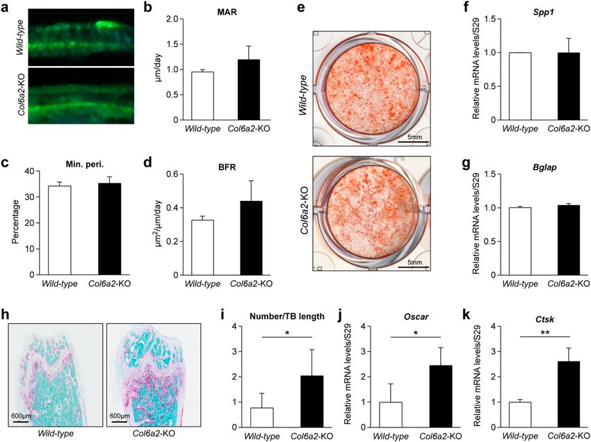
www.nature.com/scientificreports/
Figure 1. Immunoflourescence staining of ColVI in bone sections from one month-old mice WT and Col6a2-
KO femur bones (a, b) representative experiment. Relative expression of Col6a2 mRNA in WT and Col6a2-KO
femur bones (c), N = 3 WT bones, N = 3 Col6a2 bones with three technical replicates/bone. Whole body bone
mineral density (BMD) (d), lean body mass (e) and fat content (f) in wild-type (white bar) vs Col6a2-KO mice
(black bar), N = 6/genotype *p < 0.05, **p < 0.01, ***p < 0.001, ****p < 0.0001.
by the clinical presentation of patients with mutations in type I collagen chains, or in the enzymes that regulate
collagen maturation. These defects lead to the “brittle bone disease” also known as osteogenesis imperfecta
(OI)1,2. While the functions of type I collagen in mineralized tissue have been well-investigated, the roles of
other collagen types are unclear.
Type VI collagen is most often composed of three different α-chains (α1, α2, α3) with three other minor
chains (α4, α5, and α6) found at low levels3. Mutations in any one of the three major chains of human type VI
collagen cause Bethlem myopathy or Ullrich congenital muscular dystrophy (UCMD), diseases that present
with mild to severe muscle weakness, r espectively4. While type VI collagen is found in many musculoskeletal
tissues, its functions in bone are just beginning to be investigated5. Assembly of the type VI collagen triple
helix begins at the C-terminus6 and the triple helical monomer is flanked by two large multidomain globular
regions7. Dimers are assembled from head-to-tail staggered monomers. Then, dimers align side-by-side to form
tetramers, which are then secreted. Outside the cell, tetramers assemble end-to-end into microfibril structures
that appear as “beads” when visualized by the electron m icroscope4. The importance of the ColVIα2 chain in
the assembly, secretion and subsequent microfibril formation was elegantly highlighted by Tooley et al.8 who
identified a UCMD patient with compound heterozygous mutations in α2 (VI). The mutant collagen was found
to be retained intracellularly, preventing normal folding and microfibril assembly. In order to investigate the role
of ColVI in bone and its mechanistic foundation, the skeletal phenotype of mice globally deficient in the ColVI
α2 (Col6a2) protein was examined.
In this investigation, we show that collagen VI regulates trabecular bone mass in both the femur and spine by
controlling the balance between bone formation and resporption. The reduced bone mass observed in Col6α2-KO
mice arises from increased osteoclastogenesis with no apparent effect on bone formation. RNAseq analysis of
mRNA extracted from the bones of Col6α2-KO mice showed deregulation of bone remodeling pathways, and in
addition, pointed to a possible link to TNFα. Here we show that there is a direct interaction between Col6a2 and
TNFα and in vitro, that Col6a2 attenuates TNFα-induced osteoclastogenesis to subsequently influence bone mass.
Results
ColVI protein and Col6a2 mRNA expression is abundant in bone. The expression of ColVI (all
three chains) in bone was measured by immunofluorescence staining in bones from WT and Col6a2-KO mice.
In 1 month-old WT mice, prominent expression was found in the hypertrophic cartilage layer that lies adjacent
to newly formed bone in the primary ossification center (Fig. 1a). By contrast, no ColVI staining was seen in age
and sex-matched Col6a2-KO mice (Fig. 1b). RT-PCR of col6a2 mRNA extracted from whole Col6a2-KO bones
showed complete depletion of Col6a2 mRNA compared with WT counterparts (Fig. 1b,c). The Col6a2-KO mice
were slightly smaller than age (3-month) and sex (male) matched WT counterparts (not shown), and when sub-
Scientific Reports | (2020) 10:13749 | https://doi.org/10.1038/s41598-020-70730-7 2
Vol:.(1234567890)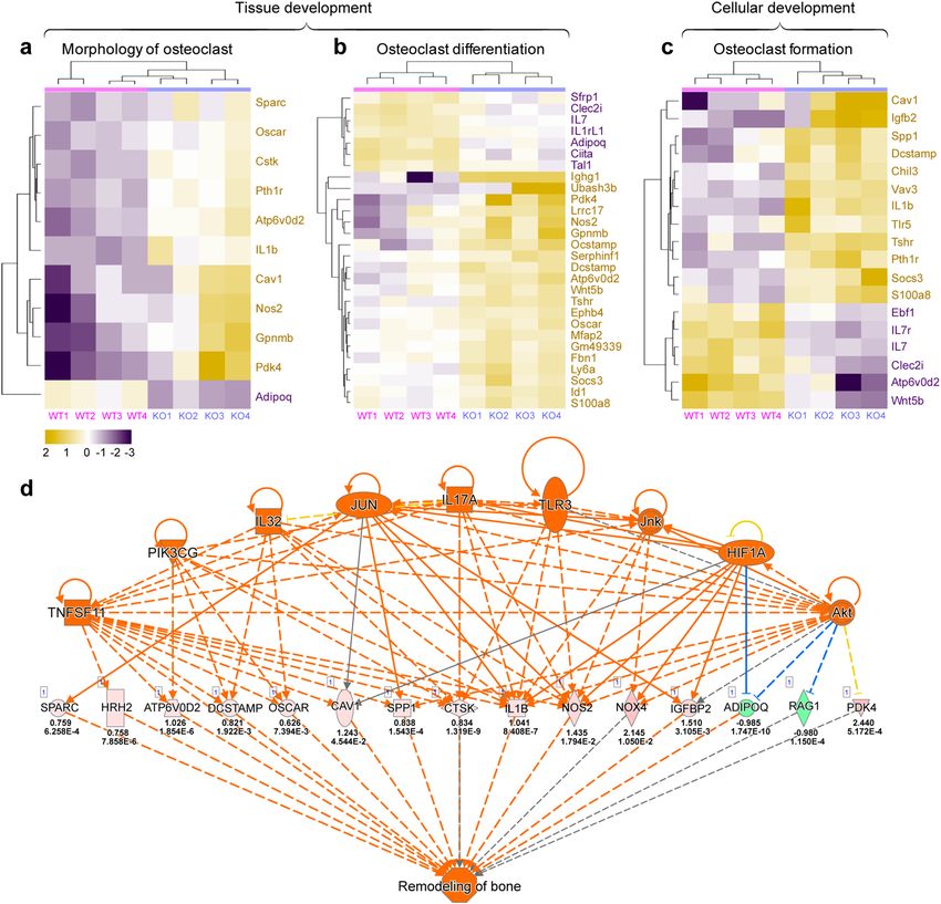
www.nature.com/scientificreports/

                            Figure 2.  Low trabecular bone mass phenotype in Col6a2-KO mice vs WT mice. (a) 3D rendering of the distal
                            femoral metaphyseal bone from 3 month-old (3 m) WT vs Col6a2-KO, representative images (b) and 6 month-
                            old (6 m) mice. Representative images (c) Quantitative µCT analysis of Bone Mineral Density (BMD), (d)
                            Bone Volume/Total Volume (BV/TV), (e) trabecular (Tb) number and (f) Tb spacing. (g) 3D rendering of L3
                            vertebral bodies from 3 month-old (3 m), and (h) 6 month-old (6 m) WT vs Col6α2-KO mice. (i) Quantitative
                            µCT analysis of BMD, (j) BV/TV, (k) Tb number and (l) Tb spacing. 3D Images from each group were obtained
                            from animals with median BV/TV. N = 6/genotype.

                            jected to DEXA analysis (Fig. 1e) showed lower whole-body Bone Mineral Density (BMD) than WT mice. The
                            Col6a2-KO mice also had lower fat body mass and lower fat content compared to age (3-month) and sex (male)
                            matched WT mice (Fig. 1f).

                            Trabecular bone is reduced in Col6a2‑KO mice.              To determine how the bones of Col6a2-KO mice are
                            affected, isolated femora and vertebrae (L3) were dissected and subjected to μCT analysis (representative 3D
                            constructions shown in Fig. 2a, b, g, h). At 3 months of age, the femora from the Col6a2-KO mice had signifi-
                            cantly lower BMD (Fig. 2c), bone volume/tissue volume (BV/TV) (Fig. 2d) and Tb number (Fig. 2e), and higher

Scientific Reports |   (2020) 10:13749 |             https://doi.org/10.1038/s41598-020-70730-7                                        3

                                                                                                                                  Vol.:(0123456789)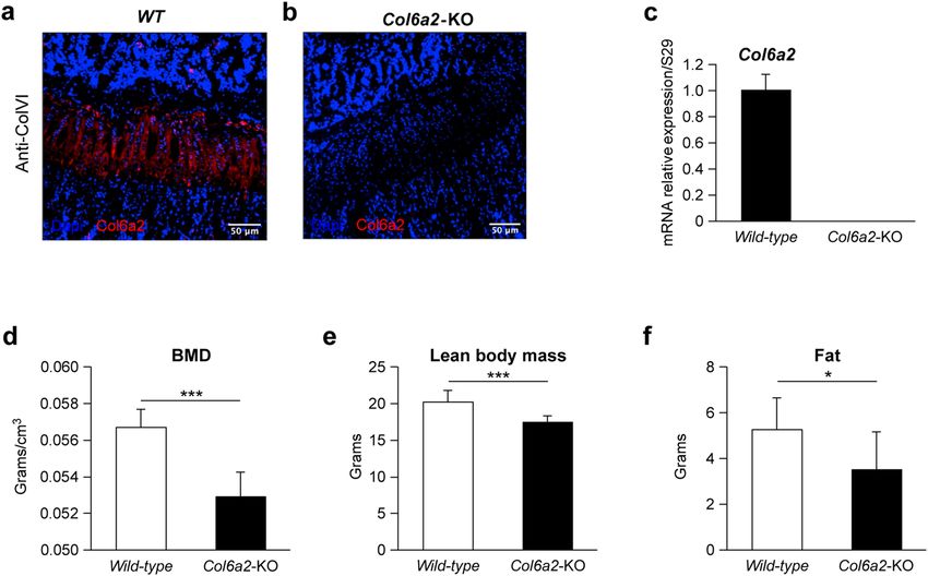
www.nature.com/scientificreports/

                                      Figure 3.  Cortical dimensions of Col6a2-KO vs. WT mice. 3D µCT reconstruction of femoral mid-diaphyseal
                                      cortical bone at 3 months of age from WT (a) and Col6a2-KO (b) mice. Images from each group were obtained
                                      from animals with medial cortical thickness bone, (a, b), representative images. Quantitative µCT analysis of
                                      (c) diaphysis diameter, (d) medullary diameter, (e) Bone Volume/Total Volume (BV/TV), (f) Cortical Thickness
                                      (Cort. Th), (g) Cortical (Cort.) porosity, and (h) Bone Mineral Density (BMD). No significant differences were
                                      found in any of the parameters tested (c–h) between WT (a) and Col6a2-KO mice. N = 8/genotype.

                                      Tb spacing (Fig. 2f) than WT mice. These changes persisted with aging and all were still evident in the same
                                      parameters when tested in 6 month-old mice (Fig. 1c–f). The L3 vertebra showed a similar pattern of low bone
                                      mass phenotype in the Col6a2-KO mice with a lower BMD (Fig. 2i), BV/TV (Fig. 2j) and Tb number (Fig. 2k)
                                      and higher Tb spacing (Fig. 2l) compared with WT mice.

                                      Cortical bone is not affected in the Col6a2‑KO mice. Beyond the trabecular compartment, the mid-
                                      diaphyseal cortical femora were assessed for changes associated with the absence of Col6a2. μCT analysis found
                                      no striking differences in the appearance of the cortices between genotypes (Fig. 3a, b, left panel WT, right panel
                                      Col6a2-KO). Quantification of the cortical area showed no significant differences in diaphysis diameter (Fig. 3c),

          Scientific Reports |   (2020) 10:13749 |               https://doi.org/10.1038/s41598-020-70730-7                                            4

Vol:.(1234567890)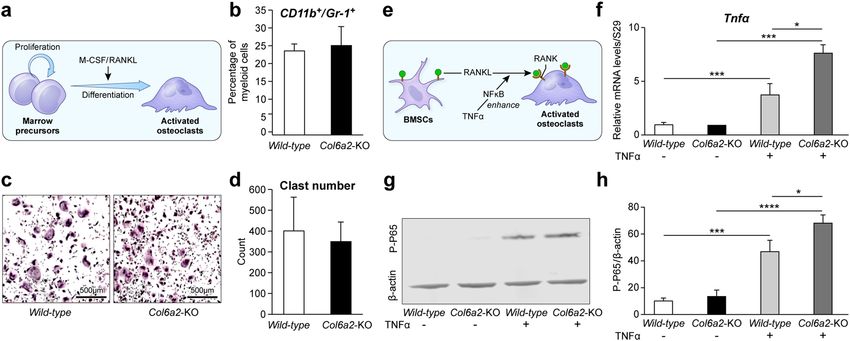
www.nature.com/scientificreports/

                            Figure 4.  Col6a2-KO have increased osteoclastogenesis with no change in bone formation parameters. (a–d)
                            Dynamic histomorphometric parameters based on fluorescent visualization of calcein fluorochrome in the
                            trabecular compartment of the distal metaphysis of the femur. (a) Top panel representative picture from WT,
                            bottom panel, from Col2a2-KO mice. (b) mineral apposition rate (MAR), (c) mineralized perimeter (Min.
                            Peri.) and (d) bone formation rate (BFR) between wild type and Col2a2-KO mice. N = 5/genotype. (e) Alizarin
                            red accumulation in BMSCs from wild-type and Col2a2-KO mice cultured in osteogenic media, representative
                            image (f) Relative mRNA expression of osteopontin (Spp1) and (g) osteocalcin (Bglap). (h) Representative
                            images of histological sections of distal femoral metaphysis stained with TRAP from WT and Col2a2-KO
                            mice, (i) quantitation of osteoclast number/trabecular bone length (N/Tb.Le) from WT and Col6a2-KO, N = 6/
                            genotype. (j) Relative mRNA expression of Oscar and (k) Ctsk in WT and Col2a2-KO derived cells, n = 3 bones/
                            genotype with biological triplicates for each bone. *p < 0.05, **p < 0.01.

                            medullary diameter (Fig. 3d), BV/TV (Fig. 3e), cortical (Cort.) thickness (Th) (Fig. 3f), Cort. porosity (Fig. 3g)
                            or BMD (Fig. 3h) in the Col6a2-KO compared with WT controls.

                            Col6a2‑KO mice have no change in bone formation parameters but have increased osteo‑
                            clastogenesis. To determine the cellular basis for the low bone mass phenotype in the Col6a2-KO mice,
                            dynamic histomorphometry was performed using double fluorochrome labeling techniques (Fig. 4a). This anal-
                            ysis demonstrated that there were no significant differences in between Col6a2-KO and WT mice in the mineral
                            apposition rate (MAR), (Fig. 4b) or Mineralizing perimeter (Min.Peri.) (Fig. 4c) or bone formation rate (BFR)
                            (Fig. 4d). Alizarin red staining of BMSCs cultured under osteogenic conditions showed no appreciable differ-
                            ences in osteoblast differentiation (Fig. 4e). Relative mRNA expression levels of the osteoblast-expressed genes
                            osteopontin (Spp1) (Fig. 4f) and osteocalcin (Bglap) (Fig. 4g) were similar in WT and Col6a2-KO-derived cells.
                            To examine osteoclastogenesis levels, bones were analyzed by TRAP staining. This revealed that the Col6a2-KO
                            mice had significantly more positive staining (pink stain) compared with WT mice (Fig. 4h). Quantitation of the
                            TRAP stain showed that the number of osteoclast/trabecular length was significantly higher in the Col6a2-KO
                            than WT mice (Fig. 4i). Relative levels of osteoclast-expressed genes were also higher in the Col6a2-KO bones
                            as judged by the expression of Osteoclast-associated Ig-like receptor (Oscar) (Fig. 4j) and Cathepsin K (Cstk)
                            (Fig. 4k).

Scientific Reports |   (2020) 10:13749 |              https://doi.org/10.1038/s41598-020-70730-7                                            5

                                                                                                                                       Vol.:(0123456789)
www.nature.com/scientificreports/
Figure 5. Relative mRNA expression patterns related to osteoclast function between WT and Col6a2-KO
mice. RNA was extracted from the femora bones from 4 separate wild-type (WT 1-4) and Col6a2-KO (KO1-4)
mice and subjected to RNAseq and differentially expressed genes were further applied to IPA. Heat maps show
relative expression levels of genes related to osteoclast (a) morphology, (b) differentiation and (c) formation,
with purple blocks being down regulated and yellow being upregulated. Individual genes are shown to the right
of the heat maps. (d) Interactome analysis of dysregulated genes and pathways in the transcriptome of wild-
type and Col6a2-KO. The orange ovals/triangle/squares show over 9 different affected regulators (top row) that
cascade to influence other sets of genes shown in pink (as up) and green (as down) (middle row). The numbers
below the genes show log2 fold change and p-values. Orange colored shapes predict activation. Orange or
blue lines lead to activation or inhibition, respectively. This latter set of genes all coalesced and integrated with
functions related to bone remodeling.
Transcriptome profiling shows dysregulation of osteoclast regulatory pathways in Col6a2‑KO
bones related to bone remodeling. To broaden our analysis of osteoclast-related genes, RNAseq was
performed on bones from 3 month-old WT and Col6a2-KO mice. Bioinformatic analysis identified a total of
1,107 genes (466 down and 641 upregulated) shown in the heatmap as yellow (up) and purple (down), with
clusters linked to the osteoclast morphology (Fig. 5a), osteoclast differentiation (Fig. 5b) or osteoclast forma-
tion (Fig. 5c). This included Oscar and Ctsk that we previously showed to be up-regulated in the Col6a2-KO
bones using conventional real-time RT-PCR (Fig. 4j,k). Futher analysis of the affected interactomes pointed to
numerous key drivers (Tnfsf11, Pik3cg, Il32, Jun, Il17a, Tlr3, Jnk, Hif1a, and Akt (Fig. 5d) that act as upstream
Scientific Reports | (2020) 10:13749 | https://doi.org/10.1038/s41598-020-70730-7 6
Vol:.(1234567890)www.nature.com/scientificreports/
Figure 6. (a) Model showing the relationship between bone marrow cell precursors and osteoclasts
highlighting the importance of M-CSF/RANKL. (b) Percent myeloid cells (osteoclast precursor) judged by
CD11b + /Gr-1 + surface markers, N = 3 biological replicates/genotype (c) Representative pictures of TRAP
staining of cultured osteoclast progenitors treated with M-CSF and RANKL in WT compared to Col6a2-KO
mice. (d) Quantitation of osteoclast number showing no differences between WT and Col6a2-KO mice,
N = 4 biological replicates/genotype. (e) Model showing potential role of TNFα in the functional coupling
of BMSCs and osteoclasts. (f) Relative mRNA levels of Tnfa in WT and Col6a2-KO BMSCs treated with or
without TNFα, N = 3 biological replicates/genotype with triplicate technical replicates. (g) levels of the TNFα
target transcription factore p-65 in WT vs Col6a2-KO mice treated with and without TNFα in the presence of
conditioned media from WT vs Col6a2-KO mice with quantitation, N = 4 biological replicates (h) using β-actin
as loading control. *p < 0.05, ***p < 0.001, ****p < 0.0001.
of effectors of osteoclastogenesis. These effectors influence expression of genes including Sparc, Hrh2, Atp6vd2,
Dcstamp, Oscar, Cav1, Spp1, Ctsk, Il1b, Nos2, Nox4, Igfbp2, Adipoq, Raq1, and Pdk4 with pink or green-colored
shapes shown as up and down-regulation, respectively, (middle row showing raw data), many of which are con-
nected to bone remodeling. These data support the hypothesis that bones from Col6a2-KO mice have overactive
osteoclastogenesis and revealed additional molecular players that could potentially contribute to the biological
outcome of low bone mass in the Col6a2-KO mice.
Myeloid osteoclast precursor number is not altered in Col6a2‑KO mice, but response to TNF
α is increased in osteoclast precurosors pretreated with BMSC‑produced Col6a2. Our TRAP
staining and RNAseq data pointed towards osteoclast involvement in the low bone mass phenotype of the
Col6a2-KO mice. To determine if this was due to an increased number of osteoclast precursors (Fig. 6a), the
percentage of myeloid cells positive for CD11b and Gr-1 was measured. This analysis showed that there were no
significant differences in the percent of CD11b+/Gr-1+ cells in the WT compared with Col6a2-KO mice (Fig. 6b).
There was also no significant difference in the number of osteoclast precursors differentiated in the presence of
M-CSF and RANKL between the WT vs Col6a2-KO mice (Fig. 6c, d). This indicates that the Col6a2 deficient
osteoclast precursor number and differentiation capacity was not the cause of the low bone mass phenotype.
Further analysis of the RNAseq data predicted that TNFα, a factor known to influence osteoclastogenesis was an
upstream regulator in the Col6a2-KO cells. (Supplemental Table 2), (Fig. 6e). To test the possibility that TNFα
activity is altered in the Col6a2-KO cells we treated WT osteoclast progenitors with or without conditioned
media from WT or Col6a2-KO bone marrow stroma cells (BMSCs) (the source of Col6a2). The Relative mRNA
levels of Tnfa in WT and Col6a2-KO BMSCs treated with or without TNFα showed that the response to TNFα
was more effective in BMSCs treated with conditioned medium from Col6a2-KO cells compared with condi-
tioned medium from WT BMSCs (Fig. 6f). The phosphorylated form of p65, which is a down-stream effector of
TNFα, was also measured. In response to TNFα, BMSCs treated with Col6a2-KO BMSC-conditioned medium
induced more phosphorylated p65 than BMSCs treated with WT conditioned medium (Fig. 6g, h). These find-
ings all point to a role for TNFα in the overactive osteoclastogenesis observed in the Col6a2-KO mice.
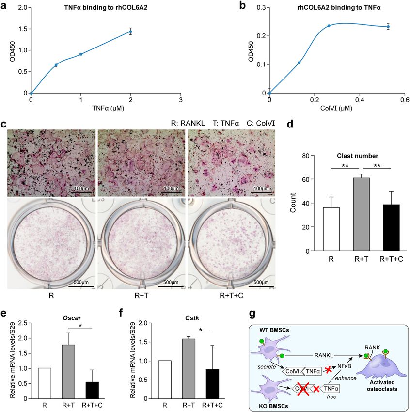
Col6a2 directly binds to TNF‑α in vitro and can reduce osteoclastogenesis in TNFα‑treated
osteoclast precursors. To determine how Col6a2 could potentially regulate TNFα activity, we tested the
possibility that Col6a2 could directly bind to TNFα and thereby harness its activity. We coated plates with a
recombinant human (rh) COL6A2 fragment (designated rhCOL6A2) and added increasing amounts of TNFα.
We found a dose-dependent binding of TNFα to the rhCOL6A2-coated plates (Fig. 7a). When the experiment
was done using TNF-α coated plates we again saw a dose response in binding of rhCOL6A2 (Fig. 7b). Next,
we treated the osteoclast precursor RAW 264.6 cell-line with either RANKL (to stimulate osteoclastogenesis),
Scientific Reports | (2020) 10:13749 | https://doi.org/10.1038/s41598-020-70730-7 7
Vol.:(0123456789)www.nature.com/scientificreports/
Figure 7. ColVI binds to TNFa and reduces TNFa induced osteoclastogeneis. (a, b) solid-phase binding assay.
(a) rhCOl6A2 was bound to plates and treated with increasing concentrations of TNFα. (b) TNFα was bound
to plates and treated with increasing concentrations of rhCOl6A2, representative graphs, Data are mean ± SE
obtained from N = 3 independent experiments. (c–g) Inhibition assay. (c) TRAP staining of cultured RAW
264.7 cells treated with RANKL, TNFα or ColVI and (d) quantitative number of osteoclasts in those cultures,
(f, g) relative mRNA levels of osteoclast markers Oscar and Cstk , N = 4 independent experiments. (h) Diagram
showing the proposed inhibitory function of ColVIα2 to reduce the effects of TNFα on osteoclastogenesis. The
scheme proposes that ColVI made by BMSCs binds to extracellular TNFα and reduces its ability to stimulate
osteoclastogenesis. *p < 0.05, **p < 0.01.
and either TNFα alone (to further enhance osteoclastogenesis) or with ColVI (total human protein), which
we predicted would dampen osteoclastogenesis. Our data showed that the presence of TNFα could stimulate
RANKL induced osteoclastogenesis (Fig. 7c), and that adding ColVI to the RANKL and TNFα-induced cultures
reduced osteoclast number (Fig. 7d), and differentiation judged by the expression of Oscar (Fig. 7e), and Cstk
(Fig. 7f). Taken together, these data suggest that ColVI produced by BMSCs binds to TNFα and reduces its
ability to stimulate osteoclastogenesis (Fig. 7g). When ColVIa2 is depleted (causing a reduction in total COLVI,
see Fig. 1), TNFα is not sequestered in the extracellular matrix and is free to enhance the actions of RANKL on
osteoclastogenesis (Fig. 7g).
Scientific Reports | (2020) 10:13749 | https://doi.org/10.1038/s41598-020-70730-7 8
Vol:.(1234567890)www.nature.com/scientificreports/
Discussion
It has been known for some time that the extracellular matrix (ECM) plays an essential role in mineralized tissue
and yet its exact roles in regulating bone homeostasis are not fully understood9. Several “key players” have been
identified that are embedded in the mineral compartment of bone that include type I collagen and non-colla-
genous proteins such as p roteoglycans10,11. Considering the uniqueness of hard tissue, many have searched for a
component in the ECM that would control bone mass accrual and subsequent biomineralization. While many
factors may contribute to bone formation and bone turnover, their precise nature and interaction with other sys-
temic or local factors is in need of clarification. In an attempt to deepen our understanding of ECM components
in bone health, we have focused on ColVI, which we and others have found to be abundant in forming bone.
By creating mice globally deficient in Col6α1, Cescon et al. described a high versatility for this protein,
showing its involvement in many tissues, including muscle, skin, adipose tissue and the nervous system5. Its role
varies depending on tissue context, and affects a wide range of processes including apoptosis, tumor growth, and
autophagy5. In the nervous system, ColVI is needed for peripheral nerve regeneration in a process dependent
on macrophage recruitment and p olarization12.
ColVI is highly expressed in the osteogenic lineage13 suggesting that it has a function in bone. When the
Col6α1KO mice were examined for bone parameters it was found there was reduced trabecular bone volume
starting at 2 months that persisted until 9 months and leveled off by 15 months of age14. The reduced trabecular
bone in the Col6α1-KO mice is accompanied by an increase in trabecular structure model index (SMI) at 2, 9
and also at 15-months of age compared with WT mice, suggesting that ColVI has a significant role in regulating
normal bone homeostasis. Other studies using the same Col6a1-KO mouse model confirm its importance in
maintaining bone mass and suggest this could arise from defects in osteoblasts located in the periosteum, the
thin layer of tissue that surrounds the bone. The osteoblasts in the Col6a1-KO are less cuboidal or “plump” and
appear more disorganized than WT counterparts15. Mice deficient in ColXII (Col12a1KO) also show a similar
osteoblast disorganization, leading to the speculation that ColVI and ColXII could interact and somehow regulate
osteoblast integrity16. In an in vitro osteoblast differentiation system, Izu et al., found that ColVI and ColXII
co-localize in bridge-like structures that are thought to be involved in osteoblast communication. Interestingly,
the matrix-rich bridge structures containing ColVI or ColXII were reduced in both the Col6α1-KO and the
Col12a1-KO, suggesting they are functionally dependent on each other for network and bridge assembly during
bone formation16. What is not known from these studies is how bone turnover could be affected by either ColVI
or ColXII. Our work builds and expands upon these investigations and shows, for the first time, a novel role for
ColVI in attenuating osteoclastogenesis and, subsequently, bone mass status.
Despite our evidence showing that Col6a2-KO mice have higher levels of osteoclasts than WT mice as
3 months of age, it must be noted that the increase in osteoclast number found in the Col6a2-KO mice did not
perfectly match the extent of bone loss found compared with WT mice and, furthermore, was not progressive
with age as would be expected. It is possible that even with the apparent increase in osteoclast numbers found that
they were not active and did not lead to increased resporption. However, further experiments will be required,
such as measurements of biochemical markers of bone turnover in the serum to fully address this question. In
addition to this, we find from IHC analysis of bones from 1 day, 1 month and 3 month-old mice that ColVI
was expressed at a higher level in young versus old animals (data not shown). Thus, the loss of Col6a2 from
birth on that occurs in our mouse model could be affecting trabecular bone formation prior to the time point
selected to measure the Mineral Apposition Rate. It is also possible that the expression of ColVI in the growth
plate during development affects bone structure and integrity. Additional experiments will need to be needed
to fully address these questions.
While it is clear that ColVI potentially has multiple functions throughout the body, its role in muscle is the
most thoroughly studied and could help inform us about its roles in the skeletal system. Numerous human muta-
tions in the ColVI genes occurring in all three alpha chains, lead to either Bethlem myopathy, at the mild end of
the disease spectrum, or Ullrich congenital muscular dystrophy, a severe debilitating disorder17. Insterstitial fibro-
blasts are the main source of ColVI in muscle tissue and efficient secretion from the cell is needed for its proper
function18. Currently, very little is known about how mutations in human ColVI genes affect bone function.
A previous study designed to test the effects of cyclosporin in ameliorating the muscular symptoms of UCMD
used whole body DEXA scans, and their analysis showed no significant differences in BMI, Lean Tissue Mass,
Fat Tissue Mass, %Fat or BMD before and after cyclosporin t reatment19. Despite the apparent negative outcome
of the investigation, the patients with mutations in either COL6A1, COL6A2 or COL6A3 all had BMD Z scores
significantly below the norm. These data imply a role for ColVI in human bone health19. COLVI levels decrease
with age in human osteoporotic b one20, further pointing to a possible role for COLVI in bone homeostasis with
aging. Reduced BMD in patients with COLVI mutations could result from the influence of the defective muscle
on bone tissue. Decreased muscle activity could reduce the biomechanical forces on bone and lead to reduced
BMD, or alternatively, patients could have defective cytokine interplay between muscle and bone. The use of ani-
mal models with tissue-specific ColVI (bone vs. muscle), or other experiments using co-cultures or conditioned
media from muscle- and bone-derived c ells21 could help resolve this important question.
The immune and skeletal systems have a close association, where immune cells mediate powerful effects
on bone t urnover22. It is well-known that chronic inflammation exacerbates bone loss. Tumor necrosis factor
alpha (TNFα), a proinflammatory agent, is often involved in inflammatory r eactions23,24. TNFα is produced as
a transmembrane protein (pro-TNFα) that is subsequently cleaved by the TNFα converting enzyme (TACE)
to a soluble f orm25. Both forms of TNFα are biologically active, though the membrane-bound form is the one
used under normal conditions, while the soluble form is associated with pathology26–29. Both forms bind as a
trimer to either TNFR1 (also known as TNFRSF1A or p55) or to TNFR2 (also known as TNFRSF1B or p75).
Scientific Reports | (2020) 10:13749 | https://doi.org/10.1038/s41598-020-70730-7 9
Vol.:(0123456789)www.nature.com/scientificreports/
Compared with the ubiquitously expressed TNFR1, which has a “death domain” to initiate apoptotic signaling,
the expression of TNFR2 is limited to cells of the hematopoietic lineage.
Numerous investigations have examined the relationship between TNFα and osteoclastogenesis. These stud-
ies show that even though TNFα on its own does not effectively induce osteoclast differentiation30–33, it plays a
major role in promoting bone resorption, usually in synergy with RANKL at the signal transduction level30,34,35.
TNFα indirectly increases osteoclastogenesis through augmentation of M-CSF and RANKL expression in stromal
cells while down-regulating osteoblastic production of O PG36. It also directly promotes RANK expression on
monocytes, thus converting them into osteoclast p recursors37–39.
TNFα also enhances proteoglycan expression, facilitating ECM remodeling during the early stages of the
inflammation process40. Moreover, BMSC-secreted SLRPs have been shown to regulate osteoclastogenesis via
their interaction with TNFα41. It is likely that a similar mechanism is used by ColVI produced by BMSCs to atten-
uate osteoclastogenesis. TNFα inhibits nitric oxide (NO) production and intracellular calcium, while strongly
reducing F-actin content in osteocytes, leading to a reduction in osteocyte stiffness, which in turn induces their
apoptosis42. Interestingly, apoptotic osteocytes are known to attract osteoclasts, pointing to the possibility that
osteocytes are also affected in the Col6a2-KO mice, but this is yet to be determined.
Despite our finding that ColVI binds to TNFα and inhibits osteoclastogenesis in vitro, we have not yet pro-
vided direct proof that this occurs in vivo. In this regard, further experiments will be needed using inhibitors
to TNFα or other genetically altered mice to prove that the association of TNFα with ColVI is valid and has an
impact in the context of an in vivo situation. We are also not sure why there were no significant effects of Col6a2
loss on cortical bone but suggest that one reason could be that there are many more osteoclasts in the trabecular
bone compared with cortical bone, at least at the 3-month time point analyzed. Still another possibility is that
trabecular bone is much more metabolically active than cortical bone and is “primed” for environmental cues
such as changes in TNFα activity. It could also be that there are other factors working in conjunction with Col6a2
that are more affected in the trabecular versus the cortical compartment, but a true understanding of differential
effects on different bone regions will only come with further investigation.
ColVI binds to a wide range of the ECM components besides TNFα. The proteoglycan, NG2, is needed
to retain ColVI at the cell m embrane43 by ColVI binding to the central nonglobular domain of the NG2 core
protein44. NG2 expression is reduced in skeletal muscle of UCMD patients suggesting it has some connection to
this muscle p athology45. NG2 is also found in growing bone and colocalizes with ColI in the ECM of the osteonal
Haversian canal46. While the role of NG2 in the osteon is not definitively understood, its location coincides with
Runx2-positive and PCNA-positive cells suggesting that the combination of ColVI and NG2 could provide an
extracellular microenvironment conducive for proliferation and differentiation of osteoblastic lineage cells46.
In addition to NG2, ColVI also binds to matrilin-1, decorin and biglycan47. Biglycan is a SLRP already known
to be important in bone48,49 that co-localizes to similar regions as ColVI in bone, making it an attractive candidate
for further investigation in this context.
When the biglycan-ColVI interaction was tested using fragments or isolated ColVI chains, the strongest bind-
ing was with the Col6a2 chain, revealing its importance in this matrix–matrix i nteraction50. The biglycan-ColVI
interaction leads to the formation of hexagonal-like networks that resemble tissue structures when visualized
by electron microscopy (EM)51. Comprehensive genotype–phenotype analysis of patients harboring mutations
in COL6A1, COL6A2 or COL6A3 shows a cluster of mutations in the N-terminal region of the triple helix that
produce severe phenotypes, indicating this could be a functional domain in the triple h elix52. Indeed, previous
EM investigations show that both biglycan and decorin bind to a domain in ColVI that is close to the interface
between the N terminus of the triple helical region and the neighboring globular d omain50. This observation
further supports the concept that the N-terminus of COLVI has key roles in its function. Unclear at this time is
how COLVI interactions with biglycan, decorin or even matrilin-1 might regulate its functions in bone.
In summary, we show for the first time, a role for Col6a2 in regulating bone mass. We further show Col6a2
regulates bone loss by modulating TNFα-induced osteoclastogenesis. Exactly which region of Col6a2 controls this
function is still unknown, as well as the possible interplay with additional binding partners such as biglycan. It
will also be interesting to determine if Col6a2 plays a role in bone healing, a process that is distinct from normal
bone turnover shown in the present investigation.
Materials and methods
Animal experiments. The Col6a2-KOmouse strain used for this research project was created from ES cell
clone 12228C-E10, generated by Regeneron Pharmaceuticals, Inc. in the KOMP Repository (https://www.komp.
org) and the Mouse Biology Program (https://www.mousebiology.org) at the University of California-Davis, and
then made into live mice that were backcrossed to the C57B6J strain for 5 generations. Animals were housed
under standard conditions (55% humidity, 12 h day night cycle, standard chow and free access to water) follow-
ing the guidelines and approval of The National Institutes of Dental and Craniofacial Research Animal Care and
Use Committee (protocol #18-865).
Bone marrow stromal cell culture (BMSCs). Mouse BMSCs from both WT and Col6a2-KO 12-week-old male
mice were isolated from the femora and tibiae by flushing the bone marrow and cultured in vitro using α-MEM
(Gibco, USA) containing 20% fetal bovine serum (FBS) (Atlanta Biologicals, USA), 1% antibiotics (penicillin
100 units/ml and streptomycin 100 mg/ml), 1% GlutaMax-I (Gibco, USA), and 55 μM β-mercaptoethanol (Life
Technologies, NY, USA). After cells reached 70% confluence (passage 0) they were trypsinized and suspended
for use in the cell culture assay.
Scientific Reports | (2020) 10:13749 | https://doi.org/10.1038/s41598-020-70730-7 10
Vol:.(1234567890)www.nature.com/scientificreports/
Osteoclast cultures. Primary osteoclast culture. Mouse bone marrow cells from 12-week-old male WT and
Col6α2 KO mice were isolated from femora and tibiae by flushing the bone marrow with 25- and 27-gauge nee-
dles, respectively, and filtered with a 70-μm cell strainer (Falcon, USA). Cells were centrifuged at 1,600 rpm for
10 min and re-suspended in α-MEM medium containing 10% FBS, 1% penicillin 50 units/ml, and 1% fungizone
1.25 μg/ml. Cells were seeded in 10 ml/10 cm2 culture dish. After 3 h, the supernatant was collected and seeded
into a new 10-cm2 culture dish for 24 h. The supernatant was re-collected and centrifuged at 1,400 rpm for 5 min.
The pellets were re-suspended for use in the osteoclastogenesis assay. The cells were plated at 500,000 cells/cm2
in a 96-well plate with 40 ng/ml of RANKL (R&D, USA) and 30 ng/ml of M-CSF (R&D, USA) to induce osteo-
clastogenesis. The induction medium was changed every other day, samples were harvested at day 5 and fixed
and stained with a TRAP staining kit (Sigma, US) following the manufacturer’s instruction. The images were
observed under an EVOS XL Core microscope (Thermo Fisher, USA) and analyzed by Image Pro 7.0 software
(Media Cybernetics, USA).
RAW 264.7 cell culture. The RAW 264.7 cell line was purchased from ATTC (TIB-7) and cultured in DMEM
(ATCC) containing 10% FBS, 1% penicillin 50 units/ml. To perform the osteoclast assay, 20,000 cells were plated
in 96 wells plate with 30 ng/ml of RANKL (R&D, USA) and simultaneously treated with or without 10 ng/ml of
TNFα (R&D, USA), and 100 ng/ml of human COLVI (SouthernBiotech, USA). The medium with factors was
replenished every other day, samples were fixed at day 5, and stained and imaged as described above.
Dual‑energy X‑ray absorptiometry (DEXA). To determine the bone mineral density, whole mouse bodies (the
skull were excluded from region of interest when analyzing) and femurs were scanned with a DEXA machine
(Lunar PIXImus densitometer, GE Healthcare) and Faxitron Ultrafocus (Faxitron, USA), respectively.
Micro‑computed tomography (μCT). Right femurs and third lumbar vertebrae (L3) from 3 and 6 month-old
WT and Col6a2 KO mice were surgically collected and fixed for 24 h at room temperature in Z-fix (170; Anatech,
LTD), then stored in 70% ethanol at 4 ºC. Samples were scanned by µCT (µCT 50, Scanco Medical AG, Bassers-
dorf, Switzerland) at a resolution of 10 μm and reconstructed using the global approach with energy at 70 kV,
intensity/beam current at 85uA, power at 6 W and Integration time 300 ms53. Trabecular and cortical bone was
quantified according to previously published g uidelines54. For trabecular bone, the following parameters were
analyzed: bone volume/tissue volume (BV/TV), trabecular thickness (Tb.Th), trabecular number (Tb.N), and
trabecular spacing (Tb.S). For cortical bone, (BV/TV), cortical thickness (Cort.th), cortical porosity (Ct. Po),
bone mineral density (BMD), diaphysis diameter (Dp.dm) and medullary diameter (Me.DM) were assessed. All
cortical bone measurements were on bones from 3 month-old mice.
TRAP analysis. Femurs were fixed in Z-fix for 24 h, rinsed with PBS overnight and decalcified with 10% EDTA
for 5 days. Samples were then washed and dehydrated through a graded ethanol series and xylene before paraffin
embedding. The blocks were sectioned at 5 μm thickness, deparaffinized, stained with H&E or performed with
tartrate resistant acid phosphatase activity (TRAP) (Wako Pure Chemical Industries, Ltd., Osaka, Japan), and
observed under an Aperio ScanScope (Leica ICC50 W, Germany).
Immunofluorescence staining. For preparation of frozen sections from non-fixed and non-decalcified hard tis-
sues, Kawamoto’s film methods were used. Briefly, samples were freeze-embedded with super cryoembedding
medium (SECTION-LAB Co. Ltd., Hiroshima, Japan), and 3 um sections were cut using a tungsten carbide
blade after attaching the adhesive film onto the sample surface. Samples were then immediately fixed with 4%
paraformaldehyde (PFA) for 5 min. For analysis of ColVI, its primary antibody (70R-CR009x, Fitzgerald, USA)
was incubated at 4 °C overnight, after blocking with 10% normal donkey serum (Jackson Immunoresearch,
USA) for 60 min at room temperature. Primary antibody was diluted to 1:50. After washing, the specimens were
incubated with secondary antibody, Alexa Fluor 647 donkey anti-rabbit IgG (Jackson Immunoresearch, USA)
for 60 min at room temperature. All images were taken by fluorescence microscope.
Dynamic histomorphometry. For in vivo calcein double labeling, 12-week old mice were injected intraperi-
toneally with a 15 mg/kg of calcein fluorochrome (Sigma-Aldrich, St. Louis, MO) at 7 days and 1 day prior to
euthanasia. The femora were collected and embedded undecalcified in methyl methacrylate, then coronally sec-
tioned. Mid-frontal sections were scanned with a fluorescent microscope (Olympus DP72, Japan) and analyzed
by Image Pro 7.0 software (Media Cybernetics, USA). For in-vivo osteoclast assessment, paraffin-embedded
femoral histological sections were stained with a tartrate-resistant acid phosphatase (TRAP) staining kit (Wako,
Osaka, Japan), following the manufacturer’s protocol, and observed using an Aperio ScanScopes (Leica ICC50
W, Germany). The number of osteoclasts was counted by Image Pro 7.0 software (Media Cybernetics, USA).
mRNA extraction and real time reverse‑transcription polymerase chain reaction (real time RT‑PCR). 2-month
old WT mouse femora were isolated, immediately frozen in liquid nitrogen, and stored at -t80 ºC. They were
then put into the center of a tissue tube (Covaris, USA), frozen in liquid nitrogen, and pulverized on the CP02
cryoPREP Automated Dry Pulverizer (Covaris, USA) to disrupt the extracellular matrix. Total tissue RNA was
extracted by TriPure (Sigma, USA) /RNeasy (Qiagen, Germany) hybrid extraction protocol. Total RNA was
extracted from cell cultures using RLT buffer (Qiagen, Germany) and isolated using the TriPure/RNAeasy sys-
tem described above. cDNA was synthesized with an iScript cDNA Synthesis Kit (Bio-Rad, USA). For both
in-vivo and in-vitro samples, RT-PCR was performed with primers, iQ SYBR (Bio-Rad, USA) and a CFX96
Scientific Reports | (2020) 10:13749 | https://doi.org/10.1038/s41598-020-70730-7 11
Vol.:(0123456789)www.nature.com/scientificreports/
Real-Time PCR detection system (Bio-Rad, USA). The relative levels of mRNA of target genes were normalized
to the housekeeping gene, S29. Primer sequences are listed in Supplementary Table 1. All experiments employed
at least three independent experiments with greater than 3 biological replicates in each experiment.
RNA‑sequencing and analysis. Total RNA was extracted from 3 month-old mouse femora as described above.
Sequencing libraries were prepared using a Nextera XT kit (Illumina), individually barcoded, pooled to a 2 nM
final concentration, and sequenced on a NextSeq500 (Illumina) using 75 × 75 single-end reads. After sequenc-
ing, the base-called demultiplexed (fastq) read qualities were determined using FastQC (v0.11.2) (https://www.
bioinformatics.babraham.ac.uk/projects/fastqc/), aligned to the GENCODE M11 mouse genome (GRCm38.
p4) and gene counts generated using STAR (v2.5.2a)55. Post-alignment qualities were generated with QoRTS
(v 1.1.6) 56. An expression matrix of raw gene counts was generated using R (https://www.R-project.org) and
filtered to remove low count genes (defined as those with less that 5 reads in at least one sample). The filtered
expression matrix was used to generate a list of differentially expressed genes between the sample groups using
three statistical methods: DESeq257, EdgeR58,59, and Limma-voom60. Differentially expressed genes (Col6a2-KO
vs. WT, FC > 1.5, FDR < 0.05) were considered for further analyses based on results from DESeq2. A total of
1,107 genes (466 down, 641 up) were applied to Ingenuity Pathway Analysis (IPA) (Qiagen). IPA-identified can-
didate genes related to biofunction and regulator effects. Heat maps were generated using shinyheatmap from
the Genomics and Computational Biology Core (https://brics.nidcr.nih.gov).
Western blotting of treated cell lysates. BMSCs harvested from WT and Col6a2-KO mice were cultured in
basal medium until 60% confluency. Subsequently, the basal medium was changed to serum-free medium, then
BMSCs were cultured for 2 days to collect conditioned medium containing Col6 secreted by BMSCs. To examine
the possible interaction of Col6VI and TNFα, BMSCs were treated with conditioned medium that was supple-
mented with 10 ng/mL TNFα (R&D systems, Minneapolis, MN) for 15 min. Treated BMSCs were lysed using
M-PER (Thermo, Waltham, MA), supplemented with a Protease Inhibitor Cocktail (Roche, Indianapolis, IN)
and PhoSTOP (Sigma-Aldrich, Saint-Louis, MO). Cell debris were removed from lysates by centrifugation at
12,000 rpm for 10 min at 4 °C. The protein concentration in the cell lysate was determined by using the Pierce
BCA Protein assay kit (Thermo). Twenty micrograms of the total protein was separated in precast polyacryla-
mide gels (NuPage; Life Technologies, Carlsbad, CA) by electrophoresis and then transferred onto nitrocellulose
membranes at 30 V for 2 h. Blots were blocked and incubated with primary antibodies against phospho-p65
(1:1,000 dilution) or β-actin (8H10D10; 1:5,000 dilution, Cell Signaling Technology, Danvers, MA), which was
used as the loading control. Blots were then washed and incubated with IRDye 800CW Goat anti-Rabbit IgG
Secondary Antibody (1:5,000 dilution; LI-COR Biosciences, Lincoln, NE) or IRDye 680LT Goat anti-Mouse IgG
Secondary Antibody (1:5,000 dilution; LI-COR Biosciences) for 1 h at room temperature. The blots were visual-
ized with a LI-COR imaging system and densiometric analysis done using Image Studio Lite software (LI-COR
Biosciences).
Solid‑phase binding assay. Recombinant human COL6A2 (abcam 169,887) or human TNFα (R&D) proteins
were pre-tested to determine the optimal concentration for coating plates used in the binding assay, which was
1 µM and 0.26 µM respectively. Bait proteins were bound to Nickel coated plates (15,242; Thermo scientific)
with 1-h incubation at room temperature. Unbound proteins were washed off and non-specific binding sites
was blocked with 1%BSA/TBS. hCCOL6A2 protein-coated plates were incubated with different concentrations
of prey proteins, mTNFα (R&D). In the reverse experiment hTNFα coated plates were incubated with different
concentration of hCOL6A2 at 4 °C overnight. Unbound proteins were washed off and binding of the prey pro-
teins to the coated plate was detected using primary antibodies raised against abTNFα (abcam 9,739) or abColVI
(70R-CR009X, Fitzgerald), combined with species-matched HRP-conjugated secondary antibodies. Plates were
read at 450 nm after HRP chromogenic substrate reaction using the TMB-peroxidase substrate system (KPL,
Gaithersburg, MD, USA) according to the manufacturer’s protocol.
Statistics. Statistical analyses were performed with unpaired Student`s t-test. A statistically significant differ-
ence was considered as p < 0.05.
Study approval. All animal studies were performed in accordance with NIH guidelines under institutionally
approved protocols.
Received: 6 April 2020; Accepted: 29 July 2020
References
1. Tauer, J. T., Robinson, M. E. & Rauch, F. Osteogenesis imperfecta: new perspectives from clinical and translational research. JBMR
Plus 3, e10174. https://doi.org/10.1002/jbm4.10174 (2019).
2. Robinson, M. E. & Rauch, F. Mendelian bone fragility disorders. Bone 126, 11–17. https://doi.org/10.1016/j.bone.2019.04.021
(2019).
3. Fitzgerald, J., Rich, C., Zhou, F. H. & Hansen, U. Three novel collagen VI chains, alpha4(VI), alpha5(VI), and alpha6(VI). J. Biol.
Chem. 283, 20170–20180. https://doi.org/10.1074/jbc.M710139200 (2008).
Scientific Reports | (2020) 10:13749 | https://doi.org/10.1038/s41598-020-70730-7 12
Vol:.(1234567890)www.nature.com/scientificreports/
4. Lamande, S. R. & Bateman, J. F. Collagen VI disorders: insights on form and function in the extracellular matrix and beyond.
Matrix Biol. 71–72, 348–367. https://doi.org/10.1016/j.matbio.2017.12.008 (2018).
5. Cescon, M., Gattazzo, F., Chen, P. & Bonaldo, P. Collagen VI at a glance. J Cell Sci 128, 3525–3531. https: //doi.org/10.1242/jcs.16974
8 (2015).
6. Ball, S., Bella, J., Kielty, C. & Shuttleworth, A. Structural basis of type VI collagen dimer formation. J. Biol. Chem. 278, 15326–15332.
https://doi.org/10.1074/jbc.M209977200 (2003).
7. Chu, M. L. et al. Characterization of three constituent chains of collagen type VI by peptide sequences and cDNA clones. Eur. J.
Biochem. 168, 309–317. https://doi.org/10.1111/j.1432-1033.1987.tb13422.x (1987).
8. Tooley, L. D. et al. Collagen VI microfibril formation is abolished by an {alpha}2(VI) von Willebrand factor type A domain mutation
in a patient with Ullrich congenital muscular dystrophy. J. Biol. Chem. 285, 33567–33576. https: //doi.org/10.1074/jbc.M110.15252
0 (2010).
9. Licini, C., Vitale-Brovarone, C. & Mattioli-Belmonte, M. Collagen and non-collagenous proteins molecular crosstalk in the patho-
physiology of osteoporosis. Cytokine Growth Factor Rev. https://doi.org/10.1016/j.cytogfr.2019.09.001 (2019).
10. Fisher, L. W., Termine, J. D. & Young, M. F. Deduced protein sequence of bone small proteoglycan I (biglycan) shows homology
with proteoglycan II (decorin) and several nonconnective tissue proteins in a variety of species. J. Biol. Chem. 264, 4571–4576
(1989).
11. Young, M. F. Skeletal biology: where matrix meets mineral. Matrix Biol. 52–54, 1–6. https://doi.org/10.1016/j.matbio.2016.04.003
(2016).
12. Chen, P. et al. Collagen VI regulates peripheral nerve regeneration by modulating macrophage recruitment and polarization. Acta
Neuropathol. 129, 97–113. https://doi.org/10.1007/s00401-014-1369-9 (2015).
13. Kohara, Y., Soeta, S., Izu, Y., Arai, K. & Amasaki, H. Distribution of type VI collagen in association with osteoblast lineages in the
groove of Ranvier during rat postnatal development. Ann Anat 208, 58–68. https://doi.org/10.1016/j.aanat.2016.07.003 (2016).
14. Christensen, S. E. et al. Altered trabecular bone structure and delayed cartilage degeneration in the knees of collagen VI null mice.
PLoS ONE 7, e33397. https://doi.org/10.1371/journal.pone.0033397 (2012).
15. Izu, Y. et al. Type VI collagen deficiency induces osteopenia with distortion of osteoblastic cell morphology. Tissue Cell 44, 1–6.
https://doi.org/10.1016/j.tice.2011.08.002 (2012).
16. Izu, Y., Ezura, Y., Koch, M., Birk, D. E. & Noda, M. Collagens VI and XII form complexes mediating osteoblast interactions during
osteogenesis. Cell Tissue Res 364, 623–635. https://doi.org/10.1007/s00441-015-2345-y (2016).
17. Bonnemann, C. G. The collagen VI-related myopathies: muscle meets its matrix. Nat. Rev. Neurol 7, 379–390. https://doi.
org/10.1038/nrneurol.2011.81 (2011).
18. Zou, Y., Zhang, R. Z., Sabatelli, P., Chu, M. L. & Bonnemann, C. G. Muscle interstitial fibroblasts are the main source of collagen
VI synthesis in skeletal muscle: implications for congenital muscular dystrophy types Ullrich and Bethlem. J. Neuropathol. Exp.
Neurol. 67, 144–154. https://doi.org/10.1097/nen.0b013e3181634ef7 (2008).
19. Merlini, L. et al. Cyclosporine A in Ullrich congenital muscular dystrophy: long-term results. Oxid. Med. Cell Longev. 2011, 139194.
https://doi.org/10.1155/2011/139194 (2011).
20. Bailey, A. J., Wotton, S. F., Sims, T. J. & Thompson, P. W. Biochemical changes in the collagen of human osteoporotic bone matrix.
Connect Tissue Res. 29, 119–132. https://doi.org/10.3109/03008209309014239 (1993).
21. Huang, J. et al. Crosstalk between MLO-Y4 osteocytes and C2C12 muscle cells is mediated by the Wnt/beta-catenin pathway.
JBMR Plus 1, 86–100. https://doi.org/10.1002/jbm4.10015 (2017).
22. Weitzmann, M. N. Bone and the immune system. Toxicol. Pathol. 45, 911–924. https: //doi.org/10.1177/019262 33177 35316 (2017).
23. Teitelbaum, S. L. Osteoclasts; culprits in inflammatory osteolysis. Arthritis Res. Ther. 8, 201. https: //doi.org/10.1186/ar1857 (2006).
24. Boyce, B. F., Schwarz, E. M. & Xing, L. Osteoclast precursors: cytokine-stimulated immunomodulators of inflammatory bone
disease. Curr. Opin. Rheumatol. 18, 427–432. https://doi.org/10.1097/01.bor.0000231913.32364.32 (2006).
25. Black, R. A. et al. A metalloproteinase disintegrin that releases tumour-necrosis factor-alpha from cells. Nature 385, 729–733. https
://doi.org/10.1038/385729a0 (1997).
26. Boyce, B. F., Xiu, Y., Li, J., Xing, L. & Yao, Z. NF-kappaB-mediated regulation of osteoclastogenesis. Endocrinol. Metab. (Seoul) 30,
35–44. https://doi.org/10.3803/EnM.2015.30.1.35 (2015).
27. Novack, D. V. Role of NF-kappaB in the skeleton. Cell Res. 21, 169–182. https://doi.org/10.1038/cr.2010.159 (2011).
28. Takayanagi, H. et al. Induction and activation of the transcription factor NFATc1 (NFAT2) integrate RANKL signaling in terminal
differentiation of osteoclasts. Dev. Cell 3, 889–901. https://doi.org/10.1016/s1534-5807(02)00369-6 (2002).
29. Asagiri, M. et al. Autoamplification of NFATc1 expression determines its essential role in bone homeostasis. J. Exp. Med. 202,
1261–1269. https://doi.org/10.1084/jem.20051150 (2005).
30. Lam, J. et al. TNF-alpha induces osteoclastogenesis by direct stimulation of macrophages exposed to permissive levels of RANK
ligand. J. Clin. Invest. 106, 1481–1488. https://doi.org/10.1172/JCI11176 (2000).
31. Kim, N. et al. Osteoclast differentiation independent of the TRANCE-RANK-TRAF6 axis. J. Exp. Med. 202, 589–595. https://doi.
org/10.1084/jem.20050978 (2005).
32. Kobayashi, K. et al. Tumor necrosis factor alpha stimulates osteoclast differentiation by a mechanism independent of the ODF/
RANKL-RANK interaction. J. Exp. Med. 191, 275–286. https://doi.org/10.1084/jem.191.2.275 (2000).
33. Azuma, Y., Kaji, K., Katogi, R., Takeshita, S. & Kudo, A. Tumor necrosis factor-alpha induces differentiation of and bone resorption
by osteoclasts. J. Biol. Chem. 275, 4858–4864. https://doi.org/10.1074/jbc.275.7.4858 (2000).
34. Zhang, Y. H., Heulsmann, A., Tondravi, M. M., Mukherjee, A. & Abu-Amer, Y. Tumor necrosis factor-alpha (TNF) stimulates
RANKL-induced osteoclastogenesis via coupling of TNF type 1 receptor and RANK signaling pathways. J. Biol. Chem. 276,
563–568. https://doi.org/10.1074/jbc.M008198200 (2001).
35. Fuller, K., Murphy, C., Kirstein, B., Fox, S. W. & Chambers, T. J. TNFalpha potently activates osteoclasts, through a direct action
independent of and strongly synergistic with RANKL. Endocrinology 143, 1108–1118. https://doi.org/10.1210/endo.143.3.8701
(2002).
36. Hofbauer, L. C. et al. Interleukin-1beta and tumor necrosis factor-alpha, but not interleukin-6, stimulate osteoprotegerin ligand
gene expression in human osteoblastic cells. Bone 25, 255–259. https://doi.org/10.1016/s8756-3282(99)00162-3 (1999).
37. Yao, Z. et al. Tumor necrosis factor-alpha increases circulating osteoclast precursor numbers by promoting their proliferation
and differentiation in the bone marrow through up-regulation of c-Fms expression. J. Biol. Chem. 281, 11846–11855. https://doi.
org/10.1074/jbc.M512624200 (2006).
38. Li, P. et al. Systemic tumor necrosis factor alpha mediates an increase in peripheral CD11bhigh osteoclast precursors in tumor
necrosis factor alpha-transgenic mice. Arthritis Rheum. 50, 265–276. https://doi.org/10.1002/art.11419 (2004).
39. Zhang, Q., Guo, R., Schwarz, E. M., Boyce, B. F. & Xing, L. TNF inhibits production of stromal cell-derived factor 1 by bone stromal
cells and increases osteoclast precursor mobilization from bone marrow to peripheral blood. Arthritis Res. Ther. 10, R37. https://
doi.org/10.1186/ar2391 (2008).
40. Tufvesson, E. & Westergren-Thorsson, G. Alteration of proteoglycan synthesis in human lung fibroblasts induced by interleukin-
1beta and tumor necrosis factor-alpha. J. Cell. Biochem. 77, 298–309. https: //doi.org/10.1002/(sici)1097-4644(200005 01)77:2%3c298
::aid-jcb12%3e3.0.co;2-d (2000).
41. Kram, V., Kilts, T. M., Bhattacharyya, N., Li, L. & Young, M. F. Small leucine rich proteoglycans, a novel link to osteoclastogenesis.
Sci. Rep. 7, 12627. https://doi.org/10.1038/s41598-017-12651-6 (2017).
Scientific Reports | (2020) 10:13749 | https://doi.org/10.1038/s41598-020-70730-7 13
Vol.:(0123456789)You can also read