Eukaryotic community composition in the sea surface microlayer across an east-west transect in the Mediterranean Sea - Biogeosciences
←
→
Page content transcription
If your browser does not render page correctly, please read the page content below
Biogeosciences, 18, 2107–2118, 2021
https://doi.org/10.5194/bg-18-2107-2021
© Author(s) 2021. This work is distributed under
the Creative Commons Attribution 4.0 License.

Eukaryotic community composition in the sea surface microlayer
across an east–west transect in the Mediterranean Sea
Birthe Zäncker1,2 , Michael Cunliffe2,3 , and Anja Engel1
1 GEOMAR,    Helmholtz Centre for Ocean Research Kiel, 24105 Kiel, Germany
2 Marine Biological Association of the UK, Plymouth, PL1 2PB, UK
3 School of Biological and Marine Sciences, University of Plymouth, Plymouth, PL4 8AA, UK

Correspondence: Anja Engel (aengel@geomar.de)

Received: 29 June 2020 – Discussion started: 21 July 2020
Revised: 6 January 2021 – Accepted: 1 February 2021 – Published: 23 March 2021

Abstract. The sea surface microlayer (SML) represents the        1   Introduction
boundary layer at the air–sea interface. Microbial eukaryotes
in the SML potentially influence air–sea gas exchange di-        The sea surface microlayer (SML) constitutes a 1 to 100 µm
rectly by taking up and producing gases and indirectly by        thick boundary layer between the ocean and the atmosphere
excreting and degrading organic matter, which may mod-           (Cunliffe and Murrell, 2010; Liss and Duce, 2005; Zhang
ify the viscoelastic properties of the SML. However, little is   et al., 2003) with distinct physical and chemical properties
known about the distribution of microbial eukaryotes in the      compared to the underlying water (Cunliffe et al., 2013;
SML. We studied the composition of the microbial commu-          Zhang et al., 2003). The SML potentially has a substantial
nity, transparent exopolymer particles and polysaccharides       influence on air–sea exchange processes, such as gas transfer
in the SML during the PEACETIME cruise along a west–             and sea spray aerosol formation (Cunliffe et al., 2013; Engel
east transect in the Mediterranean Sea, covering the west-       et al., 2017; Freney et al., 2020; Sellegri et al., 2021).
ern basin, Tyrrhenian Sea and Ionian Sea. At the stations           The microbial food web plays a crucial role in ocean bio-
located in the Ionian Sea, fungi – likely of continental ori-    geochemistry and has been vastly studied. Despite the fact
gin and delivered by atmospheric deposition – were found in      that microbes in the SML can directly and indirectly influ-
high relative abundances, making up a significant proportion     ence air–sea gas exchange, few studies have looked at the mi-
of the sequences recovered. Concomitantly, bacterial and pi-     crobial community composition in the SML and have mainly
cophytoplankton counts decreased from west to east, while        focused on bacteria (Agogué et al., 2005; Joux et al., 2006;
transparent exopolymer particle (TEP) abundance and to-          Obernosterer et al., 2008) with little attention to microbial
tal carbohydrate (TCHO) concentrations remained constant         eukaryotes (Taylor and Cunliffe, 2014). While phytoplank-
in all basins. Our results suggest that the presence of sub-     ton throughout the water column play an important role in
strates for fungi, such as Cladosporium, known to take up        the ocean as primary producers, phytoneuston in the SML
phytoplankton-derived polysaccharides, in combination with       (Apts, 1989; Hardy and Apts, 1984; Naumann, 1917) might
decreased substrate competition by bacteria, might favor fun-    have an additional crucial role by impacting air–sea gas ex-
gal dominance in the neuston of the Ionian Sea and other         change (Ploug, 2008; Upstill-Goddard et al., 2003). Early mi-
low-nutrient, low-chlorophyll (LNLC) regions.                    croscopic observations of the SML reported mostly diatoms,
                                                                 dinoflagellates and cyanobacteria (Hardy et al., 1988). More
                                                                 recent studies using 18S rRNA gene sequencing found a de-
                                                                 creased protist diversity in the SML compared to underlying
                                                                 water with chrysophytes and diatoms enriched in the SML
                                                                 (Cunliffe and Murrell, 2010; Taylor and Cunliffe, 2014).
                                                                    Not only phytoneuston, but also zooneuston, bacteri-
                                                                 oneuston and myconeuston might influence air–sea gas ex-

Published by Copernicus Publications on behalf of the European Geosciences Union.
2108 B. Zäncker et al.: Eukaryotic community composition in the sea surface microlayer
change processes by either parasitizing phytoneuston and
thus impacting the primary productivity or by degrading or-
ganic matter available in the SML and releasing CO2 . While
some studies have explored bacterioneuston diversity in the
Mediterranean Sea (Agogué et al., 2005; Joux et al., 2006),
fungi have not yet been characterized in the SML in this re-
gion. Fungi are however abundant in marine environments
(Gladfelter et al., 2019; Grossart et al., 2019; Hassett et al.,
2019), living a saprotrophic or parasitic lifestyle, and have
been found in the Mediterranean Sea before (Garzoli et al.,
2015; Gnavi et al., 2017) and in the myconeuston studied at
other locations (Taylor and Cunliffe, 2014).
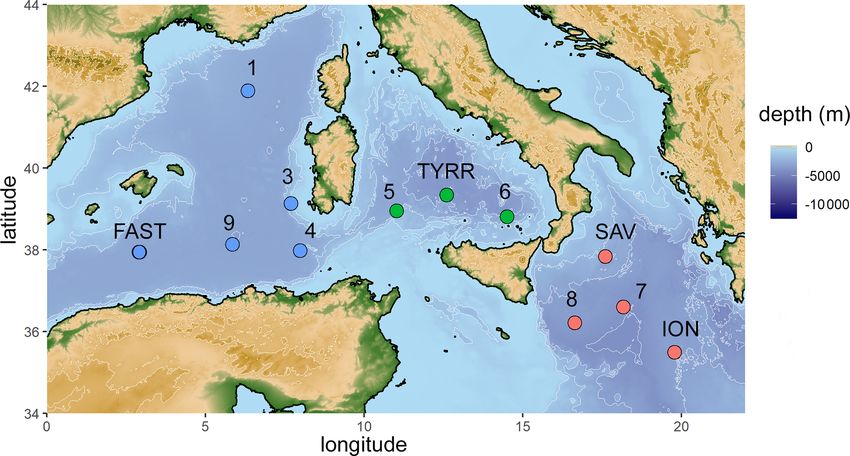
Phytoplankton and phytoneuston can release precursors Figure 1. Map of the stations sampled during the PEACETIME
such as carbohydrates which can aggregate and form gelati- cruise in the Mediterranean Sea in May/June 2017. Stations FAST
and TYRR were sampled twice. Colors represent sampled basins
nous particles such as transparent exopolymer particles
(blue: western basin, green: Tyrrhenian Sea, red: Ionian Sea).
(TEPs) (Chin et al., 1998; Engel et al., 2004; Verdugo et al.,
2004). TEPs contain mainly polysaccharides (Mopper et al.,
1995; Passow, 2002), occur ubiquitously in the ocean (All- 12 stations from 2.9 to 19.8◦ E and 35.5 to 42.0◦ N (Fig. 1).
dredge et al., 1993; Passow, 2002) and are an important struc- SML samples were collected from a zodiac using a glass
tural component of the SML (Wurl and Holmes, 2008; Cun- plate sampler (Cunliffe and Wurl, 2014; Harvey, 1966). The
liffe and Murrell, 2009). Due to their stickiness TEPs can dimensions of the silicate glass plate (50 × 26 cm) resulted
aggregate with other denser particles (Azetsu-Scott and Pas- in an effective sampling surface area of 2600 cm2 consider-
sow, 2004; Engel, 2000; Passow and Alldredge, 1995) and ing both sides. To avoid contamination during sampling, the
eventually sink out of the euphotic layer into the deep ocean, zodiac was positioned upwind and in front of the research
leading to carbon export (Engel et al., 2004). However, the vessel. The glass plate was immersed and withdrawn perpen-
rate of TEP-related carbon export does not only depend on dicular to the sea surface. With a Teflon wiper, SML samples
TEP production by phytoplankton, but also on their micro- were collected in acid-cleaned and rinsed bottles (Cunliffe
bial degradation. and Wurl, 2014). Approximately 1.5 L of SML sample was
Few studies have looked at the spatial distribution of the collected in the course of 1 h. Sampling times are listed in Ta-
microbial eukaryote communities in the SML and possi- ble 1. All sampling equipment was acid-cleaned (10 % HCl),
ble environmental drivers of community composition in the rinsed with Milli-Q and copiously rinsed with seawater from
open Mediterranean Sea, a characteristic low-nutrient, low- the respective depth once the sampling site was reached. The
chlorophyll (LNLC) region (Durrieu de Madron et al., 2011). ULW samples were collected simultaneously with two acid-
The anti-estuarine circulations at the Strait of Gibraltar and cleaned and Milli-Q-rinsed glass bottles by immersing the
the Strait of Sicily transport low-nutrient surface waters into closed bottles and opening them at approximately 20 cm.
the basins and deeper waters out of the basins, resulting in
oligotrophic conditions and ultra-oligotrophic conditions in 2.2 Gel particle determination
the western and eastern Mediterranean basin, respectively
(Krom et al., 2004; Mermex Group et al., 2011; Pujo-Pay The abundance and area of TEP was measured microscop-
et al., 2011; Tanhua et al., 2013). The present study focuses ically (Engel, 2009). The sample volume (10–30 mL) was
on TEPs as important structural components of the SML chosen according to the prevailing TEP concentrations. Sam-
and their precursors, carbohydrates, and microbial eukary- ples were filtered onto 0.4 µm Nuclepore membranes (What-
otes distribution, focusing on the myconeuston community man) and stained with 1 mL Alcian Blue solution (0.2 g L−1
composition in the SML using samples collected during the w/v) for 3 s. Filters were mounted on Cytoclear® slides and
PEACETIME cruise in the Mediterranean Sea during May stored at −20 ◦ C until analysis. Two filters per sample with
and June 2017. 30 images each were analyzed using a Zeiss Axio Scope.A1
(Zeiss) equipped with a Zeiss AxioCam MRc. The pictures
with a resolution of 1388 × 1040 pixels were saved using
2 Material and methods AxioVision LE64 Rel. 4.8 (Zeiss). All particles larger than
0.2 µ m2 were analyzed. ImageJ was subsequently used for
2.1 Sampling image analysis (Schneider et al., 2012). A filter prepared with
10 mL Milli-Q water served as a blank.
Samples were collected on board the RV Pourquoi pas? from
10 May to 11 June 2017. Water from the SML and the under-
lying water (ULW; 20 cm below the SML) was collected at
Biogeosciences, 18, 2107–2118, 2021 https://doi.org/10.5194/bg-18-2107-2021
B. Zäncker et al.: Eukaryotic community composition in the sea surface microlayer                                                    2109

Table 1. Name, position and environmental conditions at the stations sampled throughout the cruise. Temperature and salinity were collected
at 5 m water depth.

              Station      Latitude   Longitude     Local time    Wind speed      Water temperature   Salinity   Irradiation
                                                                     [m s−1 ]                 [◦ C]    [PSU]       [W m−2 ]
              S1           41.8918       6.3333          15:45            9.7                  16.4       38.2       1297.8
              S3           39.1333       7.6835          10:00            2.9                  18.7       37.2       2343.2
              S4           37.9832       7.9768          10:30            3.5                  19.8       37.1       2270.2
              TYRR_1         39.34      12.5928          11:00            3.4                  20.3       37.8       2253.1
              TYRR_2       39.3398      12.5928          12:30            2.5                  21.1       37.7       2311.1
              S6           38.8077      14.4997          09:00            5.2                  20.4       37.4       2215.5
              SAV          37.8401      18.1658          12:00            1.5                  20.1       38.5
              S7           36.6035      18.1658          07:00            2.5                  20.8       38.5         16.8
              ION_2        35.4892      19.7765          09:45            6.4                  21.1       38.8       1235.3
              S8           36.2103       16.631          07:45            1.9                  21.2       37.9       2144.0
              FAST_2        37.946       2.9102          08:30            3.1                  21.7       36.7        627.4
              FAST_6       37.0466       2.9168          08:30            5.1                  21.9       36.6       1787.1

2.3   Bacterioplankton and bacterioneuston abundance                    system (Engel and Händel, 2011). Prior to analysis, sam-
                                                                        ples were desalinated by membrane dialysis (1 kDa MWCO,
Bacterial cell numbers were determined from 2 mL samples                Spectra/Por) at 1 ◦ C for 5 h and hydrolyzed for 20 h at 100 ◦ C
fixed with 100 µL glutaraldehyde (GDA, 1 % final concen-                in HCl (0.8 M final concentration) with subsequent neutral-
tration). Samples were stored at −20 ◦ C and stained with               ization using acid evaporation (N2 , for 5 h at 50 ◦ C). Two
SYBR Green I (Molecular Probes) to determine abundance                  replicates were analyzed for each sample.
using a Becton and Dickinson (BD Biosciences) FACSCal-
ibur flow cytometer equipped with a 488 nm laser. Bacterial             2.6     DNA extraction and eukaryote 18S rRNA gene
cells were detected by the unique signature in a plot of side                   sequencing
scatter (SSC) vs. green fluorescence (FL1). Yellow-green la-
tex beads (Polysciences, 0.5 µm) were used as internal stan-            Water samples for sequencing (400 mL each) were passed
dards.                                                                  through a 100 µm pore size mesh in order to remove meta-
                                                                        zooplankton that could dominate the 18S sequences and
2.4   Picophytoplankton and picophytoneuston                            were subsequently filtered onto a Durapore membrane (Mil-
      abundance                                                         lipore, 47 mm, 0.2 µm) and immediately stored at −80 ◦ C.
                                                                        In order to improve cell accessibility for the DNA extrac-
Picophytoplankton and picophytoneuston cell numbers were                tion, filters in cryogenic tubes were immersed in liquid
determined from 2 mL samples fixed and stored as for bac-               nitrogen, and the filter was crushed with a pestle. DNA
terial abundances. Samples were filtered through a 50 µm                was extracted according to a modified protocol from Zhou
filter and analyzed with a flow cytometer (similar to                   et al. (1996) by Wietz et al. (2015). The protocol included
Sect. 2.3). Enumeration of cells was conducted using a high             bead-beating, phenol–chloroform–isoamyl alcohol purifica-
flow rate (approximately 39–41 µL min−1 ). The forward- or              tion, isopropanol precipitation and ethanol washing. An ad-
right-angle light scatter (FALS or RALS) as well as the                 ditional protein-removal step by salting was used to avoid
phycoerythrin- and Chl a-related fluorescent signal was used            protein contamination.
to distinguish the cells. Cell counts were analyzed using the              Library preparation and sequencing was conducted at
CellQuest Pro software (BD Biosciences). The method used                the Integrated Microbiome Resource at Dalhousie Univer-
here (fixative addition + slow freezing) follows recommen-              sity, Halifax, Canada, and is described in detail elsewhere
dations by Lepesteur et al. (1993).                                     (Comeau et al., 2017). Samples were PCR-amplified in two
                                                                        dilutions (1 : 1 and 1 : 10) using the 18S rRNA gene primers
2.5   Total combined carbohydrates                                      E572F and E1009R (Comeau et al., 2011). Prior to pool-
                                                                        ing, samples were cleaned up and normalized using the In-
Samples (20 mL) for total hydrolyzable carbohydrates                    vitrogen SequalPrep 96-well plate kit (Thermo Fisher Sci-
(TCHOs) > 1 kDa were filled into precombusted glass vials               entific). Sequencing was conducted according to Comeau
(8 h, 500 ◦ C) and stored at −20 ◦ C. In the home lab, TCHO             et al. (2017) on an Illumina MiSeq using 300+300 bp paired-
analysis was carried out using high-performance anion ex-               end V3 chemistry.
change chromatography with pulsed amperometric detection
(HPAEC-PAD) on a Dionex ICS 3000 ion chromatography

https://doi.org/10.5194/bg-18-2107-2021                                                         Biogeosciences, 18, 2107–2118, 2021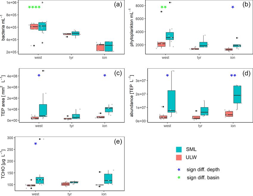
2110                                 B. Zäncker et al.: Eukaryotic community composition in the sea surface microlayer

   Sequences were processed using the DADA2 pipeline           ranean, Tyrrhenian Sea and Ionian Sea). ANOSIM showed
(Callahan et al., 2016), and sequences shorter than 400 bp,    that the differences in the eukaryotic community composi-
longer than 444 bp, with more than eight homopolymers          tion were slightly larger across basins than between SML and
or any ambiguous bases were discarded. Sequences were          ULW (p = 0.0025, R = 0.2263). However, the overall diver-
aligned with the 18S rRNA gene sequences of the SILVA          sity and evenness (based on Shannon and Pielou indices)
132 alignment database (Quast et al., 2013). Subsequently,     were not significantly different between basins (Fig. S1 in
sequences that aligned outside of most of the dataset and      the Supplement).
chimeras were removed. Sequences were classified using            A total of 16 orders were found in relative abundances
the SILVA 132 database (Quast et al., 2013) and deposited      over 5 % of the total eukaryotic community in one or more
at the European Nucleotide Archive (ENA accession num-         of all 12 stations (Fig. 3). The communities in the SML
ber PRJEB23731). Sequences were not subsampled, and se-        and ULW at most stations were similar, with Dinophyceae
quence numbers per sample ranged from 1063 (S8 SML) to         and Syndiniales (Dinoflagellata) and an unidentified Eukary-
43 027 (S5 SML). However, for principal component anal-        ote class dominating the eukaryotic community. Zooneuston
ysis (PCA), all samples were subsampled down to 1063 se-       were found in most of the SML samples but rarely (n = 2)
quences.                                                       in the ULW samples. Zooneuston were comprised of Ploim-
                                                               ida (Rotifera), Maxillopoda (Cyclopoida and Calanoida) and
2.7    Statistical analyses                                    Scyphozoa (Semaeostomeae).
                                                                  Myconeuston and mycoplankton were found in high rel-
Statistical analyses and maps were produced using R (R         ative abundances in three ULW samples and in the corre-
Core Team, 2014) and bathymetry from NOAA (National            sponding SML samples (stations 7, 8 and ION_2) in the Io-
Oceanic and Atmospheric Administration). The enrichment        nian Sea. In the ULW of station 7, fungi made up more than
factor (EF) was used to compare the concentration of param-    half (54 %) of the total number of retrieved sequences. The
eter A in the SML ([A]SML ) to the concentration in the ULW    vast majority of fungal amplicon sequence variants (ASVs)
([A]ULW ) and was calculated as follows (Eq. 1; World Health   (64 out of 69) belonged to Ascomycota and Mucoromy-
Organization, 1995):                                           cota, with the remaining five belonging to the Chytridiomy-
        [A]SML                                                 cota (n = 3), Basidiomycota and Neocallimastigomycota.
EF =           .                                         (1)   All fungal ASVs that were recovered throughout the cruise
        [A]ULW
                                                               and their relative abundance are shown in Fig. 4. While fun-
An EF > 1 indicates enrichment, EF < 1 indicates depletion     gal ASVs made up a significant amount of sequences in the
and EF = 1 indicates no difference between the SML and the     Ionian Sea (stations to the right of Fig. 4), they were barely
ULW. The significance of differences between the SML and       detectable at the other stations (p = 0.014 for differences in
ULW and between the basins of eukaryote sequences and          fungal ASVs between basins tested with PERMANOVA).
biogeochemical parameters were tested using the Kruskal–
Wallis test and PERMANOVA. Correlations were calculated        3.2   Concentrations and SML enrichments of
using Spearman’s rank correlation.                                   microorganisms and organic matter

2.8    Data obtained from the ship                             Bacterial numbers did not show any significant differences
                                                               between layers (Fig. 5a). In the SML, bacterial abundances
Wind speed, surface water salinity and temperature were ob-    ranged from 2.0×10⁵ to 1.0×10⁶ cells mL−1 with an average
tained at 5 m depth from the RV Pourquoi pas? system. Radi-    of 5.2 × 10⁵ ± 2.3 × 10⁵ cells mL−1 . In the ULW, bacterial
ation measurements were obtained with a LI-COR radiation       numbers were on average 4.6 × 10⁵ ± 1.5 × 10⁵ cells mL−1
sensor (Li-200SZ) at wavelengths of 400 to 1100 nm. All pa-    (range of 2.2 × 10⁵ to 6.9 × 10⁵ cells mL−1 ) (Fig. 5).
rameters were measured every 5 min during sampling on the         Picophytoneuston (0.2–20 µm size range) abundance was
zodiac and averaged over the sampling period for statistical   on average 3.3 × 10³ ± 1.9 × 10³ cells mL−1 in the SML, and
analyses (Table 1).                                            picophytoplankton abundance was on average 2.3 × 10³ ±
                                                               1.7 × 10³ cells mL−1 in the ULW (range of 1.4 × 10³ to
3     Results                                                  8.5 × 10³ cells mL−1 in the SML and 9.5 × 10² to 7.1 ×
                                                               103 cells mL−1 in the ULW). Overall, cell counts determined
3.1    Microbial eukaryote community composition in the        by flow cytometry were significantly higher in the SML than
       SML and ULW                                             in the ULW (p = 0.002, n = 12; Fig. 5b).
                                                                  TEP     concentration      averaged     1.4 × 10⁷ ± 9.7 ×
                                                                  6
The eukaryotic communities in the SML and the ULW were         10 particles L −1    (ranging between 3.6 × 10⁶ and
                                                                        7       −1
similar (ANOSIM, p = 0.039, R = 0.1002). However, dif-         3.7 × 10 TEP L ) in the SML. In the ULW, the average
ferences were detected in the eukaryotic community com-        TEP concentration was 3.6 × 10⁶ ± 2.1 × 10⁶ particles L−1
position (Fig. 2) of the basins sampled (western Mediter-      (ranging between 6.8 × 10⁵ and 7.5 × 10⁶ TEP L−1 )

Biogeosciences, 18, 2107–2118, 2021                                              https://doi.org/10.5194/bg-18-2107-2021
B. Zäncker et al.: Eukaryotic community composition in the sea surface microlayer 2111
Figure 2. Principal component analysis (PCA) using the eukaryotic community composition at the amplicon sequence variant (ASV) level
(see text for a detailed description) with environmental factors plotted. Colors distinguish the three different basins sampled (blue: western
basin, green: Tyrrhenian Sea, red: Ionian Sea).
Figure 3. Eukaryotic community composition at the order level (taxa over 5 % in at least one of the samples are displayed). Stations ordered
from west to east with brackets indicating the western Mediterranean (blue), the Tyrrhenian Sea (green) and the Ionian Sea (red).
in the ULW. TEP area in the SML was on av- TCHO concentrations were similar between the SML and
erage 9.7 × 10⁷ ± 1.2 × 108 mm2 L−1 (1.5 × 10⁷ to ULW (Fig. 5e), with no significant differences between
8 2 −1
4.5 × 10 mm L ). TEP area was lower in the ULW, depths (778 ± 294 nM (range 562 to 1684 nM) in the SML
with an average of 2.3 × 10⁷ ± 1.1 × 10⁷ (2.9 × 10⁶ to and 605 ± 97 nM (range 525 to 885 nM in the ULW).
3.9 × 107 mm2 L−1 ). Both TEP abundance and area were
significantly enriched in the SML (Fig. 5; p = 0.01 and
p = 0.007, respectively). While irradiation, water temper- 4 Discussion
ature and salinity did not correlate with TEP abundance or
area, wind speed did have a significant negative correlation 4.1 Eukaryotic diversity in the surface of the
with TEP abundance in the SML (R 2 = −0.73) and TEP Mediterranean Sea
area in the SML (R 2 = −0.75) and the enrichment factor for
The eukaryotic community composition between the SML
TEP area (R 2 = −0.63).
and the ULW differed only slightly, with higher horizontal
heterogeneity and significant differences between the com-
https://doi.org/10.5194/bg-18-2107-2021 Biogeosciences, 18, 2107–2118, 20212112 B. Zäncker et al.: Eukaryotic community composition in the sea surface microlayer Figure 4. Heat map of fungal relative ASV abundances in all sequences samples. Color brackets indicate the different basins as in Figs. 3 and 2. Grey corresponds to the absence of the ASV in the respective sample. munities of the western, Tyrrhenian and Ionian basins. The though diatoms most likely were not dominant in the sam- Shannon diversity did not differ significantly between depths ples, the extremely low abundance (< 1 %) of diatoms in the or basins; however, a slight decrease in species richness from samples might also indicate a bias of the primers used or re- west to east could be observed (Fig. S1), possibly related to moval of larger cells and aggregates during the pre-filtration the transition from oligotrophic to ultra-oligotrophic condi- step. Another potential bias is the dominance of dinoflagel- tions given the more pronounced water exchange with the late genera (Fig. 3). Dinoflagellates have a large number of Atlantic in the western basin (Reddaway and Bigg, 1996). 18S rRNA gene copies in comparison to other phytoplank- No diatoms were present at high relative abundances in our ton groups, and therefore their abundance in 18S rRNA gene samples. In seasonal studies in the Mediterranean Sea, di- sequencing is often overestimated (Godhe et al., 2008; Guo atom contribution can be significant during blooms in March et al., 2016). and April, but later in the year, as the water column strati- Previous studies suggested various factors that potentially fies, their contribution decreases (Marty et al., 2002). Even drive the phytoplankton community composition. In addition Biogeosciences, 18, 2107–2118, 2021 https://doi.org/10.5194/bg-18-2107-2021
B. Zäncker et al.: Eukaryotic community composition in the sea surface microlayer                                                      2113

Figure 5. Box plots of bacteria (a) and phytoplankton (b) abundance as well as area (c) and concentrations (d) of transparent exopolymer
particles (TEPs) and total carbohydrates (TCHO) (e) for each basin in the Mediterranean Sea: western basin (west), Tyrrhenian Sea (tyr) and
Ionian Sea (ion). Blue stars mark significant SML enrichment/depletion; green stars mark significant differences between the three basins
(Kruskal–Wallis tests used for significance levels). Significance levels: ∗ p < 0.05, ∗∗ p < 0.01, ∗∗∗ p < 0.001, ∗∗∗∗ p < 0.0001. Black dots
correspond to outliers and grey dots to the measured values and concentrations.

to buoyancy of cells, high levels of UV radiation could po-              scarce in the western basin and the Tyrrhenian Sea (Figs. 3
tentially cause damage by photoinhibition. While dinoflagel-             and 4). Most of the fungal ASVs present in the Ionian Sea
lates, one of the dominating phytoplankton groups, can pro-              belonged to Ascomycota and Mucoromycota. The question
duce photoprotective compounds, including mycosporine-                   arises as to what drives the higher fungal relative abundances
like amino acids (MAAs) (Carreto et al., 1990; Häder et al.,             in this region of the Mediterranean Sea. While fungi, like di-
2007), they can still be inhibited by high UV radiation                  noflagellates and other eukaryotic groups, can have varying
(Ekelund, 1991). In the present study, inhibition by UV ra-              amounts of 18S rDNA gene copy numbers, the patchy dis-
diation is not indicated in the data since phytoplankton was             tribution of fungi found in this study makes a consistent bias
enriched in the SML despite high radiation values (e.g., sta-            unlikely. Marine fungi can live a saprotrophic lifestyle, de-
tions 4 and 7; Table 1). At the same time, TEP were sig-                 grading and recycling high-molecular-weight organic matter
nificantly enriched during this sampling campaign while the              (Chrismas and Cunliffe, 2020; Cunliffe et al., 2017) and po-
phytoplankton community did not show significant differ-                 tentially competing with functionally similar bacteria. Some
ences. Previous studies suggested that TEP can protect phy-              marine fungi are also phytoplankton parasites, potentially al-
toplankton and bacteria from UV radiation (Elasri and Miller,            tering phytoplankton community composition through selec-
1999; Ortega-Retuerta et al., 2009). Process oriented studies            tive parasitism (Amend et al., 2019; Grossart et al., 2019). At
would be needed to determine whether TEP production was                  present, we have a very limited understanding of the diversity
higher in the SML due to UV protection of phytoneuston or                and functional role of fungi in the SML (myconeuston). One
whether TEP formation rates were higher in the SML due to                previous study of the coastal myconeuston in the western En-
wind and wave shear at the surface (Carlson, 1993; Cunliffe              glish Channel off Plymouth (UK) showed that the SML was
et al., 2013).                                                           dominated by both Ascomycota and Basidiomycota (Taylor
                                                                         and Cunliffe, 2014), while our results support the dominance
4.2   Fungi in the Ionian Sea                                            of Ascomycota in the SML.
                                                                            So far, not many studies have looked at fungi in LNLC
                                                                         regions. A global comparison of fungal distribution (Has-
Fungi were prevalent in the Ionian Sea (more than half of
                                                                         sett et al., 2020) has found that fungal diversity determined
the sequences retrieved at one station), whereas they were

https://doi.org/10.5194/bg-18-2107-2021                                                           Biogeosciences, 18, 2107–2118, 2021
2114 B. Zäncker et al.: Eukaryotic community composition in the sea surface microlayer by amplicon sequencing varies between different oceanic re- though overall bacterial numbers decrease, further molec- gions, with Exophiala, belonging to Ascomycota, dominat- ular analyses would be needed to determine if the bacte- ing the Ligurian Sea and an unclassified Ascomycota being rial community is changing from west to east and if certain the most abundant taxon in other regions. bacterial taxa can benefit from the ultra-oligotrophic condi- ASVs identified as Solanales (Nicotiana) had quite high tions. In contrast to bacteria and phytoplankton, spatial trends relative abundances in the easternmost stations. Since in TCHO and TEP, as well as DOC in the SML and dis- Solanales are land plants, presence of their DNA could sug- solved organic carbon (DOC) and particulate organic car- gest a terrestrial influence on the Ionian Sea, linked to wet bon (POC) in the ULW, did not show significant differences or dry deposition that occurred before and/or during our between the Ionian Sea and the other basins (Freney et al., sampling period in this basin. This is also corroborated by 2020; Trueblood et al., 2020). TEPs are often enriched in air mass trajectory backtracking using the Hybrid Single- the SML (Engel and Galgani, 2016; Jennings et al., 2017; Particle Lagrangian Integrated Trajectory (HYSPLIT) model Wurl et al., 2009; Wurl and Holmes, 2008), in particular over (Fig. S3), which showed that aerosols were most likely of oligotrophic regions (Jennings et al., 2017; Zäncker et al., continental origin (Fu et al., 2021). This is also confirmed by 2017). This is in good accordance with trends observed in the atmospheric measurements indicating that chemical compo- present study (Durrieu de Madron et al., 2011; Fogg, 1995; sition of dry and wet depositions were influenced by East- Wikner and Hagstrom, 1988). Wind speed correlated neg- ern European air masses (Desboeufs et al., 2021). Station atively with TEP abundance and area in the SML, showing FAST_2 in the western basin was highly influenced by dust that wind can negatively affect TEP concentrations at the air– input in the area (Guieu et al., 2020; Tovar-Sánchez et al., sea interface as has been previously suggested (Sun et al., 2020). This coincided not only with a strong increase in TEP 2018). abundance in the SML, but also with a distinct increase in Since exchange of water with the Atlantic is most pro- the relative abundance of unidentified dinoflagellates in the nounced in the western basin and an anti-estuarine circula- SML (Fig. 3). The details of the impact of dust input on the tion prevails in the Mediterranean Sea, nutrient limitation in- organic matter and microbial community composition in the creases eastwards. TEP production has been shown to be in- SML and ULW are discussed elsewhere (Anja Engel et al., dependent of stoichiometric ratios in the surrounding water unpublished data). However, no fungi were found at station in a previous study (Corzo et al., 2000). Since light limitation FAST_2 either in the SML or in the ULW (Fig. 4), show- rarely occurs in the SML and TEP can potentially protect ing that dust input does not necessarily deposit fungi to the phytoplankton from light damage (Elasri and Miller, 1999; surface ocean; this potentially also holds true for the Ionian Ortega-Retuerta et al., 2009), phytoplankton might still pho- Sea. In addition to atmospheric inputs, riverine inputs can tosynthesize and excrete carbohydrates that assemble to TEP. also influence the Mediterranean Sea (Martin et al., 1989). This would not only explain the lack of difference in TEP However, the Ionian Sea itself does not experience vast river- abundances between basins, but also TCHO concentrations. ine input, and riverine influence is less pronounced in the TCHO can also be produced by cell lysis (due to nutrient open sea, making riverine sources of Mycophyta unlikely. depletion). Ascomycota and Mucoromycota have been recovered from a TCHO and TEP could also provide available substrate variety of marine environments (Bovio, 2019; Grossart et al., and microhabitats for marine fungi with reduced competi- 2019; Hassett et al., 2019), implying that they also might be tion by bacteria in the Ionian Sea. Malassezia and Cladospo- thriving in the SML of the Mediterranean Sea rather than rium have been shown to assimilate carbon derived from originating solely from terrestrial sources. TEP-associated algal polysaccharides in the English Chan- Overall, the most abundant fungal ASV in the Ionian Sea nel (Cunliffe et al., 2017), highlighting that Cladosporium (ASV 8) was identified as belonging to genus Cladosporium, and other fungi might be able to profit from the decreased which has been found in marine environments before (Cun- bacterial competition in the Ionian Sea. Further, previous liffe et al., 2017). Another explanation for the high relative studies have shown that the eastern Mediterranean Sea con- abundance of fungi in the Ionian Sea might be that they tains higher concentrations of organic pollutants (Berrojalbiz are more adapted to dealing with the low-nutrient conditions et al., 2011a, b), while a Cladosporium strain has the capacity found in the more eastern basin of the Mediterranean Sea. to degrade polycyclic aromatic hydrocarbons (Birolli et al., Bacterial and microalgal numbers determined by flow cy- 2018), highlighting another potential substrate for the fungi tometry decreased significantly from west to east, with bacte- detected in the Ionian Sea. ria showing the largest decline (Tovar-Sánchez et al., 2020). While total microalgal abundances determined by flow cy- tometry were low in the SML and in the ULW, they were 5 Conclusions comparable to other studies on phytoplankton abundance in the SML of the Mediterranean Sea (Joux et al., 2006). The The present study shows that bacteria and picophytoplank- microalgal numbers between 5 and 200 m water depth (data ton numbers decrease from west to east in the Mediterranean not shown) were higher than at the air–sea interface. Even Sea; in contrast, organic matter such as microgels and TCHO Biogeosciences, 18, 2107–2118, 2021 https://doi.org/10.5194/bg-18-2107-2021
B. Zäncker et al.: Eukaryotic community composition in the sea surface microlayer                                                          2115

are still prevalent in surface waters. Our findings from the Io-          The article processing charges for this open-access
nian Sea suggest that accumulation of organic substrates in               publication were covered by a Research
the surface under oligotrophic conditions may favor certain               Centre of the Helmholtz Association.
taxa, such as fungi which can benefit from decreased compe-
tition by bacteria. LNLC regions, where phytoplankton and
bacterial counts are typically low but TEP enrichment is high             Review statement. This paper was edited by Christine Klaas and
in the SML, might be a specific ecosystem where fungi are                 reviewed by three anonymous referees.
able to thrive and to control organic matter degradation.

Data availability. All biogeochemical data will be made available         References
at the French INSU/CNRS LEFE CYBER database (data manager,
webmaster: Catherine Schmechtig, http://www.obs-vlfr.fr/proof/            Agogué, H., Casamayor, E. O., Bourrain, M., Obernos-
php/PEACETIME/peacetime.php, last access: 3 March 2021). All                terer, I., Joux, F., Herndl, G. J., and Lebaron, P.: A sur-
sequence data are available at the European Nucleotide Archive              vey on bacteria inhabiting the sea surface microlayer of
(ENA accession number PRJEB23731).                                          coastal ecosystems, FEMS Microbiol. Ecol., 54, 269–280,
                                                                            https://doi.org/10.1016/j.femsec.2005.04.002, 2005.
                                                                          Alldredge, A. L., Passow, U., and Logan, B. E.: The abundance and
Special issue statement. This article is part of the special issue “At-     significance of a class of large, transparent organic particles in
mospheric deposition in the low-nutrient–low-chlorophyll (LNLC)             the ocean, Deep-Sea Res. Pt. I, 40, 1131–1140, 1993.
ocean: effects on marine life today and in the future (ACP/BG inter-      Amend, A., Burgaud, G., Cunliffe, M., Edgcomb, V. P., Et-
journal SI)”. It is not associated with a conference.                       tinger, C. L., Gutierrez, M. H., Heitman, J., Hom, E. F. Y.,
                                                                            Ianiri, G., Jones, A. C., Kagami, M., Picard, K. T., Quandt, C. A.,
                                                                            Raghukumar, S., Riquelme, M., Stajich, J., Vargas-muñiz, J.,
Supplement. The supplement related to this article is available on-         Walker, A. K., Yarden, O., and Gladfelter, A. S.: Fungi in the
line at: https://doi.org/10.5194/bg-18-2107-2021-supplement.                Marine Environment: Open Questions and Unsolved Problems,
                                                                            MBio, 10, 1–15, 2019.
                                                                          Apts, J. T. H. C. W.: Photosynthetic carbon reduction: high
                                                                            rates in the sea-surface microlayer, Mar. Biol., 101, 411–417,
Author contributions. BZ, MC and AE wrote the paper and con-
                                                                            https://doi.org/10.1007/bf00428138, 1989.
tributed to the data analysis. BZ participated in the sample treat-
                                                                          Azetsu-Scott, K. and Passow, U.: Ascending marine parti-
ment.
                                                                            cles: Significance of transparent exopolymer particles
                                                                            (TEP) in the upper ocean, Limnol. Oceanogr., 49, 741–748,
                                                                            https://doi.org/10.4319/lo.2004.49.3.0741, 2004.
Competing interests. The authors declare that they have no conflict       Berrojalbiz, N., Dachs, J., Ojeda, M. J., Valle, M. C., Jiménez, J. C.,
of interest.                                                                Wollgast, J., Ghiani, M., Hanke, G., and Zaldivar, J. M.: Biogeo-
                                                                            chemical and physical controls on concentrations of polycyclic
                                                                            aromatic hydrocarbons in water and plankton of the Mediter-
Acknowledgements. We would like to thank the chief scientist,               ranean and Black Seas, Global Biogeochem. Cy., 25, 1–14,
Cécile Guieu and Karine Desboeufs, of the PEACETIME cruise                  https://doi.org/10.1029/2010GB003775, 2011a.
on the RV Pourquoi pas?. We would also like to thank the                  Berrojalbiz, N., Dachs, J., Vento, S. Del, Jos, M., Valle, M. C.,
captain and crew of the Pourquoi pas? for technical assistance              Castro-jim, J., Mariani, G., Wollgast, J., and Hanke, G.: Persis-
in the field. This work is a contribution of the PEACETIME                  tent Organic Pollutants in Mediterranean Seawater and Processes
project (http://peacetime-project.org, last access: 3 March 2021),          Affecting Their Accumulation in Plankton, Environ. Sci. Tech-
a joint initiative of the MERMEX and ChArMEx compo-                         nol., 45, 4315–4322, https://doi.org/10.1021/es103742w, 2011b.
nents supported by CNRS-INSU, IFREMER, CEA, and Météo-                    Birolli, W. G., Santos, D. D. A., Alvarenga, N., and Gar-
France as part of the program MISTRALS coordinated by INSU                  cia, A. C. F. S.: Biodegradation of anthracene and sev-
(https://doi.org/10.17600/17000300).                                        eral PAHs by the marine-derived fungus Cladosporium
   We thank Jon Roa for his help in analyzing the total combined            sp. CBMAI 1237 Biodegradation of anthracene and sev-
carbohydrates and Tania Klüver for analyzing the flow cytometry             eral PAHs by the marine-derived fungus Cladosporium
cell counts. We would also like to thank ISOS (Kiel, Germany) for           sp. CBMAI 1237, Mar. Pollut. Bull., 192, 525–533,
funding part of this work with a PhD miniproposal grant.                    https://doi.org/10.1016/j.marpolbul.2017.10.023, 2018.
                                                                          Bovio, E.: Marine fungi from sponges: biodiversity, chemodiversity
                                                                            and biotechnological applications, Diss. Université Côte d’Azur,
Financial support. This     research   has    been     supported            Università degli studi (Turin, Italy), 2019.
by the CNRS-INSU, IFREMER, CEA, Météo-France                              Callahan, B. J., Mcmurdie, P. J., Rosen, M. J., Han, A. W., John-
(https://doi.org/10.17600/17000300) and the Integrated School of            son, A. J., and Holmes, S. P.: DADA2: High resolution sample
Ocean Sciences (PhD miniproposal).                                          inference from Illumina amplicon data, Nat. Methods, 13, 581–
                                                                            583, https://doi.org/10.1038/nmeth.3869, 2016.

https://doi.org/10.5194/bg-18-2107-2021                                                             Biogeosciences, 18, 2107–2118, 2021
2116 B. Zäncker et al.: Eukaryotic community composition in the sea surface microlayer Carlson, D.: The Early Diagenesis of Organic Matter: Reaction Radakovitch, O., Babin, M., Baklouti, M., Bancon-Montigny, C., at the Air-Sea Interface, in: Organic Geochemistry 1, 225–268, Belviso, S., Bensoussan, N., Bonsang, B., Bouloubassi, I., Springer, Boston, MA, 1993. Brunet, C., Cadiou, J. F., Carlotti, F., Chami, M., Charmas- Carreto, J. I., Carignan, M. O., Daleo, G., and De- son, S., Charrière, B., Dachs, J., Doxaran, D., Dutay, J. C., Marco, S. G.: Ocurrence of mycosporine-like amino acids Elbaz-Poulichet, F., Eléaume, M., Eyrolles, F., Fernandez, C., in the red-tide dinoflagellate Alexandrium excavatum:uv- Fowler, S., Francour, P., Gaertner, J. C., Galzin, R., Gas- photoprotective compounds?, J. Plankton Res., 12, 909–921, parini, S., Ghiglione, J. F., Gonzalez, J. L., Goyet, C., Guidi, L., https://doi.org/10.1093/plankt/12.5.909, 1990. Guizien, K., Heimbürger, L. E., Jacquet, S. H. M., Jeffrey, W. H., Chin, W.-C., Orellana, M. V., and Verdugo, P.: Spontaneous assem- Joux, F., Le Hir, P., Leblanc, K., Lefèvre, D., Lejeusne, C., bly of marine dissolved organic matter into polymer gels, Nature, Lemé, R., Loÿe-Pilot, M. D., Mallet, M., Méjanelle, L., 391, 568–572, 1998. Mélin, F., Mellon, C., Mérigot, B., Merle, P. L., Migon, C., Chrismas, N. and Cunliffe, M.: Depth-dependent mycoplankton Miller, W. L., Mortier, L., Mostajir, B., Mousseau, L., Moutin, T., glycoside hydrolase gene activity in the open ocean – ev- Para, J., Pérez, T., Petrenko, A., Poggiale, J. C., Prieur, L., Pujo- idence from the Tara Oceans eukaryote metatranscriptomes, Pay, M., Pulido-Villena, Raimbault, P., Rees, A. P., Ridame, C., ISME J., 14, 2361–2365, https://doi.org/10.1038/s41396-020- Rontani, J. F., Ruiz Pino, D., Sicre, M. A., Taillandier, V., 0687-2, 2020. Tamburini, C., Tanaka, T., Taupier-Letage, I., Tedetti, M., Comeau, A. M., Li, W. K. W., Tremblay, J. É., Carmack, E. C., Testor, P., Thébault, H., Thouvenin, B., Touratier, F., Tronczyn- and Lovejoy, C.: Arctic ocean microbial community structure ski, J., Ulses, C., Van Wambeke, F., Vantrepotte, V., Vaz, S., before and after the 2007 record sea ice minimum, PLoS One, and Verney, R.: Marine ecosystems’ responses to climatic and 6, e27492, https://doi.org/10.1371/journal.pone.0027492, 2011. anthropogenic forcings in the Mediterranean, Prog. Oceanogr., Comeau, A. M., Douglas, G. M., and Langille, M. G. I.: 91, 97–166, https://doi.org/10.1016/j.pocean.2011.02.003, 2011. Microbiome Helper: a Custom and Streamlined Work- Ekelund, N. G. A.: The Effects of UV-B Radiation on Dinoflagel- flow for Microbiome Research, mSystems, 2, e00127-16, lates, J. Plant Physiol., 138, 274–278, 1991. https://doi.org/10.1128/mSystems.00127-16, 2017. Engel, A. and Händel, N.: A novel protocol for determining the con- Corzo, A., Morillo, J. A., and Rodriquez, S.: Production of transpar- centration and composition of sugars in particulate and in high ent exopolymer particles (TEP) in cultures of Chaetoceros calci- molecular weight dissolved organic matter (HMW-DOM) in sea- trans under nitrogen limitation, Aquat. Mar. Ecol., 23, 63–72, water, Mar. Chem., 127, 180–191, 2011. 2000. Elasri, M. O. and Miller, R. V: Study of the Response of a Biofilm Cunliffe, M. and Murrell, J. C.: The sea-surface micro- Bacterial Community to UV Radiation Study of the Response of layer is a gelatinous biofilm., ISME J., 3, 1001–1003, a Biofilm Bacterial Community to UV Radiation, Appl. Environ. https://doi.org/10.1038/ismej.2009.69, 2009. Microb., 65, 2025–2031, 1999. Cunliffe, M. and Murrell, J. C.: Eukarya 18S rRNA gene diver- Engel, A.: The role of transparent exopolymer particles (TEP) sity in the sea surface microlayer: implications for the struc- in the increase in apparent particle stickiness (alpha) during ture of the neustonic microbial loop, ISME J., 4, 455–458, the decline of a diatom bloom, J. Plankton Res., 22, 485–497, https://doi.org/10.1038/ismej.2009.133, 2010. https://doi.org/10.1093/plankt/22.3.485, 2000. Cunliffe, M. and Wurl, O.: Guide to best practices to Engel, A.: Determination of marine gel particles, in: Practical study the ocean’s surface, Plymouth, available at: https: Guidelines for the Analysis of Seawater, edited by: Wurl, O., //repository.oceanbestpractices.org/bitstream/handle/11329/261/ CRC Press Taylor & Francis Group, Boca Raton, FL, 125–142, SCOR_GuideSeaSurface_2014.pdf?sequence=1&isAllowed=y 2009. (last access: 3 March 2021), 2014. Engel, A. and Galgani, L.: The organic sea-surface microlayer in the Cunliffe, M., Engel, A., Frka, S., Gašparović, B., Gui- upwelling region off the coast of Peru and potential implications tart, C., Murrell, J. C., Salter, M., Stolle, C., Upstill- for air–sea exchange processes, Biogeosciences, 13, 989–1007, Goddard, R., and Wurl, O.: Sea surface microlayers: A https://doi.org/10.5194/bg-13-989-2016, 2016. unified physicochemical and biological perspective of Engel, A., Thoms, S., Riebesell, U., Rochelle-Newall, E., and the air-ocean interface, Prog. Oceanogr., 109, 104–116, Zondervan, I.: Polysaccharide aggregation as a potential sink https://doi.org/10.1016/j.pocean.2012.08.004, 2013. of marine dissolved organic carbon, Nature, 428, 929–932, Cunliffe, M., Hollingsworth, A., Bain, C., Sharma, V., and https://doi.org/10.1038/nature02453, 2004. Taylor, J. D.: Algal polysaccharide utilisation by sapro- Engel, A., Bange, H. W., Cunliffe, M., Burrows, S. M., trophic planktonic marine fungi, Fungal Ecol., 30, 135–138, Friedrichs, G., Galgani, L., Herrmann, H., Hertkorn, N., John- https://doi.org/10.1016/j.funeco.2017.08.009, 2017. son, M., Liss, P. S., Quinn, P. K., Schartau, M., Soloviev, A., Desboeufs, K., Fu, F., Bressac, M., Tovar-Sánchez, A., Triquet, S., Stolle, C., Upstill-Goddard, R. C., van Pinxteren, M., and Doussin, J.-F., Giorio, C., Rodri´guez-Romero, A., Wagener, T., Zäncker, B.: The Ocean’s Vital Skin: Toward an Integrated Un- Dulac, F., and Guieu, C.: Wet deposition in the remote western derstanding of the Sea Surface Microlayer, Front. Mar. Sci., 4, and central Mediterranean: A source of nutrients and trace metals 165, https://doi.org/10.3389/fmars.2017.00165, 2017. for the marine biosphere?, Atmos. Chem. Phys., in preparation, Fogg, G. E.: Some comments on picoplankton and its impor- 2021. tance in the pelagic ecosystem, Aquat. Microb. Ecol., 9, 33–39, Durrieu de Madron, X., Guieu, C., Sempéré, R., Conan, P., https://doi.org/10.3354/ame009033, 1995. Cossa, D., D’Ortenzio, F., Estournel, C., Gazeau, F., Freney, E., Sellegri, K., Nicosia, A., Trueblood, J. T., Rinaldi, M., Rabouille, C., Stemmann, L., Bonnet, S., Diaz, F., Koubbi, P., Williams, L. R., Prévôt, A. S. H., Thyssen, M., Grégori, G., Biogeosciences, 18, 2107–2118, 2021 https://doi.org/10.5194/bg-18-2107-2021
B. Zäncker et al.: Eukaryotic community composition in the sea surface microlayer                                                       2117

  Haëntjens, N., Dinasquet, J., Obernosterer, I., Van-Wambeke, F.,        functional genes, and putative ecological roles, ISME J., 13,
  Engel, A., Zäncker, B., Desboeufs, K., Asmi, E., Timmonen, H.,          1484–1496, 2019.
  and Guieu, C.: Mediterranean nascent sea spray organic aerosol       Hassett, B. T., Vonnahme, T. R., Peng, X., and Jones, E. B. G.:
  and relationships with seawater biogeochemistry, Atmos. Chem.           Global diversity and geography of planktonic marine fungi, Bot.
  Phys. Discuss. [preprint], https://doi.org/10.5194/acp-2020-406,        Mar., 63, 121–139, 2020.
  in review, 2020.                                                     Jennings, M. K., Passow, U., Wozniak, A. S., and
Fu, F., Desboeufs, K., Triquet, S., Doussin, J.-F., Giorio, C., For-      Hansell, D. A.: Distribution of transparent exopolymer
  menti, P., Feron, A., Maisonneuve, F., and Dulac, F.: Aerosol           particles (TEP) across an organic carbon gradient in the
  characterisation and quantification of trace element atmospheric        western North Atlantic Ocean, Mar. Chem., 190, 1–12,
  dry deposition fluxes in remote Mediterranean Sea during                https://doi.org/10.1016/j.marchem.2017.01.002, 2017.
  PEACETIME cruise, Atmos. Chem. Phys., in preparation, 2021.          Joux, F., Agogue, H., Obernosterer, I., Dupuy, C., Reinthaler, T.,
Garzoli, L., Gnavi, G., Tamma, F., Tosi, S., Varese, G. C.,               Herndl, G. J., and Lebaron, P.: Microbial community structure in
  and Picco, A. M.: Sink or swim: Updated knowledge                       the sea surface microlayer at two contrasting sites in the north-
  on marine fungi associated with wood substrates in the                  western Mediterranean Sea, Aquat. Microb. Ecol., 42, 91–104,
  Mediterranean Sea and hints about their potential to re-                https://doi.org/10.3354/ame042091, 2006.
  mediate hydrocarbons, Prog. Oceanogr., 137, 140–148,                 Krom, M. D., Herut, B., and Mantoura, R. F. C.: Nutri-
  https://doi.org/10.1016/j.pocean.2015.05.028, 2015.                     ent budget for the Eastern Mediterranean: Implications for
Gladfelter, A. S., James, T. Y., and Amend, A. S.: Marine fungi,          phosphorus limitation, Limnol. Oceanogr., 49, 1582–1592,
  Curr. Biol., 29, R191–R195, 2019.                                       https://doi.org/10.4319/lo.2004.49.5.1582, 2004.
Gnavi, G., Garzoli, L., Poli, A., Prigione, V., Burgaud, G.,           Lepesteur, M., Martin, J. M., and Fleury, A.: A comparative study
  and Varese, G. C.: The culturable mycobiota of Fla-                     of d methods for phytoplank flow cytometry, Mar. Ecol. Prog.
  bellia petiolata: First survey of marine fungi associated               Ser., 93, 55–63, 1993.
  to a Mediterranean green alga, PLoS One, 12, 1–20,                   Liss, P. S. and Duce, R. A.: The sea surface and global change,
  https://doi.org/10.1371/journal.pone.0175941, 2017.                     Cambridge University Press, Cambridge, 2005.
Godhe, A., Asplund, M. E., Härnström, K., Saravanan, V.,               Martin, J. M., Elbaz-Poulichet, F., Guieu, C., Loÿe-Pilot, M. D.,
  Tyagi, A., and Karunasagar, I.: Quantification of diatom and            and Han, G.: River versus atmospheric input of material to
  dinoflagellate biomasses in coastal marine seawater samples             the mediterranean sea: an overview, Mar. Chem., 28, 159–182,
  by real-time PCR, Appl. Environ. Microb., 74, 7174–7182,                https://doi.org/10.1016/0304-4203(89)90193-X, 1989.
  https://doi.org/10.1128/AEM.01298-08, 2008.                          Marty, J. C., Chiavérini, J., Pizay, M. D., and Avril, B.: Seasonal
Grossart, H.-P., Van den Wyngaert, S., Kagami, M., Wurzbacher, C.,        and interannual dynamics of nutrients and phytoplankton pig-
  Cunliffe, M., and Rojas-Jimenez, K.: Fungi in aquatic ecosys-           ments in the western Mediterranean Sea at the DYFAMED time-
  tems, Nat. Rev. Microbiol., 17, 339–354, 2019.                          series station (1991–1999), Deep-Res. Pt. II, 49, 1965–1985,
Guieu, C., D’Ortenzio, F., Dulac, F., Taillandier, V., Doglioli, A.,      https://doi.org/10.1016/S0967-0645(02)00022-X, 2002.
  Petrenko, A., Barrillon, S., Mallet, M., Nabat, P., and Des-         Mermex Group, T., Durrieu de Madron, X., Guieu, C., Sempéré, R.,
  boeufs, K.: Introduction: Process studies at the air–sea inter-         Conan, P., Cossa, D., D’Ortenzio, F., Estournel, C., Gazeau, F.,
  face after atmospheric deposition in the Mediterranean Sea              Rabouille, C., Stemmann, L., Bonnet, S., Diaz, F., Koubbi, P.,
  – objectives and strategy of the PEACETIME oceanographic                Radakovitch, O., Babin, M., Baklouti, M., Bancon-Montigny, C.,
  campaign (May–June 2017), Biogeosciences, 17, 5563–5585,                Belviso, S., Bensoussan, N., Bonsang, B., Bouloubassi, I.,
  https://doi.org/10.5194/bg-17-5563-2020, 2020.                          Brunet, C., Cadiou, J. F., Carlotti, F., Chami, M., Charmas-
Guo, L., Sui, Z., and Liu, Y.: Quantitative analysis of dinoflag-         son, S., Charrière, B., Dachs, J., Doxaran, D., Dutay, J. C.,
  ellates and diatoms community via Miseq sequencing of actin             Elbaz-Poulichet, F., Eléaume, M., Eyrolles, F., Fernandez, C.,
  gene and v9 region of 18S rDNA, Sci. Rep.-UK, 6, 1–9,                   Fowler, S., Francour, P., Gaertner, J. C., Galzin, R., Gas-
  https://doi.org/10.1038/srep34709, 2016.                                parini, S., Ghiglione, J. F., Gonzalez, J. L., Goyet, C., Guidi, L.,
Häder, D.-P., Kumar, H. D., Smith, R. C., and Worrest, R. C.: Ef-         Guizien, K., Heimbürger, L. E., Jacquet, S. H. M., Jeffrey, W. H.,
  fects of solar UV radiation on aquatic ecosystems and interac-          Joux, F., Le Hir, P., Leblanc, K., Lefèvre, D., Lejeusne, C.,
  tions with climate change, Photochem. Photobio. S., 6, 267–285,         Lemé, R., Loÿe-Pilot, M. D., Mallet, M., Méjanelle, L., Mélin, F.,
  https://doi.org/10.1039/B700020K, 2007.                                 Mellon, C., Mérigot, B., Merle, P. L., Migon, C., Miller, W. L.,
Hardy, J. T. and Apts, C. W.: The sea-surface microlayer: phy-            Mortier, L., Mostajir, B., Mousseau, L., Moutin, T., Para, J.,
  toneuston productivity and effects of atmospheric particulate           Pérez, T., Petrenko, A., Poggiale, J. C., Prieur, L., Pujo-
  matter, Mar. Biol., 82, 293–300, 1984.                                  Pay, M., Pulido-Villena, Raimbault, P., Rees, A. P., Ridame, C.,
Hardy, J. T., Coley, J. A., Antrim, L. D., and Kiesser, S. L.: A          Rontani, J. F., Ruiz Pino, D., Sicre, M. A., Taillandier, V., Tam-
  hydrophobic large-volume sampler for collecting aquatic sur-            burini, C., Tanaka, T., Taupier-Letage, I., Tedetti, M., Testor, P.,
  face microlayers: characterization and comparison with the glass        Thébault, H., Thouvenin, B., Touratier, F., Tronczynski, J.,
  plate method, Can. J. Fish. Aquat. Sci., 45, 822–826, 1988.             Ulses, C., Van Wambeke, F., Vantrepotte, V., Vaz, S., and Ver-
Harvey, G.: Microlayer collection from the sea surface: a new             ney, R.: Marine ecosystems’ responses to climatic and anthro-
  method and intial results, Limnol. Oceanogr., 11, 608–613, 1966.        pogenic forcings in the Mediterranean, Prog. Oceanogr., 91, 97–
Hassett, B. T., Borrego, E. T., Vonnahme, T. R., Rämä, T., Kolomi-        166, https://doi.org/10.1016/j.pocean.2011.02.003, 2011.
  ets, M. V., and Gradinger, R.: Arctic marine fungi: Biomass,         Mopper, K., Zhou, J., Ramana, K. S., Passow, U., Dam, H. G., and
                                                                          Drapeau, D. T.: The role of surface active carbohydratesin the

https://doi.org/10.5194/bg-18-2107-2021                                                          Biogeosciences, 18, 2107–2118, 2021
2118 B. Zäncker et al.: Eukaryotic community composition in the sea surface microlayer flocculation of a diatom bloom in a mesocosm, Deep-Sea Res. Taylor, J. D. and Cunliffe, M.: High-throughput sequenc- Pt. I, 42, 43–73, 1995. ing reveals neustonic and planktonic microbial eukary- Naumann, E.: Über das Neuston des Süsswassers, Biol. Cent., 37, ote diversity in coastal waters, J. Phycol., 50, 960–965, 98–106, 1917. https://doi.org/10.1111/jpy.12228, 2014. Obernosterer, I., Catala, P., Lami, R., Caparros, J., Ras, J., Bricaud, Tovar-Sánchez, A., Rodríguez-Romero, A., Engel, A., Zäncker, A., Dupuy, C., van Wambeke, F., and Lebaron, P.: Biochemical B., Fu, F., Marañón, E., Pérez-Lorenzo, M., Bressac, M., Wa- characteristics and bacterial community structure of the sea sur- gener, T., Triquet, S., Siour, G., Desboeufs, K., and Guieu, C.: face microlayer in the South Pacific Ocean, Biogeosciences, 5, Characterizing the surface microlayer in the Mediterranean Sea: 693–705, https://doi.org/10.5194/bg-5-693-2008, 2008. trace metal concentrations and microbial plankton abundance, Ortega-Retuerta, E., Passow, U., Duarte, C. M., and Reche, Biogeosciences, 17, 2349–2364, https://doi.org/10.5194/bg-17- I.: Effects of ultraviolet B radiation on (not so) trans- 2349-2020, 2020. parent exopolymer particles, Biogeosciences, 6, 3071–3080, Trueblood, J. V., Nicosia, A., Engel, A., Zäncker, B., Rinaldi, M., https://doi.org/10.5194/bg-6-3071-2009, 2009. Freney, E., Thyssen, M., Obernosterer, I., Dinasquet, J., Be- Passow, U.: Transparent Exopolymer Particles in losi, F., Tovar-Sánchez, A., Rodriguez-Romero, A., Santachiara, Aquatic Environments, Prog. Oceanogr., 55, 287–333, G., Guieu, C., and Sellegri, K.: A Two-Component Parame- https://doi.org/10.1016/S0079-6611(02)00138-6, 2002. terization of Marine Ice Nucleating Particles Based on Sea- Passow, U. and Alldredge, A. L.: Aggregation of a diatom bloom in water Biology and Sea Spray Aerosol Measurements in the a mesocosm: The role of transparent exopolymer particles (TEP), Mediterranean Sea, Atmos. Chem. Phys. Discuss. [preprint], Deep-Res. Pt. II, 42, 99–109, https://doi.org/10.1016/0967- https://doi.org/10.5194/acp-2020-487, in review, 2020. 0645(95)00006-C, 1995. Upstill-Goddard, R. C., Frost, T., Henry, G. R., Franklin, M., Ploug, H.: Cyanobacterial surface blooms formed by Murrell, J. C., and Owens, N. J. P.: Bacterioneuston con- Aphanizomenon sp. and Nodularia spumigena in the trol of air-water methane exchange determined with a labora- Baltic Sea: Small-scale fluxes, pH, and oxygen mi- tory gas exchange tank, Global Biogeochem. Cy., 17, 1108, croenvironments, Limnol. Oceanogr., 53, 914–921, https://doi.org/10.1029/2003GB002043, 2003. https://doi.org/10.4319/lo.2008.53.3.0914, 2008. Verdugo, P., Alldredge, A. L., Azam, F., Kirchman, D. L., Pas- Pujo-Pay, M., Conan, P., Oriol, L., Cornet-Barthaux, V., Falco, sow, U., and Santschi, P. H.: The oceanic gel phase: A C., Ghiglione, J.-F., Goyet, C., Moutin, T., and Prieur, L.: In- bridge in the DOM-POM continuum, Mar. Chem., 92, 67–85, tegrated survey of elemental stoichiometry (C, N, P) from the https://doi.org/10.1029/2002GL016046, 2004. western to eastern Mediterranean Sea, Biogeosciences, 8, 883– Wietz, M., Wemheuer, B., Simon, H., Giebel, H. A., Seibt, M. A., 899, https://doi.org/10.5194/bg-8-883-2011, 2011. Daniel, R., Brinkhoff, T., and Simon, M.: Bacterial community Quast, C., Pruesse, E., Yilmaz, P., Gerken, J., Schweer, T., dynamics during polysaccharide degradation at contrasting sites Yarza, P., Peplies, J., and Glöckner, F. O.: The SILVA ri- in the Southern and Atlantic Oceans, Environ. Microbiol., 17, bosomal RNA gene database project: Improved data process- 3822–3831, https://doi.org/10.1111/1462-2920.12842, 2015. ing and web-based tools, Nucleic Acids Res., 41, 590–596, Wikner, J. and Hagstrom, A.: Evidence for a tightly coupled https://doi.org/10.1093/nar/gks1219, 2013. nanoplanktonic predator-prey link regulating the bacterivores in R Core Team: R: A language and environment for statistical com- the marine environment, Mar. Ecol. Prog. Ser., 50, 137–145, puting, R Foundation for Statistical Computing, Vienne, Austria, https://doi.org/10.3354/meps050137, 1988. 2014. World Health Organization: The sea-surface microlayer and its role Reddaway, J. M. and Bigg, G. R.: Climatic change over the Mediter- in global change, WMO, Geneva, 1995. ranean Sea and links to the more general atmospheric circulation, Wurl, O. and Holmes, M.: The gelatinous nature of Int. J. Climatol., 16, 651–661, 1996. the sea-surface microlayer, Mar. Chem., 110, 89–97, Schneider, C. A., Rasband, W. S., and Eliceiri, K. W.: NIH Image to https://doi.org/10.1016/j.marchem.2008.02.009, 2008. ImageJ: 25 years of image analysis, Nat. Methods, 9, 671–675, Wurl, O., Miller, L., Röttgers, R., and Vagle, S.: The dis- https://doi.org/10.1038/nmeth.2089, 2012. tribution and fate of surface-active substances in the sea- Sellegri, K., Nicosia, A., Freney, E., Uitz, J., Thyssen, M., Gré- surface microlayer and water column, Mar. Chem., 115, 1–9, gori, G., Engel, A., Zäncker, B., Haëntjens, N., Mas, S., https://doi.org/10.1016/j.marchem.2009.04.007, 2009. Picard, D., Saint-Macary, A., Peltola, M., Rose, C., Zäncker, B., Bracher, A., Röttgers, R., and Engel, A.: Variations Trueblood, J., Lefevre, D., D’Anna, B., Desboeuf, K., of the Organic Matter Composition in the Sea Surface Micro- Meskhidze, N., Guieu, C., and Law, C. S.: Surface ocean layer: A Comparison between Open Ocean, Coastal, and Up- microbiota determine cloud precursors, Sci. Rep.-UK, 11, 281, welling Sites Off the Peruvian Coast, Front. Microbiol., 8, 1–17, https://doi.org/10.1038/s41598-020-78097-5, 2021. https://doi.org/10.3389/fmicb.2017.02369, 2017. Sun, C.-C., Sperling, M., and Engel, A.: Effect of wind speed on Zhang, Z., Liu, L., Liu, C., and Cai, W.: Studies on the sea sur- the size distribution of gel particles in the sea surface microlayer: face microlayer: II. The layer of sudden change of physical insights from a wind–wave channel experiment, Biogeosciences, and chemical properties, J. Colloid Interf. Sci., 264, 148–159, 15, 3577–3589, https://doi.org/10.5194/bg-15-3577-2018, 2018. https://doi.org/10.1016/S0021-9797(03)00390-4, 2003. Tanhua, T., Hainbucher, D., Schroeder, K., Cardin, V., Álvarez, M., Zhou, J., Bruns, M. A., and Tiedje, J. M.: DNA recovery from soils and Civitarese, G.: The Mediterranean Sea system: a review and of diverse composition, Appl. Environ. Microb., 62, 316–322, an introduction to the special issue, Ocean Sci., 9, 789–803, 1996. https://doi.org/10.5194/os-9-789-2013, 2013. Biogeosciences, 18, 2107–2118, 2021 https://doi.org/10.5194/bg-18-2107-2021
You can also read