Type-2 Cells as Link Between Glial and Neuronal Lineage in Adult Hippocampal Neurogenesis
←
→
Page content transcription
If your browser does not render page correctly, please read the page content below
GLIA 54:805–814 (2006)

Type-2 Cells as Link Between Glial and Neuronal Lineage
in Adult Hippocampal Neurogenesis
BARBARA STEINER,1,2 FRIEDERIKE KLEMPIN,1,2 LIPING WANG,1 MONIKA KOTT,1
HELMUT KETTENMANN,1 AND GERD KEMPERMANN1,2*
1
  Max-Delbr€
           uck-Center for Molecular Medicine (MDC) Berlin-Buch, 13125 Berlin-Buch, Germany
2
  Volkswagenstiftung Research Group, Department of Experimental Neurology, Charite University Medicine Berlin,
10117 Berlin, Germany

KEY WORDS                                                         abundant, the radial glia-like stem cell (type-1) is hardly
precursor cells; BLBP; radial glia; dentate gyrus; Sox2           dividing (Filippov et al., 2003) but its proliferation can
                                                                  be stimulated, e.g., by hypoxia or seizures (Huttmann
                                                                  et al., 2003; Kunze et al., 2006). Most of the proliferative
ABSTRACT                                                          activity in the adult SGZ is accounted for by type-2 cells
In the course of adult hippocampal neurogenesis, new cells
                                                                  as the transiently amplifying progenitor cells. Type-1
go through a series of stages associated with proliferative
activity. The most highly proliferative cell type is an inter-    cells might constitute the largely quiescent population of
mediate precursor cell, called type-2 cell. We here report        stem cells that can replenish the pool of transiently
that on the level of type-2 cells a transition takes place        amplifying progenitor cells (Seri et al., 2001, 2004). The
between features associated with the glial and the neuronal       concept of radial glia-like cells serving as stem cells in
lineage. We show that stem-cell marker Sox2 and radial            the adult hippocampus is in line with similar reports
glia marker BLBP are expressed in type-2 cells but label          from fetal brain development (Heins et al., 2002;
only a small percentage of the proliferating cells. By and        Malatesta et al., 2003; Noctor et al., 2001, 2002) and the
large, precursor cell marker Sox2 was found to be widely          adult subventricular zone (Doetsch et al., 1997; Scheffler
expressed in hippocampal astrocytes. Between 3 h and              et al., 2005).
1 week after a single injection of permanent S-phase marker
bromodeoxyuridine (BrdU), the number of BrdU-labeled
                                                                     Type-2 cells were initially identified in nestin-GFP re-
BLBP-positive cells did not change, consistent with the idea      porter gene mice, in which expression of the green fluo-
that both markers here are associated with the maintained         rescent protein (GFP) is driven by the neural enhancer
precursor cell pool. Using reporter gene mice expressing the      element of the nestin promoter (Yamaguchi et al., 2000).
green fluorescent protein (GFP) under the promoter for nes-        Nestin is an intermediate filament expressed in many, if
tin we found an overlap of GFP with markers of the neuronal       not all, neural precursor cells (Lendahl et al., 1990).
lineage, doublecortin (DCX) and transcription factor NeuroD1      Morphologically, type-2 cells are irregularly shaped cells
in type-2 cells, whereas in glial fibrillary acidic protein        with dense chromatin and brief, more or less horizontal
(GFAP)-GFP mice expression of GFP and NeuroD1 or DCX              processes. Electrophysiologically, the population of type-
was mutually exclusive. Electrophysiologically, the group of
                                                                  2 cells appeared diverse and included cells with astrocy-
type-2 cells fell into two subgroups: one with astrocytic prop-
erties and another with an early ‘‘complex’’ phenotype of         tic properties like the type-1 cells, cells with the complex
neural progenitor cells. Our data further support the exis-       phenotype initially described for oligodendrocyte precur-
tence of proliferative precursor cells that mark the transition   sor cells, and a few cells with sodium currents and thus
between glia-like states and neuronal differentiation.            first neuronal properties (Filippov et al., 2003; Fukuda
V
C 2006   Wiley-Liss, Inc.                                         et al., 2003; Wang et al., 2005). A subpopulation of type-
                                                                  2 cells was also shown to receive the first synaptic input,
                                                                  which is GABAergic (Wang et al., 2005).
                            INTRODUCTION                             From these data it became apparent that type-2 cells
                                                                  mark a transition between cells with a glial phenotype
  In adult hippocampal neurogenesis, new granule cells            (as far as the neural precursor cells are glial) and cells
are generated from precursor cells residing in the sub-           with features of the neuronal lineage. What remained
granular zone (SGZ) of the dentate gyrus (Alvarez-                unclear was the time-point of this transition.
Buylla and Lim, 2004; Kempermann et al., 2004). Simi-
lar to other stem-cell systems, such as bone marrow,
skin, or intestines, a hierarchy of cells with stem-cell            Grant sponsor: Volkswagenstiftung.
properties exist in the SGZ. Seri and Alvarez-Buylla                *Correspondence to: Gerd Kempermann, M.D., Max-Delbr€     uck-Centre for Molecu-
                                                                  lar Medicine (MDC), Berlin-Buch, Robert-R€ossle-Str. 10, 13125 Berlin, Germany.
identified a radial glia-like astrocyte as the stem cell of        E-mail: gerd.kempermann@mdc-berlin.de
the adult hippocampus (Seri et al., 2001, 2004). We later           B.S. and F.K. contributed equally to this work.
described how adult neurogenesis proceeds from this cell            Received 19 June 2006; Revised 13 July 2006; Accepted 24 July 2006
over intermediate progenitor stages to postmitotic matu-            DOI 10.1002/glia.20407
ration (Brandt et al., 2003; Filippov et al., 2003; Kem-            Published online 6 September 2006 in Wiley InterScience (www.interscience.
permann et al., 2004; Steiner et al., 2004). Although             wiley.com).

V
C 2006   Wiley-Liss, Inc.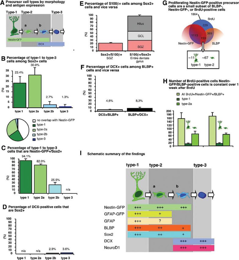
806                                                   STEINER ET AL.

  On the basis of the presence of doublecortin (DCX) in        tion containing 25% ethylene glycol, 25% glycerin, and
nestin-GFP-positive cells that lacked radial morphology,       0.05M phosphate buffer.
we had identified a transitional population and labeled
these cells type-2b cells (Filippov et al., 2003). The pres-
ent study was undertaken to further describe type-2b                            Immunofluorescence
cells and to examine the details of the transition from
glia-like to neuronal. Our hypothesis was that at this           For BrdU staining DNA was denatured in 2N HCl for
point of adult hippocampal neurogenesis a brief overlap        30 min at 37%. Hereafter, sections were rinsed in 0.1M
of glial- and stem-cell characteristics and neuronal fea-      borate buffer and washed with Tris-buffered saline
tures existed. Our rationale was that the precise charac-      (TBS). Primary and secondary antibodies were diluted
terization of type-2 cells would provide a very useful tool    in TBS containing 0.1% TritonX-100 and 3% donkey se-
in further studying how adult neurogenesis is regulated        rum (TBS1). Sections were incubated with primary
because it would further allow to distinguish between a        antibodies for 24 h at 4°C, washed with TBS and TBS1,
regulation on the level of undetermined and lineage-           and incubated with the secondary antibodies in TBS1
determined progenitor cells. In a previous study we had,       for 4 h at room temperature. Sections were then washed
for example, found that the well-documented effects of         with TBS and coverslipped in polyvinyl alcohol with dia-
physical activity on precursor cell proliferation in the       zabicyclo-octane (PVA-DABCO) as antifading agent. The
adult dentate gyrus cause the largest relative increase        primary antibodies were applied in the following concen-
on the level of type-2b cells (Kronenberg et al., 2003).       trations: anti-BrdU (rat, 1:500, Harlan Seralab), anti-
The present study was designed to investigate, if the          BLBP (rabbit, 1:2,000, kind gift from Dr. N. Heintz), anti-
type-2b stage of development is indeed as particular as        GFAP (guinnea pig, 1:1,000, Advanced ImmunoChemis-
such results seemed to indicate.                               try), anti-S100b (mouse, 1:1,000), anti-Doublecortin (goat,
                                                               1:200, Santa Cruz Biotechnologies), anti-NeuroD1 (rabbit,
           MATERIALS AND METHODS                               1:200, Chemicon), anti-NeuroD1 (goat, 1:200, Santa Cruz)
          Animals and Housing Conditions                       anti-GFP (rabbit, 1: 400, Abcam, Cambridge, UK), anti-
                                                               GFP (goat, 1:1,000, Acris Antibodies), anti-GFP (goat,
  The generation of transgenic mice expressing GFP             1:1,000, Santa Cruz Biotechnologies), anti-Sox-2 (rabbit,
driven by regulatory elements of the nestin (Yamaguchi         1:100, Santa Cruz), anti-Sox2 (rabbit, 1:1,500, Chemicon).
et al., 2000) and the GFAP gene (Nolte et al., 2001) has       The secondary antibodies were anti-rat RhodamineX,
been described elsewhere. Both strains were bred at the        anti-goat FITC, anti-rabbit FITC, anti-rabbit Cy5, anti-
animal facility of the MDC. Seven-week-old female mice         goat Cy5, anti-rabbit RhodamineX, anti-guinea pig Cy5,
were held three per cage under standard laboratory             anti-mouse RhodamineX (all:1:250, Dianova).
housing conditions with a light/dark cycle of 12 h each
and free access to food and water. Twelve nestin-GFP
animals were randomly assigned to two experimental             Quantification of Double- and Triple-Labeled Cells
groups, six per group, to allow the investigation of the
proliferating cells at two different time points after           One-in-twelve series of sections of each brain (at least
labeling the dividing precursor cells. Three adult trans-      five per group) were triple-labeled as described. Fifty
genic mice expressing GFP under the GFAP promoter              BrdU-positive cells within the granule cell layer were
were held under standard housing conditions.                   analyzed for co-expression of BrdU and glial or neuronal
                                                               markers, using a Leica TCS SP2 confocal microscope.
                                                               All images were taken in sequential scanning mode and
                    BrdU Injections                            further processed in Adobe Photoshop 7.0 for Macintosh.
                                                               Only general contrast adaptations were made and fig-
  BrdU (Sigma, Germany) was dissolved in 0.9% NaCl             ures were not otherwise manipulated.
and filtered. The animals received one single intraperito-
neal (i.p.) injection of 50 mg/kg body weight. Animals
were killed 3 h or 1 week after the BrdU injection.
                                                                               Immunohistochemistry

                  Tissue Preparation                             Sections were treated with 0.6% H2O2 to block endo-
                                                               genous tissue peroxidases. Hereafter, sections were
  Animals were deeply anesthetized with ketamine and           washed and incubated with 2N HCl for 30 min at 37°C
perfused transcardially with 4% paraformaldehyde in            and rinsed in borate buffer as described earlier. Sections
0.1M phosphate buffer. The brains were dissected from          were then incubated with the primary anti-BrdU anti-
the skulls, postfixed in 4% paraformaldehyde at 4°C             body overnight at 4°C, rinsed in TBS and TBS1 and
overnight, and transferred into 30% sucrose. Brains            incubated with the secondary antibody for 2 h at room
were cut on a dry-ice-cooled copper block with a sliding       temperature. ABC reagent (Vectastain Elite, Vector
microtome (Leica, Bensheim) into 40-lm thick coronal           Laboratories) was applied for 1 h at a concentration of
sections and stored at 220°C in a cryoprotectant solu-         9 lL/mL. Diaminobenzidine (DAB, Sigma, Germany)

GLIA DOI 10.1002/glia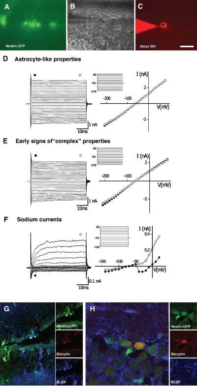
TYPE-2 CELLS IN ADULT HIPPOCAMPAL NEUROGENESIS                                    807
was used as chromogen at the concentration of 0.025           was equilibrated with 95% O2 and 5% CO2 at a rate of
mg/mL in TBS with 0.01% H2O2 and 0.04% nickel                 1–2 mL/min to a final pH of 7.4.
chloride.                                                       Patch-clamp recordings were carried out in nestin-
                                                              GFP-expressing-cells located in dentate gyrus about 10–
                                                              20 lm below the surface of the brain slice using an EPC
       Quantification of BrdU Labeled Cells                    9/2 double patch clamp amplifier. The acquisition and
                                                              the analysis of the data were performed with WinTIDA
  One-in-six series of sections (240 lm apart) from all       software (HEKA, Lambrecht/Pfalz, and Germany). In
animals were DAB-stained, and BrdU positive cells were        voltage-clamp mode, whole-cell recordings were per-
counted throughout the rostrocaudal extent of the gran-       formed with pipettes filled with the standard solution
ule cell layer. Resulting numbers were then multiplied        which consisted of (in millimolar): 130 KCl, 2MgCl2,
by six to obtain the estimated total number of BrdU-          0.5 CaCl2, 2 Na-ATP, 5 EGTA, and 10 HEPES. The pH
positive cells per granule cell layer.                        was adjusted to 7.3 with KOH. To confirm the intracellu-
                                                              lar access, Alexa Fluor 594 (10 lg/mL, Molecular Probes)
                                                              was used. The open resistance of the patch pipettes was
         Acute Brain Slices Preparation for                   6–8 MX. Patch pipettes were pulled from borosilicate
                 Electrophysiology                            capillaries (inner diameter 0.87 mm; outer diameter
                                                              1.5 mm; Hilgenberg, Malsfeld, Germany) using a P-2000
   Acute hippocampus slices from adult nestin-GFP             laser-based pipette puller (Sutter Instrument, Novato).
transgenic mice were prepared as described previously         All experiments were performed at room temperature
(Filippov et al., 2003).                                      (21–25°C). During an individual experiment the temper-
   Briefly, mice were decapitated and the brain was re-        ature fluctuated
808 STEINER ET AL.
phosphate buffer, pH 7.4, containing 0.1% Glutaralde- SGZ but most were found in the molecular layer (Figs.
hyde at 4°C overnight. For further characterization of 1E and 2C,C0 ).
the patched cells immunohistochemistry was performed.
Fixed slices were washed in TBS and incubated with
TBS1 for 30 min and incubated with primary antibodies Type-1 and Type-2 Cells Express Radial-Glia
in TBS1 for 48 h at 4°C. Primary antibodies were used Marker BLBP
in following concentrations: anti-GFP (goat, 1:1,000),
anti-BLBP (rabbit, 1:2,000). Slices were washed with We next wanted to know, up to which stage in the pro-
TBS and preincubated in TBS1 for 15 min. Hereafter, posed course of development, the putative precursor
they were incubated in secondary antibodies for 4 h. cells would express glial markers. We had shown that
Secondary antibodies used: anti-rabbit Cy5, anti-goat all nestin-GFP-positive type-1 cells were GFAP-positive
FITC (all 1:250, Dianova). Sections were then washed but that not all GFAP-cells with radial morphology
with TBS and coverslipped in PVA-DABCO. expressed nestin-GFP (Filippov et al., 2003).
Brain lipid binding protein (BLBP) is transiently
expressed in radial glia during fetal brain development
Statistical Analysis (Feng et al., 1994) and actively blocks differentiation,
presumably downstream of Notch signaling (Anthony
All numerical analysis were performed using Statview et al., 2005).
5.0.1. For all comparisons ANOVA was performed by With very few exceptions, all type-1 cells expressed
Fisher’s post hoc test when appropriate. Differences BLBP, further supporting their radial glia-like nature
were considered statistically significant at P < 0.05. (Fig. 2B). In addition, we detected a small population of
radial glia-like BLBP-positive cells that expressed GFAP
but were negative for nestin-GFP.
RESULTS We used the systemic application of S-phase marker
Type-1 and Type-2 Cells Express Precursor Cell bromodeoxyuridine (BrdU) to permanently label a cohort
Marker Sox2 of dividing cells in the SGZ and to study the prolifera-
tive activity of radial glia-like cells. BrdU-immunohisto-
Sox2 is a transcription factor required for stem-cell chemistry at 3 h after BrdU revealed that 148 6 39.65
maintenance that actively inhibits neuronal differentia- (12.05% 6 2.4%) of the BrdU-positive cells (1227.33 6
tion in neural precursor cells (Wegner and Stolt, 2005). 507.24; all numbers are means 6 standard error of the
Sox2 is found in the adult SGZ (Komitova et al., 2005) mean) co-expressed nestin-GFP and BLBP—but only
and reporter gene mice with GFP expressed under the less than 5% of the BrdU-positive nestin-GFP-positive
Sox2 promoter have been used to isolate precursor cells cells have a radial glial morphology (Filippov et al.,
from the adult dentate gyrus (D’Amour and Gage, 2003). 2003). Thus the BLBP-positive/nestin-GFP-positive ra-
We studied Sox2 expression in type-1, type-2, and dial glia-like type-1 cells represented a rarely prolifera-
type-3 cells of the adult SGZ (Fig. 1A). Of 100 Sox2-posi- tive population which did not account for the majority of
tive cells, 23.4% were type-1 cells with radial morphol- the proliferating nestin-GFP/BLBP-positive cells. Ninety-
ogy and 30.9% were type-2a cells, whereas only 2.7% five percent of all BrdU-labeled nestin-GFP-expressing
were type-2b cells, and 1.3% type-3 cells (Fig. 1B). This cells are type-2 cells (Fig. 2B, inset).
implied that more than 40% of all Sox2-positive cells did However, 133.84 6 38.23 BrdU-positive cells, which re-
not show any overlap with nestin-GFP or DCX (Fig. 1B, present nearly one half of the BLBP-positive/BrdU-posi-
inset). Inversely, however, 94.1% of all nestin-GFP-posi- tive cells (281.84 6 37.67) were negative for nestin-GFP
tive type-1 cells expressed Sox2, 82.0% of type-2a cells, 3 h after BrdU. At the same time, the vast majority of
and 25.5% of type-2b cells (Fig. 1C). An analysis based BrdU-positive/nestin-GFP-positive type-2 cells (959.24 6
on the expression of DCX revealed that only 6.5% of all 95.90) was negative for the expression of BLBP. This sug-
DCX-expressing cells co-expressed Sox2, about half of gests that BLBP-positive type-2 cells can divide actively
which were nestin-GFP-positive type-2b cells (Fig. 1D). but most do not. This, too, is consistent with the idea that
From this result we conclude that the putative precur- BLBP, like Sox2, is a maintenance gene that preserves
sor cells in the adult SGZ express stem-cell marker the precursor cell pool. The hypothesis, not testable in
Sox2. Type-1 cells, which divide rarely, almost all vivo, would be that BLBP-positive divisions are self-
expressed Sox2, consistent with the idea that the high- renewing symmetric divisions, whereas BLBP-negative
est-order stem cell is maintained and actively prevented divisions are asymmetric divisions leading to the lineage
from neuronal development. Type-3 cells, in contrast, determination of one daughter cell.
are cells with early neuronal features, and consequently, In line with this consideration we found that the per-
Sox2 cells was rare in this population. The switch from centage of BLBP-positive type-1 cells among all BrdU
high Sox2 expression to low Sox2 expression occurs at cells remained unchanged between 3 h and 1 week after
the level of the transition between type-2a and type-2b BrdU, indicating a rarely and slowly proliferating cell
that is coinciding with the up-regulation of DCX. population.
The remainder of the Sox2-positive cells were largely This held true also for the numbers of proliferating
S100b-positive astrocytes, some of which resided in the BLBP-expressing type-2 cells that also remained con-
GLIA DOI 10.1002/gliaTYPE-2 CELLS IN ADULT HIPPOCAMPAL NEUROGENESIS 809
Fig. 1. Quantitative results. (A) The scheme recapitulates the nomen- only a very small percentage of DCX-positive cells co-expresses Sox2. (E)
clature used in this study (Kempermann et al., 2004). Adult neurogenesis 40% of the Sox2 positive cells are nestin-GFP-negative. In the subgranular
originates from radial glia-like stem cells (type-1) and progresses over in- zone (SGZ) approximately 1/5 is S100b-positive (left panel). If, conversely,
termediate progenitor cell stages (type-2 and type-3). Identification of type- all S100b-positive astrocytes in the dentate gyrus are considered, essen-
1 and type-2 cells is based on the expression of nestin-GFP and morphology tially all of the co-express Sox2. One third of these are found in the SGZ,
(Filippov et al., 2003). (B) stem cell marker Sox2 is expressed in the den- almost half in the hilus. (F) Similarly to that of D we also found that a
tate gyrus. The graph shows the distribution of 100 Sox2-positive cells to small percentage of DCX-positive cells was BLBP-positive. Also the reverse
the different categories of precursor cells in vivo. Forty-one percent of was true: about 8% of all BLBP-positive cells co-expressed DCX. (G) The
Sox2-positive cells do not belong to the precursor cells; more than half of actively dividing precursor cells represent only a small subset of all BLBP-
which are S100b-positive astrocytes (cf. E). (C) If the precursor cells are positive, Nestin-GFP-positive and BrdU-positive cells in the SGZ (at 3 h af-
taken as the basis of the analysis, it is found that essentially all type-1 cells ter the injection of BrdU). (H) The size of this cell population identified in
are Sox2-positive and 4/5 of the type-2 cells. Among the doublecortin G does not change between 3 h and 1 week after BrdU, which is consistent
(DCX)-positive type-2b cell only one quarter is Sox2-positive. Because with the idea that these cells constitute an asymmetrically dividing group of
type-3 cells are by definition nestin-GFP-negative they are not listed here. cells. (I) Presents a schematic qualitative summary of our findings, highlight-
(D) The same kind of analysis based on the expression of DCX reveals that ing the transition from type-2a to type-2b cells.
GLIA DOI 10.1002/glia810 STEINER ET AL.
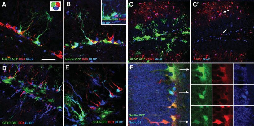
Fig. 2. Precursor cell markers and lineage markers in the adult in Fig. 1E) is full of Sox2/S100b-positive astrocytes (upper arrow in C0 ).
dentate gyrus. (A) Nestin-GFP (green), doublecortin (DCX, red), and Scale bar (in A), 100 lm. (D,E) Analysis of the GFAP-GFP transgenic
Sox2 (blue) show varying degrees of overlap in the subgranular zone. mice reveals a similar picture as the nestin-GFP mice. Radial glia-like
All radial glia-like type-1 cells are Sox2-positive. Scale bar, 40 lm. (B) cells are BLBP-positive. GFAP-GFP and DCX are mutually exclusive.
Radial glia-marker BLBP identifies both type-1 cells with their radial Few cells show an overlap between DCX and BLBP (appearing pink).
morphology and type-2 cells, which lack the radial process (type-2 cells Scale bar (in A), 40 lm. (F) nestin-GFP-positive cells can show weak
appearing yellow here). Scale bar (in A), 40 lm. Inset: BLBP-positive (upper row) to strong (lower row) co-expression of BLBP. NeuroD1 as
type-2 cells incorporate BrdU and are thus proliferative. C/C0 , essen- the earliest known neuronal lineage marker in adult neurogenesis
tially all S100b-positive astrocytes in the dentate gyrus are Sox2-posi- appears first in nestin-GFP-positive type-2 cells and is never found in
tive. Such astrocytes also exist in the SGZ (lower arrow in C0 ). The mo- type-1 cells. Scale bar (in A), 25 lm.
lecular layer (which was not included into the quantification depicted
stant between 3 h after BrdU and 1 week after BrdU ductance, while the type-2 cells are more heterogeneous
(Fig. 1H). Similarly, the numbers of new BLBP-positive/ including cells with neuronal properties. Here, we focus
nestin-GFP-negative cells did not change (3 h: 235.67 6 on cells with a type-2 property.
16.42; 1 week: 270.66 6 16.4; P 5 0.76). After establishing a whole-cell recording configuration,
We never found BLBP-positive/nestin-GFP-negative the cell was clamped to a series of de- and hyperpolariz-
cells with radial glia-like shape to be BrdU-positive. ing potentials from 2210 to 180 mV or from 2140 to
This might indicate that nestin-GFP expression in radial 120 mV at holding potential of 270 mV (see Fig. 3).
glia-like cells is related to an activated state associated One group of type-2 cells displayed passive membrane
with cell division. currents with no apparent time-dependence and a linear
In the fetal brain, radial glia is also characterized by current to voltage curve. This pattern corresponds to
the expression of GLAST and RC2. We did not find evi- type-1 cells. The other group typically had a high input
dence of GLAST and RC2 immunoreactions in the adult resistance (Rm 5 1,220 MX). The current pattern was
SGZ (not shown). characterized by the presence of small rapidly inactivat-
ing inward currents with depolarization typical for vol-
tage-gated Na21 channels followed by delayed outward
currents. With hyperpolarization, no currents were acti-
The Astrocyte-Like Electrophysiological vated. Electrophysiologically, this type of cell is similar
Phenotype Is Associated with BLBP Expression to glial precursor cells or the so-called ‘‘receptor astro-
cyte’’ (Matthias et al., 2003).
The nestin-GFP transgenic animals allowed to distin- After electrophysiological recordings Biocytin-labeled
guish the putative precursor cells in living acute slice type-2 cells were stained for BLBP to identify radial
preparations from the adult hippocampus. We dialyzed glial antigen properties in these cells. Fig. 3G shows a re-
cells with the hydrophilic fluorescent dye Alexa Fluor presentative BLBP-positive/nestin-GFP-positive cell with
via the patch-pipette to confirm the morphological fea- passive membrane conductance (Fig. 3D). In contrast,
tures and to distinguish between type-1 and type-2 cells type-2 cells with early signs of the complex type of mem-
(Filippov et al., 2003; Wang et al., 2005). In addition, brane currents (depolarization typical for voltage-gated
type-1 cells are characterized by passive membrane con- Na21 channels followed by delayed outward currents)
GLIA DOI 10.1002/gliaTYPE-2 CELLS IN ADULT HIPPOCAMPAL NEUROGENESIS 811
Technical limits precluded to conduct this study quan-
titatively. The available data, however, can serve as
another piece of evidence that BLBP-positive type-2 cells
show predominantly glial features.
GFAP-GFP Is Found in Type-2 Cells
To further corroborate the idea that at the type-2
stage a switch from glial to neuronal phenotype occurs,
we studied GFAP-GFP mice, in which the cytoplasmic
GFP allows a better appreciation of cell morphology
than the detection of GFAP protein, which is largely lim-
ited to the processes. We found GFAP-GFP cells with
both type-1 (with radial process) and type-2 morphology
(without radial process but short and delicate horizontal
processes). Because horizontal astrocytes in the SGZ are
GFAP-GFP-positive and also have horizontal processes
we costained for S100b that is expressed by horizontal
astrocytes but would not be found the in possibly GFAP-
GFP-positive type-2 cells. Indeed, we found GFAP-GFP-
positive S100b-negative cells with type-2 morphology in
the SGZ (Fig. 1C).
Lineage Specific Marker NeuroD1 Is First
Expressed in Type-2 Cells
The transcription factor NeuroD1 is one of the earliest
indications of granule cell development (Liu et al., 2000;
Pleasure et al., 2000). Three hours after BrdU, 764.48 6
77.80 BrdU-positive cells co-expressed NeuroD1 and nes-
tin-GFP, whereas 552.24 6 220.20 BrdU-positive cells
co-expressed NeuroD1 but lacked nestin-GFP expres-
sion, and 250.62 6 61.75 BrdU-positive type-2 cells were
negative for NeuroD1. This implies that about 75% of all
proliferating type-2 cells co-express NeuroD1. In co-loca-
lization studies with antibodies against BLBP and Neu-
roD1 we never found type-2 cells to be positive for both
markers. Similarly, we never found any type-1 cells to
be NeuroD1-positive. All NeuroD1-positive cells in the
SGZ and GCL were oval-shaped without processes.
DCX-positive type-2 cells (type-2b) were always Neu-
roD1-positive, supporting the idea that these cells are
Fig. 3. Electrophysiology. (A–C) In acute slice preparations type-2 pre- determined towards the neuronal lineage.
cursor cells were identified by their nestin-GFP expression. After establish-
ing the patch contact cells were filled with Alexa-594 to confirm the distinct We did not detect co-labeling of Sox-2 with NeuroD1
morphology devoid of radial processes. Scale bar, 20 lm. (D–F) Type-2 cells in type-2 cells, which is in a slight discrepancy with the
might display different electrophysiological characteristics previously
attributed to nestin-GFP-positive cells (Filippov et al., 2003; Wang et al.,
finding that Sox2 and DCX had a small overlap. Because
2005), including passive astrocyte-like properties. Roughly half of the type- not all markers can be investigated in the same immu-
2 cells investigated belonged to the astrocyte-like type (D), whereas the nohistochemical experiment and the discrepancy affects
other half showed signs of the ‘‘complex’’ type (E). These cells do not qualify
as complex cells of the definition but have clearly different properties than only a few percent, we assume that this unexplained
the astrocyte-like cells of (D). As before, single type-2 cells exhibited small overlap is due to technical limitations. This issue, how-
sodium currents. (G,H) After electrophysiological recording the cells were ever, does not contradict the principal conclusion to be
filled with biocytin and processed for immunohistochemistry. Although this
analysis was not quantitative (because only small numbers of cells could be drawn from this experiment, that NeuroD1 expression
studied) we found the astrocyte-like cell of D to be BLBP-positive (and thus first appears at the type-2 stage and is essentially
a type-2a cell), whereas the cell from E was BLBP-negative and thus a
type-2b cell (H). Scale bar (in C), 60 lm (G), and 15 lm (H). mutually exclusive with the precursor cell markers
BLBP and Sox2.
were BLBP-negative (Figs. 3E,H). Few cells showed As anticipated, at 1 week after BrdU injection the
small sodium currents (Fig. 3F), consistent with early numbers of both NeuroD1-positive/nestin-GFP-positive
signs of neuronal differentiation. (1002.56 6 144.64) and NeuroD1-positive/nestin-GFP-
GLIA DOI 10.1002/glia812 STEINER ET AL.
negative (1000.51 6 144.66) cells had increased, indicat- Some authors have suggested modifications to our de-
ing a robustly increasing population of immature neu- velopmental model of adult hippocampal neurogenesis
rons. In contrast, the numbers of nestin-GFP-positive/ (Kempermann et al., 2004): Encinas and colleagues, for
NeuroD1-negative type-2 cells decreased between 3 h example, collapsed what is type-2 and type-3 cells in our
(250.62 6 61.74) and 1 week after BrdU (134.48 6 scheme into just one intermediate progenitor cell, which
67.46), indicating a continuous maturation of these cells would be subject to the regulation of proliferation
towards a neural phenotype. (Encinas et al., 2006). Our present data corroborate that
A small number of the BrdU-positive cells in our on the stage of the intermediate precursor cells, the
experiment were negative for nestin-GFP, BLBP, Sox2, transiently amplifying progenitor cells of the adult den-
DCX, or NeuroD1 (but only two markers could be tested tate gyrus, a series of distinct developmental events
at a time). As shown previously, this population includes takes place, which, in our eyes, justifies to distinguish
astrocytes, microglia or NG2-positive glial precursors preneuronal stages (type-2a) from a transitional phase
(Steiner et al., 2004). (type-2b) from an early neuronal stage (type-3), all of
which are still associated with cell proliferation. It
should though be noted that these ‘‘stages’’ are con-
DISCUSSION structs to aid scientific theory building, here about the
transition from glial to neuronal phenotypes. In reality
In this study, we describe that in type-2 cells of the changes are more gradual than the categorization into
adult SGZ an overlap exists between the expression of ‘‘cell types’’ might suggest.
markers associated with a precursor cell stage (nestin- Besides the radial glia-like vertical astrocytes with
GFP, Sox2, BLBP), a radial glia-like phenotype (BLBP), their precursor cell function, the adult dentate gyrus
and the neuronal lineage (DCX, NeuroD1). On the basis harbors a second population of astrocytes (‘‘horizontal
of the currently favored model of neuronal development astrocytes’’), which are consistently negative for nestin-
in the adult hippocampus we consider this stage to be a GFP, but positive for both GFAP and S100b (Steiner
transient phase that characterizes the transition be- et al., 2004). Within the granule cell layer of the dentate
tween a bi- or multipotent precursor cell and a lineage- gyrus, S100b-positive cells were never found to be in cell
determined progenitor cell. cycle and are thus considered to be postmitotic. Both
In our initial study, we had chosen to distinguish S100b-positive horizontal astrocytes and S100b-negative
type-2a cells from type-2b cells based on the additional radial astrocytes are generated in the adult dentate
expression of DCX in the nestin-positive type-2b cells gyrus, presumably from the radial glia-like stem cell.
that is absent in type-2a (Filippov et al., 2003). These However, we found that upon induction of neurogenesis
data had been generated in the nestin-GFP mice. Like and gliogenesis in the adult dentate gyrus by voluntary
in the GFAP-GFP mice additionally used in the present physical activity an elusive second population of GFAP-
study, the difference in the decay of GFP compared to positive cells acutely increased in number (Steiner et al.,
the original protein nestin or GFP is not known. It is 2004). Whereas proliferation of the nestin-GFP-positive
assumed that this decay, and with it the level of detec- radial cells was unaffected (Kronenberg et al., 2003), the
tion, are different between endogenous protein and re- number of dividing GFAP-positive S100b-negative cells
porter product but very few studies have actually increased. We interpreted this finding as upregulation of
addressed this issue (Corish and Tyler-Smith, 1999; Mel- an intermediate progenitor cell in the glial lineage.
lodew et al., 2004; Verkhusha et al., 2003). Conse- However, there was no strong argument against the
quently, there was a chance that we had overestimated assumption that this astrocyte-like progenitor cell might
the size of the population of type-2b cells. On the other contribute to the neuronal lineage as well. The present
hand, type-2b cells immediately appeared as a very study could not finally solve this issue but provided fur-
interesting group of cells worth further investigation ther arguments that the lineage separation between
because they embody the initiation of neuronal develop- gliogenesis and neurogenesis in the adult dentate gyrus
ment in the course of adult hippocampal neurogenesis. occurs on the level of type-2a cells.
In a study based on a DCX-reporter gene mouse, no nes- The revised model of adult neurogenesis would thus
tin protein was detected in DCX-dsRed-positive cells look like Fig. 1G: type-1 cells are the radial glia-like
(Karl et al., 2005). However, the low expression of nestin stem cells of the SGZ, which give rise to the highly pro-
protein in the murine dentate gyrus had been the main liferative type-2a cells. Initially, type-2a cells remain
reason to use nestin-GFP mice in the first place, so that BLBP-positive and Sox2-positive. To some degree, thus,
the sensitivity of the antibody detection has to be ques- type-1 and type-2a cells share features of stem cells and
tioned for a cell type with presumably low nestin expres- radial glia-like cells. A large number of type-2 cells,
sion. Although all reports based on nestin-GFP mice however, are negative for Sox2 and BLBP. We propose
agree in essence, some differing details are noteworthy that both Sox2 and BLBP are associated with the main-
(Filippov et al., 2003; Fukuda et al., 2003; Mignone et al., tenance of the precursor cell pool. The downregulation
2004). The discrepancy between nestin-GFP expression of these markers largely coincides with the upregulation
and GFAP immunoreaction found in our study, for exam- of NeuroD1 and DCX as two markers associated with
ple, might depend on the transgenic line (Mignone et al., the neuronal lineage. These latter cells represent the
2004). type-2b cells.
GLIA DOI 10.1002/gliaTYPE-2 CELLS IN ADULT HIPPOCAMPAL NEUROGENESIS 813
The present data strengthen the initial concept (Kem- Nestin-expressing cells can repopulate a dentate gyrus
permann et al., 2004) and add further distinctive crite- depleted of all amplifying stages of neurogenesis has
ria to the important transitional phase between the pre- been convincingly demonstrated (Seri et al., 2001, 2004).
cursor cells and neuronal development, which is cap- We have here shown, at which point the lineage-deter-
tured by the category type-2b. Type-2b cells are not only mining step in the development towards a new neuron
nestin-GFP-positive but also to some degree express occurs.
BLBP or Sox2. Sox2 identifies precursor cells in the
adult brain but consistent with our in vivo report, the
protein identifies a heterogeneous population of cells ACKNOWLEDGMENTS
ex vivo (Brazel et al., 2005). In addition, Sox3 has been
identified in neural precursor cells in the adult hippo- We thank Michael Hack and Magdalena G€ otz for
campus (Wang et al., 2006). advice and material, Nathaniel Heintz for the BLBP
At the same time, type-2b cells represent the first antibody, and Ruth Zarmstorff, Irene Thun, and Signe
stage, at which neuronal lineage markers appear. The Knespel for technical assistance.
apparently transitional stage is thus not only a result
the possible persistence of a reporter gene product
(GFP) as some might have suspected after our first REFERENCES
description. In addition we have shown that the type-2b
Alvarez-Buylla A, Lim DA. 2004. For the long run: Maintaining germi-
stage is characterized by the complete lack of glucocorti- nal niches in the adult brain. Neuron 41:683–686.
coid receptor expression, although both type-2a and Anthony TE, Mason HA, Gridley T, Fishell G, Heintz N. 2005. Brain
type-3 express the receptor (Garcia et al., 2004). lipid-binding protein is a direct target of Notch signaling in radial
glial cells. Genes Dev 19:1028–1033.
The electrophysiological data confirm this concept. Brandt MD, Jessberger S, Steiner B, Kronenberg G, Reuter K, Bick-
The BLBP-positive cells showed astrocytic properties, Sander A, Von der Behrens W, Kempermann G. 2003. Transient cal-
retinin-expression defines early postmitotic step of neuronal differen-
whereas BLBP-negative cells had a complex electrophys- tiation in adult hippocampal neurogenesis of mice. Mol Cell Neurosci
iological phenotype. We propose that the first synaptic 24:603–613.
input into the cells occurs on the level of the type-2b Brazel CY, Limke TL, Osborne JK, Miura T, Cai J, Pevny L, Rao MS.
2005. Sox2 expression defines a heterogeneous population of neuro-
cells. We have shown that this input is GABAergic sphere-forming cells in the adult murine brain. Aging Cell 4:197–
(Wang et al., 2005). One would assume that the cells 207.
receiving synaptic input are BLBP/Sox2-negative but at Corish P, Tyler-Smith C. 1999. Attenuation of green fluorescent protein
half-life in mammalian cells. Protein Eng 12:1035–1040.
present this is speculation. Also, our electrophysiological D’Amour KA, Gage FH. 2003. Genetic and functional differences
analysis was not quantitative, so that an overlap of glial between multipotent neural and pluripotent embryonic stem cells.
Proc Natl Acad Sci USA 100(Suppl 1):11866–11872.
antigenic features with early neuronal electrophysiologi- Doetsch F, Garcia-Verdugo JM, Alvarez-Buylla A. 1997. Cellular compo-
cal properties might exist. GABAergic input, however, sition and three-dimensional organization of the subventricular ger-
further induces neuronal differentiation (Ge et al., 2006; minal zone in the adult mammalian brain. J Neurosci 17:5046–5061.
Encinas JM, Vaahtokari A, Enikolopov G. 2006. Fluoxetine targets early
Tozuka et al., 2005). progenitor cells in the adult brain. Proc Natl Acad Sci USA 103:8233–
Taken together, the present data support the existence 8238.
of transient amplifying progenitor cells (type-2 cells) Feng L, Hatten ME, Heintz N. 1994. Brain lipid-binding protein
(BLBP): A novel signaling system in the developing mammalian
that mark the progression from a glia-like precursor cell CNS. Neuron 12:895–908.
phenotype to characteristics of the neuronal lineage. Filippov V, Kronenberg G, Pivneva T, Reuter K, Steiner B, Wang LP,
Yamaguchi M, Kettenmann H, Kempermann G. 2003. Subpopulation
Scheffler, Walton and colleagues have studied the glia– of nestin-expressing progenitor cells in the adult murine hippocam-
neuron transition in the course of neurogenesis in an ex pus shows electrophysiological and morphological characteristics of
vivo model based on cell cultures from the early post- astrocytes. Mol Cell Neurosci 23:373–382.
Fukuda S, Kato F, Tozuka Y, Yamaguchi M, Miyamoto Y, Hisatsune T.
natal and adult subventricular zone, the second neuro- 2003. Two distinct subpopulations of nestin-positive cells in adult
genic region of the adult brain (Scheffler et al., 2005). mouse dentate gyrus. J Neurosci 23:9357–9366.
With time lapse video-microscopy they could follow the Garcia A, Steiner B, Kronenberg G, Bick-Sander A, Kempermann G.
2004. Age-dependent expression of glucocorticoid- and mineralocorti-
transition from glial to neuronal phenotype in individual coid receptors on neural precursor cell populations in the adult mu-
cells. On a phenomenological level their data are in full rine hippocampus. Aging Cell 3:363–371.
Ge S, Goh EL, Sailor KA, Kitabatake Y, Ming GL, Song H. 2006. GABA
agreement with ours: the neuron–glia transition can be regulates synaptic integration of newly generated neurons in the
identified as a critical step in neurogenesis. adult brain. Nature 439:589–593.
We caution, however, that none of the markers that Hamill OP, Marty A, Neher E, Sakmann B, Sigworth FJ. 1981. Improved
patch-clamp techniques for high-resolution current recording from cells
can be used to identify cells on the different stages of de- and cell-free membrane patches. Pflugers Arch 391:85–100.
velopment are fully sensitive and specific. Sox2, for Heins N, Malatesta P, Cecconi F, Nakafuku M, Tucker KL, Hack MA,
example, also identifies astrocytes in the dentate gyrus Chapouton P, Barde YA, Gotz M. 2002. Glial cells generate neurons:
The role of the transcription factor Pax6. Nat Neurosci 5:308–315.
for which no precursor cell properties can be assumed. Huttmann K, Sadgrove M, Wallraff A, Hinterkeuser S, Kirchhoff F,
Some markers are mutually exclusive (e.g. GFAP and Steinhauser C, Gray WP. 2003. Seizures preferentially stimulate pro-
liferation of radial glia-like astrocytes in the adult dentate gyrus:
DCX or NeuroD), whereas others might identify particu- Functional and immunocytochemical analysis. Eur J Neurosci 18:
lar functional states of a cell type. This problem not- 2769–2778.
withstanding it becomes increasingly apparent that Karl C, Couillard-Despres S, Prang P, Munding M, Kilb W, Brigadski T,
Plotz S, Mages W, Luhmann H, Winkler J, Bogdahn U, Aiqner L.
adult neurogenesis originates from a cell that lacks neu- 2005. Neuronal precursor-specific activity of a human doublecortin
ronal features but is glia-like. That resident GFAP- and regulatory sequence. J Neurochem 92:264–282.
GLIA DOI 10.1002/glia814 STEINER ET AL. Kempermann G, Jessberger S, Steiner B, Kronenberg G. 2004. Mile- lar zone have morphological and molecular characteristics of radial stones of neuronal development in the adult hippocampus. Trends glia. J Neurosci 22:3161–3173. Neurosci 27:447–452. Nolte C, Matyash M, Pivneva T, Schipke CG, Ohlemeyer C, Hanisch UK, Komitova M, Mattsson B, Johansson BB, Eriksson PS. 2005. Enriched Kirchhoff F, Kettenmann H. 2001. GFAP promoter-controlled EGFP- environment increases neural stem/progenitor cell proliferation and expressing transgenic mice: A tool to visualize astrocytes and astroglio- neurogenesis in the subventricular zone of stroke-lesioned adult rats. sis in living brain tissue. Glia 33:72–86. Stroke 36:1278–1282. Pleasure SJ, Collins AE, Lowenstein DH. 2000. Unique expression pat- Kronenberg G, Reuter K, Steiner B, Brandt MD, Jessberger S, Yamagu- terns of cell fate molecules delineate sequential stages of dentate chi M, Kempermann G. 2003. Subpopulations of proliferating cells of gyrus development. J Neurosci 20:6095–6105. the adult hippocampus respond differently to physiologic neurogenic Scheffler B, Walton NM, Lin DD, Goetz AK, Enikolopov G, Roper SN, stimuli. J Comp Neurol 467:455–463. Steindler DA. 2005. Phenotypic and functional characterization of Kunze A, Grass S, Witte OW, Yamaguchi M, Kempermann G, Redecker adult brain neuropoiesis. Proc Natl Acad Sci USA 102:9353–8. C. 2006. Proliferative response of distinct hippocampal progenitor cell Seri B, Garcia-Verdugo JM, Collado-Morente L, McEwen BS, Alvarez- populations after cortical infarcts in the adult brain. Neurobiol Dis Buylla A. 2004. Cell types, lineage, and architecture of the germinal 21:324–332. zone in the adult dentate gyrus. J Comp Neurol 478:359. Lendahl U, Zimmerman LB, McKay RDG. 1990. CNS Stem cells express Seri B, Garcia-Verdugo JM, McEwen BS, Alvarez-Buylla A. 2001. a new class of intermediate filament protein. Cell 60:585–595. Astrocytes give rise to new neurons in the adult mammalian hippo- Liu M, Pleasure SJ, Collins AE, Noebels JL, Naya FJ, Tsai MJ, Lowen- campus. J Neurosci 21:7153–60. stein DH. 2000. Loss of BETA2/NeuroD leads to malformation of the Steiner B, Kronenberg G, Jessberger S, Brandt MD, Reuter K, dentate gyrus and epilepsy. Proc Natl Acad Sci USA 97:865–870. Kempermann G. 2004. Differential regulation of gliogenesis in the Malatesta P, Hack MA, Hartfuss E, Kettenmann H, Klinkert W, context of adult hippocampal neurogenesis in mice. Glia 46:41–52. Kirchhoff F, Gotz M. 2003. Neuronal or glial progeny: Regional differ- Tozuka Y, Fukuda S, Namba T, Seki T, Hisatsune T. 2005. GABAergic ences in radial glia fate. Neuron 37:751–764. excitation promotes neuronal differentiation in adult hippocampal Matthias K, Kirchhoff F, Seifert G, Huttmann K, Matyash M, Ketten- progenitor cells. Neuron 47:803–815. mann H, Steinhauser C. 2003. Segregated expression of AMPA-type Verkhusha VV, Kuznetsova IM, Stepanenko OV, Zaraisky AG, glutamate receptors and glutamate transporters defines distinct Shavlovsky MM, Turoverov KK, Uversky VN. 2003. High stability of astrocyte populations in the mouse hippocampus. J Neurosci 23: Discosoma DsRed as compared to Aequorea EGFP. Biochemistry 42: 1750–1758. 7879–7884. Mellodew K, Suhr R, Uwanogho DA, Reuter I, Lendahl U, Hodges H, Wang LP, Kempermann G, Kettenmann H. 2005. A subpopulation of Price J. 2004. Nestin expression is lost in a neural stem cell line precursor cells in the mouse dentate gyrus receives synaptic GABAer- through a mechanism involving the proteasome and Notch signalling. gic input. Mol Cell Neurosci 29:181–189. Brain Res Dev Brain Res 151(1/2):13–23. Wang TW, Stromberg GP, Whitney JT, Brower NW, Klymkowsky MW, Mignone JL, Kukekov V, Chiang AS, Steindler D, Enikolopov G. 2004. Parent JM. 2006. Sox3 expression identifies neural progenitors Neural stem and progenitor cells in nestin-GFP transgenic mice. in persistent neonatal and adult mouse forebrain germinative zones. J Comp Neurol 469:311–324. J Comp Neurol 497:88–100. Noctor SC, Flint AC, Weissman TA, Dammerman RS, Kriegstein AR. Wegner M, Stolt CC. 2005. From stem cells to neurons and glia: A Sox- 2001. Neurons derived from radial glial cells establish radial units in ist’s view of neural development. Trends Neurosci 28:583–588. neocortex. Nature 409:714–720. Yamaguchi M, Saito H, Suzuki M, Mori K. 2000. Visualization of neuro- Noctor SC, Flint AC, Weissman TA, Wong WS, Clinton BK, Kriegstein genesis in the central nervous system using nestin promoter-GFP AR. 2002. Dividing precursor cells of the embryonic cortical ventricu- transgenic mice. Neuroreport 11:1991–1996. GLIA DOI 10.1002/glia
You can also read