Protective Mechanism of Luteinizing Hormone and Follicle-Stimulating Hormone Against Nicotine-Induced Damage of Mouse Early Folliculogenesis
←
→
Page content transcription
If your browser does not render page correctly, please read the page content below
ORIGINAL RESEARCH
                                                                                                                                        published: 07 September 2021
                                                                                                                                        doi: 10.3389/fcell.2021.723388

                                            Protective Mechanism of Luteinizing
                                            Hormone and Follicle-Stimulating
                                            Hormone Against Nicotine-Induced
                                            Damage of Mouse Early
                                            Folliculogenesis
                                            Wen-Xiang Liu 1,2† , Yan-Jie Zhang 3† , Yu-Feng Wang 2 , Francesca Gioia Klinger 4 ,
                                            Shao-Jing Tan 2 , Donatella Farini 4 , Massimo De Felici 4 , Wei Shen 2 and
                                            Shun-Feng Cheng 1,2*
                                            1
                                              College of Animal Science and Technology, Qingdao Agricultural University, Qingdao, China, 2 College of Life Sciences,
                                            Institute of Reproductive Sciences, Qingdao Agricultural University, Qingdao, China, 3 College of Veterinary Medicine,
                                            Qingdao Agricultural University, Qingdao, China, 4 Department of Biomedicine and Prevention, University of Rome Tor
                                            Vergata, Rome, Italy
                            Edited by:
                     Jennifer R. Wood,
       University of Nebraska System,       Previous studies have shown that nicotine could impair the germ cell cyst breakdown
                         United States      and the primordial follicle assembly by autophagy. In this paper, we discovered that
                       Reviewed by:         luteinizing hormone (LH) and follicle-stimulating hormone (FSH) could counteract the
                         So-Youn Kim,
       University of Nebraska Medical       damage caused by nicotine of mouse germ cell cyst breakdown. The neonatal mice
                 Center, United States      were separately intraperitoneally injected with nicotine, nicotine plus LH, nicotine plus
         A. Marie Lefrançois-Martinez,
Université Clermont Auvergne, France
                                            FSH, and saline (control) for 4 days. Compared with the nicotine group, the quality
                    *Correspondence:
                                            of oocytes and the number of follicles were remarkably increased in the nicotine plus
                      Shun-Feng Cheng       LH group or nicotine plus FSH group. LH and FSH could alleviate nicotine-induced
                   sfcheng@qau.edu.cn       oocyte autophagy by different pathways. LH reduced the nicotine-induced autophagy
 † These   authors share first authorship
                                            by restoring the phosphorylation level of adenosine 50 -monophosphate-activated protein
                    Specialty section:
                                            kinase α-1, while FSH by downregulating the phosphorylation level of Forkhead box
         This article was submitted to      class O 1. In addition, in a subsequent study of 6-week mice in different treated groups,
 Molecular and Cellular Reproduction,
                                            we found that LH and FSH supplementation significantly improved normal maturation
                a section of the journal
  Frontiers in Cell and Developmental       rates, fertilization rates, and embryo’s developmental potential of oocytes in oocytes
                                Biology     exposed to nicotine. Taken together, these results suggested that LH and FSH could
           Received: 10 June 2021           counteract the damage caused by nicotine and finally ensure normal germ cell cyst
        Accepted: 09 August 2021
     Published: 07 September 2021
                                            breakdown and early embryo development.
                              Citation:     Keywords: LH, FSH, nicotine, folliculogenesis, autophagy
      Liu W-X, Zhang Y-J, Wang Y-F,
         Klinger FG, Tan S-J, Farini D,
 De Felici M, Shen W and Cheng S-F          INTRODUCTION
        (2021) Protective Mechanism
               of Luteinizing Hormone
                                            Smoking has long been a well-established risk factor in human reproductive conditions, especially
    and Follicle-Stimulating Hormone
  Against Nicotine-Induced Damage
                                            in women who smoke during pregnancy, which is associated with various pregnancy complications
     of Mouse Early Folliculogenesis.       (Kharrazi et al., 2004). Despite a gradual decline in the number of smokers over the past few
      Front. Cell Dev. Biol. 9:723388.      decades, there was about 10–30% of women still smoking during pregnancy in western countries
     doi: 10.3389/fcell.2021.723388         (Reitan and Callinan, 2017). Nicotine inhaled by a pregnant woman after smoking can cross the

Frontiers in Cell and Developmental Biology | www.frontiersin.org                  1                                       September 2021 | Volume 9 | Article 723388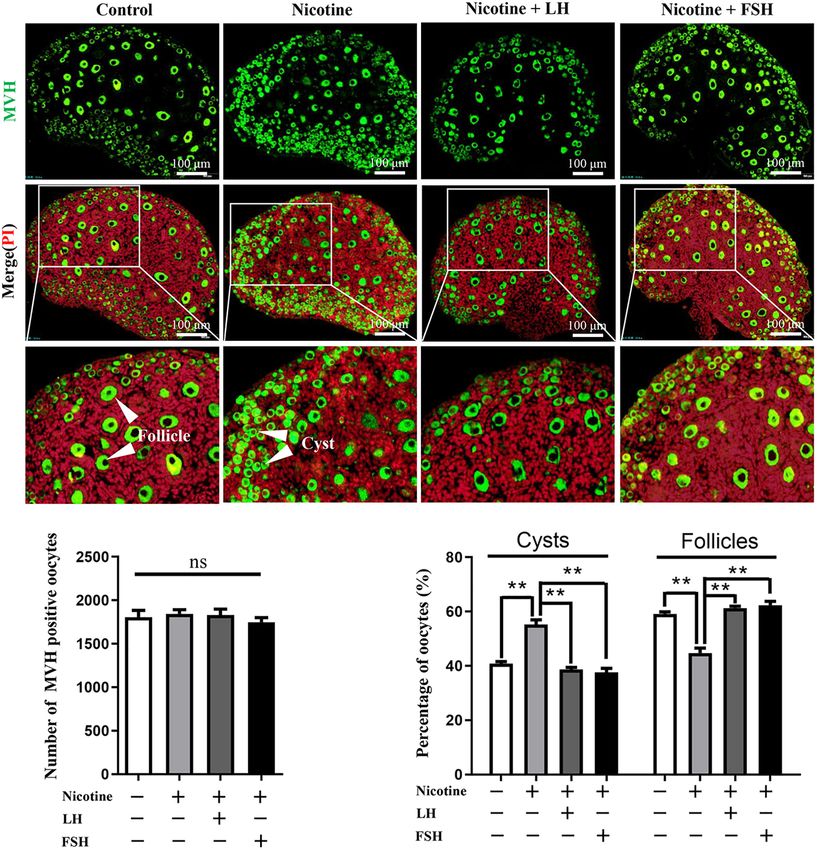
Liu et al.                                                                                                    LH/FSH Against Nicotine-Induced Damage

placenta, exposing the fetus to nicotine (Slotkin, 1998). Luck                 A recent study suggested that luteinizing hormone (LH) could
et al. (1985) proved that smoking mothers have higher levels of             inhibit oocyte apoptosis induced by external toxin exposure and
nicotine in their amniotic fluid, and consequently, the developing          maintain normal female reproductive ability in mice (Rossi et al.,
fetuses were exposed to high levels of nicotine. Evidences                  2017). A research by Shen et al. (2014) shows that follicle-
indicated that birth malformations and intellectual impairment              stimulating hormone (FSH) downregulates Forkhead box class
in later life are linked to smoking during pregnancy, with                  O 1 (FOXO1)-dependent apoptosis in mouse granulosa cells by
established causality (Little et al., 2004). In addition, nicotine is       coordinating the PI3K–AKT–FOXO1 axis. Recent studies have
known to interfere with mice cyst breakdown and primordial                  reported that FSH plays a protective role in ovarian damage and
follicle (PF) assembly (Wang Y. F. et al., 2018).                           autophagy through its downstream signal FOXO1 (Shen et al.,
    Ovarian follicles are the basic reproduction units of female            2017). Nonetheless, the protective mechanism of LH and FSH on
reproductive system in mammals. The germ cell “cysts” are clonal            early folliculogenesis still needs long-term investigation.
germ cell groups by the incomplete cytokinesis during mitosis                  Although the nicotine’s toxicity and even its reproductive
after primordial germ cell migration into the gonadal ridges,               toxicity to early ovarian development have been reported
allowing daughter cells to connect to each other by intercellular           (Wang Y. F. et al., 2018), the related rescue solutions
bridges (Wang C. et al., 2017). The germ cell cyst breakdown                are rarely mentioned. In this study, we would explore the
and PF formation are pre-requisites for the establishment of                underlying mechanism of nicotine affecting the perinatal ovarian
the ovarian reserve (Ge et al., 2019). In humans, the germ                  development and provide a new solution to prevent the damage,
cell cyst breakdown occurs around the beginning of the 20th                 thereby preserving their future fertility in reproductive age.
week of pregnancy, and the PF pool is formed before birth
(Sarraj and Drummond, 2012). However, in rodent, the germ
cell cyst breakdown mainly happens after birth, although there              MATERIALS AND METHODS
is emerging evidence that PF start to form at 17.5 days post-
coitus (Pepling and Spradling, 2001) and continue until about               Animals and Reagents
5 days post-partum (dpp) after birth. Therefore, studies of early           All CD-1 mice involved in the experiments were raised in an
folliculogenesis in mouse model were conducted mostly after                 environment with free diet, according to the regulations of the
birth (Wang Y. F. et al., 2018; Zhang et al., 2018). Germline nest          Animal Care Center of Qingdao Agricultural University, at 23◦ C,
breakdown is associated with a massive loss of oocytes that, in             50% humidity, and light and dark cycle of half day (turning lights
mouse, has been reported to occur following programmed cell                 off at 19:30 h). Male and female mice were randomly mated in the
death mainly in the form of apoptosis and autophagy (Pepling,               afternoon and checked for vaginal plugs the next morning. The
2006; Klinger et al., 2015; Wang C. et al., 2017). The process of           day of mouse birth is designated as 0 dpp. All studies involving
early folliculogenesis is very sensitive to toxic substances, and           mice were approved by the ethics Committee of Qingdao
adverse conditions in this process may lead to infertility and              Agriculture University (QAU; Agreement No. 2019-066).
female gametogenesis abnormalities (Mu et al., 2015).                           Nicotine (Sigma, 613207, St. Louis, MO, United States)
    Autophagy is an evolutionally conserved process by which                was dissolved in phosphate buffer solution (PBS) as described
damaged organelles, misfolded proteins, etc. are transported                in our previous study (Wang Y. F. et al., 2018). Low-dose
to lysosomes for degradation and reuse by lysosomes (Parzych                nicotine [1 mg/kg body weight/day (mg/kg)] was injected
and Klionsky, 2014). There are many ways to detect and                      intraperitoneally into 0-dpp female pups before 11:00 a.m.
monitor autophagosome number and autophagosomes flux,                       every day, and the same volume of PBS was administered to
including transmission electron microscopy (TEM) and                        control pups. LH (Sigma, L5269, purified LH protein from sheep
detection of autophagy-related protein such as BECLIN1                      pituitary) [50, 100, or 200 mIU/kg body weight/day (mIU/kg)]
and the autophagosome membrane-associated light chain                       or FSH (Sigma, 5925-FS, recombinant human FSH protein; 50,
3 (LC3; Mizushima et al., 2010). Among them, LC3 is a                       100, or 200 mIU/kg) were dissolved in PBS and injected into
special autophagy protein, and LC3-I in cytoplasmic form                    pups intraperitoneally soon after nicotine injection, respectively.
will transform to LC3-II in membrane-bound lipidated form                   The neonatal mice were continuously injected intraperitoneally
in the process of cell autophagy, which usually reflects the                for 4 days and euthanized, and their ovaries were collected for
autophagy level in the form of the ratio of LC3-II and LC3-I                the experiment. At this point, there are still plenty of oocytes
proteins (Mizushima et al., 2010). At birth and/or puberty,                 remaining in the ovarian cysts. Consistent with previous studies
autophagy formed by exposure to harmful substances during                   (Wang Y. F. et al., 2018; Liu et al., 2019; Liu W. X. et al., 2021; Tian
the establishment of ovarian reserves (mainly before birth)                 et al., 2021; Wang et al., 2021), the effect of the drug on germ cell
may lead to a shortage of germ cells and an insufficient PF                 cyst breakdown and PF formation could be measured by counting
pool (Ge et al., 2019). The research of Zhang et al. (2018)                 the proportion of oocytes in cysts and follicles. In addition,
indicates that fetal–neonatal toxic exposure damages mouse                  newborn mice were fed normally after 4 days of intraperitoneal
ovarian development and impairs primordial folliculogenesis                 injection. Follicles at all levels were tested at 3 weeks of age,
by inducing autophagy. Furthermore, the accumulation                        and in vitro maturation (IVM), spindle staining, and in vitro
of autophagy in starving mice at birth has been shown                       fertilization (IVF) experiments were performed after 6 weeks.
to be the primary cause of damage to cyst breakdown                             In this study, a total of 447 newborn female pups were
(Wang Y. Y. et al., 2017).                                                  used. This included the following: 287 newborn female pups

Frontiers in Cell and Developmental Biology | www.frontiersin.org       2                                   September 2021 | Volume 9 | Article 723388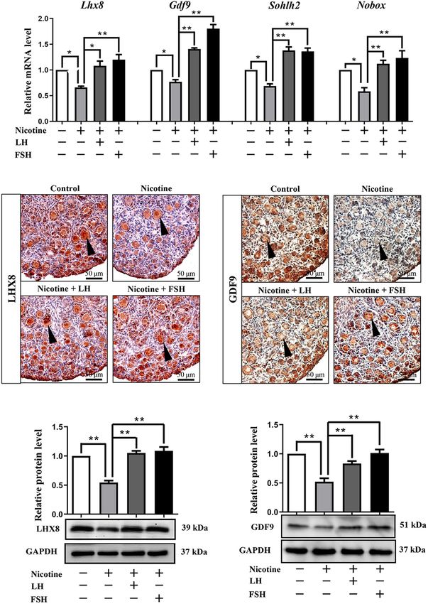
Liu et al.                                                                                             LH/FSH Against Nicotine-Induced Damage

were sacrificed for investigating 4-dpp mice female status; 64          transferred onto a polyvinylidene fluoride membrane (Millipore,
newborn female mice were used for the 3- and 6-week studies;            ISEQ00010, United States) and blocked in TBST containing
and 96 newborn female pups were used for nicotine receptor,             6% BSA for 4 h. The membranes were incubated with
LH receptor, and FSH receptor (FSHR) experiment. Notably, the           primary antibody (Supplementary Table 1) at 4◦ C, and then
ovaries of female pups from the same nest were evenly distributed       horseradish peroxidase-conjugated corresponding secondary
among the groups to eliminate the differences.                          antibody (Beyotime) was incubated at room temperature for
                                                                        120 min on the second day. Ultimately, the BeyoECL Plus Kit
Immunofluorescence                                                      (Beyotime, P0018) was used for chemiluminescence. The relative
Collected ovaries were fixed in 4% paraformaldehyde                     expression level of the target protein to GAPDH was calculated by
(Solaibio, P1110, Beijing, China) overnight, then treated               the software AlphaView SA (ProteinSimple, CA, United States)
according to the standard procedure for paraffin-embedded               and normalization method.
tissues. Continuous sections every 5 µm were applied
for immunofluorescence after antigen retrieval. Sections                Transmission Electron Microscopy
were blocked for 45 min, and then incubated overnight                   As previously described (Wang Y. F. et al., 2018), TEM
with primary antibodies (Supplementary Table 1). Then,                  observations were processed with standard methods. Ovaries
the sections were incubated with secondary antibodies                   were collected and then fixed in 2.5% glutaraldehyde for
(Supplementary Table 1), and nucleic acid was stained                   24 h. Serial sections were obtained by Leica ultramicrotome
with propidium iodide (PI; Solaibio, P8080-10). Oocytes                 (Leica EM UC7, Wetzlar, Germany), and the samples
were indicated by positive staining for mouse vasa homolog              were stained with uranyl acetate and lead citrate. Finally,
(MVH), a germ cell-specific protein. The aggregation of                 HT7700 (Hitachi, Tokyo, Japan) was used to capture and
two or more germ cells was defined as cysts and others as               observe the images. At least three replicates and at least 30
follicles (Pepling, 2006). Oocytes in the cysts or follicles were       oocytes in each group were analyzed to count the number
counted every five sections in a double-blind manner with the           of autophagosomes.
Image-Pro Plus software 6.0 (Media Cybernetics, Rockville,
MD, United States).                                                     In vitro Maturation and Spindle Staining
                                                                        Briefly, after 2 days of treatment with pregnant mares serum
Immunohistochemistry                                                    gonadotropin (PMSG), oocytes were collected and isolated in M2
As previously described (Liu W. X. et al., 2021), the rehydrated        medium (Macgene, CE003, Beijing, China) supplemented with
sections were antigen retrieved with sodium citrate and treated         2.5 µmol/l milrinone (Sigma, M4659) from 6-week-old mice in
in 3% H2 O2 for 10 min. Next, sections were blocked with                each group. Oocytes in germinal vesicle (GV) stage were obtained
BDT and incubated with primary antibodies (Supplementary                by washing with M2 medium for three times. Finally, GV stage
Table 1). Biotin-labeled secondary antibodies (Supplementary            oocytes were cultured in M16 medium under mineral oil (Sigma,
Table 1) were incubated at 25◦ C for 60 min on the second               M5904) and IVM at 37◦ C in 5% CO2 atmosphere.
day, followed by staining using a DAB kit (ZSGB-BIO, ZLI-                   MII stage oocytes were collected and fixed with 4% PFA for
9017, Beijing, China) and hematoxylin. Vectashield (Vector,             0.5 h. Then, oocytes were blocked in blocking fluid, and then they
Shanghai, China, H-1000) mounting medium was used to                    were incubated with anti−α−tubulin antibody (Supplementary
seal the cover slips, and images were analyzed using a                  Table 1) for 1 h; oocytes were incubated with secondary
BX51 microscope.                                                        antibodies (Supplementary Table 1) for 1 h. The chromosome
                                                                        was incubated with DAPI (Beyotime, C1022) for 15 min. After
Quantitative Real-Time PCR                                              washing, a laser−scanning confocal microscope (Leica TCS SP5
As previously described (Wang Y. F. et al., 2018), the total RNA        II, Mannheim, Germany) was used to captured representative
was isolated from six ovary tissues with the RNAprep Pure Micro         images; 30 oocytes were analyzed in each group.
Kit (Aidlab, RN07, Beijing, China). The cDNA was synthesized
by TransScript One-Step gDNA Removal and cDNA Synthesis                 In vitro Fertilization
SuperMix (TransGen, AT311-03, Beijing, China). Quantitative             As previously described (Zhang et al., 2020), after 2 days of
real-time PCR (RT-qPCR) amplification with specific primers             treatment with PMSG and 12 h of treatment with human
(Supplementary Table 2) was performed using LightCycler                 chorionic gonadotropin (HCG), cumulus–oocyte complexes
480 (Roche, Germany). The relative transcript abundance was             (COCs) were collected from the ampulla of the 6-week-old mice
calculated by the 2−11Ct method and normalized according to             ovary. Sperm from the caudal epididymis of fertile male mice
the housekeeping gene Gapdh.                                            at 8 weeks old were released into the balanced human tubal
                                                                        fluid to capacitate, and then 10-µl capacitated sperms were
Western Blot                                                            mixed with COC from each mouse for fertilization in vitro. After
Proteins were extracted with RIPA (Beyotime, P0013C, Nantong,           fertilization, every 20 to 30 oocytes were transferred to balanced
China). The 5X SDS was mixed with the samples and boiled                60-µl KSOM medium (EMD Millipore Corp., Billerica, MA,
for 5 min in water for protein denaturation, and then the               United States) droplet for two-cell embryos, four-cell embryos,
SDS-PAGE was used to separate proteins. The proteins were               and blastocyst culture.

Frontiers in Cell and Developmental Biology | www.frontiersin.org   3                                 September 2021 | Volume 9 | Article 723388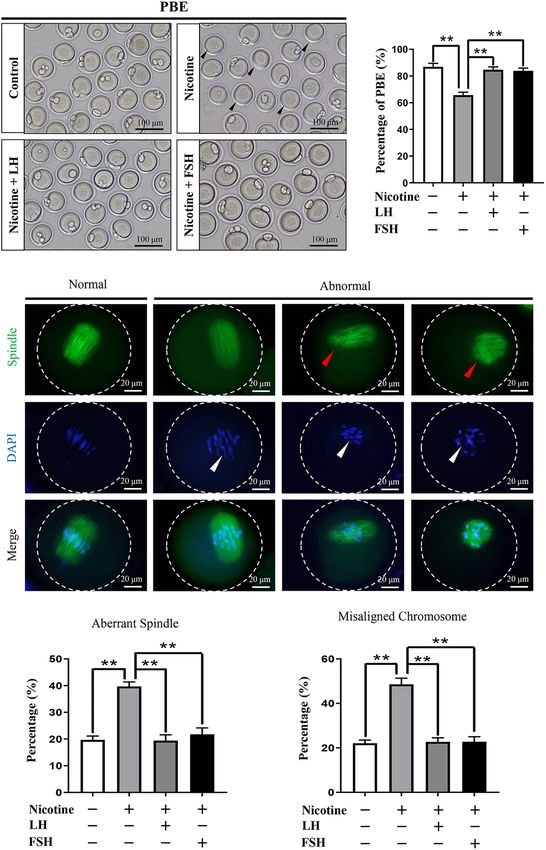
Liu et al.                                                                                               LH/FSH Against Nicotine-Induced Damage

Hematoxylin–Eosin Staining                                                  were expressed in the oocytes of the nicotine group, nicotine
Liver tissues were fixed in 4% paraformaldehyde, paraffin                   plus LH group, and nicotine plus FSH group (Figures 2B,C).
embedded, cut into 5-µm sections, and subsequently stained with             Moreover, protein expression levels were confirmed by western
hematoxylin–eosin (H&E) for histopathological analysis.                     blot analysis, and LHX8 and GDF9 protein expression levels
                                                                            were significantly decreased after nicotine exposure, while the
Statistical Analyses                                                        nicotine plus LH group and nicotine plus FSH group achieved
GraphPad Prism 8 (GraphPad Software, San Diego, CA,                         significant recovery (Figures 2D,E). In addition, we added the
United States) was applied for data analysis with one-way analysis          LH group and FSH group separately, we performed analyses
of variance followed by Tukey’s test for multiple comparisons;              on the ovaries of mice pups treated with LH and FSH without
Student’s t-test was used when only two pairs of data were                  nicotine in the same conditions. The statistical results showed
compared; P < 0.05 means statistically significant difference               that 100 mIU/kg LH and 100 mIU/kg FSH did not affect
(*), P < 0.01 means extremely significant difference (**) and               the total number of oocytes and germ cell cyst breakdown
P > 0.05 means not significant (ns). The data were expressed                (Supplementary Figures 3A–C), and the protein expression of
as mean ± standard error (S.E.), and all the experiments were               oocyte-specific genes LHX8 and GDF9 treated with LH and FSH
repeated at least three times.                                              alone did not change significantly compared with the control
                                                                            group (Supplementary Figures 3D–G).
                                                                               The RT-qPCR results of the expression of nicotinic
RESULTS                                                                     acetylcholine receptors (nAChRs) in the ovary from postnatal
                                                                            day 4 mouse revealed that among all of the 16 nAChRs subunits
LH and FSH Prevents Nicotine-Induced                                        (Dani, 2015), only seven subunits, namely, a4, a5, a7, a10,
                                                                            β2, and β4, were specifically affected by nicotine, with a4 and
Follicular Dysplasia in vivo                                                a10 mRNA levels showing a significantly increased expression
Neonatal mice were intraperitoneally injected with 1 mg/kg                  (Supplementary Figure 4A). The gene expressions of luteinizing
nicotine for four consecutive days in the presence of 50–                   hormone receptor (Lhr) was significantly activated by LH from
200 mIU/kg LH or 50–200 mIU/kg FSH. The statistical results                 the second day of treatment (Supplementary Figure 4B), and
showed that 100 mIU/kg LH or 100 mIU/kg FSH were the                        the increase was most significant on the 4 days (Supplementary
lowest concentrations for inhibiting the effect of nicotine on the          Figures 4C–E). Similarly, follicle-stimulating hormone receptor
cyst breakdown of the ovary without changing the total oocyte               (FSHR) is also significantly activated by FSH (Supplementary
number (Figure 1, Supplementary Figure 1, and Supplementary                 Figures 4F–H).
Table 3). Follicular counts showed that the ovaries in the
nicotine-treated group, compared with the control group, had
a lower proportion of oocytes in the follicles (44.77 ± 1.77%               LH and FSH Alleviate Nicotine-Induced
vs 59.16 ± 0.72% for the control group, Figure 1C and                       Autophagy in Ovarian Cells by Different
Supplementary Table 3); the rate of oocyte in follicles in the              Ways
nicotine plus LH or FSH treatment group was significantly                   Transmission electron microscopic observations revealed that
increased (61.28 ± 0.69% and 62.35 ± 1.44% separately,                      compared with the nicotine exposure group, oocytes in
Figure 1C and Supplementary Table 3). So, in vivo studies were              cysts (control = 1.74 ± 0.38, nicotine = 8.46 ± 0.75,
carried out with the dose of 100 mIU/kg LH or 100 mIU/kg                    nicotine + LH = 1.68 ± 0.34, and nicotine + FSH = 2.23 ± 0.49)
FSH. Notably, there were no significant changes in body                     and follicles (control = 1.17 ± 0.20, nicotine = 7.29 ± 0.56,
length, body weight, or ovarian diameter after intraperitoneal              nicotine + LH = 1.30 ± 0.06, and nicotine + FSH = 1.51 ± 0.19)
injection in each treatment group (Supplementary Figures 2A–                had fewer autophagosomes in the control and LH- or FSH-
D). Moreover, there was no significant difference in liver                  treated ovaries (Figures 3A–C). In addition, we also found
index and liver morphology among all treatment groups                       autophagosomes in ovarian somatic cells in the nicotine-treated
(Supplementary Figures 2E,F).                                               group, while mitochondria in the other groups were normal
                                                                            (Figure 3A). Further research results show that somatic cells
LH and FSH Rescues the Impaired                                             and oocytes in cysts or follicles in the nicotine-treated group
Oocyte-Specific Gene Expression                                             have stronger BECLIN1-positive signals and more LC3B-positive
In fact, many previous studies have confirmed that the                      spots, contrary to the other groups (Figures 3D,E). Results
obstruction of germ cell cyst breakdown and PF assembly                     of western blot analysis showed that the autophagy markers
are related to insufficient oocyte-specific gene expression                 BECLIN1 protein and ratio of LC3-II/LC3-I were not up-
(Wang Y. Y. et al., 2017; Wang et al., 2021; Wang J. J. et al., 2018;       regulated in the control group, nicotine plus LH group, and
Liu J. C. et al., 2021). Interestingly, LH or FSH could reduce the          nicotine plus FSH group (Figures 3F,G). This suggested LH and
effects of nicotine on specific gene expressions in oocytes, such           FSH protect against nicotine-induced damage of mouse germ cell
as factor in the LIM homeobox 8 (Lhx8), growth differentiation              cyst breakdown by inhibiting autophagy.
factor-9 (Gdf9), spermatogenesis and oogenesis helix-loop-helix                Further studies showed that LH and FSH can attenuate the
2 (Sohlh2), and newborn ovary homeobox (Nobox; Figure 2A).                  downregulation of the phosphorylation of autophagy-related
Immunohistochemistry analysis showed that LHX8 and GDF9                     proteins AKT and mTOR by nicotine (Figures 4A,B). We

Frontiers in Cell and Developmental Biology | www.frontiersin.org       4                               September 2021 | Volume 9 | Article 723388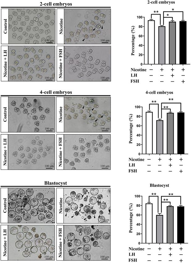
Liu et al.                                                                                                                  LH/FSH Against Nicotine-Induced Damage

  FIGURE 1 | LH and FSH counteract the delay of cyst breakdown in nicotine-exposed ovaries. (A) Representative image of germ cell cyst breakdown and primordial
  follicle assembly alignment in the control, nicotine, nicotine plus LH, and nicotine plus FSH groups. Scale bar, 100 µm. (B) The number of MVH-positive oocytes in
  the ovary in each group. (C) The percentage of oocytes in cysts and follicles in each group. n = 27 newborn female pups in figure and Supplementary Figure 1.
  The data are presented as means ± S.E. of three independent experiments (each in triplicate). **P < 0.01, ns P > 0.05. Abbreviations: LH, luteinizing hormone; FSH,
  follicle-stimulating hormone; MVH, mouse vasa homolog; and S.E., standard error.

found that nicotine was able to significantly increase the                            (PI3KC3) were significantly decreased by nicotine, while the
phosphorylation level of adenosine 50 -monophosphate (AMP)-                           protein phosphorylation levels of PI3KC3 and FOXO1 were
activated protein kinase α-1 (AMPKα1), an upstream signaling                          restored almost to the control in the nicotine plus FSH-treated
of AKT and mTOR, and this increase was drastically reduced in                         ovaries (Figures 4E,F). This evidence suggested that FSH
the ovaries co-treated with LH but not in those co-treated with                       eliminates the nicotine-induced autophagy by downregulating
FSH (Figures 4C,D). This indicates that LH reduced the nicotine-                      the phosphorylation level of FOXO1.
induced autophagy by restoring the phosphorylation level of
AMPKα 1.
   Forkhead box class O 1 is a key downstream effector of FSH
                                                                                      LH or FSH Improves Meiotic Maturation
signal, and FOXO1 is also involved in the upregulation                                and Spindle Assembly of
of autophagy-related genes and promoting autophagy                                    Nicotine-Exposed Mouse Oocytes
flux (Sengupta et al., 2009). Interestingly, the protein                              To test whether early exposure to nicotine affects ovary
phosphorylation levels of phosphoinositide-3-kinase class 3                           development, we measured the different classes of follicles at

Frontiers in Cell and Developmental Biology | www.frontiersin.org                 5                                       September 2021 | Volume 9 | Article 723388
Liu et al. LH/FSH Against Nicotine-Induced Damage FIGURE 2 | LH and FSH counteract the decreased expression of oocyte-specific transcription factors in nicotine-exposed ovaries. (A) Quantitative real-time PCR (RT-qPCR) for Lhx8, Gdf9, Sohlh2, and Nobox mRNA levels in control, nicotine, and nicotine plus LH or FSH (n = 36 newborn female pups). (B) Representative images of IHC for LHX8 (black arrow) in tissue sections of the ovaries in each group. Scale bar, 50 µm. (C) Representative images of immunohistochemistry (IHC) for the GDF9 (black arrow) in tissue sections of the ovaries in each group (n = 12 newborn female pups in B,C). Scale bar, 50 µm. (D) Relative protein level of LHX8 of the ovaries in each group. (E) Relative protein level of GDF9 of the ovaries in each group (n = 36 newborn female pups in D,E). The data are presented as means ± S.E. of three independent experiments (each in triplicate). *P < 0.05, **P < 0.01. Frontiers in Cell and Developmental Biology | www.frontiersin.org 6 September 2021 | Volume 9 | Article 723388
Liu et al. LH/FSH Against Nicotine-Induced Damage
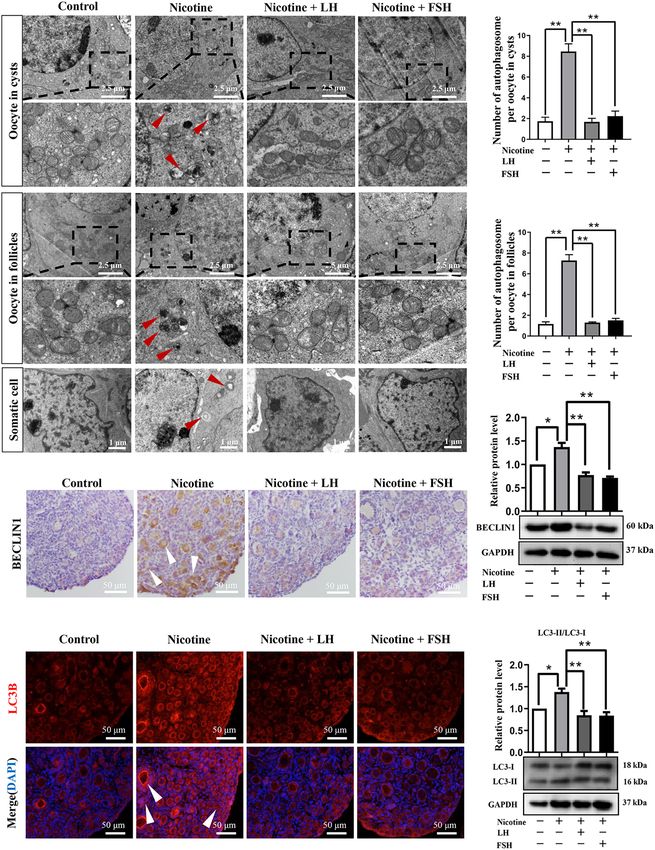
FIGURE 3 | LH and FSH suppressed autophagy in nicotine-exposed ovarian cells. (A) Autophagosomes (black arrow) in the oocyte (cysts or follicles) and somatic
cytoplasm in each group’s ovaries with transmission electron microscopy (TEM). (B) The number of autophagosome in one oocyte in cysts of each group’s ovaries
(control, n = 90; nicotine, n = 90; nicotine + LH, n = 90; and nicotine + FSH, n = 90, n = total number of oocytes from three replicate experiments). (C) The number
(Continued)
Frontiers in Cell and Developmental Biology | www.frontiersin.org 7 September 2021 | Volume 9 | Article 723388Liu et al. LH/FSH Against Nicotine-Induced Damage FIGURE 3 | Continued of autophagosome in one oocyte in follicles of each group’s ovaries (control, n = 90; nicotine, n = 90; nicotine + LH, n = 90; and nicotine + FSH, n = 90; n = total number of oocytes from three replicate experiments; n = 12 newborn female pups in A–C). (D) Representative images of IHC for the BECLIN1 in tissue sections of the ovaries in each group. The white arrows indicate BECLIN1-positive somatic cells and oocytes in cysts or follicles. Scale bar, 50 µm. (E) Representative image of immunofluorescence (IF) for the LC3B in tissue sections of the ovaries in each group. The white arrows indicate BECLIN1-positive somatic cells and oocytes in cysts or follicles (n = 12 newborn female pups in D,E). Scale bar, 50 µm. (F) Relative protein level of BECLIN-1 in each group. (G) Relative protein level of LC3-II/LC3-I in each group (n = 36 newborn female pups in F,G). The data are presented as means ± S.E. of three independent experiments (each in triplicate). *P < 0.05, **P < 0.01. FIGURE 4 | LH or FSH alleviates nicotine-induced autophagy in ovarian cells in different ways. (A) p-AKT expression relative to AKT in the control, nicotine, nicotine plus LH, and nicotine plus FSH groups, respectively. (B) p-mTOR expression relative to mTOR in the control, nicotine, nicotine plus LH, and nicotine plus FSH groups, respectively. (C) p-AMPKα1 expression relative to AMPKα1 in the control, nicotine, nicotine plus LH, and nicotine plus FSH groups, respectively. (D) p-AMPKα1 expression relative to AMPKα1 in the control, nicotine, nicotine plus LH, and nicotine plus FSH groups, respectively. (E) p-PIK3C3 expression relative to PIK3C3 in the control, nicotine, nicotine plus LH, and nicotine plus FSH groups, respectively. (F) p-FOXO1 expression relative to FOXO1 in the control, nicotine, nicotine plus LH, and nicotine plus FSH groups, respectively, (n = 72 newborn female pups in figure). The data are presented as means ± S.E. of three independent experiments (each in triplicate). *P < 0.05, **P < 0.01, and ns P > 0.05. 21 dpp after nicotine treatment. In this regard, we observed nicotine exposure were able to recover normal PF pool. In that exposure to nicotine resulted in a significant reduction order to assess the meiotic maturation of oocytes in each in the total follicle number and PF number compared to treatment group, the newborn mice in each group were the control group, while the LH and FSH supplementation injected intraperitoneally for 4 days and fed for 6 weeks for groups recovered significantly (Supplementary Figure 5). These further experiment. We detected the rate of first polar body results verified that the ovaries treated with LH or FSH after extrusion (PBE). As Figure 5A shows, the proportion of PBE Frontiers in Cell and Developmental Biology | www.frontiersin.org 8 September 2021 | Volume 9 | Article 723388
Liu et al.                                                                                                                  LH/FSH Against Nicotine-Induced Damage

  FIGURE 5 | Effects of LH or FSH on the meiotic maturation in nicotine-induced oocytes. (A) Representative images of oocytes with polar body extrusion (PBE;
  metaphase II of meiosis: MII) in each group. Scale bar, 100 µm. The black arrows indicate abnormal PBE. (B) The rates of PBE in each group (control, n = 90;
  nicotine, n = 90; nicotine + LH, n = 90; nicotine + FSH, n = 90; and n = total number of oocytes from three replicate experiments). (n = 16 newborn female pups in
                                                                                                                                                         (Continued)

Frontiers in Cell and Developmental Biology | www.frontiersin.org                  9                                       September 2021 | Volume 9 | Article 723388
Liu et al. LH/FSH Against Nicotine-Induced Damage
FIGURE 5 | Continued
A,B). (C) Representative images of spindle (green) morphologies and chromosome (blue) alignment. Scale bar, 20 µm. The arrows indicate aberrant spindles (red
arrows) and misaligned chromosomes (white arrows). (D) The rates of aberrant spindles (control = 19.70 ± 1.47%, n = 90; nicotine = 39.68 ± 1.73%, n = 90;
nicotine + LH = 19.42 ± 2.16%, n = 90; and nicotine + FSH = 21.79 ± 2.39%, n = 90; n = total number of oocytes from three replicate experiments). (E) The rates
of misaligned chromosomes (control = 22.08 ± 1.43%, n = 90; nicotine = 48.58 ± 2.78%, n = 90; nicotine + LH = 22.70 ± 1.81%, n = 90; and
nicotine + FSH = 22.77 ± 2.23%, n = 90; n = total number of oocytes from three replicate experiments; n = 16 newborn female pups in C–E). The data are
presented as means ± S.E. of three independent experiments (each in triplicate). **P < 0.01.
for nicotine-treated mice was significantly reduced compared studies have shown that cigarette smoke exposure in mice
with the control group after 12-h culture, while FSH and LH decreases the PF pool and induces autophagy in ovarian cells in
restored the rate to control levels (control = 86.93 ± 2.66%, preference to apoptosis (Gannon et al., 2012; Furlong et al., 2015).
nicotine = 65.73 ± 2.23%, nicotine + LH = 84.75 ± 2.10%, and Moreover, high levels of autophagy due to nicotine exposure
nicotine + FSH = 83.93 ± 2.05%; Figures 5A,B). The normal were responsible for the abnormal germ cell cyst breakdown in
spindle is fusiform, and the chromosome is arranged regularly perinatal mice (Liu et al., 2020). However, in many countries,
and linearly. In general, abnormalities in spindle assembly and some pregnant women are still exposed to smoke, often causing
chromosome separation are likely to cause oocyte arrest (Bennabi irreversible damage to the fetal reproductive system (Salihu and
et al., 2016); thus, we detected the spindle assembly and the Wilson, 2007; Ye et al., 2010; Lange et al., 2018). Since germ cell
chromosome arrangement in oocytes (Figures 5C–E). The result cyst breakdown and PF formation in mice mainly happens in the
showed that the rates of aberrant spindles and misaligned first 5 days after birth (Pepling and Spradling, 2001), the nicotine
chromosomes in the nicotine-exposed group were significantly exposure model of neonatal mouse was used to simulate the PF
higher than those in the control, while FSH and LH restored the formation period of human fetus in order to seek reasonable
rate to control levels (Figures 5D,E). rescue measures. We show here for the first time that specific
doses of LH or FSH supplementation can inhibit nicotine-
LH or FSH Improves the Potential of induced autophagy activation, thereby maintaining normal early
Fertilization and Early Embryo folliculogenesis.
In this paper, a nicotine concentration of 1 mg/kg body
Development of Nicotine-Exposed weight/day was given intraperitoneally as the in vivo nicotine
Mouse Oocytes exposure group. The experiment of Holloway et al. (2006) showed
The quality of oocyte influences fertilization and embryo’s that the steady-state levels of cotinine, the main metabolite
developmental potential (Wang and Sun, 2007). In order of nicotine, in serum was 135.9 ± 7.86 ng/ml after rats were
to observe the fertility rate in each treatment group, injected with 1 mg/kg bw/day nicotine for 14 consecutive days.
we measured the proportion of development of two-cell Importantly, this cotinine concentration was within the range
embryos, four-cell embryos, and blastocyst after IVF in each reported by human pregnant smokers (21.5–228.1 ng/ml; George
treatment group. Figure 6A indicates that the ratio of the et al., 2006). In addition, Wang et al. (2012) proved that mice
developed two−cell embryos was significantly decreased with treated with 1.5 mg/kg bw/day nicotine for 6 weeks did not
nicotine treatment and recovered to normal levels with the affect hemodynamic parameters or metabolic indices in the
administration of LH or FSH (control = 86.93 ± 2.66%, mice. In addition, experiments of Wang Y. F. et al. (2018)
nicotine = 65.73 ± 2.23%, nicotine + LH = 84.75 ± 2.10%, show that 1 mg/kg bw/day nicotine was associated with a
and nicotine + FSH = 83.93 ± 2.05%; Figures 6A,B). significant reduction of germ cell cyst breakdown but did not
Similar to the statistics of four-cell embryos and blastocyst affect the total oocyte number. The optimal dose of FSH and
results, the development rate of oocytes in nicotine-treated LH was 100 mIU/kg for 4 days in the in vivo drug treatment
mice was significantly lower than that of the control group. The research group of Rossi et al. (2017) reported that
group and recovered to normal levels with LH or FSH LH (200 mIU per mouse) can protect female ovarian reserve
supplementation (four-cell embryos: control = 86.47 ± 1.88%, and fertility by inhibiting cisplatin-disrupted prepuberal mice
nicotine = 71.73 ± 2.17%, nicotine + LH = 87.40 ± 1.48%, oocytes. Furthermore, Shen et al. (2017) proved that 3-week-
and nicotine + FSH = 88.27 ± 1.49%; blastocyst: old mouse ovarian granulosa cells cultured in the presence of
control = 84.51 ± 2.26%, nicotine = 59.93 ± 3.13%, 50 mg/kg FSH can repress autophagy. Our results proved that LH
nicotine + LH = 78.55 ± 1.36%, and and FSH can maintain the percentage of PF by reducing the level
nicotine + FSH = 78.15 ± 2.07%; Figures 6C–F). of nicotine-induced autophagy.
Autophagy is reported to maintain cellular homeostasis by
eliminating misfolded proteins or defective organelles, while
DISCUSSION excess autophagy induced by stimulation can lead to extensive
degradation of the required components for cell survival (Levine
Based on our previous research, nicotine, the main component and Kroemer, 2008). The accumulation of autophagy at birth
of cigarettes, is a toxic substance that could induce a high level has been shown to be the primary cause of damage to germ
of autophagy in ovarian cells, causing a disorder of mouse early cell cyst breakdown (Wang Y. Y. et al., 2017). Importantly,
folliculogenesis (Wang Y. F. et al., 2018). Importantly, previous the timely elimination of autophagosome is an effective method
Frontiers in Cell and Developmental Biology | www.frontiersin.org 10 September 2021 | Volume 9 | Article 723388Liu et al. LH/FSH Against Nicotine-Induced Damage FIGURE 6 | Effects of LH or FSH on fertilization and early embryo development in nicotine-induced oocytes. (A) Representative images of two-cell embryos in each group. Scale bar, 100 µm. The black arrows indicate two-cell embryos of developmental failure. (B) The rates of two-cell embryos in each group (control, n = 90; nicotine, n = 90; nicotine + LH, n = 90; and nicotine + FSH, n = 90; n = total number of oocytes from three replicate experiments). (C) Representative images of four-cell embryos in each group. Scale bar, 100 µm. The black arrows indicate four-cell embryos of developmental failure. (D) The rates of four-cell embryos in each group (control, n = 90; nicotine, n = 90; nicotine + LH, n = 90; and nicotine + FSH, n = 90; n = total number of oocytes from three replicate experiments). (E) Representative images of blastocyst in each group. Scale bar, 100 µm. The black arrows indicate blastocyst of developmental failure. (F) The rates of blastocyst in each group (control, n = 90; nicotine, n = 90; nicotine + LH, n = 90; and nicotine + FSH, n = 90; n = total number of oocytes from three replicate experiments; n = 16 newborn female pups in figure). The data are presented as means ± S.E. of three independent experiments (each in triplicate). *P < 0.05, **P < 0.01. Frontiers in Cell and Developmental Biology | www.frontiersin.org 11 September 2021 | Volume 9 | Article 723388
Liu et al.                                                                                                       LH/FSH Against Nicotine-Induced Damage

to treat damage caused by toxic exposure (Shen et al., 2017;              AUTHOR CONTRIBUTIONS
Wang Y. F. et al., 2018). This paper revealed that LH and FSH
could significantly reduce the expression level of autophagy-             W-XL and Y-JZ conducted the animal experiments. W-XL,
related proteins, such as BECLIN1 and LC3-II, which is caused by          Y-FW, and S-JT analyzed the data. FK, MD, and DF wrote the
nicotine. Certainly, the number of autophagosome also remained            manuscript. S-FC and WS designed the manuscript. All authors
at normal levels in the LH or FSH treatment group (Figure 3).             contributed to the article and approved the submitted version.
   Studies have reported the role of LH and FSH in protecting
ovaries from toxins (Rossi et al., 2017; Shen et al., 2017).
Our study showed that LH inhibited nicotine-induced                       FUNDING
downregulation of the phosphorylation level of AMPKα1,
while FSH did not. This evidence suggested that LH and                    This work was supported by the National Natural Science
FSH alleviated nicotine-induced oocytes autophagy by                      Foundation of China (32072734), Shandong Province Key
different ways. Many previous studies have shown that                     Research and Development Program (2019GSF107082),
FSH can protect ovarian cells against autophagy (Shen                     and Taishan Scholar Construction Foundation of Shandong
et al., 2016, 2017). Consistent with the results of Shen                  Province (ts20190946).
et al. (2017), our data demonstrated that FSH inhibited the
phosphorylation level of FOXO1 expression via the PI3K
pathway, thereby blocking the induction of downstream                     SUPPLEMENTARY MATERIAL
autophagic genes.
                                                                          The Supplementary Material for this article can be found
   The meiotic maturation, fertilization, and embryo quality
                                                                          online at: https://www.frontiersin.org/articles/10.3389/fcell.2021.
are important indicators of oocyte quality (Shi et al., 2014).
                                                                          723388/full#supplementary-material
Our results showed that nicotine exposure resulted in spindle
defects and chromosome misalignment of oocytes and a                      Supplementary Figure 1 | Determination of the optimum concentrations of LH
significant decrease in the rate of PBE, while the nicotine plus          and FSH to delay cyst breakdown in nicotine-exposed ovaries in vivo. (A) The
LH group and nicotine plus FSH group achieved significant                 number of MVH-positive oocytes in mouse ovary after intraperitoneal injection of
recovery (Figure 5). Moreover, we discovered that LH or                   1 mg/kg nicotine plus 0–200 mIU/kg LH at increasing dosage for 4 days. (B) The
                                                                          percentage of oocytes in cysts and follicles after treatment with nicotine and LH.
FSH significantly improved the potential of fertilization and             (C) The number of MVH-positive oocytes in mouse ovary after intraperitoneal
early embryo development of nicotine-exposed mouse oocytes                injection of 1 mg/kg nicotine plus 0–300 mIU/kg FSH at increasing dosage. (D)
(Figure 6). These results provide robust evidence indicating              The percentage of oocytes in cysts and follicles after treatment with nicotine and
that LH and FSH could rescue reproductive toxicity of nicotine            FSH. The data are presented as means ± S.E. of three independent experiments
                                                                          (each in triplicate). ∗ P < 0.05, ∗∗ P < 0.01, and ns P > 0.05.
during mouse oocyte maturation. Nicotine has been widely
reported to have negative effects on the function of the anterior         Supplementary Figure 2 | The development of mice in each group after
pituitary and the secretion of LH or FSH (Moshtaghi-Kashanian             treatment was detected. (A) Representative image of mice and the ovary. (B–D)
                                                                          The body length, body weight, and ovary diameter of fetuses in each group
et al., 2005; Chen et al., 2008). Therefore, the mechanism of
                                                                          (n = 60 newborn female pups). (E) The liver index (liver weight/body weight) in
LH or FSH restoring nicotine on oogenesis still needs to be               each group. (F) Representative images of H&E in tissue sections of the liver in
further explored.                                                         each group. Scale bar, 50 µm. n = 60 newborn female pups. The data are
   In conclusion, by investigating the inhibition of LH and FSH           presented as means ± S.E. of three independent experiments (each in triplicate).
                                                                          ∗ P < 0.05, ∗∗ P < 0.01, and ns P > 0.05.
on the autophagy mechanism of nicotine in ovarian oocytes, we
revealed two potential therapeutic agents that provide theoretical        Supplementary Figure 3 | Treatment of LH and FSH alone did not affect the
basis for clinical treatment of fetal ovarian developmental               expression of cyst breakdown and oocyte-specific transcription factors. (A)
disorders associated with autophagy damage caused by smoking              Representative image of germ cell cyst breakdown and primordial follicle assembly
                                                                          alignment in the control, LH, and FSH groups. Scale bar, 100 µm. (B) The number
in pregnant women.
                                                                          of MVH-positive oocytes in the ovary in the control, LH, and FSH groups. (C) The
                                                                          percentage of oocytes in follicles in the control, LH, and FSH groups (n = 9
                                                                          newborn female pups in A–C). (D) Representative images of IHC for the LHX8 in
                                                                          tissue sections of ovaries in the control, LH, and FSH groups. Scale bar, 50 µm.
DATA AVAILABILITY STATEMENT                                               (E) Representative images of IHC for the GDF9 in tissue sections of ovaries in the
                                                                          control, LH, and FSH groups (n = 9 newborn female pups in D,E). Scale bar,
The raw data supporting the conclusions of this article will be           50 µm. (F) Relative protein level of LHX8 of ovaries in the control, LH, and FSH
made available by the authors, without undue reservation.                 groups. (G) Relative protein level of GDF9 of ovaries in the control, LH, and FSH
                                                                          groups (n = 27 newborn female pups in F,G). The data are presented as
                                                                          means ± S.E. of three independent experiments (each in triplicate). ∗ P < 0.05,
                                                                          ∗∗ P < 0.01, and ns P > 0.05.

ETHICS STATEMENT                                                          Supplementary Figure 4 | Detection of related receptor expression. (A) Relative
                                                                          changes of nAChR in the mRNA expression of mice treated with nicotine for
The animal study was reviewed and approved by all studies                 4 days (n = 18 newborn female pups in A,C,F). (B) Relative changes of Lhr in the
                                                                          mRNA expression of mice treated with LH for 0, 1, 2, and 3 days, respectively,
involving mice were approved by the Ethics Committee of                   (n = 72 newborn female pups). (C) Relative changes of Lhr in the mRNA
Qingdao Agriculture University (QAU; Agreement No. 2019-                  expression of mice treated with LH for 4 days. (D) Representative image of IF for
066).                                                                     the LHR in tissue sections of ovaries in the control and LH groups. (E) Relative

Frontiers in Cell and Developmental Biology | www.frontiersin.org    12                                         September 2021 | Volume 9 | Article 723388
Liu et al. LH/FSH Against Nicotine-Induced Damage
protein level of LHR in the control and LH groups. (F) Relative changes of Fshr in of MVH-positive oocytes (dark brown) in 21-day ovaries. (B) Representative IHC of
the mRNA expression of mice treated with FSH for 4 days. (G) Representative the different classes of follicles. The primordial and primary follicle of a follicle
image of IF for the FSHR in tissue sections of ovaries in the control and FSH containing an intact MVH-positive oocyte was surrounded by a single layer of flat
groups. (H) Relative protein level of FSHR in the control and FSH groups. The data or cuboidal granulosa cells; the secondary follicles were two or multiple layers of
are presented as means ± S.E. of three independent experiments (each in granulosa cells; and the antral follicle was a kind of follicle with a fluid-filled cavity
triplicate). ∗ P < 0.05, ∗∗ P < 0.01, and ns P > 0.05. adjacent to the oocyte. (C) Quantification of the number of total, primordial,
primary, secondary, and antral follicles in 21-day ovaries (n = 16 newborn female
Supplementary Figure 5 | An analysis of folliculogenesis of 21-day ovaries after pups). The data are presented as means ± S.E. of three independent experiments
intraperitoneal injection for 4 days in each treatment group. (A) Representative IHC (each in triplicate). ∗ P < 0.05, ∗∗ P < 0.01, and ns P > 0.05.
REFERENCES Luck, W., Nau, H., Hansen, R., and Steldinger, R. (1985). Extent of nicotine and
cotinine transfer to the human fetus, placenta and amniotic fluid of smoking
Bennabi, I., Terret, M. E., and Verlhac, M. H. (2016). Meiotic spindle assembly and mothers. Dev. Pharmacol. Ther. 8, 384–395. doi: 10.1159/000457063
chromosome segregation in oocytes. J. Cell. Biol. 215, 611–619. doi: 10.1083/jcb. Mizushima, N., Yoshimori, T., and Levine, B. (2010). Methods in mammalian
201607062 autophagy research. Cell 140, 313–326. doi: 10.1016/j.cell.2010.01.028
Chen, H., Fu, Y., and Sharp, B. M. (2008). Chronic nicotine self-administration Moshtaghi-Kashanian, G. R., Esmaeeli, F., and Dabiri, S. (2005). Enhanced
augments hypothalamic-pituitary-adrenal responses to mild acute stress. prolactin levels in opium smokers. Addict. Biol. 10, 345–349. doi: 10.1080/
Neuropsychopharmacology 33, 721–730. doi: 10.1038/sj.npp.1301466 13556210500351263
Dani, J. A. (2015). Neuronal nicotinic acetylcholine receptor structure and function Mu, X., Liao, X., Chen, X., Li, Y., Wang, M., Shen, C., et al. (2015). DEHP exposure
and response to nicotine. Int. Rev. Neurobiol. 124, 3–19. doi: 10.1016/bs.irn. impairs mouse oocyte cyst breakdown and primordial follicle assembly through
2015.07.001 estrogen receptor-dependent and independent mechanisms. J. Hazard. Mater.
Furlong, H. C., Stampfli, M. R., Gannon, A. M., and Foster, W. G. (2015). Cigarette 298, 232–240. doi: 10.1016/j.jhazmat.2015.05.052
smoke exposure triggers the autophagic cascade via activation of the AMPK Parzych, K. R., and Klionsky, D. J. (2014). An overview of autophagy: morphology,
pathway in mice. Biol. Reprod. 93:93. doi: 10.1095/biolreprod.115.132183 mechanism, and regulation. Antioxid. Redox Signal. 20, 460–473. doi: 10.1089/
Gannon, A. M., Stampfli, M. R., and Foster, W. G. (2012). Cigarette smoke exposure ars.2013.5371
leads to follicle loss via an alternative ovarian cell death pathway in a mouse Pepling, M. E. (2006). From primordial germ cell to primordial follicle: mammalian
model. Toxicol. Sci. 125, 274–284. doi: 10.1093/toxsci/kfr279 female germ cell development. Genesis 44, 622–632. doi: 10.1002/dvg.20258
Ge, W., Li, L., Dyce, P. W., De Felici, M., and Shen, W. (2019). Establishment Pepling, M. E., and Spradling, A. C. (2001). Mouse ovarian germ cell cysts undergo
and depletion of the ovarian reserve: physiology and impact of environmental programmed breakdown to form primordial follicles. Dev. Biol. 234, 339–351.
chemicals. Cell. Mol. Life Sci. 76, 1729–1746. doi: 10.1007/s00018-019-03028-1 doi: 10.1006/dbio.2001.0269
George, L., Granath, F., Johansson, A. L., and Cnattingius, S. (2006). Self-reported Reitan, T., and Callinan, S. (2017). Changes in smoking rates among
nicotine exposure and plasma levels of cotinine in early and late pregnancy. pregnant women and the general female population in Australia, Finland,
Acta Obstet. Gynecol. Scand. 85, 1331–1337. doi: 10.1080/00016340600935433 Norway, and Sweden. Nicotine Tob. Res. 19, 282–289. doi: 10.1093/ntr/nt
Holloway, A. C., Kellenberger, L. D., and Petrik, J. J. (2006). Fetal and neonatal w188
exposure to nicotine disrupts ovarian function and fertility in adult female rats. Rossi, V., Lispi, M., Longobardi, S., Mattei, M., Di Rella, F., Salustri, A., et al.
Endocrine 30, 213–216. doi: 10.1385/ENDO:30:2:213 (2017). LH prevents cisplatin-induced apoptosis in oocytes and preserves
Kharrazi, M., DeLorenze, G. N., Kaufman, F. L., Eskenazi, B., Bernert, J. T. female fertility in mouse. Cell Death Differ. 24, 72–82. doi: 10.1038/cdd.2016.97
Jr., Graham, S., et al. (2004). Environmental tobacco smoke and pregnancy Salihu, H. M., and Wilson, R. E. (2007). Epidemiology of prenatal smoking and
outcome. Epidemiology 15, 660–670. perinatal outcomes. Early Hum. Dev. 83, 713–720. doi: 10.1016/j.earlhumdev.
Klinger, F. G., Rossi, V., and De Felici, M. (2015). Multifaceted programmed cell 2007.08.002
death in the mammalian fetal ovary. Int. J. Dev. Biol. 59, 51–54. doi: 10.1387/ Sarraj, M. A., and Drummond, A. E. (2012). Mammalian foetal ovarian
ijdb.150063fk development: consequences for health and disease. Reproduction 143, 151–163.
Lange, S., Probst, C., Rehm, J., and Popova, S. (2018). National, regional, and doi: 10.1530/REP-11-0247
global prevalence of smoking during pregnancy in the general population: a Sengupta, A., Molkentin, J. D., and Yutzey, K. E. (2009). FoxO transcription factors
systematic review and meta-analysis. Lancet Glob. Health 6, e769–e776. doi: promote autophagy in cardiomyocytes. J. Biol. Chem. 284, 28319–28331. doi:
10.1016/S2214-109X(18)30223-7 10.1074/jbc.M109.024406
Levine, B., and Kroemer, G. (2008). Autophagy in the pathogenesis of disease. Cell Shen, M., Jiang, Y., Guan, Z., Cao, Y., Li, L., Liu, H., et al. (2017). Protective
132, 27–42. doi: 10.1016/j.cell.2007.12.018 mechanism of FSH against oxidative damage in mouse ovarian granulosa cells
Little, J., Cardy, A., and Munger, R. G. (2004). Tobacco smoking and oral clefts: a by repressing autophagy. Autophagy 13, 1364–1385. doi: 10.1080/15548627.
meta-analysis. Bull. World Health Organ. 82, 213–218. 2017.1327941
Liu, J. C., Li, L., Yan, H. C., Zhang, T., Zhang, P., Sun, Z. Y., et al. Shen, M., Jiang, Y., Guan, Z., Cao, Y., Sun, S. C., and Liu, H. (2016). FSH protects
(2019). Identification of oxidative stress-related Xdh gene as a di(2- mouse granulosa cells from oxidative damage by repressing mitophagy. Sci. Rep.
ethylhexyl)phthalate (DEHP) target and the use of melatonin to alleviate 6:38090. doi: 10.1038/srep38090
the DEHP-induced impairments in newborn mouse ovaries. J. Pineal Res. Shen, M., Liu, Z., Li, B., Teng, Y., Zhang, J., Tang, Y., et al. (2014). Involvement of
67:e12577. doi: 10.1111/jpi.12577 FoxO1 in the effects of follicle-stimulating hormone on inhibition of apoptosis
Liu, J. C., Yan, Z. H., Li, B., Yan, H. C., De Felici, M., and Shen, W. (2021). Di (2- in mouse granulosa cells. Cell Death Dis. 5:e1475. doi: 10.1038/cddis.2014.400
ethylhexyl) phthalate impairs primordial follicle assembly by increasing PDE3A Shi, W., Xu, B., Wu, L. M., Jin, R. T., Luan, H. B., Luo, L. H., et al. (2014). Oocytes
expression in oocytes. Environ. Pollut. 270:116088. doi: 10.1016/j.envpol.2020. with a dark zona pellucida demonstrate lower fertilization, implantation and
116088 clinical pregnancy rates in IVF/ICSI cycles. PLoS One 9:e89409. doi: 10.1371/
Liu, W. X., Donatella, F., Tan, S. J., Ge, W., Wang, J. J., Sun, X. F., et al. (2021). journal.pone.0089409
Detrimental effect of bisphenol S in mouse germ cell cyst breakdown and Slotkin, T. A. (1998). Fetal nicotine or cocaine exposure: which one is worse?
primordial follicle assembly. Chemosphere 264(Pt 1):128445. doi: 10.1016/j. J. Pharmacol. Exp. Ther. 285, 931–945.
chemosphere.2020.128445 Tian, Y., Zhang, M. Y., Zhao, A. H., Kong, L., Wang, J. J., Shen, W.,
Liu, W. X., Tan, S. J., Wang, Y. F., Li, L., Sun, X. F., Liu, J., et al. (2020). et al. (2021). Single-cell transcriptomic profiling provides insights
Melatonin ameliorates murine fetal oocyte meiotic dysfunction in F1 and F2 into the toxic effects of Zearalenone exposure on primordial
offspring caused by nicotine exposure during pregnancy. Environ. Pollut. 263(Pt follicle assembly. Theranostics 11, 5197–5213. doi: 10.7150/thno.5
A):114519. doi: 10.1016/j.envpol.2020.114519 8433
Frontiers in Cell and Developmental Biology | www.frontiersin.org 13 September 2021 | Volume 9 | Article 723388Liu et al. LH/FSH Against Nicotine-Induced Damage Wang, C., Zhou, B., and Xia, G. (2017). Mechanisms controlling germline cyst fertility in females. Hum. Reprod. 25, 2901–2906. doi: 10.1093/humrep/deq breakdown and primordial follicle formation. Cell. Mol. Life Sci. 74, 2547–2566. 235 doi: 10.1007/s00018-017-2480-6 Zhang, M. Y., Tian, Y., Yan, Z. H., Li, W. D., Zang, C. J., Li, L., et al. (2020). Wang, J. J., Tian, Y., Li, M. H., Feng, Y. Q., Kong, L., Zhang, F. L., et al. (2021). Maternal bisphenol S exposure affects the reproductive capacity of F1 and F2 Single-cell transcriptome dissection of the toxic impact of Di (2-ethylhexyl) offspring in mice. Environ. Pollut. 267:115382. doi: 10.1016/j.envpol.2020.115 phthalate on primordial follicle assembly. Theranostics 11, 4992–5009. doi: 382 10.7150/thno.55006 Zhang, Y., Mu, X., Gao, R., Geng, Y., Liu, X., Chen, X., et al. (2018). Foetal-neonatal Wang, J. J., Yu, X. W., Wu, R. Y., Sun, X. F., Cheng, S. F., Ge, W., et al. (2018). exposure of Di (2-ethylhexyl) phthalate disrupts ovarian development in mice Starvation during pregnancy impairs fetal oogenesis and folliculogenesis in by inducing autophagy. J. Hazard. Mater. 358, 101–112. doi: 10.1016/j.jhazmat. offspring in the mouse. Cell Death Dis. 9:452. doi: 10.1038/s41419-018- 2018.06.042 0492-2 Wang, Q., and Sun, Q. Y. (2007). Evaluation of oocyte quality: morphological, Conflict of Interest: The authors declare that the research was conducted in the cellular and molecular predictors. Reprod. Fertil. Dev. 19, 1–12. doi: 10.1071/ absence of any commercial or financial relationships that could be construed as a rd06103 potential conflict of interest. Wang, S., Zhang, C., Zhang, M., Liang, B., Zhu, H., Lee, J., et al. (2012). Activation of AMP-activated protein kinase alpha2 by nicotine instigates formation of Publisher’s Note: All claims expressed in this article are solely those of the authors abdominal aortic aneurysms in mice in vivo. Nat. Med. 18, 902–910. doi: 10. and do not necessarily represent those of their affiliated organizations, or those of 1038/nm.2711 the publisher, the editors and the reviewers. Any product that may be evaluated in Wang, Y. F., Sun, X. F., Han, Z. L., Li, L., Ge, W., Zhao, Y., et al. (2018). this article, or claim that may be made by its manufacturer, is not guaranteed or Protective effects of melatonin against nicotine-induced disorder of mouse early endorsed by the publisher. folliculogenesis. Aging (Albany N. Y.) 10, 463–480. doi: 10.18632/aging.101 405 Copyright © 2021 Liu, Zhang, Wang, Klinger, Tan, Farini, De Felici, Shen and Cheng. Wang, Y. Y., Sun, Y. C., Sun, X. F., Cheng, S. F., Li, B., Zhang, X. F., et al. (2017). This is an open-access article distributed under the terms of the Creative Commons Starvation at birth impairs germ cell cyst breakdown and increases autophagy Attribution License (CC BY). The use, distribution or reproduction in other forums and apoptosis in mouse oocytes. Cell Death Dis. 8:e2613. doi: 10.1038/cddis. is permitted, provided the original author(s) and the copyright owner(s) are credited 2017.3 and that the original publication in this journal is cited, in accordance with accepted Ye, X., Skjaerven, R., Basso, O., Baird, D. D., Eggesbo, M., Cupul Uicab, L. A., academic practice. No use, distribution or reproduction is permitted which does not et al. (2010). In utero exposure to tobacco smoke and subsequent reduced comply with these terms. Frontiers in Cell and Developmental Biology | www.frontiersin.org 14 September 2021 | Volume 9 | Article 723388
You can also read