Hyaluronic Acid versus Alpha-2-Macroglobulin Intra-Articular Injections for Amelioration of Knee Posttraumatic Osteoarthritis: A Rat Model
←
→
Page content transcription
If your browser does not render page correctly, please read the page content below
Hyaluronic Acid versus Alpha-2-Macroglobulin Intra-
Articular Injections for Amelioration of Knee
Posttraumatic Osteoarthritis: A Rat Model
Shaowei Wang ( dreamkobe@163.com )
Shanxi Medical University Second A liated Hospital https://orcid.org/0000-0002-1267-9554
Mengbo Zhu
Shanxi Medical University Second A liated Hospital
Yanjing Guo
Shanxi Medical University
Ruijia Yang
Shanxi Medical University
Yaqiong Chang
Shanxi Medical University Second A liated Hospital
Bin Zhao
Shanxi Medical University Second A liated Hospital
Zhenyu Wang
Shanxi Medical University Second A liated Hospital
Lei Wei
Brown University
Research article
Keywords: Osteoarthritis, Alpha-2-macroglobulin, Hyaluronic Acid, PTOA, cartilage degeneration, Rat ACLT
OA model
DOI: https://doi.org/10.21203/rs.3.rs-36942/v1
License: This work is licensed under a Creative Commons Attribution 4.0 International License.
Read Full License
Page 1/21
Abstract
Background: The study was performed to evaluate whether intra-articular injection of A2M has better
effect than current commonly used Hyaluronic Acid (HA) injection therapy to attenuate cartilage
degeneration in a rat anterior cruciate ligament transection (ACLT) osteoarthritis (OA) model.

Method: In vivo effects of A2M and HA on cartilage degeneration were evaluated in rat surgery induced
ACLT OA models. 100 rats were randomly divided into four groups: (a) Sham surgery + saline (Sham + S),
(b) ACLT + A2M, (c) ACLT+HA, or (d) ACLT + saline (ACLT+S). The animals were sacri ced at 12 weeks
after surgery. Histological staining was performed to assess cartilage damage. The concentration of
MMP-13 and sGAG in synovial uid lavages was measured using ELISA and spectrophotometric
quantitative determination. OA-related gene expression was quanti ed by qPCR.

Result: Indian ink staining showed that articular cartilage surface treated by A2M was relatively intact
compared with the animals treated by ACLT with saline or HA injection. Histological staining indicated
that early supplemental intra-articular injection of A2M attenuated OA pathogenesis in the rat ACLT
model compared with the animals treated with saline and HA. However, supplemental intra-articular
injection of HA showed no signi cant effect on cartilage protection for post traumatic OA compared with
saline treatment. Elisa results showed A2M reduced the concentration of MMP-13 in synovial uid
compared with HA treatment group and other groups. RT-qPCR indicated that supplemental intra-articular
A2M inhibits catabolism and enhances anabolic metabolism, while there was no signi cant difference in
the expression of OA-related genes between ACLT+HA group and ACLT+S group.

Conclusion: In rat model, intra-articular injection of A2M had obvious protective effects on cartilage
degeneration compared with HA treatment. Major indexes of joint degeneration decreased, providing
strong evidence for its intra-articular inhibitory effect. Meanwhile, we found no signi cant alleviation of
articular cartilage pathogenesis in HA treated group, which suggests that the e cacy of HA is
questionable and possibly transient, although it is extensively used to improve syndromes.

Background
Osteoarthritis (OA) is a condition characterized by the progressive destruction of the articular cartilage
that lines the knee joints, the subchondral bone surfaces, and synovium [1]. Initially, joints pain is the
main symptom of the osteoarthritis. As the disease continue to advance, patients would undergo the
reduction in function, might even the loss of ability to complete activities [2, 3]. A wide range of studies
have con rmed that joint injury is the critical risk factor for development of OA [4]. Intra-articular
in ammation induced by joint injury frequently leads to chronic progressive cartilage degeneration.,
namely post-traumatic OA. In the acute phase of (0–1 day post injury), relatively high levels of
in ammatory mediators, such as IL-1β IL-6 IL-8 TNFα, can be observed in synovial uid (SF) and joint
tissues [5]. Those in ammatory mediators maintain a high level for a long term after trauma. The
overexpression of SF IL-1, the cardinal inducer of the intra articular in ammation, lasts for three months

                                                  Page 2/21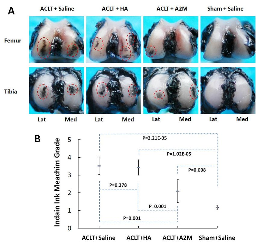
after injury, and the IL-6 and IL-8 lasts for six [6]. Although, levels of protective cytokines also increase
after joint injury, this protection mechanism occurs only in early stage of injury. SF IL-10 level drops to
normal in two weeks post trauma, and SF level of IL-1 receptor antagonist (IL-1Ra) is below normal after
three weeks [7].

Additionally, Matrix Metalloproteinases (MMPS) produced by chondrocyte and synovial cell result in the
loss of the normal cartilage tissue, exacerbating the progressive cartilage degeneration. Intensive studies
demonstrated that the MMPs levels dramatically increase in injured joint, and this fact has been validated
in various animal models [8–12]. Besides the degeneration of cartilage, Monocyte chemoattractant
protein 1(MCP-1) contributes to the joint pain in PTOA [13, 14]. Therefore, the development of PTOA can
be considered as the result of persistent in ammation after trauma. In ammatory mediators leading to
the destruction of cartilage and the lack of in ammation control nally result in the syndrome of PTOA
[15].

Hence it is assumed that by removing the destructive in ammatory mediators and enzymes we can
reduce the chronic in ammation and cartilage degeneration caused by intra-articular injury. A2M is
known as a broad-spectrum proteinase inhibitor found in both serum and synovial uid, which could be
used to attenuate cartilage degeneration. Proteinases inducing chronic in ammation can be captured by
A2M molecules, and the A2M-proteinase complex will be purged soon from serum. This protective effect
of A2M is bene ting from the special molecular structure, which is capable of blocking almost all kinds
of proteinases [16]. Unfortunately, the concentration of A2M in synovial uid is much lower than that in
serum of normal people, as well as OA patients. Thus, endogenous A2M in joint is not enough to diminish
intra-joint in ammation [17, 18].

One widely used clinical therapy is visco-supplementation, and the most common way of this direction is
injecting HA into the joint as the supplementation of the natural joint lubricant [19]. Hyaluronic Acid (HA)
received FDA approval in 1997 for treatment of Osteoarthritis (OA) in the United States. In 2009, the
American Academy of Orthopaedic Surgeons (AAOS) conducted a meta-analysis regarding HA’s
treatment and found the evidence for e cacy was considered inconclusive [20, 21]. As of early 2014, the
AAOS did not nd adequate evidence to support listing HA as indicated for treatment of knee OA [22].
However, by using the same thinking, it is possible that injection of A2M instead of HA as the intra-
articular supplementation would be helpful to retard the cartilage degeneration in knee OA process, and
A2M would make intra-articular injection therapy with more effectivity and biosafety. The objective of this
study is to compare the therapeutic effects of A2M and HA on OA in rats with knee arthritis.

Materials And Methods
This study was approved by the Institutional Review Board and the Institutional Animal Care and Use
Committee of the Shanxi Medical University.

Animals
                                                    Page 3/21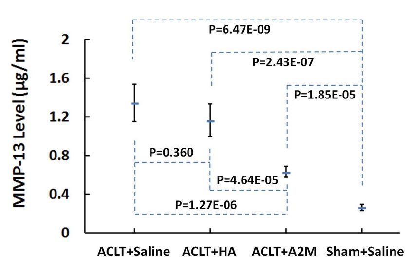
One hundred 10-week-old male wistar rats (180–230 g/each) were obtained from animal center of Shanxi
Medical University (Shanxi, China). The study was performed under a protocol approved by the Shanxi
Medical University Animal Research Committee (ARC). Seventy- ve animals received anterior cruciate
ligament transection (ACLT) operations on the right knee joint and divided into 3 groups (n = 25 per
group): (1) ACLT with intra-articular saline injection (ACLT + Saline), (2) ACLT with intra-articular
Hyaluronic Acid injection (ACLT + HA), (3) ACLT with intra-articular A2M injection (ACLT + A2M). The rest
twenty- ve animals were received sham operation on the right knee joint with intra-articular saline
injection (Sham + Saline). A2M (Cat.10602442001, Roche life science, USA) was dissolved in saline to
treat rats in a dose of 2 IU/kg (20µL). [17] Intra-articular injections were performed immediately following
and 3 days after ACLT, and then weekly for 6 weeks. All the animals were administrated 20µL liquid in
their right knee joint and were euthanized using a standard CO2 chamber 12 weeks after the operation. In
each group, 10 animals were used for Indian ink staining for a gross evaluation of cartilage lesion; 15
animals’ tibiae were studied histologically, while their femurs were used for real-time polymerase chain
reaction (PCR) assessment.

Surgical procedures
ACLT was surgically performed in the right hind knee joints of the animals as previously described. [23]
Once anesthetized, a 1.5 cm midline incision was made over the anterior knee. The skin was mobilized to
expose the patellar tendon. An incision through the joint capsule was made immediately lateral to the
tendon. The ACL was cut with a scalpel. Manual laxity testing veri ed its functional loss. The joint
capsule, fascia, and skin were closed in layers. The right hind knee joints of rats in the sham group were
sham-operated using the same approach, but without ACLT. Postoperative analgesia using buprenorphine
hydrochloride (0.05 mg/kg SQ) was maintained for at least 3 days. Animals were allowed to bear weight
on the limbs as tolerated.

Histology
After the rats were killed with carbon dioxide, gross morphologic lesions on the femur condyle and tibia
plateau (n = 10 per group) were visualized with India-ink staining [24]. Tibiae in rest set of knee joints (n =
15 per group) were xed in 10% formalin for 72 hours, followed by decalci cation in 10% EDTA solution,
while the femurs were used for used for real-time qPCR assessment. Then, tibiae were hemisected in the
midsagittal plane, and each half was embedded in a single block of Paraplast X-tra (Sigma-Aldrich, St
Louis, MO, USA). Serial 6-µm-thick sections were cut at intervals of 0 µm, 100 µm, and 200 µm and
collected on positively charged glass slides (Superfrost Plus; Fisher Scienti c). Safranin-O/fast green was
performed and the severity of cartilage damage was assessed using the OARSI Assessment System (OA
score = grade × stage; range, 0 to 24) [25]. Three independent blinded observers scored each section, and
the scores for all of the sections were averaged within each joint.

Immunohistochemistry staining
Immunohistochemistry was carried out using a Histostain-SP kit (Cat. 959943B Invitrogen) to detect the
distribution of types II collagen and MMP-13 in cartilage. The sections were digested by 5 mg/mL
                                                   Page 4/21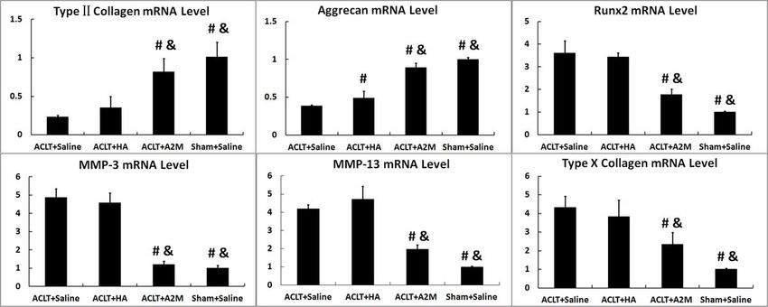
hyaluronidase in phosphate buffered saline (PBS) (Sigma-Aldrich) for 20 minutes. Nonspeci c protein
binding was blocked by incubation with a serum blocking solution. Thereafter, the sections were
incubated with speci c antibodies against rat types II collagen (Santa Cruz Biotechnology) and MMP-13
(Santa Cruz Biotechnology) respectively, at 4 °C overnight. The negative control sections were incubated
with isotype-matched control serum (2 µg/mL) (R&D Systems, Minneapolis, MN, USA) in PBS.
Subsequently, the sections were treated sequentially with biotinylated secondary antibody and SP
conjugate and then developed in DAB chromogen. Photomicrographs were taken with a Nikon E800
microscope (Nikon, Melville, NY, USA).

Rat articular cartilage Real-time qPCR
The cartilage samples from rats’ femur condyle were ground with a mortar and pestle while liquid
nitrogen was supplied. Total RNA was isolated from rat cartilage tissues using an RNeasy isolation kit
(Qiagen, Valencia, CA) according to manufacturer’s instruction. Cartilage samples from 5 rats were
dissected using a scalpel and pooled together, thus there were 3 pooled samples per group. Total RNA
was reverse transcribed into rst-strand complementary cDNA using the iScript™ cDNA synthesis kit (Bio-
Rad, Hercules, CA, USA). RT-PCR was performed on a 96-well plate ABI Prism 7500 (Applied Biosystems,
Foster City, CA) using SuperReal PreMix reagent (Qiagen, Valencia, CA) according to the manufacturer’s
protocols. The total volume (20µL) of each PCR reaction contained 10µL SuperReal PreMix, 7µL ddH2O,
2µL cDNA, and 1µL with 10 µM of each of the forward and reverse primer (Table 1). Ampli cation
conditions were as follows: 2-min preincubation at 50o; 10-min at 95oC for enzyme activation; and 40
cycles at 95oC denaturation for 10 s, 55oC annealing for 30 s and 72oC extension for 30 s. Gene
expression was normalized with 18 s mRNA levels. The comparative threshold cycle (Ct) method, that is,
the 2-ΔΔCt method was used to calculate fold ampli cation. [26]

                                                Page 5/21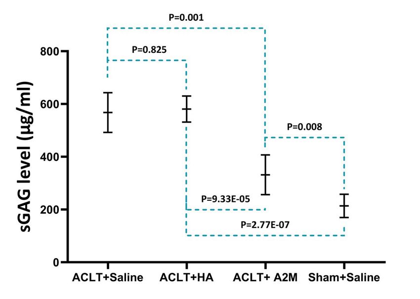
Table 1
                              Primers for real-time qPCR used in this study
                  Gene                Primer     Sequence 5' to 3'

                  Type II Collagen    Forward    AAG-GGA-CAC-CGA-GGT-TTC-ACT-GG

                                      Reverse    GGG-CCT-GTT-TCT-CCT-GAG-CGT

                  Aggrecan            Forward    CAG-TGC-GAT-GCA-GGC-TGG-CT

                                      Reverse    CCT-CCG-GCA-CTC-GTT-GGC-TG

                  Runx2               Forward    CCG-CAC-GAC-AAC-CGC-ACC-AT

                                      Reverse    CGC-TCC-GGC-CCA-CAA-ATC-TC

                  MMP-3               Forward    TTG-TCC-TTC-GAT-GCA-GTC-AG

                                      Reverse    AGA-CGG-CCA-AAA-TGA-AGA-GA

                  MMP-13              Forward    GGA-CCT-TCT-GGT-CTT-CTG-GC

                                      Reverse    GGA-TGC-TTA-GGG-TTG-GGG-TC

                  Type X Collagen     Forward    CCA-GGT-GTC-CCA-GGA-TTC-CC

                                      Reverse    CAA-GCG-GCA-TCC-CAG-AAA-GC

                  18S RNA             Forward    CGG-CTA-CCA-CAT-CCA-AGG-AA

                                      Reverse    GCT-GGA-ATT-ACC-GCG-GCT

Synovial Fluid Collection and Analyses
Synovial uid lavages were collected from the knees as soon as euthanasia. [23] The knee was shaved
and prepped with betadine. 50 µL of isotonic saline was injected into right knees of each animal through
the inferior patellar tendon. The joint capsule was visibly distended. The knee was then manually cycled
through exion and extension 10 times to distribute the uid within the joint before collection via joint
aspiration. Another twice procedures were repeated. This technique typically yielded 80–100 µl of uid
from each animal. Then the synovial uid was centrifuged at 2,000 g for 10 minutes to remove cells and
debris and then was frozen at -80˚C until analysis. Once thawed, MMP-13 and sulfated-
glycosaminoglycans (sGAG) were measured in the synovial uid lavages. MMP-13 content was
measured in the SF samples using ELISA according to the instructions of the manufacturer (Uscn Life
Science). Colorimetric density on the developed plates was determined using a microplate reader (Model
BF10000; Packard) set to 450 nm. ELISA analysis of each sample was performed in duplicate. For
evaluation of matrix PG release, we used the metachromatic dye 1, 9-dimethylmethylene blue (DMMB)
(SigmaAldrich, Gillingham, UK) assay to quantify the amount of sGAG in the SF samples according to
manufacturer’s instruction. The concentrations of sGAG were obtained from a spectrophotometric reader
at 540 nm (Thermo Scienti c Microplate Reader, UK).

                                                Page 6/21
Statistical analysis
Statistical analyses were performed using SPSS 13.0 software (SPSS Inc., Chicago, IL, USA). One-way
ANOVA were used to compare the concentrations of MMP-13 and levels of mRNA in different groups. All
data are expressed as mean ± SD. p values less than 0.05 were considered statistically signi cant.
Results
Gross morphological cartilage lesion and brillation in the rat tibia plateau were visualized by Indian ink
staining. We found that intra-articular HA could not attenuate posttraumatic OA macroscopically
compared to A2M treatment. Comparing with the HA-treated group, there was a signi cant decrease in
the OA score in A2M-treated rats as compared with rats that underwent ACLT and HA/saline treatment.
After treated by A2M, decreased India ink staining and a smoother surface were observed compared to
that in the HA-treated groups (Fig. 1A). The Indian ink staining Meacham grading system indicated that
A2M-treated group had less cartilage lesion and brillation than HA-treated or saline-treated group (p =
0.001), but more than in the sham control group (p = 0.008) (Fig. 1B). And there is no signi cant
difference between HA-treated group and saline-treated group (p = 0.378).
Then we value the effect of various treatments by the Histological staining. It is obviously that the
supplemental intra-articular A2M has the advantage over HA in attenuation of posttraumatic OA
pathogenesis in a rat model of ACLT. Firstly, we found a signi cant decrease in the OA score in A2M-
treated rats as compared with rats that underwent ACLT and saline/HA treatment. After treatment with
A2M, stronger Safranin O staining, more intact surface and more cellularity but less chondrocyte cloning,
and less brillation were observed than in the saline/HA-treated groups, but weaker staining than that
from control rats underwent sham operation (Fig. 2A). Cartilage from rats administered the injection of
HA had almost similar staining and surface compared with cartilage from rats without treatment (Saline
group) (Fig. 2A). OARSI histologic grading system scores in A2M-treated group suggested mild
degeneration (mean ± SD, 9.82 ± 4.0), while cartilage damage in saline-treated group and HA-treated
group were signi cantly more severe (18.2 ± 3.2 in the ACLT + saline group and 16.1 ± 3.8 in the ACLT +
HA group; P < 0.01). It should be noted that there was no signi cant statistical difference between ACLT +
saline group and ACLT + HA group (P = 0.062). The cartilage from rats that underwent sham operation
had the least amount of damage (0.43 ± 0.4; P < 0.01) (Fig. 2B).
Next, Immunohistochemistry was performed to determine the expression of type II collagen and MMP-13.
Type II collagen content was higher in cartilage in the ACLT + A2M and Sham + Saline groups than in the
ACLT + HA group and ACLT + Saline group (Fig. 3A). In contrast, MMP-13 was elevated in cartilage of the
HA-treated group compared to A2M-treated cartilage, in response to this, the group with higher MMP-13
level suffered critical cartilage injury. (Figs. 3B).
Additionally, Cartilage damage was associated with a change in the levels of MMP-13 in joint lavage
uid. The mean ± SD MMP-13 level of rats that underwent ACLT and A2M treatment was 0.63 ±
Page 7/210.05 µg/ml, which was higher than that in sham operation group(0.266 ± 0.03 µg/ml; P = 1.85E-05), while
much lower than that in rats administered injection of HA (1.168 ± 0.18 µg/ml; P = 4.65E-05) and rats
administered injection of saline (1.34 ± 0.19 µg/ml, P = 1.27E-06). However, there was no statistical
difference between ACLT + HA group and ACLT + Saline group (Fig. 4).
Besides, we examined the sulphated glycosaminoglycan (sGAG) levels in synovial uid groups with
different treatment strategies (Fig. 5). sGAG is an integral component part in cartilage matrix. However,
aGAG detected in synovial uid indicates the destruction of the articular cartilage. The sGAG level in A2M-
treated group was 331.68 ± 75.47 µg/ml, which was higher than the sham group (213.74 ± 44.14 µg/ml;
P = 0.008). But it is noteworthy that the synovial uid sGAG level of the A2M group was signi cant lower
than no-treatment group (567.70 ± 75.23 µg/ml; P = 0.001) (Fig. 5). This result demonstrated the
protective effect of supplemental intra-articular A2M injection. And we found ACLT operated rats in group
with saline or HA possessed higher quantities of synovial uid sGAG (576.56 ± 44.14 µg/ml) than those
treated by A2M (P = 9.33E-05), also much higher than rats in the control group too (P = 2.77E-07) (Fig. 5).
Meanwhile there was no statistical difference between ACLT + saline group and ACLT + HA group (P =
0.825) (Fig. 5).
Also, we performed qPCR to investigate the expression of related gene. Real-time qPCR results indicated
that the expression of mRNA for type II collagen and aggrecan increased in the group with supplemental
intra-articular A2M, while the levels of mRNA for Runx2, MMP-3, MMP-13, and type X collagen went down
(Fig. 6). By looking at HA treated group only, the levels of aggrecan and type II collagen mRNA were
obviously low, in contrary, the level of mRNA for Runx2, MMP-3, MMP-13, and type X collagen were over
expressed (Fig. 6). Type II collagen mRNA level in rats that underwent ACLT and A2M treatment was
signi cantly higher than that in rats underwent ACLT with HA or saline injection, and there was no
signi cant difference between the latter two groups. In the groups underwent sham operation and treated
by A2M, the aggrecan mRNA levels were signi cantly higher than the groups with saline and HA injection.
In contrast, levels of mRNA for Runx2, MMP-3, MMP-13, and type X collagen in rats that underwent ACLT
and saline/HA treatment were much higher than other two groups. (Fig. 6).
Discussion
Post-trauma Osteoarthritis is a sort of long course disease initiated by joint injury and consequent intra-
articular in ammation, and its process is irreversible [27, 28]. Thus, to delay the cartilaginous
degeneration is the main purpose of non-surgical treatment for osteoarthritis. Chronic in ammation is a
critical risk factor of the cartilage destruction during the process of osteoarthritis, therefore, it is believed
that the reduction of the intra-articular in ammation will retard the degeneration of cartilage [28].
Accordingly, we assumed that supplemental intra-articular injection of A2M, a autogeneic broad spectrum
proteinase inhibitor, could ameliorate cartilage destruction induced by in ammation.
We set four groups to compare the protective effect of A2M and HA, two experimental groups received
ACLT were treated with A2M and HA injection separately. Results of India ink staining and Safranin-
Page 8/21O/fast green staining have been consistent. All groups received ACLT showed articular cartilage defects
and outcropping of subchondral bone, which all were typical pathological changes of osteoarthritis [29].
However, there was little normal cartilage remaining in HA treated group, indicate the cartilage damages
in HA group and non-treated group had no signi cant difference, hence the protective effect of HA is
weak. This phenomenon echoed the results of clinical trials and meta-analyses, proving the uncertainty
of curative effect of HA [30–32].
In A2M treated group, cartilage damage occurred similarly but obviously slighter than the HA group and
non-treatment group. It could be observed that A2M-treated group had relatively intact surface and more
chondrocyte cloning than non-treatment group and HA-treated group. Although the cartilage was
inevitable after supplemental A2M injection, the protective effect of A2M was still very evident. Worthy of
note was that OARSI histologic grading system scores in A2M-treated group also showed signi cant
decrease, suggesting mild degeneration.
The major pathological changes of PTOA is the chronic in ammation secondary to joint injury. Due to the
increasingly producing of pro-in ammatory cytokine and matrix metalloproteinase, the in ammatory
damage of the articular tissue outrun regulation. Thus, relatively forceful way to analyze and compare the
protective effects between HA and A2M is to study whether the intervention can inhibit the destructive
cytokine and enzyme.
Accordingly, we examined more biological indicator of cartilage degeneration. Immunohistochemistry
was carried out to test distribution of types II collagen and MMP-13 in cartilage. Type II collagen is
produced by chondrocyte, and the level of type II collagen in degenerated cartilage with lower
chondrocyte cloning will reduce [33]. While MMP-13 is a kind of enzyme catalyzing destruction of
cartilage. MMP-13 induces the degradation of proteoglycan and type II collagen; thus it is used as an
important indicator of cartilage damage. [34–36] in A2M injection group we detected higher type II
collagen level and lower MMP-13 level, hence the protective effect of supplemental A2M injection could
be con rmed. Besides we examined the sGAG concentration in synovial uid from each group.
Comparing to the non-treatment group, the synovial uid sGAG level of the A2M group was lower.
Quantitatively assessing sGAG level in synovial uid could re ect the destruction of the cartilage
extracellular matrix [37]. It is probably that the protective effect of A2M lead to lower sGAG in the group
treated by this novel therapy.
In qPCR result, it is obviously that the supplemental A2M injection reduced the levels of mRNA for Runx2,
MMP-3, MMP-13, and type X collagen. However, levels of mRNA for all that above in rats that underwent
ACLT and saline/HA treatment were much higher than other two groups. These data suggested that A2M
can protect cartilage in vivo by decreasing gene expression of catabolic factors and hypertrophic
markers, as well as by increasing anabolic gene expression.
It is also apparently that the HA failed to achieved desired therapeutic effect. Immunohistochemistry
Staining showed that the cartilage degeneration in rat knees with HA injection were still severe,
meanwhile, the MMP-13 level was extremely higher than the sham operation group and A2M group.
Page 9/21According to the quantitative assay of MMP13 level in synovial uid, no signi cant disparity was found
between HA injection group and non-treatment group. Similarly, there was no diversity between the sGAG
level of HA group and non-treatment group. The result of qPCR also indicated that the HA treatment failed
to prevent cartilage destruction.
As a lot of research, HA cannot display the protective effect to the injured joint tissue. A commonly
accepted view is that the intra articular injection of HA can work as visco-supplementation [19]. But as
early as the 1980s, a study suggested that injecting HA could not raise the speci c viscosities of synovial
uid in experimental damaged joint [38]. One of the important reasons is the high molecular weight
HA(HMW-HA) will be hydrolyzed to low molecular weight HA(LMW-HA) in the wound healing process, and
LMW-HA cannot increase the speci c viscosities of synovial uid [39]. Furthermore, many studies believe
that the LWM-HA is an active pro-in ammatory stimulator, even inducing metalloproteinases production
[40–42].
Besides, there is a sort of proteoglycans called aggrecan on articular cartilage which binds to HA,
providing elasticity of cartilage [43]. When PTOA happens, aggrecan on joint cartilage is dissociated,
leading to loss of HA and erosion of cartilage, and nally cartilage degeneration [44]. And there is no
evidence that supplement of HA without aggrecan will contribute to cartilage repairment. Additionally,
previous study suggest that the HA have no in uence on proteoglycan concentration in PTOA [45]. And
our research also came to conclusion that there was no signi cant difference on mRNA level of aggrecan
between the HA treated group and non-treatment group.
Although, intra-articular injection of HA supplies HMW-HA, but the change is temporary. Through the HA
injection therapy, synovial uid is di cult to recover to normal standard. While any change in molecular
weight and concentration of HA in synovial uid will aggravate the articular cartilage damage. For the
reasons above, the HA treatment has been no longer recommended by American Academy of
Orthopaedic Surgeons (AAOS) [22].
The reason we've decided to used A2M as an alternative therapy to replace uncertain HA therapy. Not
only because A2M is able to protect articular cartilage against chronic in ammation, but also A2M has
high biosafety and less side effects.
There are many studies working on solve the in ammation caused cartilage destruction in osteoarthritis.
In this direction, existing and examining therapies are intra-articular calcium channel blocking
anesthetics, corticosteroid injection, mesenchymal stem/stromal cells, and platelet-rich plasma [46, 47].
However, some of treatments mentioned above cannot slow down the cartilage degeneration; some are
not suitable for prolonged use in clinics. A2M, as a novel therapeutic drug for knee OA, has a pretty good
prospect in solving the shortcomings mentioned above. Compared with other drugs above, A2M is an
autologous proteinase inhibitor so that it has no autoimmune rejection. Additionally, A2M injection
contains only one kind of active ingredient, but inhibits various in ammatory factors and degenerative
proteinase, due to broad-spectrum anti-in ammation effect of A2M. Therefore, A2M injection eliminates
above-mentioned limitations. As a result, the safety is guaranteed and risk is controllable.
Page 10/21New developed technologies allowed us to concentrate A2M molecule from autologous plasma of
patients, then inject A2M-rich preparation into joint to compensate for the low level of A2M in synovial
uid. [48] Furthermore, the variants of A2M called CYT-108 can be synthesized according to A2M’s
molecular properties. Doubtless synthetic A2M is more economic and convenient, and the inhibitory
effect of CYT-108 A2M far exceeded wild-type A2M [49].
Conclusion
This paper proposed to examine the uncertainty of HA treatment in animal models. And test the
articularly protective effect in osteoarthritis of A2M in the meantime. The results demonstrated that the
A2M can postpone retrograde degeneration of joint cartilage effectively. And, as expected, HA treatment
did not conduct protective effect obviously. These observations indicate that A2M is a promising anti-
in ammation treatment for osteoarthritis, and it can be used as the replacement for HA.
Abbreviations
PTOA: Post-Traumatic Osteoarthritis
HA: Hyaluronic Acid
ACLT: Anterior Cruciate Ligament Transection
A2M : Alpha-2-macroglobulin
SF: Synovial Fluid
MMPS: Matrix Metalloproteinases
MCP: Monocyte chemoattractant protein
sGAG: sulfated-Glycosaminoglycans
DMMB: Dimethylmethylene Blue
HMW-HA: High Molecular Weight HA
LMW-HA: Low Molecular Weight HA
Declarations
Availability of data and materials
All data generated and analyzed in this study are disclosed in this article.
Page 11/21Funding
This project was supported by the National Natural Science Foundation of China (Grant No. 81572207,
81201435), and Cultivate Scienti c Research Excellence Programs of Higher Education Institutions in
Shanxi (CSREP), China.
Authors’ contributions
MZ participated in the study design, wrote the manuscript. YG, RY and ZW performed most of the
experiments. BZ and YC analyzed data and participated in the interpretation of the data and the revision
of the manuscript. SW and LW conceived the in vivo study and participated in its design and data
analysis and revised the manuscript. All authors have read and approved the nal manuscript.
Con ict of interest
The authors declare that they have no con ict of interest.
Ethics approval and consent to participate
This study did not need consent from any individuals/patients because no humans were involved in this
study. Ethical approval was needed for this study and it was approved by the Institutional Review Board
and the Institutional Animal Care and Use Committee of the Shanxi Medical University (approved protocol
2018LL039)
Disclosures
We have nothing to disclose.
References
1. Minguzzi M, Cetrullo S, D'Adamo S, Silvestri Y, Flamigni F, Borzi RM: Emerging Players at the
Intersection of Chondrocyte Loss of Maturational Arrest, Oxidative Stress, Senescence and Low-
Grade In ammation in Osteoarthritis. Oxid Med Cell Longev 2018, 2018:3075293.
2. Pap T, Korb-Pap A: Cartilage damage in osteoarthritis and rheumatoid arthritis--two unequal siblings.
Nat Rev Rheumatol 2015, 11(10):606-615.
3. Heinegard D, Saxne T: The role of the cartilage matrix in osteoarthritis. Nat Rev Rheumatol 2011,
7(1):50-56.
Page 12/214. Gelber AC, Hochberg MC, Mead LA, Wang NY, Wigley FM, Klag MJ: Joint injury in young adults and
risk for subsequent knee and hip osteoarthritis. Ann Intern Med 2000, 133(5):321-328.
5. Sward P, Struglics A, Englund M, Roos HP, Frobell RB: Soft tissue knee injury with concomitant
osteochondral fracture is associated with higher degree of acute joint in ammation. Am J Sports
Med 2014, 42(5):1096-1102.
6. Elsaid KA, Fleming BC, Oksendahl HL, Machan JT, Fadale PD, Hulstyn MJ, Shalvoy R, Jay GD:
Decreased lubricin concentrations and markers of joint in ammation in the synovial uid of patients
with anterior cruciate ligament injury. Arthritis Rheum 2008, 58(6):1707-1715.
7. Cameron ML, Fu FH, Paessler HH, Schneider M, Evans CH: Synovial uid cytokine concentrations as
possible prognostic indicators in the ACL-de cient knee. Knee Surg Sports Traumatol Arthrosc 1994,
2(1):38-44.
8. Tang Z, Yang L, Xue R, Zhang J, Wang Y, Chen PC, Sung KL: Differential expression of matrix
metalloproteinases and tissue inhibitors of metalloproteinases in anterior cruciate ligament and
medial collateral ligament broblasts after a mechanical injury: involvement of the p65 subunit of
NF-kappaB. Wound Repair Regen 2009, 17(5):709-716.
9. Attia E, Brown H, Henshaw R, George S, Hanna n JA: Patterns of gene expression in a rabbit partial
anterior cruciate ligament transection model: the potential role of mechanical forces. Am J Sports
Med 2010, 38(2):348-356.
10. Aini H, Ochi H, Iwata M, Okawa A, Koga D, Okazaki M, Sano A, Asou Y: Procyanidin B3 prevents
articular cartilage degeneration and heterotopic cartilage formation in a mouse surgical
osteoarthritis model. PLoS One 2012, 7(5):e37728.
11. Haslauer CM, Elsaid KA, Fleming BC, Proffen BL, Johnson VM, Murray MM: Loss of extracellular
matrix from articular cartilage is mediated by the synovium and ligament after anterior cruciate
ligament injury. Osteoarthritis Cartilage 2013, 21(12):1950-1957.
12. Nagai T, Sato M, Kobayashi M, Yokoyama M, Tani Y, Mochida J: Bevacizumab, an anti-vascular
endothelial growth factor antibody, inhibits osteoarthritis. Arthritis Res Ther 2014, 16(5):427.
13. Cuellar JM, Scuderi GJ, Cuellar VG, Golish SR, Yeomans DC: Diagnostic utility of cytokine biomarkers
in the evaluation of acute knee pain. J Bone Joint Surg Am 2009, 91(10):2313-2320.
14. Cuellar VG, Cuellar JM, Golish SR, Yeomans DC, Scuderi GJ: Cytokine pro ling in acute anterior
cruciate ligament injury. Arthroscopy 2010, 26(10):1296-1301.
15. Lieberthal J, Sambamurthy N, Scanzello CR: In ammation in joint injury and post-traumatic
osteoarthritis. Osteoarthritis Cartilage 2015, 23(11):1825-1834.
16. Sottrup-Jensen L: Alpha-macroglobulins: structure, shape, and mechanism of proteinase complex
formation. J Biol Chem 1989, 264(20):11539-11542.
17. Wang S, Wei X, Zhou J, Zhang J, Li K, Chen Q, Terek R, Fleming BC, Goldring MB, Ehrlich MG et al:
Identi cation of α2-Macroglobulin as a Master Inhibitor of Cartilage-Degrading Factors That
Attenuates the Progression of Posttraumatic Osteoarthritis. Arthritis & Rheumatology 2014,
66(7):1843-1853.
Page 13/2118. Demirag B, Sarisozen B, Durak K, Bilgen OF, Ozturk C: The effect of alpha-2 macroglobulin on the
healing of ruptured anterior cruciate ligament in rabbits. Connect Tissue Res 2004, 45(1):23-27.
19. Altman RD, Manjoo A, Fierlinger A, Niazi F, Nicholls M: The mechanism of action for hyaluronic acid
treatment in the osteoarthritic knee: a systematic review. BMC Musculoskelet Disord 2015, 16:321.
20. Lo GH, LaValley M, McAlindon T, Felson DT: Intra-articular hyaluronic acid in treatment of knee
osteoarthritis: a meta-analysis. JAMA 2003, 290(23):3115-3121.
21. Arrich J, Piribauer F, Mad P, Schmid D, Klaushofer K, Mullner M: Intra-articular hyaluronic acid for the
treatment of osteoarthritis of the knee: systematic review and meta-analysis. CMAJ 2005,
172(8):1039-1043.
22. Cook R, Davidson P, White A, Centre ND: Partial knee replacement could be rst choice for some
patients with osteoarthritis. BMJ 2019, 367:l5994.
23. Wei L, Fleming BC, Sun X, Teeple E, Wu W, Jay GD, Elsaid KA, Luo J, Machan JT, Chen Q: Comparison
of differential biomarkers of osteoarthritis with and without posttraumatic injury in the Hartley
guinea pig model. J Orthop Res 2010, 28(7):900-906.
24. Meachim G: Light microscopy of Indian ink preparations of brillated cartilage. Ann Rheum Dis 1972,
31(6):457-464.
25. Pritzker KP, Gay S, Jimenez SA, Ostergaard K, Pelletier JP, Revell PA, Salter D, van den Berg WB:
Osteoarthritis cartilage histopathology: grading and staging. Osteoarthritis Cartilage 2006, 14(1):13-
29.
26. Wang S, Wei X, Zhou J, Zhang J, Li K, Chen Q, Terek R, Fleming BC, Goldring MB, Ehrlich MG et al:
Identi cation of alpha2-macroglobulin as a master inhibitor of cartilage-degrading factors that
attenuates the progression of posttraumatic osteoarthritis. Arthritis Rheumatol 2014, 66(7):1843-
1853.
27. Yamada EF, Bobinski F, Martins DF, Palandi J, Folmer V, da Silva MD: Photobiomodulation therapy in
knee osteoarthritis reduces oxidative stress and in ammatory cytokines in rats. J Biophotonics 2020,
13(1):e201900204.
28. Irie K, Uchiyama E, Iwaso H: Intraarticular in ammatory cytokines in acute anterior cruciate ligament
injured knee. Knee 2003, 10(1):93-96.
29. Takeda H, Nakagawa T, Nakamura K, Engebretsen L: Prevention and management of knee
osteoarthritis and knee cartilage injury in sports. Br J Sports Med 2011, 45(4):304-309.
30. Bannuru RR, McAlindon TE, Sullivan MC, Wong JB, Kent DM, Schmid CH: Effectiveness and
Implications of Alternative Placebo Treatments: A Systematic Review and Network Meta-analysis of
Osteoarthritis Trials. Ann Intern Med 2015, 163(5):365-372.
31. Bannuru RR, Schmid CH, Kent DM, Vaysbrot EE, Wong JB, McAlindon TE: Comparative effectiveness
of pharmacologic interventions for knee osteoarthritis: a systematic review and network meta-
analysis. Ann Intern Med 2015, 162(1):46-54.
32. Niemela TM, Tulamo RM, Hielm-Bjorkman AK: A randomised, double-blinded, placebo-controlled
clinical study on intra-articular hyaluronan treatment in equine lameness originating from the
Page 14/21metacarpophalangeal joint. BMC Vet Res 2016, 12:60.
33. Poole AR, Kobayashi M, Yasuda T, Laverty S, Mwale F, Kojima T, Sakai T, Wahl C, El-Maadawy S,
Webb G et al: Type II collagen degradation and its regulation in articular cartilage in osteoarthritis.
Ann Rheum Dis 2002, 61 Suppl 2:ii78-81.
34. Gu R, Shi Y, Huang W, Lao C, Zou Z, Pan S, Huang Z: Theobromine mitigates IL-1beta-induced
oxidative stress, in ammatory response, and degradation of type II collagen in human chondrocytes.
Int Immunopharmacol 2020, 82:106226.
35. Alamgeer, Hasan UH, Uttra AM, Qasim S, Ikram J, Saleem M, Niazi ZR: Phytochemicals targeting
matrix metalloproteinases regulating tissue degradation in in ammation and rheumatoid arthritis.
Phytomedicine 2020, 66:153134.
36. Burrage PS, Mix KS, Brinckerhoff CE: Matrix metalloproteinases: role in arthritis. Front Biosci 2006,
11:529-543.
37. Jin Y, Chen X, Gao ZY, Liu K, Hou Y, Zheng J: The role of miR-320a and IL-1beta in human
chondrocyte degradation. Bone Joint Res 2017, 6(4):196-203.
38. Hilbert BJ, Rowley G, Antonas KN, McGill CA, Reynoldson JA, Hawkins CD: Changes in the synovia
after the intra-articular injection of sodium hyaluronate into normal horse joints and after arthrotomy
and experimental cartilage damage. Aust Vet J 1985, 62(6):182-184.
39. Aviad AD, Houpt JB: The molecular weight of therapeutic hyaluronan (sodium hyaluronate): how
signi cant is it? J Rheumatol 1994, 21(2):297-301.
40. Chen WY, Abatangelo G: Functions of hyaluronan in wound repair. Wound Repair Regen 1999,
7(2):79-89.
41. Stern R, Asari AA, Sugahara KN: Hyaluronan fragments: an information-rich system. Eur J Cell Biol
2006, 85(8):699-715.
42. Jiang D, Liang J, Noble PW: Hyaluronan in tissue injury and repair. Annu Rev Cell Dev Biol 2007,
23:435-461.
43. Tortorella MD, Malfait AM, Deccico C, Arner E: The role of ADAM-TS4 (aggrecanase-1) and ADAM-
TS5 (aggrecanase-2) in a model of cartilage degradation. Osteoarthritis Cartilage 2001, 9(6):539-
552.
44. Tortorella MD, Burn TC, Pratta MA, Abbaszade I, Hollis JM, Liu R, Rosenfeld SA, Copeland RA,
Decicco CP, Wynn R et al: Puri cation and cloning of aggrecanase-1: a member of the ADAMTS
family of proteins. Science 1999, 284(5420):1664-1666.
45. Brandt KD, Smith GN, Jr., Simon LS: Intraarticular injection of hyaluronan as treatment for knee
osteoarthritis: what is the evidence? Arthritis Rheum 2000, 43(6):1192-1203.
46. Xie X, Zhang C, Tuan RS: Biology of platelet-rich plasma and its clinical application in cartilage
repair. Arthritis Res Ther 2014, 16(1):204.
47. Richards MM, Maxwell JS, Weng L, Angelos MG, Golzarian J: Intra-articular treatment of knee
osteoarthritis: from anti-in ammatories to products of regenerative medicine. Phys Sportsmed 2016,
Page 15/2144(2):101-108.
48. Cuellar JM, Cuellar VG, Scuderi GJ: alpha2-Macroglobulin: Autologous Protease Inhibition
Technology. Phys Med Rehabil Clin N Am 2016, 27(4):909-918.
49. Zhang Y, Wei X, Browning S, Scuderi G, Hanna LS, Wei L: Targeted designed variants of alpha-2-
macroglobulin (A2M) attenuate cartilage degeneration in a rat model of osteoarthritis induced by
anterior cruciate ligament transection. Arthritis Res Ther 2017, 19(1):175.
Figures
Page 16/21Figure 1
Intra-articular HA could not attenuate posttraumatic OA macroscopically. (A) Decreased India ink staining
and a smoother surface were detected in the articular cartilage of A2M-treated animals as compared to
ACLT and saline/HA treatment groups. (B) The Indian ink Meachim Grade score indicated that cartilage
damage was much severe in rats that underwent ACLT and saline/HA treatment, while cartilage in rats
that underwent sham operation had the least damage. Cartilage damage was also reduced in rats that
received A2M as compared to rats that received the HA treatment. Values are the mean±SD.
Figure 2
Page 17/21Posttraumatic OA rats modeled by ACLT suggested that the A2M has more advantage than HA in
attenuating the injury. (A) A smoother surface with stronger Safranin O staining was detected in the
articular cartilage of A2M-treated animals as compared to HA-treated animals and untreated controls (the
bottom panels are higher-magni cation views of the boxed areas in the top panels). (B) The
Osteoarthritis Research Society International Osteoarthritis Cartilage Histopathology Assessment System
(OOCHAS) score indicated that cartilage damage was mild in rats that underwent A2M treatment, while
cartilage in rats that underwent sham operation had the least damage. Cartilage damage was aggravated
in rats that received the ACLT operation and HA/Saline treatment. And there was no statistical
signi cance between ACLT+saline group and ACLT+HA group. Values are the mean±SD.
Figure 3
MMP-13 expression was reduced in A2M treated rats, but type II collagen was preserved. (A) Type II
collagen expression in articular cartilage was higher in the A2M-treated and the sham-operated rats than
Page 18/21in rats that underwent ACLT and HA treatment. In contrast, MMP-13 staining was elevated in rats that
underwent ACLT and HA treatment, but was lower in the A2M-treated and sham-operated rats, which is
consistent with reduced OA damage in these rats. In A and B, the bottom panels are higher-magni cation
views of the boxed areas in the top panels (20x objective lens).
Figure 4
In HA-treated rats, the concentration of MMP-13 in SF was much higher than that in rats that underwent
ACLT and A2M treatment and was similar to that in ACLT and saline treated rats. Values are the
mean±SD.
Page 19/21Figure 5
ACLT rats treated by HA had shown more articular cartilage destruction than those treated by A2M. The
sGAG concentrations were tested separately in four groups. In HA-treated rats, the concentration of sGAG
in SF was much higher than that in rats that underwent ACLT and A2M treatment. No statistical
difference was found between ACLT+Saline group and ACLT+ HA group. Values are the mean±SD.
Page 20/21Figure 6 Supplemental intra-articular A2M has the advantage over HA in inhibiting catabolism and enhancing anabolic metabolism in a rat model of ACLT. Levels of mRNA for type II collagen and aggrecan were increased in rats that were administered A2M as compared to rats that underwent ACLT and saline/HA treatment, suggesting that A2M has a positive impact on anabolic metabolism. In contrast, Runx2, MMP- 3, MMP-13, and type X collagen showed the opposite pattern. These genes were expressed at a lower level in rats that were administered A2M as compared to rats that underwent ACLT and saline/HA treatment. Values are the mean±SEM. #: P
You can also read