Transcriptome analysis of chicken intraepithelial lymphocyte natural killer cells infected with very virulent infectious bursal disease virus - Nature
←
→
Page content transcription
If your browser does not render page correctly, please read the page content below
www.nature.com/scientificreports
OPEN Transcriptome analysis of chicken
intraepithelial lymphocyte natural
killer cells infected with very
virulent infectious bursal disease
virus
Sook Yee Boo1, Sheau Wei Tan1, Noorjahan Banu Alitheen2, Chai Ling Ho2,
Abdul Rahman Omar1 & Swee Keong Yeap1,3*
The infectious bursal disease (IBD) is an acute immunosuppressive viral disease that significantly
affects the economics of the poultry industry. The IBD virus (IBDV) was known to infect B lymphocytes
and activate macrophage and T lymphocytes, but there are limited studies on the impact of IBDV
infection on chicken intraepithelial lymphocyte natural killer (IEL-NK) cells. This study employed
an mRNA sequencing approach to investigate the early regulation of gene expression patterns in
chicken IEL-NK cells after infection with very virulent IBDV strain UPM0081. A total of 12,141 genes
were expressed in uninfected chicken IEL-NK cells, and most of the genes with high expression were
involved in the metabolic pathway, whereas most of the low expressed genes were involved in the
cytokine-cytokine receptor pathway. A total of 1,266 genes were differentially expressed (DE) at
3 day-post-infection (dpi), and these DE genes were involved in inflammation, antiviral response
and interferon stimulation. The innate immune response was activated as several genes involved in
inflammation, antiviral response and recruitment of NK cells to the infected area were up-regulated.
This is the first study to examine the whole transcriptome profile of chicken NK cells towards IBDV
infection and provides better insight into the early immune response of chicken NK cells.
Infectious bursal disease (IBD) is an acute immunosuppressive viral disease that affects the poultry industry with
economic importance1. IBD is caused by the IBD virus (IBDV), a dsRNA virus belonging to the genus Avibir-
navirus in the family B irnaviridae2. IBDV is organized into 2 segments, A and B, of about 6 kb in size. Segment
A is the larger fragment and encodes the VP5, non-structural protein and another polyprotein whose post-
translational cleavage gives rise to VP2, VP3 and VP4 structural p roteins3. IBDV isolates are divided into two
serotypes, 1 and 2, which both infect chickens, but the typical clinical disease is only associated with serotype 14.
Infection usually occurs through the oral route. The virus replicates in avian immune cells such as the mac-
rophages and the lymphoid cells of the duodenum, jejunum and caecum5. It then spreads to the bursa of Fabricius
(BF) where it extensively replicates and destroys the immature B lymphocytes, thus causing a compromise in the
antibody mediated immune responses in the affected chicken. The clinical severity and mortality due to IBDV
depend on the presence of pre-existing maternal antibodies, age and the chicken’s genetic b ackground6. Acute
IBDV infections are characterized by severe clinical signs and high mortality. The incubation period is very short,
about 2 to 3 days. In acute cases, the birds are exhausted, prostrated, dehydrated, suffer from aqueous diarrhoea
and their feathers are ruffled. Mortality commences on the third day of infection, reaches a peak and then drops
rapidly, and the surviving chickens recover a state of apparent health after 5 to 7 days7.
The first 3 days are the critical period for the immune response system of the chicken to respond against
virus, and the innate immune response is the first layer of the defence system against a virus or bacteria. It is thus
important to understand how chicken immune cells response to IBDV infection. There are number of immune
1
Laboratory of Vaccines and Immunotherapeutics, Institute of Bioscience, Universiti Putra Malaysia,
43400 Serdang, Selangor, Malaysia. 2Faculty of Biotechnology and Biomolecular Sciences, Universiti Putra
Malaysia, 43400 Serdang, Selangor, Malaysia. 3China‑ASEAN College of Marine Sciences, Xiamen University
Malaysia, Bandar Sunsuria, 43900 Sepang, Selangor, Malaysia. *email: skyeap@xmu.edu.my
Scientific Reports | (2020) 10:18348 | https://doi.org/10.1038/s41598-020-75340-x 1
Vol.:(0123456789)
www.nature.com/scientificreports/
dpi Raw reads Trimmed reads Mapped in pair Mapped in broken pair Reads not mapped
R1 69,496,834 58,911,860 47,957,944 (81.41%) 3,765,047 (6.39%) 7,188,869 (12.2%)
0 R2 67,751,426 57,247,830 44,087,180 (77%) 2,598,916 (4.54%) 10,561,734 (18.45%)
R3 68,100,290 57,898,240 44,020,552 (76%) 4,188,992 (7.24%) 9,688,696 (16.73%)
R1 64,510,988 54,944,488 42,957,964 (73.55%) 2,372,524 (4.32%) 9,614,000 (17.5%)
3 R2 64,643,682 55,052,202 40,490,580 (73.55%) 6,073,371 (11.03%) 8,488,251 (15.42%)
R3 62,733,848 53,080,454 38,669,076 (72.85%) 4,879,719 (9.19%) 9,531,659 (17.96%)
Table 1. Summary of raw, trimmed and mapped reads.
cells involved in the innate immune response, including dendritic, macrophage and natural killer (NK) cells.
NK cells play a key role in innate host defence against viruses. The primary physiological role of NK cells is to
provide a crucial initial defence against pathological organisms during the time (from day 0 to day 5) that the
adaptive immune system is still being m ustered8. Their major function is to recognize and kill virally infected and
neoplastic cells. When the ligand of NK cells interacts with cell-surface receptors, they produce several cytokines
such as IFN-γ, which have an immunoregulatory r ole9.
To date, studies have been conducted to check on the expression profile of mRNA in B c ells10, macrophages11,
dendritic cells12 and embryonic fibroblast c ells13, but there is limited understanding of the role of NK cells in
IBDV infection. A study conducted by Jahromi et al.14 reported the expression profile for several groups of
activator and suppressor NK cell receptors on the surface of 28.4+ IEL-NK cells using a qPCR approach and
demonstrated that very virulent IBDV (vvIBDV) suppressed the activator receptors at 1-day post infection
(dpi), but an overexpression of the surface activator was observed at 3 dpi. As the actual regulation of IBDV on
NK cells was uncertain15, it is important to evaluate the response of IEL-NK cells against the vvIBDV infection,
particularly at 3 dpi, to elucidate the actual role and response of IEL-NK cells in the innate immune response of
chickens against this virus, which may help to establish foundations for improved prevention, including a vac-
cination strategy against IBDV. In this study, the whole transcriptome profiling of chicken IEL-NK cells infected
by vvIBDV at 3 dpi was performed to gain insight into how such cells respond to vvIBDV.
Results
Isolation and enrichment of 28.4+ IEL‑NK cells. IEL-NK cells were isolated from duodenum samples
collected from uninfected chickens and chickens infected with vvIBDV at 3 dpi. The total number of IEL-NK
cells was 19.48 million for control samples, 13.95 million cells for the infected chicken for 3 days. After the
enrichment process using CD3 and 28.4 markers, the percentage of C D3−/28.4+ IEL-NK cells at 3 dpi were
higher than in the uninfected control group, which was 42.38%.
Measurement of viral load. The viral load titre of the samples was determined by amplifying the VP4
region in the vvIBDV strain UPM0081 using the RT-qPCR method. There was no amplification in the unin-
fected IEL-NK cells while the viral load was log10 of 7.10 ± 0.65 at 3 dpi.
Identification of differentially expressed genes for infection group on day 3. The number of raw
paired-end reads per sample generated from HiSeq2500 was within the range of 62 million to 69 million reads.
The raw reads proceeded to adapter and quality trimming. The number of trimmed paired-end reads was within
the range of 53 million to 58 million reads. The trimmed reads were mapped to chicken genome Galgal4 down-
loaded from the Ensembl database. The percentage of trimmed reads mapped in pairs was 72% to 81%, whereas
the reads mapped in broken pairs was the range of 4.3% to 11%. The percentage of trimmed reads not mapped
to the genome was within the range of 12.2% to 18.5% (Table 1).
Differential expression analysis was performed to find out the differential expressed (DE) genes on dpi 3 as
compared to the uninfected samples. The DE gene was defined as a gene with a fold change ≥ 2 or ≤ − 2 and a
False Discovery Rate (FDR) corrected p value < 0.05. At 3 dpi, there were 516 genes up-regulated, whereas 750
genes were down-regulated.
GO and KEGG pathway enrichment analysis. The functions and pathways of all DE genes were ana-
lysed based on the GO and KEGG pathway. The DE genes at 3 dpi, in comparison to the uninfected samples, were
enriched in a few GOs, as shown in Table 2. In terms of biological process, most of the DE genes were involved in
the metabolic process, transmembrane transport, fatty acid beta-oxidation and inflammatory response. In terms
of cellular components, most of the DE genes were involved in the extracellular exosome, integral component
of membrane, extracellular space and peroxisome. In terms of molecular function, most of the DE genes were
involved in neurotransmitter, oxidoreductase activity or haem binding.
KEGG Pathway enrichment analysis showed that most of the DE genes at 3 dpi were clustered into metabolic
pathways or pathways of valine, leucine and isoleucine degradation, biosynthesis of antibiotics and tryptophan
metabolism (Table 3). Meanwhile, a total of 20 DE genes were involved in the cytokine-cytokine receptor inter-
action pathway (Fig. 1a). Out of the 20 DE genes, 12 genes were down-regulated and 8 genes were up-regulated
after infection by vvIBDV at 1 dpi and 3 dpi. Moreover, 7 DE genes (4 down-regulated and 3 upregulated) were
Scientific Reports | (2020) 10:18348 | https://doi.org/10.1038/s41598-020-75340-x 2
Vol:.(1234567890)
www.nature.com/scientificreports/

                                   Gene ontology          Term ID             Description                                              p value
                                                          GO:0,008,152        Metabolic process                                        4.47E−04
                                                          GO:0,055,085        Transmembrane transport                                  5.35E−04
                                   Biological process     GO:0,033,539        Fatty acid beta-oxidation using acyl-CoA dehydrogenase   0.0035
                                                          GO:0,006,954        Inflammatory response                                    0.0044
                                                          GO:0,005,975        Carbohydrate metabolic process                           0.0105
                                                          GO:0,070,062        Extracellular exosome                                    2.05E−05
                                                          GO:0,016,021        Integral component of membrane                           1.97E−04
                                   Cellular component     GO:0,005,615        Extracellular space                                      5.79E−04
                                                          GO:0,005,777        Peroxisome                                               0.0015
                                                          GO:0,009,986        Cell surface                                             0.0063
                                                          GO:0,005,328        Neurotransmitter:sodium symporter activity               0.0020
                                                          GO:0,016,491        Oxidoreductase activity                                  0.0055
                                   Molecular function     GO:0,020,037        Heme binding                                             0.0086
                                                          GO:0,004,252        Serine-type endopeptidase activity                       0.0098
                                                          GO:0,005,506        Iron ion binding                                         0.0135

                                  Table 2.  Top five enriched GOs for DE genes at 3 dpi.

                                   ID           Description                                      Number of genes   p value
                                   gga01100     Metabolic pathways                               146               2.51E−12
                                   gga00280     Valine, leucine and isoleucine degradation       17                1.76E−07
                                   gga01130     Biosynthesis of antibiotics                      34                1.05E−05
                                   gga00380     Tryptophan metabolism                            13                4.44E−05
                                   gga01200     Carbon metabolism                                21                8.53E−05
                                   gga00561     Glycerolipid metabolism                          13                0.0010
                                   gga00071     Fatty acid degradation                           10                0.0011
                                   gga00040     Pentose and glucuronate interconversions         7                 0.0018
                                   gga00010     Glycolysis / Gluconeogenesis                     12                0.0025
                                   gga00640     Propanoate metabolism                            8                 0.0033

                                  Table 3.  Top 10 enriched KEGG pathways for DE genes at 3 dpi.

                                  involved in the Toll-Like Receptor (TLR) signalling pathway (Fig. 1b). There were only 2 DE genes involved in
                                  the apoptosis pathway (Fig. 1c).

                                  Apoptosis and inflammatory. Some of the genes related to apoptosis and inflammation were differen-
                                  tially expressed after vvIBDV infection. Of the apoptosis-related genes, CASP1 was up-regulated at 3 dpi with
                                  a fold change of 2.14. All of the inflammation-related genes up-regulated at 3 dpi and these genes are CCL19,
                                  CCL20, IL17A, IL22, LIPA, P2RX7, tac1, TLR3 and TNFSF4.

                                  Cytokine, chemokines and interferon stimulation. Most of the genes for chemokines/chemokine
                                  receptors (CCR2, CCR6, CCR7, CSF2RB, CX3CR1 and CXCR4) and cytokines/cytokines receptors (IL1R2, IL8,
                                  IL18, IL20RA and IL22RA2) were down-regulated after infection by vvIBDV at 3 dpi. However, the genes for
                                  CCL19 and CCL20 were found to be up-regulated with a fold change of 2.53 and 2.09 while the genes for IL22
                                  cytokines were up-regulated with a fold change of 3.76 at 3 dpi. Other than the genes for chemokines and
                                  cytokines, several genes for TLRs were also differentially expressed as a result of the vvIBDV infection. TLR7 was
                                  down-regulated, while TLR3 was the only TLR gene up-regulated at 3 dpi, with a fold change of 2.22.
                                      The IFN-stimulated genes (ISGs) are involved in antiviral defence. Some of the ISGs genes such as IFIT5,
                                  IFITM5, MX1, RSAD2 and SAMHD1 were up-regulated at 3 dpi. Among these five genes, RSAD2 had the highest
                                  fold change of 5.26. Additionally, the tumour necrosis factor related genes, TNFRSF9 and TNFSF4, were up-
                                  regulated at 3 dpi with a fold change of 2.28 and 4.86, respectively. The list of DE genes related to DNA replication,
                                  cell cycle, inflammation, cytokine, chemokine and interferon stimulation is summarized in supplementary file 1.

                                  Avian NK cell surface receptors. The gene expression level for avian NK cells surface receptors such as
                                  CD69, CHIR-AB1, B-Lec and B-NK was identified through the RNA-Seq and RT-qPCR results. The expression of
                                  CD69, B-NK and B-Lec genes were up-regulated at 3 dpi compared to the uninfected samples. However, the gene
                                  expression for the bifunctional marker, CHIR-AB1, was similar to uninfected samples (Table 4).

Scientific Reports |   (2020) 10:18348 |                   https://doi.org/10.1038/s41598-020-75340-x                                                  3

                                                                                                                                                  Vol.:(0123456789)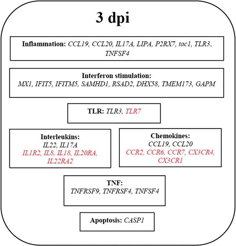
www.nature.com/scientificreports/
Figure 1. KEGG pathway enrichment analysis of the host response towards vvIBDV infection. These KEGG
pathways were generated using DE genes from IEL-NK cells obtained from RNA-Seq. (a) Cytokine-cytokine
receptor interaction pathway. (b) Toll-like receptor signalling pathway. (c) Apoptosis pathway. The down-
regulated DE genes are highlighted in blue and up-regulated DE genes are highlighted in red. Pathways were
adopted from KEGG pathway d atabase59. The green and white coloured boxes are default colours generated by
the software, depicting genes that were not the DE on RNA-Seq, and unidentified genes in the organism-specific
pathway, respectively.
Scientific Reports | (2020) 10:18348 | https://doi.org/10.1038/s41598-020-75340-x 4
Vol:.(1234567890)www.nature.com/scientificreports/
Figure 1. (continued)
Scientific Reports | (2020) 10:18348 | https://doi.org/10.1038/s41598-020-75340-x 5
Vol.:(0123456789)www.nature.com/scientificreports/
Figure 1. (continued)
RNA‑Seq result validated using NanoString technology. NanoString Technology was used to vali-
date the expression level of 21 DE genes involved in 6 pathways related to the innate immune response against
vvIBDV infection (Fig. 2). From the 21 DE genes, 19 genes showed similar expression profile to the RNA-Seq
results. Two genes could not be compared to the RNA-Seq results, as the expression levels of these two genes
(CX3CL1 and CD86) were not significant due to the high p-value (> 0.05).
Discussion
In model organisms such as humans, rats and mice, NK cells have been well c haracterized16,17. The number of
studies focusing on avian NK cells is, however, very limited, mainly due to the unavailability of specific mAbs
against avian NK cells. The avian NK cell–specific 28.4 mAb was discovered by Göbel et al.18 and this mAb can
be used to isolate 28.4+ IEL-NK cells. Further study from Gobel’s group found that the 28.4+ cells are predomi-
nantly present in the chicken duodenum, and these 28.4+ cells play important roles against pathogenic organ-
D3−/28.4+ IEL-NK cells were isolated from uninfected chickens and chickens infected
isms. In this study, the C
with vvIBDV for 3 days.
To confirm if the IEL-NK cells were activated or inhibited by vvIBDV at 3 dpi, the expression of chicken NK
cell receptors, including CD69, B-Lec, CHIR-AB1 and B-NK, were examined. CD69 is one of the cell surface
receptors expressed on human and mouse haematopoietic leukocytes19. CD69 acts as the activating receptor of
NK cells and the expression of CD69 is low in unstimulated human NK cells20. B-Lec also acts as an activating
receptor on human NK cells that contain an endocytosis motif21. CHIR-AB1 is a receptor that combines inhibi-
tory and activating features. It is found expressed on NK cells that are present in the IEL, peripheral blood and
spleen22. Meanwhile, B-NK is an inhibitory receptor on the surface of NK cells with an ITIM signalling motif21.
Pathogenicity of other types of virus, such as avian influenza virus and Newcastle disease virus (NDV), has
influenced the activation of duodenum and lung NK cells23,24. Jansen et al.24 has observed that there is elevated
activation of chicken lung NK cells following infection with a low pathogenic avian influenza virus (LPAI),
H9N2, whereas there is decreased activation for lung NK cells after infection with a high pathogenic avian
influenza virus, H5N1. According to the study by Abdolmaleki et al.23, the expression of chicken NK cell recep-
tors such as CD69, B-Lec, NK-lysin and IFN-γ were down-regulated after infection with velogenic NDV strains,
whereas chickens infected with a vaccine strain of NDV showed minor effects on both the expression of their
surface receptors and the total population of C D3−/28.4+ IEL-NK cells. A previous study by Jahromi et al.14 has
reported that, when infected with vvIBDV, all the activator markers (i.e. CD69 and B-Lec) and the bi-functional
marker CHIR-AB1 were upregulated, while the inhibitory marker B-NK was not significantly changed. In this
Scientific Reports | (2020) 10:18348 | https://doi.org/10.1038/s41598-020-75340-x 6
Vol:.(1234567890)www.nature.com/scientificreports/
study, expression of CD69 and B-Lec had the same trend as the previous report, while the bi-functional marker
CHIR-AB1 was not significantly regulated. Overexpression of CD69 and B-Lec suggests that the IEL-NK cells
were activated by the vvIBDV at 3 dpi.
The DE genes in the response of CD3−/28.4+ IEL-NK to vvIBDV infection, which mainly involved the Toll
signalling pathway, inflammatory response, interferon stimulation and cytokine and chemokines pathways, have
supported the indication of IEL-NK cell activation as proposed by the upregulation of the activation markers.
The DE genes important for the innate immune response towards vvIBDV are summarized in Fig. 3.
Jahromi et al.14 have reported the viral load of C D3−/28.4+ IEL-NK cells isolated from a chicken infected by
5.4
ELD50 10 of the vvIBDV strain UPM0081, but have concluded that vvIBDV entered the cells but was not able
to replicate effectively. Detection of a low viral copy number in the IEL-NK cells in this study was similar to the
finding by Jahromi et al. 14. When the host is infected by a virus, the NOD-like receptors or TLRs on the surface
of immune cells involved in the innate immune response rapidly sense and recognize the conserved features of
the virus entering the host. These receptors are differentially expressed among different immune cells, which
is responsible for the pro and anti-inflammatory responses. The innate immune system recognizes a specific
pathogen by replying on germline-encoded PRRs that have evolved to detect Pathogen-Associated Molecular
Patterns (PAMPs), which are a component of foreign p athogens25,26. TLR3 is a receptor that recognizes dsRNA
and induces antiviral responses by triggering the production of inflammatory cytokines and type I interferon.
The TLR recognition mechanism was clarified by the structural analysis of human TLR3 ectodomain bound
to dsRNA27,28. TLR3 is mainly expressed in the surface of the endosome of immune cells and fibroblasts for
dsRNA virus r ecognition27,28. The expression level of TLR3 varies among different immune cells, but TLR3 was
upregulated in most of the studies related to IBDV infection29,30 and similar results were found in this study.
The presence of viral particles in the IEL-NK cells may contributed to the upregulation of TLR3, which was
reported in this study and by Jahromi et al.14. Sensing of the dsRNA virus by TLR3 induced the production of
IFN and initiates signalling pathways, such as NFkB and the MAPK cascades, which resulted in the expression
of pro-inflammatory m ediators31.
Inflammation is a protective response towards any harmful stimuli, such as virus or bacterial infection and
physical injury, by triggering the migration of immune cells to the infected or injured area. In this study, some
of the inflammatory related genes (CCL19, CCL20, IL17A, IL22, LIPA, P2X7, tac1, TLR3 and TNFSF4) were
upregulated at 3 dpi (Additional file 1). Upregulation of chemokines, CCL19 and CCL20, were observed, which
help in the migration of NK cells to the infected area. Wang et al.32 had the same finding, where the mRNA
levels of CCL19 were upregulated after infection with vvIBDV. Their data suggest that CCL19 acts as a chicken
peripheral white blood cells (PWBC) chemotactic factor and facilitates the infiltration of PWBC into the bursa
after IBDV infection33. Interleukins such as IL17A and IL22, which were upregulated at 3 dpi, are important to
maintain mucosal immunity against virus infection and include induction of antiviral proteins, recruitment of
neutrophils to infected areas and enhancement of mucosal barrier repair. IL22 was identified to be regulating
mucosal epithelial cell function, maintaining barrier integrity and protecting against bacterial and viral infec-
tion in the gut and lung34–36.
ISGs are documented to be involved in antiviral defence by targeting any step in a virus life cycle to limit
virus replication and enhance IFN p roduction37. ISGs such as MX1, IFIT5, IFITM5, SAMHD1, RSAD2, DHX58,
TMEM173 and GAPM were upregulated at 3 dpi (Additional file 1). These ISGs have been suggested to play
important role in IBDV antiviral activities. MX1 is a hydrolase enzyme with antiviral characteristics which are
induced by interferon I and III. It blocks the replication and transcription of the virus to protect the host from
virus infection38. IFN-induced proteins with tetratricopeptide repeats (IFITs) are a family of antiviral proteins
which induces IFN signalling. The IFIT family consists of 4 canonical human members (IFIT1, IFIT2, IFIT3
and IFIT5) which are induced upon stimulation with IFN and virus i nfection39. Zhang et al.40 has reported that
the expression of mRNA and protein for IFIT5 was increased after detecting the presence of RNA virus in the
host body, which indicates that it plays a role in the innate immune response to virus infection. Sterile alpha
motif and HD-domain-containing protein 1 (SAMHD1) blocks the replication of retrovirus and certain DNA
viruses by reducing the intracellular pool of d NTP41–43. The expression of radical S-adenosyl methionine domain-
containing protein 2 (RSAD2) was up-regulated as part of the antiviral defence response against viruses such as
bovine respiratory syncytial virus (BRSV) and hepatitis C virus (HCV) 44,45.
TNFRSF4 (OX40) and TNFSF4 (OX40L) were upregulated at 3 dpi compared with the uninfected samples.
TNFRSF4 is a receptor for TNFSF4, and these two genes play an important role for NK cell proliferation. TNFSF4
was expressed in human NK cells after activation by ligation; the activated NK receptors then signal through the
ITAM-bearing DAP12 adapter proteins. It was reported that TNFSF4 was up-regulated in a duck infected by
avian influenza virus reservoir s pecies46 and human dendritic cells infected with Ebola v irus47. These findings
suggest that TNFSF4 was being expressed in human NK cells and the expression level increased after infection
by the virus. TNFRSF4 previously was known only as expressed in T l ymphocytes48. However, the recent studies
reported by Pollmann et al.49 have shown that TNFRSF4 was not expressed on naïve NK cells, but its expression
increased after NK cells were activated by monocyte-derived cells stimulated by HCV. Monocyte-derived cells
and the OX40/OX40L axis are triggered by the cell-to-cell contact mediated mechanism of NK cell activation
and proliferated in response to HCV. This study has thus proposed that vvIBDV promoted the expression of
TNFRSF4 in IEL-NK.
The percentage of C D3−/28.4+ IEL-NK cells was increased, but the total number of IEL-NK cells per vvIBDV
in infected chicken was reduced compared to the healthy control chicken at 3 d pi15. The same phenomena were
observed by Jahromi et al.14, where a higher percentage but lower total number of CD3−/28.4+ IEL-NK cells was
recorded at 3 dpi compared to the uninfected control group. The reduced number of the total IEL-NK cells per
chicken was mainly due to the drastic reduction of IEL cells in the infected chicken. The above evidence shows
that NK cells were activated at 3 dpi to suppress the virus activity after sensing the dsRNA of vvIBDV, which may
Scientific Reports | (2020) 10:18348 | https://doi.org/10.1038/s41598-020-75340-x 7
Vol.:(0123456789)www.nature.com/scientificreports/
Gene 3 dpi
CD69 (activator) 3.0a
CHIR-AB1 (bifunctional marker) 1.3a
B-Lec (activator) 2.5b
B-NK (repressor) 4.67b
Table 4. Fold change of NK cell receptors infected with ELD50 103 of vvIBDV strain UPM0081. a According to
RT-qPCR result. b According to RNA-Seq result.
10
8
6
Fold change
4
2
0
-2
-4
-6
-8
-10
TLR3
TMEM173
IL1R2
FAP1
CD86
IKKe
IL-3R
IL18
CCR6
CCR7
IL8
CCL19
CCL20
DHX58
CXCR4
STAT1
TLR7/8
CCR2
IL22
Tiam1
CX3CR1
Genes
RNA-Seq NanoString
Figure 2. The fold change of DE genes analysed with RNA-Seq and NanoString.
Figure 3. Schematic diagram showing the gene expression profile of DE genes at 3 dpi. The genes highlighted
in red indicate down-regulated genes.
Scientific Reports | (2020) 10:18348 | https://doi.org/10.1038/s41598-020-75340-x 8
Vol:.(1234567890)www.nature.com/scientificreports/
No Pathways DE genes
IL18, IL22, IL1R2, IL8 (CXCLi2), CCL19, CCL20, CCR2, CCR6, CCR7, CXCR4-201,
1 Cytokine-cytokine receptor interaction
CX3CR1
2 Toll-like receptor signaling pathway TLR3, TLR7/8, STAT1, IKKe, CD86
3 Apoptosis IL-3R, FAP1
4 RIG-Like receptor signaling pathway LGP2, MITA
5 Chemokine signaling pathway Tiam1
Table 5. DE genes being validated using NanoString technology.
contribute to the higher percentage of the IEL-NK cells in the vvIBDV infected chicken. However, immunosup-
pressive and apoptosis related gene expressions were also observed in the IEL-NK cells infected with vvIBDV. For
example, IL18 was down-regulated at 1 and 3 dpi. IL18 plays a key role in inducing IFN-γ production, the pro-
liferation of activated T lymphocytes and activation of NK c ells50. Both IL12 and IL18 stimulate the production
of IFN-γ by NK cells during viral infection51,52. Down-regulation of IL18 suggests that the NK cells were being
deactivated and the innate immune response was suppressed. Although expression of IL-22 was upregulated,
expression of its receptor IL22RA2 was also found to be downregulated. In addition, some of the chemokine
receptors such as CX3CR1, CX3CR4, CCR2, CCR6 and CCR7 were down-regulated after IBDV infection. The
chemokines CCR2 and CX3CR1 regulate NK cell recruitment upon inflammation53. Furthermore, downregula-
tion of these proinflammatory cytokines was also supported by the concurrent detection of upregulation of the
IEL-NK cells inhibitory marker, B-NK. Moreover, CASP1 was found upregulated at 3 dpi. Caspases are a family
of conserved cysteine proteases that play important roles in regulating a poptosis54,55. Severe inflammation may
have contributed to this e ffect56 and can be the factor contributing to the lower total number of IEL cells in the
vvIBDV infected chicken than in the healthy control chicken.
In conclusion, innate immunity represented by the IEL-NK cells was activated as several genes involved in
inflammation, antiviral response and recruitment of NK cells to the infected area were up-regulated. However,
concurrent immune suppression also occurred, particularly in the downregulation of T cell activating cytokines
and several chemokines, which may limit the antiviral effect of the IEL-NK cells. To connect the role of IEL-NK
cells with the gut adaptive immunity of chicken after vvIBDV infection and elucidate the complete gut immune
response of chickens against infection, further study is required.
Materials and methods
Ethical considerations. Based on the reference number UPM/IACUC/AUP-R051/2014, all animal experi-
ments were approved by the local animal care authority and pathogenicity study by the Institutional Animal
Care and Use Committee (IACUC), Faculty of Veterinary Medicine, Universiti Putra Malaysia (UPM), following
the ethical guidelines for the care and use of lab animals by the committee.
Chickens and viruses. Nine-day-old SPF embryonated chicken eggs were purchased from Veterinary
Research Institute, Ipoh, Perak, and incubated under sterile conditions at the Laboratory of Vaccines and Immu-
notherapeutics, Institute of Bioscience (IBS), UPM. Once hatched, the chickens were transferred to a Biosafety
Level-2 (BSL-2) animal house facility, where they were fed with pelleted feed and supplied with water ad libitum.
The vvIBDV strain UPM0081 was kindly provided by Prof Dr Abdul Rahman Omar from IBS, UPM, Malaysia.
At 4 weeks of age, 48 SPF chickens were randomly divided into two groups (i.e. 24 chickens in each group):
the control group (without viral infection/healthy group) and the chickens infected with vvIBDV for 3 days.
Chickens from the infection groups were challenged with the vvIBDV strain UPM0081 stock at a dosage of
103 EID50 in a volume of 0.1 ml through eye-nose drops. The chickens in the control group were treated with
1 × Phosphate-Buffered Saline (PBS) (Sigma, St Louis, MO, USA), pH 7.4. All of the chickens were housed under
a 12-h dark–light cycle and sterilised tap water and a standard pellet-diet were provided throughout the study. At
3 days post-infection (dpi), all chickens from each group were sacrificed under anaesthesia using carbon dioxide
for duodenum collection. The duodenal loops were harvested and submerged in sterile-cold Roswell Park Memo-
rial Institute (RPMI) 1640 medium (R6504, Sigma, St Louis, MO, USA) before IEL isolation was carried out.
CD3−28.4+IEL‑NK cell isolation. The isolation of IEL-NK cells from the duodenum was carried out as
described by Jahromi14. About 2 × 10⁸ isolated IEL cells were resuspended in 100 µl PBS-BSA-EDTA buffer and
10 μl of CD3 Phycoerythrin (PE) mAb (8200-09, Southern Biotech, Birmingham, Alabama, USA) was added.
The CD3− cells that passed through the MACS BS column (Miltenyi Biotec, Bergisch Gladbach, Germany) were
collected and then labelled with 28.4 mAb (kindly provided by Professor Thomas Göbel, Germany). The purity
and quantity of the CD3−28.4+ IEL-NK cells were measured using a flow cytometer (BD FACSCalibur, San Jose,
CA, USA) as presented by Boo et al.57. All CD3−28.4+ IEL-NK cells obtained from eight chickens were pooled
together as one biological replicate, giving a total of three biological replicates per group.
RNA extraction and mRNA isolation. Total RNA was extracted from IEL-NK cells using the Trizol
method following the manufacturer’s instructions and checked for RNA integrity number to inspect RNA integ-
rity by Agilent 2100 Bioanalyzer (Agilent technologies, Santa Clara, CA, US). The mRNA was isolated from total
Scientific Reports | (2020) 10:18348 | https://doi.org/10.1038/s41598-020-75340-x 9
Vol.:(0123456789)www.nature.com/scientificreports/
RNA using the NEBNext Poly(A) mRNA Magnetic Isolation Module (E7490S, NEB, Ipswich, MA, USA) fol-
lowing the manufacturer’s instructions. The mRNA samples were used for NanoString, RNA-Seq and RT-qPCR
assay.
cDNA synthesis and viral load determination. The extracted RNA was reverse transcribed into cDNA
using the NEXscript cDNA synthesis kit (Geneslabs, Gyeonggi-do, Korea). Briefly, 4 µl of 5 × RT buffer, 1 µl of
RTase enzyme and up to 1 µg of RNA sample were mixed together. The total volume was adjusted to 20 µl using
nuclease free water. The mixture was placed in a thermocycler at 50 °C for 60 min followed by 95 °C for 5 min
to inactivate the reverse transcriptase enzyme. The final cDNA product was used for the viral load test. SYBR
green-based real-time PCR assay was employed to quantify the viral load of IBDV in C D−/28.4+ IEL-NK cells
at various time points following vvIBDV infection. The assay uses primers that target the VP4 gene of IBDV58.
The PCR reaction was performed in a CFX96 Real Time System (BioRad, Hercules, California, USA) as follows:
95 °C for 3 min followed by 40 cycles of denaturation at 95 °C for 30 s, annealing at 60 °C for 20 s, extension at
72 °C for 40 s and melt curve analysis was carried out at 70 °C to 95 °C with increments of 0.5 °C every 5 s per
step.
Library preparation and sequencing. The library preparation was conducted using the ScriptSeq v2
library prep kit (Illumina, San Diego, CA, USA) following the manufacturer’s instruction with modifications. At
least 50 ng of mRNA was used for the library preparation. The quality and quantity of the final library were being
checked using Qubit (Thermo Fisher Scientific, Waltham, MA, USA), qPCR by CFX96 Real Time System (Bio-
Rad, Hercules, California, USA) and Agilent 2100 Bioanalyzer (Agilent technologies, Santa Clara, CA, US). The
final libraries were loaded into the Illumina cBot system for cluster generation, followed by sequencing (2 × 101
cycles) in the Illumina HiSeq2500 according to standard protocols. The data for each sample were not less than
60 million reads and the percentage of Q30 was more than 90%.
Differential expression analysis. After the sequencing was complete, the obtained BCL files were trans-
formed into FASTQ files using the BCL2FASTQ conversion software. FastQC was used to check the quality of
the reads and adapter dimer contamination. The raw reads were loaded into the CLC Genomics Workbench
and cleaned to remove the low-quality reads, including adapter and sequences shorter than 50 nt with Q < 30 at
3′ end. The clean reads were mapped to the chicken genome (GalGal4, Ensembl release 85). The differentially
expressed (DE) genes were identified with fold changes ≥ 2 and FDR corrected p value < 0.05. The raw sequenc-
ing data and processed data files have been deposited in the Gene Expression Omnibus (GEO) at the NCBI
under accession number GSE123920.
Functional annotation and pathway enrichment analysis. The DE genes were analysed using the
web-based tools in DAVID to identify enriched GO terms and KEGG pathways, to group functionally related
genes and to cluster the annotation terms.
NanoString nCounter assay. NanoString nCounter Elements probes for the 21 DE genes stated in Table 5
were designed by the NanoString Bioinformatics team (NanoString Technologies, Seattle, WA, USA) and syn-
thesized by Integrated DNA Technologies (IDT) in Singapore. The list of genes was selected from key pathways
related to innate immune response such as cytokine-cytokine receptor interaction, TLR signalling, apoptosis
and other pathways. The gene expression of 21 genes was measured using a multiplexed hybridization assay and
specific fluorescent barcode probes with no amplification step; 15 reference genes were included in the panel for
normalization.
Briefly, total RNA was diluted in nuclease free water to 40 ng/µl, making a final assay concentration of 200 ng.
Samples were incubated 16–21 h at 67 °C per the manufacturer’s standard protocol to ensure hybridization with
reporter and capture probes. After hybridization, the samples were processed in the Prep Station (NanoString
Technologies, Seattle, WA, USA) and counted in the digital analyser (NanoString Technologies, Seattle, WA,
USA); nSolver Analysis Software version 4.0 (NanoString Technologies, Seattle, WA, USA) was used to perform
data quality checks, spike-in-control normalization and reference gene normalization. Datasets from triplicates
were grouped and fold change estimates were calculated by building ratios between the infected group and the
control group at 3 dpi.
Data availability
The authors declare that all the data in this manuscript are available.
Received: 18 August 2020; Accepted: 14 October 2020
References
1. Berg, T. P. Acute infectious bursal disease in poultry: a review. Avian Pathol. 29(3), 175–194 (2000).
2. Nagarajan, M. M. & Kibenge, F. S. Infectious bursal disease virus: a review of molecular basis for variations in antigenicity and
virulence. Can. J. Vet. Res. 61(2), 81 (1997).
3. Mosley, Y. Y., Wu, C. C. & Lin, T. L. IBDV particles packaged with only segment A dsRNA. Virology 488, 68–72 (2016).
4. Mahgoub, H. A. An overview of infectious bursal disease. Arch. Virol. 157(11), 2047–2057 (2012).
5. Müller, R., Käufer, I., Reinacher, M. & Weiss, E. Immunofluorescent studies of early virus propagation after oral infection with
infectious bursal disease virus (IBDV). Zentralbl Veterinärmed B. 26(5), 345–352 (1979).
Scientific Reports | (2020) 10:18348 | https://doi.org/10.1038/s41598-020-75340-x 10
Vol:.(1234567890)www.nature.com/scientificreports/
6. Sharma, J. M., Kim, I. J., Rautenschlein, S. & Yeh, H. Y. Infectious bursal disease virus of chickens: pathogenesis and immunosup-
pression. Dev. Comp. Immunol. 24(2–3), 223–235 (2000).
7. Ingrao, F., Rauw, F., Lambrecht, B. & van den Berg, T. Infectious Bursal disease: a complex host–pathogen interaction. Dev. Comp.
Immunol. 41(3), 429–438 (2013).
8. Yokoyama, W. M. The role of natural killer cells in innate immunity to infection. In Innate Immunity (ed. Ezekowitz, R. A. B &
Hoffmann, J. A.) 321–339 (Humana Press, Totowa, 2003).
9. Mandal, A. & Viswanathan, C. Natural killer cells: in health and disease. Hematol. Oncol. Stem Cell Ther. 8(2), 47–55 (2015).
10. Quan, R. et al. Transcriptional profiles in bursal B-lymphoid DT40 cells infected with very virulent infectious bursal disease virus.
Virol. J. 14(1), 7 (2017).
11. Rasoli, M. et al. Differential modulation of immune response and cytokine profiles in the bursae and spleen of chickens infected
with very virulent infectious bursal disease virus. BMC Vet. Res. 11(1), 75 (2015).
12. Yasmin, A. R. et al. In vitro characterization of chicken bone marrow-derived dendritic cells following infection with very virulent
infectious bursal disease virus. Avian Dis. 44(6), 452–462 (2015).
13. Wong, R. T., Hon, C. C., Zeng, F. & Leung, F. C. Screening of differentially expressed transcripts in infectious bursal disease virus-
induced apoptotic chicken embryonic fibroblasts by using cDNA microarrays. J. Gen. Virol. 88(6), 1785–1796 (2007).
14. Jahromi, M. Z. et al. Differential activation of intraepithelial lymphocyte-natural killer cells in chickens infected with very virulent
and vaccine strains of infectious bursal disease virus. Dev. Comp. Immunol. 87, 116–123 (2018).
15. Rehman, Z. U., Meng, C., Umar, S., Munir, M. & Ding, C. Interaction of infectious bursal disease virus with the immune system
of poultry. Worlds Poult. Sci. J. 72, 805–820 (2016).
16. Vivier, E., Tomasello, E., Baratin, M., Walzer, T. & Ugolini, S. Functions of natural killer cells. Nat. Immunol. 9(5), 503 (2008).
17. Cerwenka, A. & Lanier, L. L. Natural killer cells, viruses and cancer. Nat. Rev. Immunol. 1(1), 41 (2001).
18. Göbel, T. W., Kaspers, B. & Stangassinger, M. NK and T cells constitute two major, functionally distinct intestinal epithelial lym-
phocyte subsets in the chicken. Int. Immunol. 13(6), 757–762 (2001).
19. Testi, R., D’Ambrosio, D., De Maria, R. & Santoni, A. The CD69 receptor: a multipurpose cell-surface trigger for hematopoietic
cells. Immunol. Today 15(10), 479–483 (1994).
20. Agaugué, S., Marcenaro, E., Ferranti, B., Moretta, L. & Moretta, A. Human natural killer cells exposed to IL-2, IL-12, IL-18, or IL-4
differently modulate priming of naive T cells by monocyte-derived dendritic cells. Blood 112(5), 1776–1783 (2008).
21. Rogers, S. L. et al. Characterization of the chicken C-type lectin-like receptors B-NK and B-lec suggests that the NK complex and
the MHC share a common ancestral region. J. Immunol. 174(6), 3475–3483 (2005).
22. Viertlboeck, B. C., Wortmann, A., Schmitt, R., Plachý, J. & Göbel, T. W. Chicken C-type lectin-like receptor B-NK, expressed on
NK and T cell subsets, binds to a ligand on activated splenocytes. Mol. Immunol. 45(5), 1398–1404 (2008).
23. Abdolmaleki, M. et al. Effects of Newcastle Disease Virus infection on chicken intestinal intraepithelial natural Killer cells. Front.
Immunol. 9, 1386 (2018).
24. Jansen, C. A. et al. Identification of new populations of chicken natural killer (NK) cells. Dev. Comp. Immunol. 34(7), 759–767
(2010).
25. Akira, S. Toll-like receptors and innate immunity. Adv. Immunol. 78, 1–56 (2001).
26. Janeway, C. A. Jr. & Medzhitov, R. Innate immune recognition. Annu. Rev. Immunol. 20(1), 197–216 (2002).
27. Choe, J., Kelker, M. S. & Wilson, I. A. Crystal structure of human toll-like receptor 3 (TLR3) ectodomain. Science 309(5734),
581–585 (2005).
28. Bell, J. K. et al. The molecular structure of the TLR3 extracellular domain. J. Endotoxin Res. 12(6), 375–378 (2006).
29. Ou, C. et al. Transcription profiles of the responses of chicken bursae of Fabricius to IBDV in different timing phases. Virol. J..
14(1), 93 (2017).
30. Farhanah, M. I. et al. Bursal immunopathology responses of specific-pathogen-free chickens and red jungle fowl infected with
very virulent infectious bursal disease virus. Adv. Virol. 163(8), 2085–2097 (2018).
31. Broom, L. J. & Kogut, M. H. Inflammation: friend or foe for animal production?. Poult. Sci. 97, 510–514 (2018).
32. Wang, Q. et al. CC chemokine ligand 19 might act as the main bursal T cell chemoattractant factor during IBDV infection. Poult.
Sci. 98(2), 688–694 (2018).
33. Meyer, L. et al. Transcriptomic profiling of a chicken lung epithelial cell line (CLEC213) reveals a mitochondrial respiratory chain
activity boost during influenza virus infection. PLoS ONE 12(4), e0176355 (2017).
34. Aujla, S. J. et al. IL-22 mediates mucosal host defense against Gram-negative bacterial pneumonia. Nat. Med. 14(3), 275 (2008).
35. Zheng, Y. et al. Interleukin-22 mediates early host defense against attaching and effacing bacterial pathogens. Nat. Med. 14(3), 282
(2008).
36. Guo, H. & Topham, D. J. Interleukin-22 (IL-22) production by pulmonary Natural Killer cells and the potential role of IL-22 during
primary influenza virus infection. J. Virol. 84(15), 7750–7759 (2010).
37. Schoggins, J. W. & Rice, C. Interferon-stimulated genes and their antiviral effector functions. Curr. Opin. Virol. 1(6), 519–525
(2011).
38. Verhelst, J., Parthoens, E., Schepens, B., Fiers, W. & Saelens, X. Interferon-inducible protein Mx1 inhibits influenza virus by inter-
fering with functional viral ribonucleoprotein complex assembly. J. Virol. 86(24), 13445–13455 (2012).
39. Vladimer, G. I., Górna, M. W. & Superti-Furga, G. IFITs: emerging roles as key anti-viral proteins. Front. Immunol. 5, 94 (2014).
40. Zhang, B., Liu, X., Chen, W. & Chen, L. IFIT5 potentiates anti-viral response through enhancing innate immune signaling pathways.
Acta Biochim. Biophys. Sin. 45(10), 867–874 (2013).
41. Berger, A. et al. SAMHD1-deficient CD14+ cells from individuals with Aicardi-Goutieres syndrome are highly susceptible to
HIV-1 infection. PLoS Pathog. 7(12), e1002425 (2011).
42. Laguette, N. et al. SAMHD1 is the dendritic-and myeloid-cell-specific HIV-1 restriction factor counteracted by Vpx. Nature
474(7353), 654 (2011).
43. Lahouassa, H. et al. SAMHD1 restricts the replication of human immunodeficiency virus type 1 by depleting the intracellular pool
of deoxynucleoside triphosphates. Nat. Immunol. 13(3), 223 (2012).
44. Hinson, E. R. & Cresswell, P. The antiviral protein, viperin, localizes to lipid droplets via its N-terminal amphipathic α-helix. Proc.
Natl. Acad. Sci. 106(48), 20452–20457 (2009).
45. Lin, J. D. et al. Distinct roles of type I and type III interferons in intestinal immunity to homologous and heterologous rotavirus
infections. PLoS Pathog. 12(4), e1005600 (2016).
46. Huang, Y. et al. The duck genome and transcriptome provide insight into an avian influenza virus reservoir species. Nat. Genet.
45(7), 776 (2013).
47. Melanson, V. R., Kalina, W. V. & Williams, P. Ebola virus infection induces irregular dendritic cell gene expression. Viral Immunol.
28(1), 42–50 (2015).
48. Humphreys, I. R. et al. A critical role for OX40 in T cell–mediated immunopathology during lung viral infection. J. Exp. Med.
198(8), 1237–1242 (2003).
49. Pollmann, J. et al. Hepatitis C virus-induced natural killer cell proliferation involves monocyte-derived cells and the OX40/OX40L
axis. J. Hepatol. 68(3), 421–430 (2018).
50. Takeda, K. et al. Defective NK cell activity and Th1 response in IL-18–deficient mice. Immunity 8(3), 383–390 (1998).
Scientific Reports | (2020) 10:18348 | https://doi.org/10.1038/s41598-020-75340-x 11
Vol.:(0123456789)www.nature.com/scientificreports/
51. Biron, C. A., Nguyen, K. B., Pien, G. C., Cousens, L. P. & Salazar-Mather, T. P. Natural killer cells in antiviral defense: function and
regulation by innate cytokines. Annu. Rev. Immunol. 17(1), 189–220 (1999).
52. Akira, S. The role of IL-18 in innate immunity. Curr. Opin. Immunol. 12(1), 59–63 (2000).
53. Baekkevold, E. S. et al. The CCR7 ligand elc (CCL19) is transcytosed in high endothelial venules and mediates T cell recruitment.
J. Exp. Med. 193(9), 1105–1112 (2001).
54. Earnshaw, W. C., Martins, L. M. & Kaufmann, S. H. Mammalian caspases: structure, activation, substrates, and functions during
apoptosis. Annu. Rev. Biochem. 68(1), 383–424 (1999).
55. Suzuki, Y., Nakabayashi, Y. & Takahashi, R. Ubiquitin-protein ligase activity of X-linked inhibitor of apoptosis protein promotes
proteasomal degradation of caspase-3 and enhances its anti-apoptotic effect in Fas-induced cell death. Proc. Natl. Acad. Sci. 98(15),
8662–8667 (2001).
56. Barton, G. M. A calculated response: control of inflammation by the innate immune system. J. Clin. Investig. 118(2), 413–420
(2008).
57. Boo, S. Y. et al. Identification of reference genes in chicken intraepithelial lymphocyte natural killer cells infected with very-virulent
Infectious Bursal Disease Virus. Sci. Rep. 10(1), 8561 (2020).
58. Kong, L. L., Omar, A. R., Bejo, M. H., Ideris, A. & Tan, S. W. Development of SYBR green I based one-step real-time RT-PCR
assay for the detection and differentiation of very virulent and classical strains of infectious bursal disease virus. J. Virol. Methods
161(2), 271–279 (2009).
59. Kanehisa, M. & Goto, S. KEGG: Kyoto Encyclopedia of Genes and Genomes. Nucleic Acids Res. 28, 27–30 (2000).
Acknowledgements
Authors wish to acknowledge Ministry of Science and Technology, Malaysia (e-Science fund, Grant No: 02-01-
04-SF1922), Universiti Putra Malaysia (Putra Grant GP-IPB; Vote No: 9425702) and Xiamen University Malaysia
(Xiamen University Malaysia Research Fund; Grant No. XMUMRF/2018-C1/ICAM/0002) for funded this work.
Author contributions
Conceptualization and investigation, S.Y.B. and S.K.Y.; writing – original draft preparation, S.Y.B.; writing –
review and editing, S.K.Y. and S.W.T.; supervision, S.K.Y., S.W.T., C.L.H., N.B.A. and A.R.O.; funding acquisition,
S.K.Y. and S.W.T.
Competing interests
The authors declare no competing interests.
Additional information
Supplementary information is available for this paper at https://doi.org/10.1038/s41598-020-75340-x.
Correspondence and requests for materials should be addressed to S.K.Y.
Reprints and permissions information is available at www.nature.com/reprints.
Publisher’s note Springer Nature remains neutral with regard to jurisdictional claims in published maps and
institutional affiliations.
Open Access This article is licensed under a Creative Commons Attribution 4.0 International
License, which permits use, sharing, adaptation, distribution and reproduction in any medium or
format, as long as you give appropriate credit to the original author(s) and the source, provide a link to the
Creative Commons licence, and indicate if changes were made. The images or other third party material in this
article are included in the article’s Creative Commons licence, unless indicated otherwise in a credit line to the
material. If material is not included in the article’s Creative Commons licence and your intended use is not
permitted by statutory regulation or exceeds the permitted use, you will need to obtain permission directly from
the copyright holder. To view a copy of this licence, visit http://creativecommons.org/licenses/by/4.0/.
© The Author(s) 2020
Scientific Reports | (2020) 10:18348 | https://doi.org/10.1038/s41598-020-75340-x 12
Vol:.(1234567890)You can also read