SPARC promotes pancreatic cancer cell proliferation and migration through autocrine secretion into the extracellular milieu - Spandidos Publications
←
→
Page content transcription
If your browser does not render page correctly, please read the page content below
ONCOLOGY LETTERS 21: 485, 2021

    SPARC promotes pancreatic cancer cell proliferation and
migration through autocrine secretion into the extracellular milieu
                                        KEHUA PAN1*, XINCE HUANG2* and XIUFEN JIA1

                          Departments of 1Radiology and 2Hepatobiliary Surgery, The First Affiliated Hospital of
                                 Wenzhou Medical University, Wenzhou, Zhejiang 325000, P.R. China

                                           Received August 2, 2020; Accepted March 24, 2021

                                                       DOI: 10.3892/ol.2021.12746

Abstract. SPARC is a secreted glycoprotein that plays a               Introduction
complex and multifaceted role in tumour formation and
progression. However, whether SPARC is an oncogene or                 Secreted protein acidic and rich in cysteine (SPARC) is a
a tumour suppressor is still unclear. Moreover, SPARC                 protein encoded by a single gene in human chromosome
demonstrates potential in clinical pancreatic adenocarcinoma          5q31.1 (1). Mature SPARC has 286 amino acids with three
(PAAD) treatment, although it has been identified as an onco‑         distinct functional domains, including an N‑terminal acidic
gene in some studies and a tumor suppressor in others. In the         domain, a follistatin‑like domain and a C‑terminal domain (2).
present study, a pan‑cancer analysis of SPARC was carried             There are two calcium binding sites on this protein, which are
out using The Cancer genome Atlas data, which demonstrated            the N‑terminus acidic domain that binds 5 to 8 Ca 2+ with a
that SPARC was an oncogene in most cancer types and a                 low affinity and a EF‑hand motifs located in the C‑terminus
cancer suppressor in others. In addition, SPARC expression            domain that bind a Ca 2+ ion with a high affinity (1). As a
was significantly upregulated in PAAD and associated with             secreted glycoprotein, SPARC binds several types of extracel‑
poor prognosis. SPARC also promoted the proliferation and             lular components, such as collagen, fibrin and minerals, and
migration of PANC‑1 and SW1990 cell lines in vitro. SPARC             plays essential roles in physiological and pathological condi‑
was detected in the culture supernatant of PAAD cells and             tions, such as cataract formation, wound and defective organ
pancreatic acinar AR42J cells. SPARC regulated PAAD cell              healing, as well as tumorigenesis (1‑3).
proliferation only when secreted into the extracellular milieu,           SPARC plays a complex and multifaceted role in tumours,
thus explaining why the prognosis of patients with PAAD is            including tumorigenesis, cellular malignant proliferation,
correlated with the SPARC expression of both tumour cells             drug resistance and metastasis (4,5). The function of SPARC
and stromal cells. Collectively, the present findings demon‑          is associated with tumour type, cellular origin and the unique
strated that the function of SPARC was associated with tumour         cancer milieu at both primary and metastatic sites. Different
type and that SPARC may represent an important oncogene in            or even contradictory functions have been reported for SPARC
PAAD that merits further study.                                       in different tumour types (4‑7). Moreover, the expression of
                                                                      SPARC has been reported to be associated with the prognosis of
                                                                      multiple tumours. Indeed, high expression of SPARC indicates
                                                                      good prognosis in diffuse large B‑cell lymphoma (DLBL) (8)
Correspondence to: Dr Xiufen Jia, Department of Radiology,            but poor prognosis in bladder urothelial carcinoma (BLAD),
The First Affiliated Hospital of Wenzhou Medical University,          breast invasive carcinoma (BRAD) and colon adenocarcinoma
Nanbaixiang, Ouhai, Wenzhou, Zhejiang 325000, P.R. China              (COAD) (9‑11). Thus, SPARC appears to be oncogene in some
E‑mail: jiaxiufen@126.com                                             tumours and a tumour suppressor in others.
                                                                          Pancreatic adenocarcinoma (PAAD) is the most common
*
    Contributed equally                                               pathological type of pancreatic cancer and one of the malig‑
                                                                      nant diseases with the worst prognosis worldwide, with a
Abbreviations: SPARC, secreted protein acidic and rich in cysteine;   median survival time of 6‑10 months for locally advanced
PAAD, pancreatic adenocarcinoma; TCGA, The Cancer Genome
                                                                      disease and 3‑5 months for metastatic disease (12). SPARC
Atlas; DLBL, diffuse large B‑cell lymphoma; BRAD, breast invasive
                                                                      has demonstrated prognostic and therapeutic potential, as it is
carcinoma; COAD, colon adenocarcinoma; CCK‑8, Cell Counting
Kit‑8; MC3, Multi‑Center Mutation‑Calling in Multiple Cancers;        expressed by peritumoral fibroblasts, but not PAAD cells, and
MMP, matrix metalloproteinase; EMT, epithelial‑to‑mesenchymal         is associated with poor prognosis for patients with PAAD (13).
transition; IHC, immunohistochemistry                                 Another study demonstrated that increased SPARC expres‑
                                                                      sion in primary PAAD cells is also associated with poorer
Key words: SPARC, pan‑cancer, pancreatic cancer, proliferation,       overall and disease‑free survival (14). However, SPARC has
migration, extracellular milieu                                       also been reported to inhibit proliferation, promote apoptosis
                                                                      and enhance the chemosensitivity of pancreatic cancer cells
                                                                      to gemcitabine (15‑17). In general, SPARC presents as an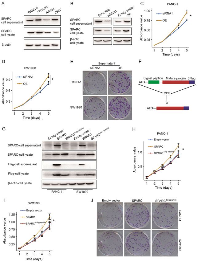
2                  PAN et al: SPARC PROMOTES PANCREATIC CANCER PROGRESSION BY AUTOCRINE SECRETION

oncogene in clinical studies and is associated with poor prog‑     and matched, para‑cancer specimens were collected for RNA
nosis in patients with PAAD (13,14). However, in biological        and protein extraction in accordance with institutional proto‑
experimental studies, SPARC is generally presented as a            cols. Para‑cancer specimens were resected 0.3‑0.5 cm from
tumour suppressor of PAAD (15‑17).                                 the tumour tissue. The patients age ranged between 52 and
    Therefore, several questions remain to be addressed.           77 years, with a mean age of 62.9 years and a median age of
First, a systematic pan‑cancer analysis of SPARC is needed         64 years. The patients included 10 men and 7 women. The
to determine whether it is an oncogene, a tumour suppressor,       ethics of the study were reviewed and approved by the board
or both. Secondly, the function of SPARC in PAAD needs             of Wenzhou Medical University.
further clarification, and the contradicting results of clinical
and biological research need to be unified. Finally, while         Cell culture and transfection. The PANC‑1 and SW1990
stroma‑derived SPARC is associated with poor prognosis in          human pancreatic cancer cell lines, the 293T human
patients with PAAD, cancer cell‑derived SPARC inhibits the         embryonic kidney cell line and the AR42J rat immortal‑
proliferation and chemoresistance of PAAD cells (13,15,17).        ized pancreatic acinar cell line were purchased from the
Whether SPARC produced by tumour stromal cells vs. PAAD            Institute of Biochemistry and Cell Biology, The Chinese
cells has the same effects on tumour growth and progression        Academy of Science. PANC‑1 and 293T cells were cultured
must be investigated. In the present study, a pan‑cancer anal‑     in high‑glucose DMEM medium (Gibco; Thermo Fisher
ysis of SPARC expression using The Cancer Genome Atlas             Scientific, Inc.) with 10% FBS (Sigma‑Aldrich; Merck KGaA),
(TCGA) data was carried out. This analysis confirmed that          100 U/ml penicillin and 100 µg/ml streptomycin. SW1990 and
SPARC was an oncogene in some cancer types, but a tumour           AR42J were cultured in 1640 medium (Gibco; Thermo Fisher
suppressor in others. Consistent with clinical data, this study    Scientific, Inc.) with the same supplements. Lipofectamine®
also confirmed SPARC to be an oncogene in PAAD. This               3000 (Invitrogen; Thermo Fisher Scientific, Inc.) and polyeth‑
has important clinical implications for patients with PAAD.        yleneimine (Sigma‑Aldrich; Merck KGaA) were used for the
Moreover, SPARC was found to promote PAAD cell prolifera‑          transfection of the PAAD cells and 293T cells, respectively,
tion and migration only when secreted.                             according to the manufacturers' protocols.

Materials and methods                                              Plasmid construction. The pLenti‑CMV‑EGFP‑3Flag vector
                                                                   was treated with EcoRI and BamHI at 37˚C for linearization.
Datasets. Mutation, RNA Sequencing (RNASeq) and                    Full‑length SPARC cDNA (NCBI, accession no. NM_003118.4;
clinical data of 10,182 patients were collected from TCGA          https://www.ncbi.nlm.nih.gov/nuccore/NM_003118.4) was
(https://portal.gdc.cancer.gov/) with 33 cancer types:             amplified from the cDNA of PANC‑1 cells and cloned into the
Adrenocortical carcinoma (ACC), bladder urothelial carci‑          linearized vector using a seamless clone kit (cat. no. D7010S;
noma (BLCA), BRCA, cervical squamous cell carcinoma                Beyotime Institute of Biotechnology) according to the manu‑
(CESC), cholangiocarcinoma (CHOL), COAD, DLBL,                     facturer's protocol.
oesophageal carcinoma (ESCA), glioblastoma multiforme                  For pLenti‑CMV‑SPARC∆ sig peptide ‑Flag, the N‑terminal
(GBM), head and neck squamous carcinoma (HNSC), kidney             17 amino acids are the signal peptide of the SPARC protein
chromophobe (KICH), kidney renal clear cell carcinoma              and are essential for autocrine secretion (19). Using the
(KIRC), kidney renal papillary cell carcinoma (KIRP), bone         full‑length cDNA of SPARC as a template, we obtained cDNA
marrow acute myeloid leukaemia (LAML), brain low grade             with deleted codons for signal peptide via polymerase chain
glioma (LGG), liver hepatocellular carcinoma (LIHC),               reaction (PCR) amplification while retaining the start codon.
lung adenocarcinoma (LUAD), lung squamous carcinoma                This SPARCΔsig peptide cDNA was cloned into the linearized
(LUSC), pleura mesothelioma (MESO), ovarian serous cyst‑           pLenti‑CMV‑EGFP‑3Flag using the aforementioned seamless
adenocarcinoma (OV), PAAD, adrenal phechromocytoma and             clone kit according to the manufacturer's protocol.
paraganglioma (PCPG), prostate adenocarcinoma (PRAD),
rectum adenocarcinoma (READ), soft tissue sarcoma (SARC),          Silencing of SPARC by small interfering (si)RNA. SPARC
skin cutaneous melanoma (SKCM), stomach adenocarcinoma             was silenced using two siRNA molecules (Shanghai Biosun
(STAD), testicular germ cell tumours (TGCT), thyroid               Sci&Tech Co., Ltd.). The sequences of the SPARC siRNAs
carcinoma (THCA), thymoma (THYM), uterine corpus endo‑             and the scrambled negative control siRNA are listed in Table I.
metrial carcinoma (UCEC), uterine carcinosarcoma (UCS)             The powder of SPARC and scrambled siRNAs was dissolved in
and uveal melanoma (UVM). For mutation data, the dataset           RNase‑free water to a concentration of 20 µM. Subsequently,
produced by the Multi‑Center Mutation Calling in Multiple          5 µl of the siRNA solution was mixed with 150 µl FBS‑free
Cancers (MC3) project was downloaded instead of the respec‑        medium at room temperature for 5 min. At the same time, for
tive maf files of 33 types of cancer (18) (https://gdc.cancer.     each group of siRNA, 5 µl Lipofectamine 3000 (Thermo Fisher
gov/about‑data/publications/mc3‑2017).                             Scientific, Inc.) was mixed with 150 µl FBS‑free medium at
                                                                   room temperature for 5 min. The siRNA solution was mixed
Patients and samples. All surgically removed PAAD and              with the Lipofectamine 3000 solution at room temperature for
para‑cancer specimens were obtained from the Department            20 min, and the transfection mixture was added to a 6‑cm dish
of Hepatobiliary Surgery of The First Affiliated Hospital,         containing PANC‑1 and SW1990 cells at 30‑40% confluency
Wenzhou Medical University, between September 2016 and             and 1.7 ml FBS‑free medium. The final siRNA concentration
July 2019. Verbal informed consent was obtained from the           was 50 pM. The medium for transfection was replaced by
patients. A total of 17 pairs of pathologically diagnosed PAAD     medium containing FBS and antibiotics 5 h later, and cells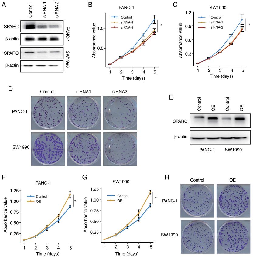
ONCOLOGY LETTERS 21: 485, 2021                                                    3

Table I. Primers and siRNA molecules.                            transfection mixture was added to the 10‑cm dishes with
                                                                 293T cells at 50‑60% confluency. 293T cells were incubated
A, Primers                                                       at 37˚C, and the transfection medium was replaced 6 h later.
                                                                 Virus‑containing medium was collected 48 h after transfec‑
Primer name                      Sequence, 5'‑3'                 tion and was centrifugated at 3,000 x g and 4˚C for 5 min
                                                                 to remove cell debris. For lentivirus infection, PAAD cells
SPARC‑mRNA‑F        CGAAGAGGAGGTGGTGGCGGAAA                      were cultured in 6‑cm dishes at 40‑50% confluency, and the
SPARC‑mRNA‑R        GGTTGTTGTCCTCATCCCTCTCATAC                   virus‑containing medium was supplemented with 5 µg/ml
GAPDH‑F             CTCTCTGCTCCTCCTGTTCGACAG                     polybrene to infect PAAD cells at 37˚C for 24 h. After 48 h
GAPDH‑R             AGGGGTCTTACTCCTTGGAGGCCA                     of infection, the infected cells were positively selected
                                                                 with 2.5 µg/ml puromycin to eliminate uninfected cells to
                                                                 generate stable cell lines.
B, siRNA

siRNA name                       Sequence, 5'‑3'                 RNA isolation and reverse transcription quantitative‑PCR
                                                                 (RT‑qPCR). The total RNA from either PAAD or para‑cancer
siRNA‑SPARC‑1       UUAUCUAAUGUAUUCCUCCUG                        tissues was extracted using TRI Reagent (Sigma‑Aldrich;
siRNA‑SPARC‑2       UAGUUCUUCUCGAAGUCCCGG                        Merck KGaA) and chloroform according to the manufacturer's
                                                                 protocol. The RevertAid First Strand cDNA Synthesis kit
Negative control    UUCUCCGAACGUGUCACGUTT
                                                                 (Thermo Fisher Scientific, Inc.) was used for first‑strand cDNA
F, forward; R, reverse; siRNA, small interfering RNA; SPARC,     synthesis. Briefly, 1 µg total RNA was mixed with oligo‑dT
secreted protein acidic and rich in cysteine.                    primer and nuclease‑free water to a total volume of 12 µl, and
                                                                 the mixture was denatured at 65˚C for 5 min. Subsequently,
                                                                 reaction buffer, RNase inhibitor, dNTP, and reverse transcrip‑
                                                                 tase were added to a total final volume of 20 µl. The mixture
                                                                 was incubated at 25˚C for 5 min, cDNA was synthesized at
were lysed for RNA or protein extraction 48 h later. For Cell    42˚C for 60 min, and finally reverse transcriptase was dena‑
Counting Kit‑8 (CCK‑8) assay, cells were re‑cultured in a        tured at 70˚C for 5 min.
96‑well plate 24 h after transfection.                               qPCR was performed using the Quantstudio™ DX
                                                                 Real‑Time PCR Instrument (Applied Biosystems; Thermo
Overexpression (OE) of SPARC. pLenti‑CMV‑SPARC‑Flag              Fisher Scientific, Inc.) using the Fast SYBR Green Master
and pLenti‑CMV‑SPARCΔsig peptide‑Flag vector were                Mix (Applied Biosystems; Thermo Fisher Scientific, Inc.).
constructed as aforementioned. A pLenti‑CMV‑EGFP‑3Flag           cDNA was mixed with SYBR Green and primers to a final
vector with deleted EGFP cDNA was used as control. For           volume of 20 µl. The mixture was incubated at 95˚C for
transfection of PAAD cells, 3 µg of control or OE vector was     10 min for pre‑denaturation, followed by 40 cycles at 95˚C
mixed with 150 µl FBS‑free medium at room temperature            for 15 sec and 62˚C for 30 sec. The relative mRNA expression
for 5 min. At the same time, for each group of vectors, 5 µl     levels were normalized to the expression level of GAPDH
Lipofectamine 3000 was mixed with 150 µl FBS‑free medium         using the 2‑ΔΔCq method (20). The primers used for qPCR are
at room temperature for 5 min. Subsequently, the medium          listed in Table I.
containing vector was mixed with the medium containing
Lipofectamine 3000 at room temperature for 20 min, and the       CCK‑8 and clone formation assay. The proliferation of PAAD
transfection mixture was added to a 6‑cm dish containing         cells was determined using a CCK‑8 assay. PANC‑1 and
PANC‑1 and SW1990 cells at 30‑40% confluency and 1.7 ml          SW1990 cells were seeded in a 96‑well plate at a density of
FBS‑free medium. The medium for transfection was replaced        2x10³ cells/well in triplicate. After a 12‑h incubation, the cells
by medium containing FBS and antibiotics 5 h later, and          adhered to the bottom of the plate, and this time was defined
cells were lysed for RNA or protein extraction 48 h later. For   as day 0. At days 0‑3 and 4, 100 µl FBS‑free medium with 10%
CCK‑8 assay, cells were re‑cultured in a 96‑well plate 24 h      CCK‑8 reagent (Shanghai Yeasen Biotechnology Co., Ltd.)
after transfection.                                              were added to the wells. After incubation at 37˚C for 2 h, the
                                                                 absorbance was measured with a microplate reader (BioTek
Silencing of SPARC by short hairpin (sh)RNA. The sequence        Instruments, Inc.; Agilent Technologies, Inc.). A well with
of siRNA1 was used for the construction of SPARC shRNA           100 µl FBS‑free medium and 10% CCK‑8 reagent without
using a pLKO.1‑puro plasmid (Sigma‑Aldrich; Merck KGaA).         cells was used to determine the background absorbance. Cell
The pLKO.1‑puro plasmid without shRNA insert was used            viability was determined by subtracting the background absor‑
as a negative control. For lentivirus production, 4 µg of the    bance from the experimental wells.
shRNA vector, 3 µg of psPAX and 2 µg of pMD2.G were                  For the clone formation assay, 5x10² cells/well of PANC‑1
mixed with 150 µl FBS‑free medium at room temperature for        and SW1990 cells were seeded into 6‑well plates and cultured
5 min. A total of 30 µl of polyethylenimine (Sigma‑Aldrich;      at 37˚C for 14 days. The medium was changed every three
Merck KGaA) was mixed with 150 µl FBS‑free medium                days to avoid bias due to different evaporation rates. The cells
at room temperature for 5 min. Subsequently, the medium          were then fixed with methanol for 15 min at room temperature
containing vector was mixed with the medium containing           and stained with crystal violet for 5 min at room temperature
polyethylenimine at room temperature for 20 min. The             (Beyotime Institute of Biotechnology).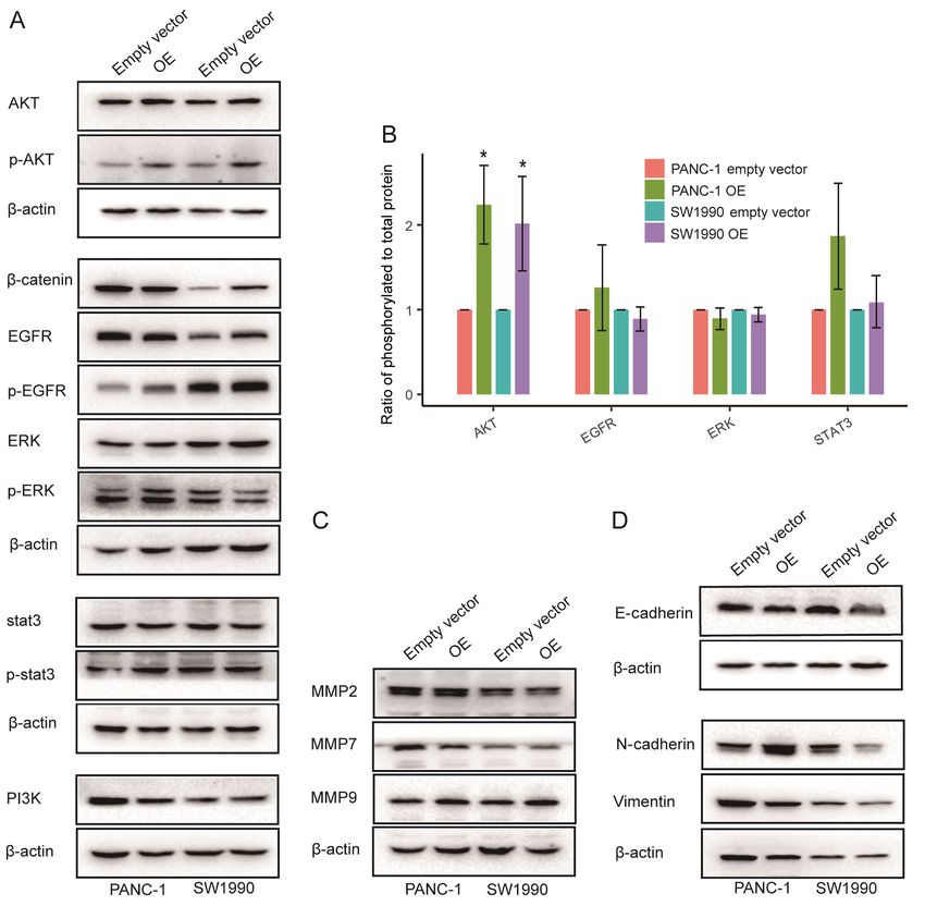
4                   PAN et al: SPARC PROMOTES PANCREATIC CANCER PROGRESSION BY AUTOCRINE SECRETION

Table II. Antibodies.

Antibody name                                                                   Supplier                      Cat. no.          Dilution

SPARC                                                              Abcam                                   ab207743            1:2,000
β‑actin                                                            Sigma‑Aldrich, Merck KGaA               A1978               1:5,000
Flag                                                               Sigma‑Aldrich, Merck KGaA               SAB4200071          1:5,000
AKT                                                                Cell Signaling Technology, Inc.         4691                1:1,000
p‑AKT                                                              Cell Signaling Technology, Inc.         4060                1:1,000
β‑catenin                                                          Abcam                                   ab32572             1:5,000
EGFR                                                               Abcam                                   ab32077             1:1,000
p‑EGFR                                                             Cell Signaling Technology, Inc.         3777                1:1,000
ERK                                                                Abcam                                   ab184699            1:2,000
p‑ERK                                                              Abcam                                   ab214036            1:2,000
Stat3                                                              Bioworld                                AP0365              1:1,000
p‑stat3                                                            Abcam                                   ab76315             1:2,000
p‑NFκB                                                             Abcam                                   ab76302             1:1,000
PI3K                                                               Abcam                                   ab32089             1:1,000
MMP2                                                               Abcam                                   Ab92536             1:1,000
MMP7                                                               Santa Cruz Biotechnology, Inc.          sc‑515703           1:100
MMP9                                                               Santa Cruz Biotechnology, Inc.          sc‑393859           1:200
E‑Cadherin                                                         Santa Cruz Biotechnology, Inc.          sc‑21791            1:200
N‑Cadherin                                                         Abcam                                   ab98952             1:2,000
Vimentin                                                           Santa Cruz Biotechnology, Inc.          sc‑6260             1:200
Goat anti rabbit secondary antibody, HRP‑conjugated                Sigma‑Aldrich, Merck KGaA               SAB3700885          1:10,000
Goat anti mouse secondary antibody, HRP‑conjugated                 Sigma‑Aldrich, Merck KGaA               SAB3700885          1:10,000

SPARC, secreted protein acidic and rich in cysteine; p, phosphorylated.

Transwell assay. The migration of the PANC‑1 and SW1990                   at 4˚C and for 1 h with HRP‑conjugated secondary antibodies
cells was assessed using 24‑well BioCoat cell culture inserts             (1:10,000) at 37˚C (Table II). Amersham ECL Prime (Cytiva)
(BD Biosciences) with an 8‑µm porosity polyethylene tere‑                 was used to detect bands. The densities of the specific protein
phthalate membrane. The lower compartment contained                       bands were visualized and captured using ImageQuant™ 400
RPMI‑1640 (for SW1990) or DMEM (for PANC‑1) with 10%                      (GE Healthcare Life Sciences) and analysed using Image Lab
FBS as a chemoattractant. A total of 2x10⁴ cells in 0.2 ml                3.0 software (Bio‑Rad Laboratories).
FBS‑free medium were plated in the upper compartment and
incubated at 37˚C for 48 h. After incubation, the cells were              Collection of cellular supernatant and preparation for
fixed with methanol for 15 min at room temperature and                    western blot analysis. The 293T cells (5x10⁶) transfected with
stained with crystal violet for 5 min at room temperature.                a SPARC siRNA or OE vector, were seeded in two 10‑cm
The images of Transwell assay were acquired using the Leica               dishes. A volume of 8 ml medium containing 10% FBS was
DMIL‑LED inverted light laboratory microscope (Leica                      added to each dish. The supernatant was collected 24 h later,
Microsystems GmbH; magnification, x400).                                  centrifuged at 12,000 x g and 4˚C for 5 min, then added to
                                                                          96‑well plates containing the PANC‑1 and SW1990 cells.
Western blot analysis. Cells were lysed using a radioim‑                      Western blot analysis was used to determine the difference
munoprecipitation assay lysis buffer with 10% phosphatase                 of SPARC concentration in the supernatant. The supernatant
inhibitor (Shanghai Yeasen Biotechnology Co., Ltd.) and 1%                was collected and centrifuged at 12,000 x g and 4˚C for
phenylmethylsulfonyl fluoride (Sigma‑Aldrich; Merck KGaA)                 5 min; then, 5X SDS‑PAGE loading buffer was added. After
for 30 min at 4˚C. Total protein lysate was then collected, and           denaturation at 100˚C for 5 min, the samples were used for
the concentration was determined with a BCA Protein Assay                 the western blot assay. To determine the concentration of the
kit (Beyotime Institute of Biotechnology). After denaturation             SPARC‑Flag and SPARCΔsig peptide‑Flag fusion protein in the
at 100˚C for 5 min, 20 µg of protein samples from each group              supernatant, the same number of PANC‑1 or SW1990 cells
were resolved by SDS‑PAGE using 10% gels, then electro‑                   (2x10 6), transfected with the same amount of the control,
phoretically transferred to a PVDF. The PVDF membrane                     SPARC‑Flag, or SPARCΔsig peptide‑Flag expression vector (2 µg),
was blocked with 5% skimmed milk (Shanghai Yeasen                         were seeded in 6 cm‑dishes. The supernatant was collected
Biotechnology Co., Ltd.) for 1.5 h at room temperature. The               24 h later for western blot analysis, which was carried out as
membrane was incubated overnight with primary antibodies                  aforementioned.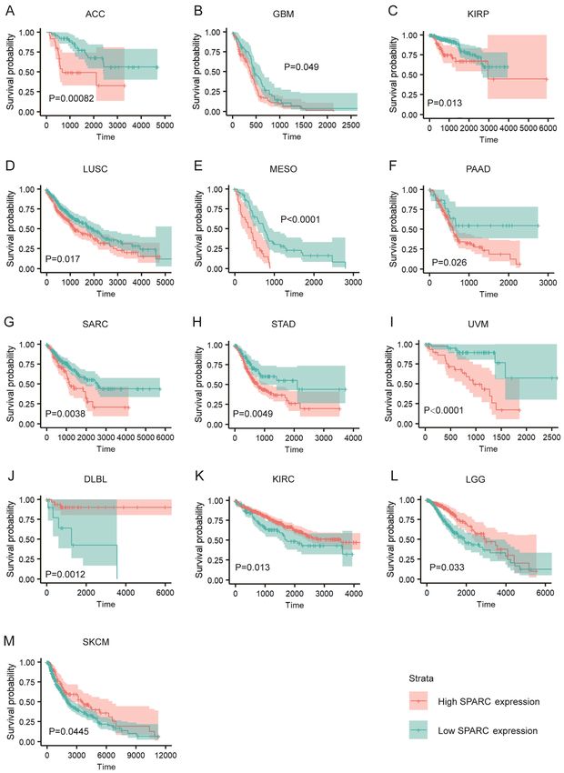
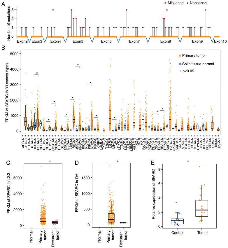
ONCOLOGY LETTERS 21: 485, 2021                                                                  5

Figure 1. SPARC drives tumorigenesis through abnormal changes in expression but not gene mutations. (A) Single‑nucleotide variations are uniformly distrib‑
uted across the coding region of the SPARC gene (TCGA data). (B) Expression pattern of SPARC in 33 cancer types. SPARC expression is generally increased
in various cancer types. Unpaired, two‑tailed t‑test (TCGA data). (C) Expression of SPARC decreased significantly in recurrent LGG, compared with the
primary tumour. Unpaired, two‑tailed t‑test (TCGA data). (D) Expression of SPARC decreased significantly in recurrent OV, compared with the primary
tumour. Unpaired, two‑tailed t‑test (TCGA data). (E) Reverse transcription‑quantitative PCR demonstrated that SPARC expression increased significantly in
PAAD, compared with the paired para‑cancer tissue. n=17, paired, two‑tailed t‑test. *P
6                    PAN et al: SPARC PROMOTES PANCREATIC CANCER PROGRESSION BY AUTOCRINE SECRETION

Figure 2. SPARC is associated with prognosis of various cancers. (A‑I) High SPARC expression is associated with worse prognosis in ACC, GBM, KIRP,
LUSC, MESO, PAAD, SARC, STAD and UVM (TGCA data). (J‑M) High SPARC expression is associated with improved prognosis in DLBL, KIRC,
LGG and SKCM. (TCGA data). The Kaplan‑Meier method with log‑rank tests (ACC, GBM, LUSC, MESO, PAAD, SARC, STAD, UVM, DLBL, KIRC) or
Cramér‑von Mises tests (KIRP, LGG, SKCM) were used for comparison of survival time between two groups. SPARC, secreted protein acidic and rich in
cysteine; ACC, adrenocortical carcinoma; GBM, glioblastoma multiforme; KIRP, kidney renal papillary cell carcinoma; LUSC, lung squamous carcinoma;
MESO, pleura mesothelioma; SARC, soft tissue sarcoma; STAD, stomach adenocarcinoma; UVM, uveal melanoma; DLBL, diffuse large B‑cell lymphoma;
KIRC, kidney renal clear cell carcinoma; LGG, brain low‑grade glioma; SKCM, skin cutaneous melanoma.

minimal. The cut‑off value was achieved using the R pack‑                  the bias caused by the difference in the number of people
ages ‘survival’ and ‘survminer’, with a loop function. To avoid            in the two groups, each group comprising ≥25% of the total
ONCOLOGY LETTERS 21: 485, 2021 7
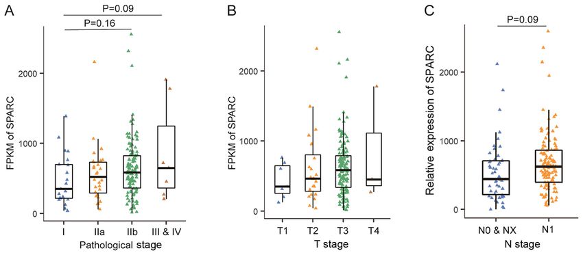
Figure 3. SPARC is not associated with the pathological stages of PAAD. (A) SPARC expression is not associated with PAAD pathological stage. One‑way
ANOVA with Tukey's post‑hoc test (TCGA data). (B) SPARC expression is not associated with PAAD T stage. One‑way ANOVA with Tukey's post‑hoc Tukey's
test (TCGA data). (C) SPARC expression is not associated with PAAD N stage. Unpaired, two‑tailed t‑test (TCGA data). PAAD, pancreatic adenocarcinoma;
SPARC, secreted protein acidic and rich in cysteine; T, tumour stage; N, lymph node metastasis.
population. R‑3.4 (https://www.r‑project.org/) was used for and OV tumours compared with primary tumour tissues
the statistical analysis. P10,000 cancer specimens across 33 cancer tissues, compared with normal tissue (Fig. 1E).
types were analysed. The mutation frequency of SPARC was
very low, and only 101 single nucleotide variations were identi‑ SPARC is associated with the prognosis of various cancers.
fied in the coding region of SPARC, including 21 synonymous, The expression level of SPARC was associated with the
71 missense, 8 nonsense and 1 splice site mutations. The 80 prognosis of various tumour types. Of the 33 cancer types
non‑synonymous mutations were uniformly distributed across in TCGA dataset, high expression of SPARC was associated
64 coding base sites, with 1‑3 mutations at each site, and no with significantly worse prognosis in 9 cancer types, ACC,
prominent recurrent mutations were found (Fig. 1A). It is GBM, KIRP, LUSC, MESO, PAAD, SARC, STAD and UVM
likely that these mutations were only passengers and mutations (Fig. 2A‑I). However, SPARC may be a tumour suppressor
of SPARC had no significant relationship with tumorigenesis. gene in a few cancer types. Indeed, high SPARC expression
Interestingly, no insertion‑deletion of the SPARC gene was was associated with improved prognosis in DLBL, KIRC,
found in the MC3 dataset, whether in‑frame or frame‑shift. LGG and SKCM (Fig. 2J‑M).
The low mutation frequency and high conservation suggested High expression of SPARC was associated with the worse
that the normal function of SPARC was essential in tumour prognosis of PAAD (Fig. 2F). Further analysis revealed that
development. Thus, SPARC might be a potential negative patients with high or low SPARC expression levels displayed
selection gene during tumorigenesis (21). similar survival times in the short term after diagnosis
Since mutations in the SPARC gene had no significant rela‑ (8 PAN et al: SPARC PROMOTES PANCREATIC CANCER PROGRESSION BY AUTOCRINE SECRETION Figure 4. SPARC promotes the proliferation of PAAD cells. (A) Silencing of SPARC was achieved using two siRNA molecules. (B and C) CCK‑8 assays showed that SPARC silencing impaired the proliferation of (B) PANC‑1 cells and (C) SW1990 cells. n=3. *P
ONCOLOGY LETTERS 21: 485, 2021 9
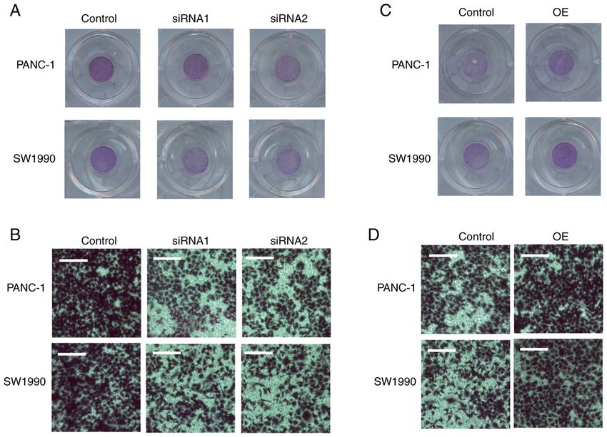
Figure 5. SPARC promotes the migration of PAAD cells. (A and B) Transwell assays showed that SPARC silencing impaired the migration of (A) PANC‑1 and
(B) SW1990 cells. (C and D) Transwell assays showed that SPARC OE increased the migration of (C) PANC‑1 and (D) SW1990 cells. Scale bar, 100 µm. OE,
overexpression; SPARC, secreted protein acidic and rich in cysteine; siRNA, small interfering RNA.
SPARC promotes the migration of PAAD cells. A Transwell supernatant from the SPARC OE cells significantly promoted
assay was performed to determine whether SPARC could the proliferation of the PAAD cells, compared with the control
affect the migration of PAAD cells. The ability of the PAAD group treated with a supernatant of SPARC‑silenced 293T
cells to cross the Transwell membrane was significantly cells (Fig. 6C and D). PAAD cells treated with exogenous
impaired after SPARC was silenced (Fig. 5A and B). The SPARC also showed increased clone formation (Fig. 6E).
changes in the migration ability of the PAAD cells following These results confirmed that SPARC from the extracellular
SPARC OE were also assessed. The migration of PAAD cells milieu can promote the proliferation of PAAD cells.
increased following SPARC OE (Fig. 5C and D). Furthermore, it was hypothesised that this autocrine‑
extracellular milieu‑cancer cell pathway may be necessary for
SPARC regulates pancreatic cancer cell proliferation through SPARC function in cancer cells. As SPARC has a well‑defined
autocrine secretion into the extracellular milieu. Considering signal peptide, which is its N‑terminal 17 amino acid resi‑
that SPARC is an exocrine protein, western blot analysis was dues, an expression vector that expresses the SPARC protein
carried out in the culture supernatant of PAAD cells. Clear with deletion of signal peptide, referred to as pLenti‑CMV‑
SPARC bands were detected in the supernatant of PAAD SPARC ∆ sig peptide , was constr ucted (Fig. 6F). In both
cells, immortalized normal acinar AR42J cells and 293T cells, PANC‑1 and SW1990 cells, endogenous SPARC was
confirming that SPARC was secreted into the extracellular almost undetectable compared with the SPARC‑Flag or
milieu (Fig. 6A). SPARC∆ sig peptide ‑Flag fusion protein. Using an anti‑Flag
It was hypothesised that the SPARC protein could affect antibody, it was demonstrated that the SPARC‑Flag fusion
the proliferation and migration of PAAD cells when produced protein, but not the SPARC∆ sig peptide‑Flag fusion protein, can
either by cancer cells or stromal cells and secreted to the be effectively secreted to the extracellular milieu (Fig. 6G).
stroma. This would explain why the prognosis of patients with The endogenous SPARC expression of the PANC‑1 and
PAAD is associated with SPARC expression levels of both SW1990 cells was silenced by stably transfecting with
tumour cells and stroma cells (13,14). Therefore, the role of the SPARC shRNA. Then, the control vector, SPARC OE vector
SPARC protein in the extracellular milieu was examined. It and SPARC∆ sig peptide vector were transfected into the PAAD
was confirmed that the SPARC concentration in the superna‑ cells with the silencing of endogenous SPARC, and the prolif‑
tant of 293T cells was significantly decreased following siRNA eration of the PAAD cells was examined. Only the wildtype
silencing and increased by the SPARC OE vector, compared SPARC, but not SPARC∆ sig peptide, effectively promoted the prolif‑
with scramble RNA or control vector, respectively (Fig. 6B). eration of PAAD cells (Fig. 6H and I). A clone‑formation assay
Before being treated with the supernatant from 293T cells further confirmed this result (Fig. 6J). Thus, SPARC promotes
treated with siRNA and OE vector, the PANC‑1 and SW1990 the proliferation of PAAD cells through autocrine secretion
cells were stably transfected with SPARC shRNA. The into the extracellular milieu.10 PAN et al: SPARC PROMOTES PANCREATIC CANCER PROGRESSION BY AUTOCRINE SECRETION Figure 6. SPARC regulates pancreatic cancer cell proliferation through autocrine secretion into the extracellular milieu. (A) SPARC is detected in cell lysate and supernatant of PAAD cells, immortalized normal acinar AR42Jccells, and 293T cells. (B) Supernatant of 293T cells transfected with the SPARC siRNA has significant decreased concentration of SPARC protein, compared with cells transfected with scramble siRNA. The supernatant of 293T cells transfected with SPARC OE vector has increased concentration of SPARC protein, compared with the empty vector group. (C) Proliferation of PANC‑1 cells trans‑ fected with SPARC OE supernatant increased significantly, compared with the cells treated with the SPARC‑silenced supernatant (n=3). *P
ONCOLOGY LETTERS 21: 485, 2021                                                              11

Figure 7. Mechanism of action of the SPARC protein. (A) SPARC significantly upregulates the phosphorylation AKT, but not STAT3, EGFR, ERK and
β‑catenin in PAAD cells. (B) Ratio of phosphorylated to total protein of AKT increased significantly in SPARC over‑expressed PAAD cells. (C and D) SPARC
had no significant and repeatable effect on the expression of the MMP2, MMP7, MMP9 and EMT related genes. *P
12                 PAN et al: SPARC PROMOTES PANCREATIC CANCER PROGRESSION BY AUTOCRINE SECRETION

addition, SPARC predicts improved prognosis in DLBL but             or other types of stromal cell infiltration. The subcutaneous
poor prognosis in melanoma (8,27). To construct a compre‑           xenografts are mainly composed of cancer cells. Thus, an
hensive picture of SPARC's function in cancer, a pan‑cancer         in vivo experiment may be useful in determining the effect of
analysis of SPARC was carried out using a public dataset from       SPARC on in vivo growth of PAAD, but it cannot be used to
TCGA covering 33 cancer types. By analysing the association         distinguish the cellular origin of SPARC. The lack of IHC and
between SPARC and the prognosis of different tumours, it was        in vivo experiments are a limitation of the present study, and
found that the function of SPARC seemed to mainly depend on         the above questions should be explored in further studies.
the pathological cancer type rather than the organic or cellular        The mechanisms underlying SPARC functions are
origin. Moreover, SPARC predicted the distinct prognosis of         complex. SPARC has been reported to facilitate the prolifera‑
LGG and GBM, which both stem from glial cells and occur in          tion and metastasis of hepatocellular carcinoma via the ERK
the brain or spine cord (28,29), and KIRC and KIRP, which both      signalling pathway, promote cervical cancer cell proliferation
stem from renal tubular epithelial cells (30). Meanwhile, SPARC     by modulating the bax/bcl‑2 ratio, and induce the migration of
seems associated with poor prognosis of adenocarcinoma              non‑small cell lung cancer via WNK1/Snail signalling (35‑37).
(PAAD and STAD), whereas for COAD, LUAD and READ, the               In acute myeloid leukaemia, SPARC interacts with inte‑
results did not reach significance. In addition, consistent with    grin‑linked kinase (ILK) and promotes ILK signalling (38).
a previous report (8), SPARC was found to predict improved          Since AKT is an important downstream effector of ILK (39), it
prognosis of lymphatic and hematopoietic cancer types, such as      was hypothesised that SPARC may also promote AKT activa‑
DLBL and LAML. Due to the influence of the mRNA turn‑               tion by interacting with ILK in PAAD cells.
over rate, ribosome‑bound mRNA abundance and the protein                In conclusion, the present study partly answered the afore‑
turnover rate, mRNA abundance does not always accurately            mentioned controversial roles of SPARC. Overall, SPARC
reflect protein content (31). Nevertheless, according to existing   exhibits regulatory potential and may play a role in PAAD
reports, the mRNA and protein expression of the SPARC gene          progression, and its significance in cancer therapy merits
in tumour tissues show a consistent trend (32,33). To our knowl‑    further study.
edge, this study is the first to clarify the function of SPARC
at the pan‑cancer level, which could therefore help explain the     Acknowledgements
inconsistencies seen in previous studies.
    It was demonstrated that SPARC acts as an oncogene in           The authors would like to thank Dr Yi Zhou (The First
PAAD. Moreover, SPARC is associated with poor prognosis             Affiliated Hospital of Wenzhou Medical University, Wenzhou,
of PAAD, and in the present in vitro research, we found that it     China) for her help in finishing supplementary western blot
promotes the proliferation and migration of PAAD cells. To a        assay and for providing guidance for survival analysis.
certain extent, the in vitro results of our work contradict some
previous reports, in which SPARC was proposed to inhibit            Funding
the proliferation and increase the chemosensitivity of PAAD
cells (15‑17). Thus, it may be proposed that cell type and          The present study was supported by The Foundation of Wenzhou
experimental technique differences may be the direct cause          Science & Technology Bureau (grant no. 2020Y0255).
of this inconsistency. However, the root cause may also be the
versatility of SPARC. In PAAD, SPARC may have different             Availability of data and materials
functions in patients with different genomic alterations and
molecular profiles. The possibility that SPARC may act as a         The datasets used and/or analyzed during the current study
tumor suppressor in some patients and some PAAD cell lines          are available from the corresponding author on reasonable
cannot be excluded, since subtypes of PAAD with signifi‑            request. The datasets generated and/or analyzed during the
cant molecular and clinical difference have been previously         current study are available in TCGA repository (https://portal.
found (34).                                                         gdc.cancer.gov/).
    Another question regarding SPARC and PAAD is whether
the SPARC proteins that affect tumour prognosis come from           Authors' contributions
tumour cells or tumour stromal cells. Our work confirmed
that SPARC regulated PAAD proliferation only when secreted          KP, XH and XJ performed the biological experiments, data
extracellularly. Since the SPARC that affects the proliferation     analysis, statistical work and manuscript preparation. KP and
of PAAD cells comes from the extracellular milieu, it may be        XJ collected TCGA data and performed pan‑cancer analysis
inferred that the SPARC from both stroma cells and tumor            of SPARC. XJ supervised the experimental work and finished
cells can promote PAAD cell proliferation. In TCGA data             manuscript proofreading. XJ designed the study and guaran‑
used on this study, SPARC expression was determined via             tees its integrity. KP, XH and XJ confirm the authenticity of
RNASeq; however, this method could not distinguish the              all the raw data. All authors have read and approved the final
cellular origin of SPARC. The optimal method to elucidate           manuscript.
the source of SPARC may be immunohistochemistry (IHC),
which requires paraffin‑embedded clinical PAAD specimen             Ethics approval and consent to participate
or xenograft tissue. Unfortunately, there were not enough
PAAD specimens to carry out IHC in the study. In addition,          The present study was reviewed and approved by The Board
according to our past experience, the subcutaneous injection of     of Wenzhou Medical University. Verbal informed consent was
PAAD cells in node mice does not result in massive fibroblasts      obtained from the patients.
ONCOLOGY LETTERS 21: 485, 2021 13
Patient consent for publication 19. Almagro Armenteros JJ, Tsirigos KD, Sønderby CK, Petersen TN,
Winther O, Brunak S, von Heijne G and Nielsen H: SignalP 5.0
improves signal peptide predictions using deep neural networks.
Not applicable. Nat Biotechnol 37: 420‑423, 2019.
20. Livak KJ and Schmittgen TD: Analysis of relative gene expres‑
sion data using real‑time quantitative PCR and the 2(‑Delta Delta
Competing interests C(T)) method. Methods 25: 402‑408, 2001.
21. Martincorena I, Raine KM, Gerstung M, Dawson KJ, Haase K, Van
The authors declare that they have competing interests. Loo P, Davies H, Stratton MR and Campbell PJ: Universal patterns of
selection in cancer and somatic tissues. Cell 171: 1029‑1041.e21, 2017.
22. Chio IIC, Jafarnejad SM, Ponz‑Sarvise M, Park Y, Rivera K,
References Palm W, Wilson J, Sangar V, Hao Y, Öhlund D, et al: NRF2
promotes tumor maintenance by modulating mRNA translation
1. Termine JD, Kleinman HK, Whitson SW, Conn KM, in pancreatic cancer. Cell 166: 963‑976, 2016.
McGarvey ML and Martin GR: Osteonectin, a bone‑specific 23. Zhang H, Pan YZ, Cheung M, Cao M, Yu C, Chen L, Zhan L,
protein linking mineral to collagen. Cell 26: 99‑105, 1981. He ZW, Sun CY: LAMB3 mediates apoptotic, proliferative, inva‑
2. Mayer U, Aumailley M, Mann K, Timpl R and Engel J: sive, and metastatic behaviors in pancreatic cancer by regulating
Calcium‑dependent binding of basement membrane protein the PI3K/Akt signaling pathway. Cell Death Dis 10: 230, 2019.
BM‑40 (osteonectin, SPARC) to basement membrane collagen 24. Bergamaschi A, Tagliabue E, Sørlie T, Naume B, Triulzi T,
type IV. Eur J Biochem 198: 141‑150, 1991. Orlandi R, Russnes HG, Nesland JM, Tammi R, Auvinen P, et al:
3. Gaudet P, Livstone MS, Lewis SE and Thomas PD: Extracellular matrix signature identifies breast cancer subgroups
Phylogenetic‑based propagation of functional annotations within with different clinical outcome. J Pathol 214: 357‑367, 2008.
the Gene Ontology consortium. Brief Bioinform 12: 449‑462, 2011. 25. Huang Y, Zhang J, Zhao YY, Jiang W, Xue C, Xu F, Zhao HY,
4. Nagaraju GP, Dontula R, El‑Rayes BF and Lakka SS: Molecular Zhang Y, Zhao LP, Hu ZH, et al: SPARC expression and prognostic
mechanisms underlying the divergent roles of SPARC in human value in non‑small cell lung cancer. Chin J Cancer 31: 541‑548, 2012.
carcinogenesis. Carcinogenesis 35: 967‑973, 2014. 26. Koukourakis MI, Giatromanolaki A, Brekken RA, Sivridis E,
5. Said N: Roles of SPARC in urothelial carcinogenesis, progres‑ Gatter KC, Harris AL and Sage EH: Enhanced expression of
sion and metastasis. Oncotarget 7: 67574‑67585, 2016. SPARC/osteonectin in the tumor‑associated stroma of non‑small
6. Said N, Frierson HF, Sanchez‑Carbayo M, Brekken RA and cell lung cancer is correlated with markers of hypoxia/acidity and
Theodorescu D: Loss of SPARC in bladder cancer enhances with poor prognosis of patients. Cancer Res 63: 5376‑5380, 2003.
carcinogenesis and progression. J Clin Invest 123: 751‑766, 2013. 27. Massi D, Franchi A, Borgognoni L, Reali UM and Santucci M:
7. Chin D, Boyle GM, Williams RM, Ferguson K, Pandeya N, Osteonectin expression correlates with clinical outcome in thin
Pedley J, Campbell CM, Theile DR, Parsons PG and Coman WB: cutaneous malignant melanomas. Hum Pathol 30: 339‑344, 1999.
Novel markers for poor prognosis in head and neck cancer. Int J 28. Cancer Genome Atlas Research Network: Comprehensive
Cancer 113: 789‑797, 2005. genomic characterization defines human glioblastoma genes and
8. Meyer PN, Fu K, Greiner T, Smith L, Delabie J, Gascoyne R, core pathways. Nature 455: 1061‑1068, 2008.
Ott G, Rosenwald A, Braziel R, Campo E, et al: The stromal cell 29. Perry A and Wesseling P: Histologic classification of gliomas.
marker SPARC predicts for survival in patients with diffuse large Handb Clin Neurol 134: 71‑95, 2016.
B‑cell lymphoma treated with rituximab. Am J Clin Pathol 135: 30. Ricketts CJ, De Cubas AA, Fan H, Smith CC, Lang M, Reznik E,
54‑61, 2011. Bowlby R, Gibb EA, Akbani R, Beroukhim R, et al: The cancer
9. Yamanaka M, Kanda K, Li NC, Fukumori T, Oka N, Kanayama HO genome atlas comprehensive molecular characterization of renal
and Kagawa S: Analysis of the gene expression of SPARC and its cell carcinoma. Cell Rep 23: 313‑326.e5, 2018.
prognostic value for bladder cancer. J Urol 166: 2495‑2499, 2001. 31. Maier T, Güell M and Serrano L: Correlation of mRNA
10. Jones C, Mackay A, Grigoriadis A, Cossu A, Reis‑Filho JS, and protein in complex biological samples. FEBS Lett 583:
Fulford L, Dexter T, Davies S, Bulmer K, Ford E, et al: Expression 3966‑3973, 2009.
profiling of purified normal human luminal and myoepithelial 32. Zhang S, Jin J, Tian X and Wu L: hsa‑miR‑29c‑3p regulates
breast cells: Identification of novel prognostic markers for breast biological function of colorectal cancer by targeting SPARC.
cancer. Cancer Res 64: 3037‑3045, 2004. Oncotarget 8: 104508‑104524, 2017.
11. Liang JF, Wang HK, Xiao H, Li N, Cheng CX, Zhao YZ, Ma YB, 33. Qu X, Gao D, Ren Q, Jiang X, Bai J and Sheng L: miR‑211
Gao JZ, Bai RB and Zheng HX: Relationship and prognostic inhibits proliferation, invasion and migration of cervical cancer
significance of SPARC and VEGF protein expression in colon via targeting SPARC. Oncol Lett 16: 853‑860, 2018.
cancer. J Exp Clin Cancer Res 29: 71, 2010. 34. Cancer Genome Atlas Research Network. Electronic address:
12. Hariharan D, Saied A and Kocher HM: Analysis of mortality andrew_aguirre@dfci.harvard.edu; Cancer Genome Atlas
rates for pancreatic cancer across the world. HPB (Oxford) 10: Research Network: Integrated genomic characterization of pancre‑
58‑62, 2008. atic ductal adenocarcinoma. Cancer Cell 32: 185‑203.e13, 2017.
13. Infante JR, Matsubayashi H, Sato N, Tonascia J, Klein AP, 35. Liu Y, Feng Y, Wang X, Yang X, Hu Y, Li Y, Zhang Q, Huang Y,
Riall TA, Yeo C, Iacobuzio‑Donahue C and Goggins M: Shi K, Ran C, et al: SPARC negatively correlates with prognosis
Peritumoral fibroblast SPARC expression and patient outcome after transarterial chemoembolization and facilitates prolifera‑
with resectable pancreatic adenocarcinoma. J Clin Oncol 25: tion and metastasis of hepatocellular carcinoma via ERK/MMP
319‑325, 2007. signaling pathways. Front Oncol 10: 813, 2020.
14. Yu XZ, Guo ZY, Di Y, Yang F, Ouyang Q, Fu DL and Jin C: The 36. Chen J, Shi D, Liu X, Fang S, Zhang J and Zhao Y: Targeting
relationship between SPARC expression in primary tumor and SPARC by lentivirus‑mediated RNA interference inhibits
metastatic lymph node of resected pancreatic cancer patients and cervical cancer cell growth and metastasis. BMC Cancer 12:
patients' survival. Hepatobiliary Pancreat Dis Int 16: 104‑109, 2017. 464, 2012.
15. Fan X, Mao Z, Ma X, Cui L, Qu J, Lv L, Dang S, Wang X and 37. Hung JY, Yen MC, Jian SF, Wu CY, Chang WA, Liu KT,
Zhang J: Secreted protein acidic and rich in cysteine enhances Hsu YL, Chong IW and Kuo PL: Secreted protein acidic and
the chemosensitivity of pancreatic cancer cells to gemcitabine. rich in cysteine (SPARC) induces cell migration and epithelial
Tumour Biol 37: 2267‑2273, 2016. mesenchymal transition through WNK1/snail in non‑small cell
16. Xiao Y, Zhang H, Ma Q, Huang R, Lu J, Liang X, Liu X, lung cancer. Oncotarget 8: 63691‑63702, 2017.
Zhang Z, Yu L, Pang J, et al: YAP1‑mediated pancreatic stel‑ 38. Alachkar H, Santhanam R, Maharry K, Metzeler KH,
late cell activation inhibits pancreatic cancer cell proliferation. Huang X, Kohlschmidt J, Mendler JH, Benito JM, Hickey C,
Cancer Lett 462: 51‑60, 2019. Neviani P, et al: SPARC promotes leukemic cell growth and
17. Mao Z, Ma X, Fan X, Cui L, Zhu T, Qu J, Zhang J and Wang X: predicts acute myeloid leukemia outcome. J Clin Invest 124:
Secreted protein acidic and rich in cysteine inhibits the growth of 1512‑1524, 2014.
human pancreatic cancer cells with G1 arrest induction. Tumour 39. Lynch DK, Ellis CA, Edwards PA and Hiles ID: Integrin‑linked
Biol 35: 10185‑10193, 2014. kinase regulates phosphorylation of serine 473 of protein kinase B
18. Ellrott K, Bailey MH, Saksena G, Covington KR, Kandoth C, by an indirect mechanism. Oncogene 18: 8024‑8032, 1999.
Stewart C, Hess J, Ma S, Chiotti KE, McLellan M, et al: Scalable
open science approach for mutation calling of tumor exomes This work is licensed under a Creative Commons
using multiple genomic pipelines. Cell Syst 6: 271‑281.e7, 2018. Attribution-NonCommercial-NoDerivatives 4.0
International (CC BY-NC-ND 4.0) License.You can also read