Cyst Fluid Telomerase Activity Predicts the Histologic Grade of Cystic Neoplasms of the Pancreas
←
→
Page content transcription
If your browser does not render page correctly, please read the page content below
Published OnlineFirst May 26, 2016; DOI: 10.1158/1078-0432.CCR-16-0311

   Biology of Human Tumors                                                                                                            Clinical
                                                                                                                                      Cancer
                                                                                                                                      Research
 Cyst Fluid Telomerase Activity Predicts the
 Histologic Grade of Cystic Neoplasms
 of the Pancreas
 Tatsuo Hata1, Marco Dal Molin1, Masaya Suenaga1, Jun Yu1, Meredith Pittman1,
 Matthew Weiss2, Marcia I. Canto2,3, Christopher Wolfgang2, Anne Marie Lennon2,3,
 Ralph H. Hruban1,4, and Michael Goggins1,3,4

 Abstract
    Purpose: Pancreatic cysts frequently pose clinical dilemmas.                  thawed, surgical cyst fluids from cystic neoplasms with high-
 On one hand, cysts with high-grade dysplasia offer opportunities                 grade dysplasia  associated invasive cancer had higher telo-
 for cure, on the other hand, those with low-grade dysplasia are                  merase activity [median (interquartile range), 1,158 (295.9–
 easily over treated. Cyst fluid markers have the potential to                     13,033)] copies/mL of cyst fluid than those without [19.74
 improve the evaluation of these cysts. Because telomerase activity               (2.58–233.6) copies/mL; P < 0.001)]. Elevated cyst fluid telo-
 is commonly activated in malignant cells, we evaluated the                       merase activity had a diagnostic accuracy for invasive cancer/
 diagnostic performance of cyst fluid telomerase activity measure-                 high-grade dysplasia of 88.1% (discovery), 88.6% (validation),
 ments for predicting histologic grade.                                           and 88.2% (merged). Among cysts classified preoperatively as
    Experimental Design: Telomerase activity was measured using                   having "worrisome features," cyst fluid telomerase activity had
 telomerase repeat amplification with digital-droplet PCR in sur-                  high diagnostic performance (sensitivity 73.7%, specificity
 gically aspirated cyst fluid samples from 219 patients who under-                 90.6%, accuracy, 86.1%). In multivariate analysis, telomerase
 went pancreatic resection for a cystic lesion (184 discovery, 35                 activity independently predicted the presence of invasive can-
 validation) and 36 patients who underwent endoscopic ultra-                      cer/high-grade dysplasia.
 sound fine-needle aspiration. Methodologic and clinical factors                     Conclusions: Cyst fluid telomerase activity can be a useful
 associated with telomerase activity were examined.                               predictor of the neoplastic grade of pancreatic cysts. Clin Cancer
    Results: Telomerase activity was reduced in samples that had                  Res; 22(20); 5141–51. 2016 AACR.
 undergone prior thawing. Among 119 samples not previously                          See related commentary by Allen et al., p. 4966

 Introduction                                                                     IPMN with low-grade dysplasia (LGD) can be managed with
                                                                                  surveillance, whereas cysts with high-grade dysplasia (HGD)
    The evaluation and management of patients with pancre-
                                                                                  or an associated low-stage invasive cancer should be surgi-
 atic cysts can be a significant clinical challenge (1). A number
                                                                                  cally resected (3, 4). Serous cystic neoplasms (SCN) are
 of different pathologies with a wide spectrum of malignant
                                                                                  virtually always benign, and some SCNs can be difficult to
 potential can produce a cyst in the pancreas (1). Most
                                                                                  distinguish from IPMNs and other cysts with malignant
 neoplastic pancreatic cysts are intraductal papillary mucinous
                                                                                  potential (5). Mucinous cystic neoplasms (MCN) have sig-
 neoplasms (IPMN). Although the vast majority of IPMNs do
                                                                                  nificant malignant potential and are usually resected without
 not progress, some IPMNs do progress to invasive cancer (2).
                                                                                  any surveillance (3).
 Patients with good performance status suspected to have an
                                                                                      Guidelines for the management of a patient with a pancreatic
                                                                                  cyst currently are based on the patient's clinical symptoms and
 1                                                                                imaging findings. The most recent international consensus guide-
   Department of Pathology, The Sol Goldman Pancreatic Cancer
 Research Center, Johns Hopkins Medical Institutions, Baltimore, Mary-            lines (ICG 2012) used clinical and imaging findings based on CT
 land. 2Department of Surgery, The Sol Goldman Pancreatic Cancer                  or MRI to classify pancreatic cysts into those with "high-risk
 Research Center, Johns Hopkins Medical Institutions, Baltimore, Mary-            stigmata," "worrisome features," or "low-risk" (3). These guide-
 land. 3Department of Medicine, The Sol Goldman Pancreatic Cancer
 Research Center, Johns Hopkins Medical Institutions, Baltimore, Mary-            lines recommend surgical intervention for cases with "high-risk
 land. 4Department of Oncology, The Sol Goldman Pancreatic Cancer                 stigmata," whereas those with "worrisome features" should be
 Research Center, Johns Hopkins Medical Institutions, Baltimore,                  further evaluated by endoscopic ultrasonography (EUS; ref. 6).
 Maryland.
                                                                                  These guidelines are effective, but still some patients who undergo
 Note: Supplementary data for this article are available at Clinical Cancer       pancreatic resection for a cyst with "worrisome features" (such as a
 Research Online (http://clincancerres.aacrjournals.org/).
                                                                                  history of pancreatitis, a cyst size of 30 mm, a thickened cyst
 Corresponding Author: Michael Goggins, Johns Hopkins School of Medicine, 351     wall, or a nonenhancing mural nodule) will have a lesion with
 CRB2, 1550 Orleans Street, Baltimore, MD 21231. Phone: 410-955-3511; Fax: 410-   little or no malignant potential (such as IPMNs with low-grade
 614-0671; E-mail: mgoggins@jhmi.edu
                                                                                  dysplasia or an SCN thought preoperatively to be mucinous
 doi: 10.1158/1078-0432.CCR-16-0311                                               neoplasm; refs. 7, 8). Indeed, recent evidence indicates that most
 2016 American Association for Cancer Research.                                  patients with worrisome features who do not undergo pancreatic

 www.aacrjournals.org                                                                                                                                    5141

Downloaded from clincancerres.aacrjournals.org on January 26, 2021. © 2016 American Association for Cancer Research.
Published OnlineFirst May 26, 2016; DOI: 10.1158/1078-0432.CCR-16-0311
Hata et al.
isolating individual DNA molecules from a sample into nanoliter-
Translational Relevance sized droplets by emulsification so that thousands of individual
The surveillance of patients with pancreatic cysts represents PCR reactions can occur (40–42). Simple, precise, and reproduc-
an important opportunity to prevent the progression of pre- ible quantification of the absolute amount of target DNA can be
invasive cysts to pancreatic cancer. Pancreatic cyst evaluation determined by counting the number of positive droplets after
relies on pancreatic imaging but cannot adequately evaluate PCR.
the neoplastic nature of pancreatic cysts. Pancreatic cyst fluid In the current study, we evaluated the diagnostic accuracy of
analysis has the potential to improve the evaluation of pan- pancreatic cyst fluid telomerase measurements determined by
creatic cysts, but better markers are needed. In this study, we ddPCR (dd-TRAP) as an approach to distinguishing pancreatic
evaluated the ability of cyst fluid telomerase activity to predict cysts with high-grade dysplasia and/or invasive cancer from those
neoplastic grade of pancreatic cysts. We find cyst fluid telo- with benign features or lower grades of dysplasia.
merase activity can distinguish pancreatic cysts with high-
grade dysplasia and/or invasive cancer from those with lower Materials and Methods
grades of dysplasia with high accuracy. Cyst fluid telomerase
Patients and specimens
activity measurements were similarly useful when preopera-
Two hundred thirty-three patients (194 in a discovery set, 39
tive evaluation was worrisome, but lacked definitive evidence
validation set) who had undergone surgical resection for a cystic
to suggest high-grade dysplasia or invasive cancer (sensitivity
pancreatic lesion at Johns Hopkins Hospital between 2008 and
73.7%, specificity 90.6%, and accuracy, 86.1%).
March 2016 and had pancreatic cyst fluid collected from their
surgical resection specimen in the pathology laboratory as well as
36 subjects who had their pancreatic cyst fluid collected by fine-
needle aspiration during endoscopic ultrasound were studied.
resection will not progress to death over the next 5 years (9). At the
Ten cases in the discovery set and four cases in the validation set
same time, once an invasive cancer arises progression may be
were excluded because their cyst fluid sample was inadequate for
rapid (10), and recurrence after resection and diminished survival
telomerase analysis (insufficient volume of sample available or
is common even for patients with small cancers (11).
degraded protein). Patient information, including demographics,
Cytologic assessment of cystic tumors of the pancreas and
clinical symptoms, preoperative imaging findings, cyst fluid cytol-
carcinoembryonic antigen (CEA) measurements have clinical
ogy, and CEA values were obtained from hospital records. Pan-
utility, but they are both imperfect (12, 13). The detection of
creatic imaging findings were classified on the basis of pancreatic
somatic mutations and copy number alterations in cyst fluid
CT and/or MRI results into "high-risk stigmata," "worrisome
samples using next-generation sequencing has shown promise
features," and "low-risk" following the ICG 2012 algorithm
as an approach to classify pancreatic cysts (14, 15). Other pan-
(3). The diagnostic accuracy of cyst fluid CEA was evaluated using
creatic cyst fluid markers have been evaluated for their clinical
the cut-off of 192 ng/mL as described previously (43). The
utility, but to date none have been shown to reliably predict
decision to operate was based on the clinical estimated risk of
neoplastic grade (16–21). The development of additional cyst
cancer, or for symptoms thought to be caused by the cyst. Cases
fluid markers that can predict the grade of neoplasia in a pancre-
that underwent surgical resection and were classified as low-risk
atic cyst would have clinical utility. One marker that has been
had a variety of indications for resection, including suspected
shown to indicate evidence of cancer in a variety of diagnostic
MCN, elevated cyst fluid CEA, significant increase in cyst diameter
settings is telomerase (22).
but not 3 cm, symptoms suspected to be due to the cyst and
Telomerase is not expressed in most normal somatic cells,
resection before the development of the 2012 consensus
but is usually expressed during neoplastic development and
guidelines.
plays an important role in the maintenance of telomere length
The pathologic features of each surgically resected neoplasm
and in the immortalization of neoplastic cells (23, 24). Telo-
were reviewed by pancreatic pathologists (R.H. Hruban and
merase activity can be measured directly using the telomerase
M. Pittman). Surgical cyst fluid samples were aspirated from the
repeat amplification protocol (TRAP) assay (25–28). In one
resected cyst in the surgical pathology suite immediately after the
study, telomerase activity, measured using gel-TRAP in fine-
surgical resection using a fine-needle sterile syringe and stored
needle biopsies from the walls of 19 pancreatic cystic neo-
immediately at 4 C; aspirated endoscopic ultrasound fine-needle
plasms and 10 pancreatic pseudocysts, was reported to be
aspiration (EUS-FNA) samples placed immediately on ice. Sam-
useful in distinguishing malignant cysts from benign cysts and
ples were transferred on ice to the laboratory generally within
pseudocysts (29). An alternative to measuring telomerase activ-
2 hours, aliquoted and stored at –80 C. All cyst fluid samples used
ity is to measure the expression of one of its RNA components,
in this study had been prospectively collected in our fluid and
TERT, whose expression closely correlates with telomerase
tissue bio-bank in a standardized fashion. All experiments and
activity and can be more readily assayed in clinical samples
data analysis were conducted in a blinded fashion, without any
(30). TERT RNA levels in pancreatic juice samples collected
prior knowledge of pathologic diagnosis. All elements of this
from the pancreatic duct can accurately differentiate benign
study were approved by the institutional review board of Johns
from malignant pancreatic lesions (31–33). Studies have also
Hopkins Medical Institutions and written informed consent was
measured TERT RNA in resected pancreatic cystic neoplasm
obtained from all patients.
specimens as a marker of telomerase activity (34).
The original TRAP assay has been modified to incorporate real-
time quantitative PCR (27, 35–38), and more recently, using Cell culture
digital-droplet PCR (ddPCR) to quantify telomerase activity more Human pancreatic cancer cell lines MIA PaCa-2, BxPC-3, Hs
reliably and to avoid gel-based methods (39). ddPCR involves 766T, PANC-1, AsPC-1, CFPAC-1, Capan-1, Capan-2, and
5142 Clin Cancer Res; 22(20) October 15, 2016 Clinical Cancer Research
Downloaded from clincancerres.aacrjournals.org on January 26, 2021. © 2016 American Association for Cancer Research.Published OnlineFirst May 26, 2016; DOI: 10.1158/1078-0432.CCR-16-0311
Telomerase Activity in Pancreatic Cyst Fluid
SU.86.86 were obtained from the ATCC and A38-5 was analyzed by gel electrophoresis in 0.5 Tris-borate-EDTA
obtained from the investigator who created the line (Dr. James buffer on 12% polyacrylamide nondenaturing gels and visual-
Eshleman, Johns Hopkins University, Baltimore, MD). An HPV- ized with ethidium bromide staining. The images were then
E6/E7 immortalized human pancreatic duct epithelial cell line, processed and quantified by densitometry using ImageJ soft-
HPDE, was kindly provided by Dr Ming-Sound Tsao (Univer- ware (NIH, Bethesda, MD). Telomerase activity measured by
sity of Toronto, Ontario, Canada). These cancer cell lines were the ratio of the intensity of 6-bp ladder to that of an internal
tested for Mycoplasma and most recently authenticated using control (IC) was calculated on the basis of the following
genetic markers by the Johns Hopkins Genetics Core facility. formula: [(intensity of sample's 6-bp ladder)(background
HPDE was authenticated by testing for genetic markers of HPV intensity between the sample lanes)]/intensity of sample's IC
E6 and E7. The authentication was performed within a few band (27).
weeks of completing this study's experiments. All cell lines,
except for HPDE, were cultured in DMEM (Life Technologies,
Digital droplet TRAP assay
Inc.) supplemented with 10% FBS (Mediatech, Inc.) and 1%
For ddPCR, each 20-mL reaction mixture contained 1
antibiotics (penicillin/streptomycin; Life Technologies) and
ddPCR EvaGreen Supermix (Bio-Rad), 50 nmol/L of TS and
incubated at 37 C in a humidified atmosphere of 5% CO2 in
ACX primer, 2 mL of extension reaction mixture (the same as for
air. HPDE cells were cultured in keratinocyte serum–free medi-
the gel-TRAP assay described above). The 20-mL droplet ddPCR
um supplemented by bovine pituitary extract and EGF (Life
reaction mixture was then loaded into the DG8 disposable
Technologies).
droplet generator cartridge (Bio-Rad), and placed into the
Protein extraction QX200 Droplet Generator (Bio-Rad). After droplet generation,
Cyst fluid was inspected for turbidity to estimate the protein the water-in-oil droplet emulsions were transferred to a 96-well
concentration before extraction and quantification. Cells were polypropylene PCR plate (twin.tec PCR plate; Eppendorf). The
pelleted down by centrifugation at 5,000 rpm for 5 minutes. plate was heat-sealed with foil using a PX1 PCR Plate Sealer
Supernatant was aspirated and discarded. Precipitated cells were (Bio-Rad) and placed in a Veriti Thermal Cycler (Applied
lysed in NP-40 buffer (10 mmol/L Tris-HCl, pH 8.0, 1 mmol/L Biosystems). PCR conditions were 95 C for 5 minutes (1 cycle),
MgCl2, 1 mmol/L EDTA, 1% (v/v) NP-40, 0.25 mmol/L sodium was followed by 37 cycles of 95 C for 30 seconds, 54 C for 30
deoxycholate, 10% (v/v) glycerol, 150 mmol/L NaCl, 5 mmol/L seconds, 72 C for 30 seconds. A dye-stabilization step was also
b-mercaptoethanol, 0.1 mmol/L AEBSF (4-[2-aminoethyl] ben- included at the end of PCR (4 C for 5 minutes then, 95 C for 5
zenesulfonyl fluoride hydrochloride) for a 30 minutes on ice (39) minutes, and finally at 12 C indefinite hold). The temperature
and the lysate was then centrifuged at 15,000 rpm for 20 minutes ramp rate was set to 2.5 C/second, and the lid was heated to
at 4 C and the supernatant collected as the cell extract. Protein 105 C, according to the manufacturer's recommendations.
concentration was measured by BCA method using Pierce BCA After thermal cycling, droplet reading was performed on a
Protein Assay Kit—Reducing Agent Compatible (Thermo Fisher QX200 ddPCR Droplet Reader (Bio-Rad). Analysis was done
scientific) and stored at –80 C until ready for further analyses. To using QuantaSoft 1.7.4 Analysis software (Bio-Rad) and the
ensure that protein was sufficiently extracted from cell pellets, target concentration (copies/mL; PCR scale) was then computed
protein extraction procedures were repeated by adding equal using Poisson statistics. In the current study, to best reflect the
volumes of NP-40 lysis buffer to any remaining pellet until there nature of cyst fluid, telomerase products were calculated per
was no longer any lysed protein detectable. Total yield of lysed microliter of cyst fluid. We evaluated the reproducibility of dd-
protein was then calculated per original cyst fluid volume. For cyst TRAP measurements of cyst fluid using three patients' cyst fluid
fluid samples, 1 to 5 mg of protein extract (depending on the yield samples by performing multiple dd-TRAP on 4 aliquots of cyst
of extracted protein from the original cyst fluid) was applied to fluid obtained each sample and found similar results across
further analysis. replicates (coefficient of variation 20%, see Supplementary
Fig. S1).
Gel-TRAP assay
The telomerase repeat amplification protocol (TRAP) assay Statistical analysis
measures endogenous telomerase within cell extracts by the The nonparametric Mann–Whitney U test was used to compare
detection of added TTAGGG hexameric repeats catalyzed onto continuous variables. The Fisher exact test was used to compare
the 30 end of an oligo (the telomeric TS primer; refs. 26, 39). categorical variables. Correlations between two different variables
One mg of protein extract from cell pellets was applied and were assessed by scatter plot and R2 value. The diagnostic accuracy
incubated with 50 mL of extension reaction mixture containing of telomerase activity for predicting the presence of either invasive
1 TRAP reaction buffer (10 concentration: 200 nmol/L cancer or high-grade dysplasia was assessed by receiver operating
Tris-HCl, pH 8.3, 15 nmol/L MgCl2), 0.4 mg/mL BSA, TS characteristics (ROC) curve analysis. The cutoff value was defined
primer (200 nmol/L; 50 -AATCCGTCGAGCAGAGTT-30 ), dNTP as the result with the highest sensitivity and specificity that
(2.5 mmol/L each, Thermo Fisher Scientific) at 25 C for lay closest to the top left corner of the curve. Separate analyses
60 minutes. were performed for discovery and validation samples followed
The second step of the TRAP protocol, amplifying extension by combined analysis. A multivariate analysis using the logistic
reaction products by PCR, was performed with a gel-TRAP PCR regression model was performed, including the variables with
reaction mixture; 25 mL of this mixture contained 1 Thermo- statistical significance by univariate analysis. Statistical analysis
Pol Reaction Buffer (New England Biolabs), 12.5 ng of TS was performed using GraphPad Prism V6.0 (GraphPad Software)
primer, 0.125 mL of TRAP primer mix (TS, ACX, and TSNT) as and JMP Pro 11.0.0 statistical software (SAS Institute Inc.). A
described previously (27). After PCR, reaction mixtures were P value of less than 0.05 was considered to statistically significant.
www.aacrjournals.org Clin Cancer Res; 22(20) October 15, 2016 5143
Downloaded from clincancerres.aacrjournals.org on January 26, 2021. © 2016 American Association for Cancer Research.Published OnlineFirst May 26, 2016; DOI: 10.1158/1078-0432.CCR-16-0311
Hata et al.
Results 119 patients (84 discovery, 35 validation) whose samples had
not undergone prior freeze/thawing.
Evaluation of the dd-TRAP assay
Among the 84 patients in the discovery set, higher levels
Abundant telomerase activity was detected in protein
of telomerase activity were detected in the cyst fluid of IPMN
extracts of the pancreatic cancer cell line MIAPaCa-2 and
cases with HGD compared with IPMN cases with intermediate-
was almost completely lost when cell extracts were treated
grade dysplasia (IGD), cases with SCN, or cases with a pseu-
with RNase A (37 C for 20 minutes) or heat (95 C for 5
docyst (P < 0.001; Supplementary Fig. S8A). We also compared
minutes) before PCR (Supplementary Fig. S2A). Supplemen-
cyst fluid telomerase activity in the most relevant diagnostic
tary Figure S2B shows the number of droplets in each lane
groups; cases with IPMN and HGD an associated invasive
(16,000–18,000 droplets). Supplementary Fig. S2C shows the
cancer versus those with IPMN and either IGD or low-grade
concentration of telomerase products corresponding to Sup-
dysplasia (LGD; P < 0.001), and versus SCN (P < 0.022),
plementary Fig. S2C after the computational analysis using
respectively (Supplementary Fig. S8B). There was no significant
Poisson statistics. After ddPCR, DNA from the droplets was
difference in telomerase activity between cyst fluids from
extracted and analyzed by gel electrophoresis to confirm the
IPMNs with an associated invasive cancer and those from
presence of a DNA ladder generated by telomerase (Supple-
IPMNs with HGD only (P ¼ 0.280, Supplementary Fig.
mentary Fig. S2D).
S8A). The median value and interquartile range of telomerase
We next detected the dynamic range and limit of detection of
activity levels in each group are shown in Supplementary Fig.
the dd-TRAP assay and compared it with the conventional gel-
S8C. The telomerase activity level (cutoff value; 730 copies/mL
TRAP assay using serial dilutions of MIAPaCa-2 cell extracts. The
of cyst fluid) with the best overall accuracy for distinguishing
dd-TRAP assay had a greater dynamic range and lower limit of
cyst fluids with invasive cancer/HGD versus those from IGD/
detection than the gel-TRAP assay. The gel-TRAP assay had a
LGD among all 84 cases had an area under the ROC curve
dynamic range from 8 ng to 500 ng (gel electrophoresis and
(AUC) of 0.890; for the 58 IPMN cases the AUC was 0.853
densitometry, Supplementary Fig. S3A and S3B), whereas the dd-
(Supplementary Fig. S8D and S8E). In this series, telomerase
TRAP could detect telomerase activity between 0.8 ng and 2,500 ng
activity had higher diagnostic sensitivity (83.3%) than other
of MIAPaCa-2 protein extract (Supplementary Fig. S3C and S3D).
clinical parameters for distinguishing IPMNs with invasive
The limit of detection of the dd-TRAP assay was estimated to be
cancer or HGD from IPMNs with lower grade dysplasia (Sup-
approximately 9 telomerase-positive copies (Supplementary
plementary Table S3). We also measured telomerase activity in
Fig. S3E) and the dynamic range of the assay to be between 10
a validation set in surgically aspirated samples from 35 addi-
and 34,000 copies with high linearity (R2 ¼ 0.99657, Supple-
tional patients. In the validation set, telomerase activity had
mentary Fig. S3F).
similarly high diagnostic performance; (AUC of 0.929, Sup-
We compared telomerase activity measured by gel-TRAP and
plementary Fig. S9) for distinguishing samples from patients
dd-TRAP in 10 pancreatic cancer cell lines and one immortal-
with invasive cancer or HGD from samples from all other cases
ized pancreatic ductal epithelial cell (HPDE) with varying
with lower grades of dysplasia and samples from the IPMN
levels of telomerase activity (44), using levels in MIAPaCa-2
cases alone (AUC of 0.889, Supplementary Fig. S9; and results
cell as a reference (Fig. 1A and B). All cell lines, including
in Supplementary Materials).
HPDE, had telomerase activity as has been previously reported
Among discovery set cases whose cyst fluid samples had
(Fig. 1B and C; ref. 45). There was a close correlation between
undergone multiple (2 or 3) rounds of thawing and re-freezing,
the telomerase activity measured by gel-TRAP and dd-TRAP
we found the differences in telomerase activity between the
(R2 ¼ 0.91498, Fig. 1D) with a wider detectable range again
IPMN-invasive cancer/HGD group versus the IPMN IGD/LGD
noted for the dd-TRAP assay. Further description of the telo-
group were still statistically significant and had similar diag-
merase activity assay is provided in Supplementary Materials
nostic accuracy to samples not previously thawed (Supplemen-
(and Supplementary Figs. S4–S6).
tary Fig. S10). Furthermore, the diagnostic performance of cyst
fluid telomerase activity in the discovery set analyzed without
Pancreatic cystic fluid telomerase activity regard for sample thawing was very similar to subset of cyst
We next evaluated the diagnostic performance of the dd- fluids without multiple freeze/thaws: telomerase activity was
TRAP assay in pancreatic cyst fluid in 219 patients (Supple- significantly higher in the IPMN HGD/invasive cancer cases
mentary Table S1). First, 184 patients in the discovery set were compared with IPMN cases with IGD, cases with LGD, and
studied; their cyst fluid characteristics, imaging findings, and cases with SCN, and diagnostic accuracy was only slightly less
final pathologies are provided in Supplementary Table S2. (AUC 0.83 for IPMN HGD/cancer vs. IPMN IGD/LGD; Sup-
Because the dd-TRAP assay measures enzymatic activity and plementary Fig. S11).
therefore needs samples of optimal quality, we determined Among the discovery set cases, the diagnostic accuracy of
whether cyst telomerase activity was affected by length of telomerase activity was not increased significantly by combin-
storage at –80 C or by the effects of freeze/thawing (some ing this measurement with clinical parameters that predict
samples from each diagnostic group had previously undergone neoplastic progression of pancreatic cysts (Supplementary
one or more freeze/thaws; see Supplementary Results in Sup- Table S4).
plementary Materials). We found evidence that prior freeze/ We also determined whether there was evidence for inhibi-
thawing, but not length of storage, was associated with reduced tors of telomerase activity in cyst fluid samples with HGD yet
telomerase activity (Supplementary Fig. S7). Therefore, we low telomerase activity by mixing extracts from two such cyst
stratified cyst fluid sample analyses according to their freeze/ fluid samples with various dilutions of telomerase-positive
thaw status and analyzed the difference of telomerase activity pancreatic cancer cell line extract. There was no evidence of
across diagnostic groups. Table 1 describes the characteristics of inhibition of telomerase activity found when cell line extracts
5144 Clin Cancer Res; 22(20) October 15, 2016 Clinical Cancer Research
Downloaded from clincancerres.aacrjournals.org on January 26, 2021. © 2016 American Association for Cancer Research.Published OnlineFirst May 26, 2016; DOI: 10.1158/1078-0432.CCR-16-0311
Telomerase Activity in Pancreatic Cyst Fluid
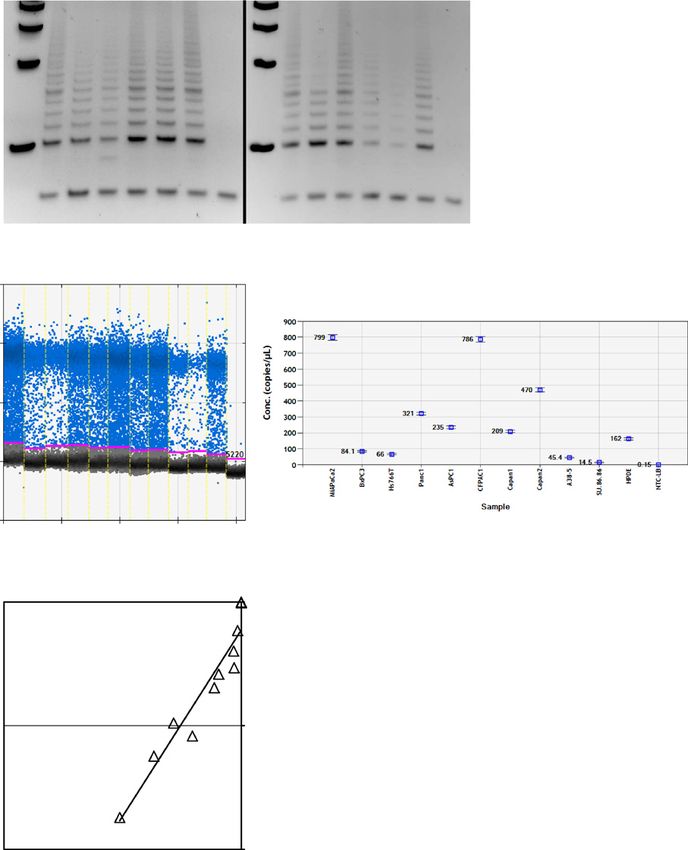
A M 1 2 3 4 5 6 N M 1 7 8 9 10 11 N 1 MIA PaCa-2
200 2 BxPC-3
150 3 Hs 766T
4 PANC-1
100 5 AsPC-1
6 CFPAC-1
7 Capan-1
8 Capan-2
50 9 A38-5
10 SU.86.86
IC 11 HPDE
N NTC-LB
B C
1 2 3 4 5 6 7 8 9 10 11 N
20,000
Fluorescence amplitude
15,000
10,000
5,000
0
0 50,000 100,000 150,000 200,000
Event number
D
1.00
1
6
Relative telomerase activity (dd-TRAP)
8
R² = 0.915
4
7
5
11
2
0.10
3
9
10
0.01
0.01 0.10 1.00
Relative telomerase activity (gel-TRAP)
Figure 1.
A and B, representative gel image and 1-D plot graph of gel-TRAP assay (A) and dd-TRAP assay (B) using pancreatic cancer cell lines. C, concentration of telomerase
products determined by dd-TRAP assay. Error bar, Poisson 95% confidence limits. D, correlation of telomerase activity level by gel-TRAP and dd-TRAP
assays in 10 pancreatic cancer cell lines and HPDE-immortalized pancreatic ductal epithelial cell line. Relative telomerase activity was calculated using the level in MIA
PaCa-2 cells set at 1.0 for reference. R2 means coefficient of determination. N, nontemplate control lysis buffer.
www.aacrjournals.org Clin Cancer Res; 22(20) October 15, 2016 5145
Downloaded from clincancerres.aacrjournals.org on January 26, 2021. © 2016 American Association for Cancer Research.Published OnlineFirst May 26, 2016; DOI: 10.1158/1078-0432.CCR-16-0311
Hata et al.
Table 1. Patient and cyst characteristics (patients whose cyst fluid samples had not undergone prior thawing)
Total IPMN MCN SCN PanNET Pseudocyst Simple cyst
Characteristics (n ¼ 119) (n ¼ 74) (n ¼ 10) (n ¼ 25) (n ¼ 4) (n ¼ 5) (n ¼ 1)
Male/female (n) 55/64 42/32 0/10 6/19 3/1 4/1 0/1
Age (median, range), y 65 (29–87) 69 (42–87) 52 (30–61) 63 (29–77) 57 (43–75) 73 (54–82) 64
Symptoms (n)
Abdominal pain 20 11 2 5 0 2 0
Pancreatitis 14 12 1 1 0 0 0
Jaundice 1 1 0 0 0 0 0
Cyst location (n)
Head and uncinate/body and tail 54/65 40/34 0/10 11/14 1/3 2/3 0/1
Cyst size, median (range), cm 2.5 (0.5–11.5) 2.0 (0.5–9.0) 2.6 (1.6–7.0) 2.5 (1.5–10.0) 5.5 (2.5–7.6) 3.8 (2.5–11.5) 1.5
Mural nodule (n)a
Absent/present 76/43 45/29 10/0 16/9 0/4 4/1 1/0
Communication with MPD (n)a
Absent/present 73/46 33/41 10/0 20/5 4/0 5/0 1/0
Dilatation of MPD 10 mm (n)a
Absent/present 105/14 60/14 10/0 25/0 4/0 5/0 1/0
Dilatation of MPD 5 mm (n)a
Absent/present 84/35 42/32 10/0 24/1 3/1 4/1 1/0
CT/MRI findings (n)
Worrisome features 72 42 5 16 4 5 0
High-risk stigmata 21 19 0 2 0 0 0
No concerning features 26 13 5 7 0 0 1
Cyst fluid color (n)
Bloody/sero-bloody/brown/straw/clear 26/51/13/5/24 17/35/4/3/15 0/2/2/0/6 7/12/4/0/2 2/1/1/0/0 0/1/2/2/0 0/0/0/0/1
Cyst fluid appearance (n)
Serous/mucinous 63/56 23/51 9/1 23/2 3/1 4/1 1/0
Original cyst volume (median, range), mL 139 (10–1,000) 92.5 (10–800) 250 (50–600) 250 (120–1,000) 125 (80–200) 50 (50–200) 400
EUS cytology (n ¼ 45), n
Nondiagnostic 6 2 1 3 0 0 0
Benign/atypia/cancer 20/9/10 12/8/10 3/0/0 4/1/0 1/0/0 0/0/0 0/0/0
Cyst fluid CEA (n ¼ 23), nPublished OnlineFirst May 26, 2016; DOI: 10.1158/1078-0432.CCR-16-0311
Telomerase Activity in Pancreatic Cyst Fluid
A ***
***
100,000
***
10,000
(copies/µL cyst fluid)
Telomerase product
1,000
100
10
1
0.1
0
IPMN IPMN IPMN IPMN MCN MCN PanNET SCN Pseudocyst Simple
Cancer HGD IGD LGD IGD LGD cyst
B C n Median IQR
***
*** IPMN
100,000
*** Cancer 11 1055 (56.7–17345)
HGD 20 1315 (830.2–11980)
IGD 36 31.19 (3.303–452.2)
10,000
LGD 7 109.8 (1.76–246.5)
(copies/µL cyst fluid)
Telomerase product
1,000
MCN
100 IGD 3 7.9 (2.04–50.68)
LGD 7 7.36 (1.98–37.59)
PanNET 4 377.3 (110.2–511)
10
SCN 25 9.2 (2.97–46.03)
Pseudocyst 5 5.97 (0–186)
1
Simple cyst 1 3.84
0.1
IPMN Cancer, HGD 31 1158 (295.9–13033)
0 IPMN IGD, LGD 43 70.68 (3.25–425.9)
IPMN Cancer IPMN IGD MCN IGD SCN MCN IGD, LGD 10 7.63 (2.025–40.86)
IPMN HGD IPMN LGD MCN LGD
D Cancer and HGD vs. others
E IPMN Cancer and HGD vs. others
100 100
80 80
Sensitivity%
Sensitivity%
60 60
40 40
20 AUC 0.893 20 AUC 0.849
SE 0.0334 SE 0.0448
95% CI 0.827–0.959 95% CI 0.761–0.936
0 0
0 20 40 60 80 100 0 20 40 60 80 100
100%–Specificity% 100%–Specificity%
Figure 2.
A, absolute quantification of telomerase activity per microliter of original cyst fluid samples among 119 samples from the discovery and validation set that
had not undergone any prior thawing. The longer horizontal bar represents the median value and shorter ones represent values of the 75th and 25th percentiles,
respectively. B, comparison of telomerase activity of IPMN cases classified by their surgical indication (invasive cancer and HGD vs. IGD and LGD) and MCN
and SCN cases. C, telomerase activity levels per microliter of cyst fluid samples in each group. D and E, ROC curve analysis for the diagnostic accuracy of telomerase
activity in predicting malignancy among all 119 cases (D) and 74 IPMN cases (E). HGD, high-grade dysplasia; IGD, intermediate-grade dysplasia; LGD,
low-grade dysplasia; MCN, mucinous cystic neoplasm; SCN, serous cystic neoplasm; PanNET, pancreatic neuroendocrine tumor; IQR, interquartile range; AUC,
area under the curve; SE, standard error; CI, confidence interval. N.S., not significant; , P < 0.001.
www.aacrjournals.org Clin Cancer Res; 22(20) October 15, 2016 5147
Downloaded from clincancerres.aacrjournals.org on January 26, 2021. © 2016 American Association for Cancer Research.Published OnlineFirst May 26, 2016; DOI: 10.1158/1078-0432.CCR-16-0311
Hata et al.
Table 2. The diagnostic accuracy of telomerase activity, imaging, and clinical factors for predicting high-grade dysplasia/invasive cancer
Findings Cutoff Sensitivity (%; 95% CI) Specificity (%; 95% CI) Accuracy (%) PPV (%) NPV (%)
All cases (n ¼ 119)a
Cyst appearance Mucinous 83.9 (66.3–94.6) 63.6 (52.7–73.6) 68.9 44.8 91.8
Cyst size 30 mm 32.3 (16.7–51.4) 64.8 (53.9–74.7) 56.3 24.4 73.1
MPD dilatation 10 mm 25.8 (11.9–44.6) 93.2 (85.8–97.5) 84.0 57.1 78.1
MPD dilatation 5 mm 58.1 (39.1–75.5) 80.7 (70.9–88.3) 74.8 51.4 84.5
Mural nodule Present 58.1 (39.1–75.5) 71.6 (61.0–80.7) 68.1 41.9 82.9
Telomerase activity 730 copies/mL cyst fluid 74.2 (55.4–82.1) 93.2 (85.8–97.5) 88.2 79.3 91.1
IPMN cases (n ¼ 74)
Cyst appearance Mucinous 83.9 (66.3–94.6) 41.9 (27.0–57.9) 59.5 51.0 78.3
Cyst size 30 mm 32.3 (16.7–51.4) 24.4 (58.8–86.5) 56.8 47.6 60.4
MPD dilatation 10 mm 25.8 (11.9–44.6) 86.1 (72.1–94.7) 60.8 57.1 61.7
MPD dilatation 5 mm 58.1 (39.1–75.5) 67.4 (51.5–80.9) 63.5 56.3 69.1
Mural nodule Present 58.6 (39.1–75.5) 74.4 (58.8–86.5) 67.6 62.1 71.1
Telomerase activity 730 copies/mL cyst fluid 74.2 (55.4–88.1) 86.1 (72.1–94.7) 81.1 79.3 82.2
Abbreviations: CI, confidence interval; MPD, main pancreatic duct; NPV, negative predictive value; PPV, positive predictive value.
a
Discovery and validation sets combined (samples without prior thawing).
cut-off value, determined by the discovery set results, the series were factors associated with having an IPMN). Table 4
diagnostic performance for predicting the presence of high- shows that the higher diagnostic performance in the subgroup
grade dysplasia/invasive cancer was somewhat higher when all of patients whose preoperative evaluation was "worrisome
119 cases were considered (AUC, 0.893; sensitivity, 74.2%; features" was similar in the combined (discovery and valida-
specificity, 93.2%, true-positives 23, false-positives 6, false- tion) set (AUC, 0.900; sensitivity, 73.7; specificity, 90.6%;
negatives 8, true-negatives 82; Fig. 2D; Table 2) than among accuracy, 86.1%) as it was in the discovery set (AUC, 0.927;
the 74 cases with IPMN (AUC, 0.849; sensitivity, 74.2%; Supplementary Table S5).
specificity, 86.1%, true-positives 23, false-positives 6, false- We also performed telomerase activity analysis on pancreatic
negatives 8, true-negatives, 37; Fig. 2E; Table 2). Table 3 shows samples obtained by EUS-FNA preoperatively from 36 cases
that the level of telomerase activity as a predictor of malignancy who underwent subsequent pancreatic resection. A summary of
(invasive cancer/HGD) was independent of other factors asso- the endoscopic findings, cyst fluid analyses, and pathologic
ciated with malignancy by multivariate analysis (which in this diagnoses for these cases are provided in Supplementary Table
Table 3. Univariate and multivariate analyses of factors predictive of invasive cancer or high-grade dysplasia in pancreatic cysts among the combined discovery and
validation setsa
Cancer, HGD Others Univariate Multivariate
Variable (n ¼ 31) (n ¼ 88) P OR (95% CI) P
AgePublished OnlineFirst May 26, 2016; DOI: 10.1158/1078-0432.CCR-16-0311
Telomerase Activity in Pancreatic Cyst Fluid
Table 4. Subgroup analysis of diagnostic performance of telomerase activity among 119 merged cases without prior thawing
Sensitivity Specificity Accuracy PPV NPV
Subgroup n AUC Cutoffa TP FN FP TN (%; 95% CI) (%; 95% CI) (%) (%) (%)
All cases (n ¼ 119)
High-risk stigmata 21 0.796 730 9 3 1 8 75.0 (42.8–94.5) 88.9 (51.8–99.7) 81.0 90.0 72.7
Worrisome features 72 0.900 730 14 5 5 48 73.7 (48.8–90.9) 90.6 (79.3–96.9) 86.1 73.7 90.6
IPMN cases (n ¼ 74)
High-risk stigmata 19 0.774 730 9 3 1 6 75.0 (42.8–94.5) 85.7 (42.1–99.6) 78.9 90.0 66.7
Worrisome features 42 0.842 730 14 5 5 18 73.7 (48.8–90.9) 78.3 (56.3–92.5) 76.2 73.7 78.3
Abbreviations: AUC, area under the curve; FN, false negative; FP, false positive; NPV, negative predictive value; PPV, positive predictive value; TN, true negative;
TP, true positive.
a
Value of telomerase activity in pancreatic cyst fluid (copies/mL of cyst fluid).
S6. In these samples, the telomerase activity levels correlated to further evaluate its diagnostic performance. Our study
well with those found in the surgical cyst fluid samples (Sup- population consisted of patients who underwent resection of
plementary Fig. S15). their pancreatic cysts and the most important clinical setting in
which pancreatic cyst fluid markers such as telomerase activity
should be evaluated is when surgical resection is being con-
Discussion sidered, but it will also be valuable to know if cyst fluid
In the current study, we demonstrate that measurement of markers can help evaluate the risk of future neoplastic pro-
telomerase activity in pancreatic cyst fluid samples using dd-TRAP gression of pancreatic cysts that do not currently meet indica-
adds diagnostic utility for predicting whether or not a pancreatic tions for surgical resection or in patients whose pancreatic cysts
cystic lesion harbors high-grade dysplasia and/or an associated do meet criteria for surgical resection but elect to undergo
invasive cancer. The need to determine the neoplastic grade of a surveillance. The diagnostic utility of telomerase activity might
pancreatic cyst is usually the most important question clinicians also be strengthened by combining the results of cyst fluid
need to answer when evaluating patients with pancreatic cysts and analysis obtained at multiple time points during patient
a pancreatic cyst fluid test that could do this reliably would likely surveillance.
have high clinical utility. In conclusion, quantification of pancreatic cyst fluid telomerase
The diagnostic utility of cyst fluid telomerase activity levels activity using dd-TRAP can accurately predict the presence or
for distinguishing samples obtained from cystic neoplasms absence of invasive cancer/high-grade dysplasia.
with HGD invasive cancer versus cysts with LGD compares
favorably to other clinically used measures such as cyst fluid Disclosure of Potential Conflicts of Interest
cytology and cyst fluid CEA, which have lower diagnostic A.M. Lennon is a consultant/advisory board member for NovoNordisc and
accuracy (14). We recently evaluated a panel of genetic bio- Olympus. R.H. Hruban is an employee of MiDiagnostics and reports receiving
markers in cyst fluid and found that a combination of molec- royalty payments from Myriad Genetics for the PALB2 invention in a relation-
ular markers had excellent accuracy for classifying pancreatic ship supervised by Johns Hopkins University. No potential conflicts of interest
were disclosed by the other authors.
cysts and for predicting their grade of dysplasia (15, 46).
Telomerase activity measured by dd-TRAP had similar accuracy
(AUCcombined ¼ 0.849) to our recently reported molecular Authors' Contributions
marker model for the predicting the histologic grade of IPMNs Conception and design: T. Hata, M. Weiss, M.I. Canto, M. Goggins
Development of methodology: T. Hata, M. Suenaga, M. Goggins
(invasive cancer/high-grade dysplasia vs. intermediate-grade Acquisition of data (provided animals, acquired and managed patients,
dysplasia or low-grade dysplasia). Given the high prevalence provided facilities, etc.): T. Hata, J. Yu, M. Pittman, M. Weiss, M.I. Canto,
of asymptomatic cysts in the general population and in high- C. Wolfgang, A. Marie Lennon, R.H. Hruban, M. Goggins
risk groups, the ability to reliably stratify pancreatic cysts into Analysis and interpretation of data (e.g., statistical analysis, biostatistics,
high-risk cysts (requiring surgery or close surveillance) versus computational analysis): T. Hata, M.D. Molin, M.I. Canto, C. Wolfgang,
low-risk cysts (less frequent surveillance) is vital for optimal M. Goggins
Writing, review, and/or revision of the manuscript: T. Hata, M.D. Molin, J. Yu,
patient management (47–50). M. Weiss, M.I. Canto, C. Wolfgang, A. Marie Lennon, R.H. Hruban, M. Goggins
The diagnostic performance of telomerase activity was similarly Administrative, technical, or material support (i.e., reporting or organizing
high in cases whose pancreatic cyst evaluation was classified as data, constructing databases): M.D. Molin, R.H. Hruban
having "worrisome features" (AUCcombined ¼ 0.842). This is the Study supervision: M.I. Canto, M. Goggins
group that most needs pancreatic cyst fluid markers since cysts
considered low-risk are not recommended to have FNA and cysts Grant Support
with high-risk stigmata are generally recommended for surgical This work was supported by Susan Wojcicki and Dennis Troper, NIH grants
resection without further examination (3). (CA62924 and R01CA176828), the Lustgarten Foundation for Pancreatic
Although this study had the advantage that all patients Cancer Research, the Pancreatic Cancer Action Network, the Rolfe Pancreatic
Cancer Foundation and Hugh and Rachel Victor.
underwent surgical resection and therefore had defined histol-
The costs of publication of this article were defrayed in part by the payment of
ogy, most of the samples analyzed in this study were obtained page charges. This article must therefore be hereby marked advertisement in
from surgical resection specimens. Prospective validation stud- accordance with 18 U.S.C. Section 1734 solely to indicate this fact.
ies measuring telomerase activity in larger numbers of EUS-
FNA cyst fluid samples, particularly those from patients whose Received February 4, 2016; revised April 26, 2016; accepted May 10, 2016;
cysts pose diagnostic and management dilemmas, are needed published OnlineFirst May 26, 2016.
www.aacrjournals.org Clin Cancer Res; 22(20) October 15, 2016 5149
Downloaded from clincancerres.aacrjournals.org on January 26, 2021. © 2016 American Association for Cancer Research.Published OnlineFirst May 26, 2016; DOI: 10.1158/1078-0432.CCR-16-0311
Hata et al.
References
1. Brugge WR. Diagnosis and management of cystic lesions of the pancreas. 19. Cao Z, Maupin K, Curnutte B, Fallon B, Feasley CL, Brouhard E, et al.
J Gastrointest Oncol 2015;6:375–88. Specific glycoforms of MUC5AC and endorepellin accurately distinguish
2. Lennon AM, Manos LL, Hruban RH, Ali SZ, Fishman EK, Kamel IR, et al. mucinous from nonmucinous pancreatic cysts. Mol Cell Proteomics
Role of a multidisciplinary clinic in the management of patients with 2013;12:2724–34.
pancreatic cysts: a single-center cohort study. Ann Surg Oncol 2014;21: 20. Hong SM, Omura N, Vincent A, Li A, Knight S, Yu J, et al. Genome-wide
3668–74. CpG island profiling of intraductal papillary mucinous neoplasms of the
3. Tanaka M, Fernandez-del Castillo C, Adsay V, Chari S, Falconi M, Jang JY, pancreas. Clin Cancer Res 2012;18:700–12.
et al. International consensus guidelines 2012 for the management of 21. Das KK, Xiao H, Geng X, Fernandez-Del-Castillo C, Morales-Oyarvide V,
IPMN and MCN of the pancreas. Pancreatology 2012;12:183–97. Daglilar E, et al. mAb Das-1 is specific for high-risk and malignant
4. Lennon AM, Wolfgang C. Cystic neoplasms of the pancreas. J Gastrointest intraductal papillary mucinous neoplasm (IPMN). Gut 2014;63:1626–34.
Surg 2013;17:645–53. 22. Balcom JHt, Keck T, Warshaw AL, Antoniu B, Graeme-Cook F, Fernandez-
5. Jais B, Rebours V, Malleo G, Salvia R, Fontana M, Maggino L, et al. Serous del Castillo C. Telomerase activity in periampullary tumors correlates with
cystic neoplasm of the pancreas: a multinational study of 2622 patients aggressive malignancy. Ann Surg 2001;234:344–50.
under the auspices of the international association of pancreatology and 23. Maser RS, DePinho RA. Connecting chromosomes, crisis, and cancer.
european pancreatic club (european study group on cystic tumors of the Science 2002;297:565–9.
pancreas). Gut 2016;65:305–12. 24. Shay JW. Role of telomeres and telomerase in aging and cancer. Cancer
6. Ridtitid W, DeWitt JM, Schmidt CM, Roch A, Stuart JS, Sherman S, et al. Discov. 2016Mar 30. [Epub ahead of print].
Management of branch-duct intraductal papillary mucinous neoplasms: 25. Kim NW, Piatyszek MA, Prowse KR, Harley CB, West MD, Ho PL, et al.
a large single-center study to assess predictors of malignancy and long- Specific association of human telomerase activity with immortal cells and
term outcomes. Gastrointest Endosc 2016 Feb 18. [Epub ahead of cancer. Science 1994;266:2011–5.
print]. 26. Kim NW, Wu F. Advances in quantification and characterization of telo-
7. Valsangkar NP, Morales-Oyarvide V, Thayer SP, Ferrone CR, Wargo JA, merase activity by the telomeric repeat amplification protocol (TRAP).
Warshaw AL, et al. 851 resected cystic tumors of the pancreas: a 33-year Nucleic Acids Res 1997;25:2595–7.
experience at the Massachusetts General Hospital. Surgery 2012;152: 27. Herbert BS, Hochreiter AE, Wright WE, Shay JW. Nonradioactive detection
S4–12. of telomerase activity using the telomeric repeat amplification protocol.
8. Sahora K, Mino-Kenudson M, Brugge W, Thayer SP, Ferrone CR, Sahani D, Nat Protoc 2006;1:1583–90.
et al. Branch duct intraductal papillary mucinous neoplasms: does cyst size 28. Krupp G, Kuhne K, Tamm S, Klapper W, Heidorn K, Rott A, et al. Molecular
change the tip of the scale? A critical analysis of the revised international basis of artifacts in the detection of telomerase activity and a modified
consensus guidelines in a large single-institutional series. Ann Surg primer for a more robust `TRAP' assay. Nucleic Acids Res 1997;25:919–21.
2013;258:466–75. 29. Yeh TS, Cheng AJ, Chen TC, Jan YY, Hwang TL, Jeng LB, et al. Telomerase
9. Crippa S, Bassi C, Salvia R, Malleo G, Marchegiani G, Rebours V, et al. Low activity is a useful marker to distinguish malignant pancreatic cystic tumors
progression of intraductal papillary mucinous neoplasms with worrisome from benign neoplasms and pseudocysts. J Surg Res 1999;87:171–7.
features and high-risk stigmata undergoing non-operative management: a 30. Saji M, Xydas S, Westra WH, Liang CK, Clark DP, Udelsman R, et al. Human
mid-term follow-up analysis. Gut 2016 Jan 7. [Epub ahead of print]. telomerase reverse transcriptase (hTERT) gene expression in thyroid neo-
10. Yu J, Blackford A, Dal Molin M, Wolfgang C, Goggins M. Time to plasms [In Process Citation]. Clin Cancer Res 1999;5:1483–9.
progression of pancreatic ductal adenocarcinoma from low-to-high 31. Ohuchida K, Mizumoto K, Ogura Y, Ishikawa N, Nagai E, Yamaguchi K,
tumour stages. Gut 2015;64:1783–89. et al. Quantitative assessment of telomerase activity and human telomerase
11. Winter JM, Jiang W, Basturk O, Mino-Kenudson M, Fong ZV, Tan WP, et al. reverse transcriptase messenger RNA levels in pancreatic juice samples for
Recurrence and survival after resection of small intraductal papillary the diagnosis of pancreatic cancer. Clin Cancer Res 2005;11:2285–92.
mucinous neoplasm-associated carcinomas (less than or equal to 20 mm 32. Hashimoto Y, Murakami Y, Uemura K, Hayashidani Y, Sudo T, Ohge H,
invasive component): a multi-institutional analysis. Ann Surg 2016;263: et al. Detection of human telomerase reverse transcriptase (hTERT) expres-
793–801. sion in tissue and pancreatic juice from pancreatic cancer. Surgery
12. Woolf KM, Liang H, Sletten ZJ, Russell DK, Bonfiglio TA, Zhou Z. False- 2008;143:113–25.
negative rate of endoscopic ultrasound-guided fine-needle aspiration for 33. Nakashima A, Murakami Y, Uemura K, Hayashidani Y, Sudo T, Hashimoto
pancreatic solid and cystic lesions with matched surgical resections as the Y, et al. Usefulness of human telomerase reverse transcriptase in pancreatic
gold standard: one institution's experience. Cancer Cytopathol juice as a biomarker of pancreatic malignancy. Pancreas 2009;38:527–33.
2013;121:449–58. 34. Hashimoto Y, Murakami Y, Uemura K, Hayashidani Y, Sudo T, Ohge H,
13. Thornton GD, McPhail MJ, Nayagam S, Hewitt MJ, Vlavianos P, Monahan et al. Telomere shortening and telomerase expression during multistage
KJ. Endoscopic ultrasound guided fine needle aspiration for the diagnosis carcinogenesis of intraductal papillary mucinous neoplasms of the pan-
of pancreatic cystic neoplasms: a meta-analysis. Pancreatology 2013;13: creas. J Gastrointest Surg 2008;12:17–28.
48–57. 35. Jakupciak JP.Real-time telomerase activity measurements for detection of
14. Maker AV, Carrara S, Jamieson NB, Pelaez-Luna M, Lennon AM, Dal Molin cancer. Expert Rev Mol Diagn 2005;5:745–53.
M, et al. Cyst fluid biomarkers for intraductal papillary mucinous neo- 36. Shim WY, Park KH, Jeung HC, Kim YT, Kim TS, Hyung WJ, et al. Quan-
plasms of the pancreas: a critical review from the international expert titative detection of telomerase activity by real-time TRAP assay in the body
meeting on pancreatic branch-duct-intraductal papillary mucinous neo- fluids of cancer patients. Int J Mol Med 2005;16:857–63.
plasms. J Am Coll Surg 2015;220:243–53. 37. Hou M, Xu D, Bjorkholm M, Gruber A. Real-time quantitative telomeric
15. Springer S, Wang Y, Molin MD, Masica DL, Jiao Y, Kinde I, et al. A repeat amplification protocol assay for the detection of telomerase activity.
combination of molecular markers and clinical features improve the Clin Chem 2001;47:519–24.
classification of pancreatic cysts. Gastroenterology 2015;4:01067–7. 38. Wege H, Chui MS, Le HT, Tran JM, Zern MA. SYBR Green real-time
16. DiMaio CJ, Weis-Garcia F, Bagiella E, Tang LH, Allen PJ. Pancreatic cyst telomeric repeat amplification protocol for the rapid quantification of
fluid concentration of high-mobility group A2 protein acts as a differential telomerase activity. Nucleic Acids Res 2003;31:E3.
biomarker of dysplasia in intraductal papillary mucinous neoplasm. 39. Ludlow AT, Robin JD, Sayed M, Litterst CM, Shelton DN, Shay JW, et al.
Gastrointest Endosc. 2016;83:1205–9. Quantitative telomerase enzyme activity determination using droplet
17. Yip-Schneider MT, Wu H, Dumas RP, Hancock BA, Agaram N, Radovich M, digital PCR with single cell resolution. Nucleic Acids Res 2014;42:e104.
et al. Vascular endothelial growth factor, a novel and highly accurate 40. Pinheiro LB, Coleman VA, Hindson CM, Herrmann J, Hindson BJ, Bhat S,
pancreatic fluid biomarker for serous pancreatic cysts. J Am Coll Surg et al. Evaluation of a droplet digital polymerase chain reaction format for
2014;218:608–17. DNA copy number quantification. Anal Chem 2012;84:1003–11.
18. Jabbar KS, Verbeke C, Hyltander AG, Sjovall H, Hansson GC, Sadik R. 41. Hindson BJ, Ness KD, Masquelier DA, Belgrader P, Heredia NJ, Makarewicz
Proteomic mucin profiling for the identification of cystic precursors of AJ, et al. High-throughput droplet digital PCR system for absolute quan-
pancreatic cancer. J Natl Cancer Inst 2014;106:djt439. titation of DNA copy number. Anal Chem 2011;83:8604–10.
5150 Clin Cancer Res; 22(20) October 15, 2016 Clinical Cancer Research
Downloaded from clincancerres.aacrjournals.org on January 26, 2021. © 2016 American Association for Cancer Research.Published OnlineFirst May 26, 2016; DOI: 10.1158/1078-0432.CCR-16-0311
Telomerase Activity in Pancreatic Cyst Fluid
42. Dube S, Qin J, Ramakrishnan R. Mathematical analysis of copy number profiles of intraductal papillary neoplasms of the pancreas. J Pathol
variation in a DNA sample using digital PCR on a nanofluidic device. PLoS 2014;233:217–27.
ONE 2008;3:e2876. 47. Canto MI, Hruban RH, Fishman EK, Kamel IR, Schulick R, Zhang Z, et al.
43. Brugge WR, Lewandrowski K, Lee-Lewandrowski E, Centeno BA, Szydlo Frequent detection of pancreatic lesions in asymptomatic high-risk indi-
T, Regan S, et al. Diagnosis of pancreatic cystic neoplasms: a report of viduals. Gastroenterology 2012;142:796–804.
the cooperative pancreatic cyst study. Gastroenterology 2004;126: 48. de Jong K, Nio CY, Hermans JJ, Dijkgraaf MG, Gouma DJ, van Eijck CH,
1330–6. et al. High prevalence of pancreatic cysts detected by screening magnetic
44. Sato N, Maehara N, Mizumoto K, Nagai E, Yasoshima T, Hirata K, et al. resonance imaging examinations. Clin Gastroenterol Hepatol 2010;
Telomerase activity of cultured human pancreatic carcinoma cell lines 8:806–11.
correlates with their potential for migration and invasion. Cancer 49. Vege SS, Ziring B, Jain R, Moayyedi P. American gastroenterological
2001;91:496–504. association institute guideline on the diagnosis and management of
45. Ouyang H, Mou L, Luk C, Liu N, Karaskova J, Squire J, et al. Immortal asymptomatic neoplastic pancreatic cysts. Gastroenterology 2015;148:
human pancreatic duct epithelial cell lines with near normal genotype and 819–22.
phenotype. Am J Pathol 2000;157:1623–31. 50. Scheiman JM, Hwang JH, Moayyedi P. American gastroenterological asso-
46. Amato E, Molin MD, Mafficini A, Yu J, Malleo G, Rusev B, et al. Targeted ciation technical review on the diagnosis and management of asymptom-
next-generation sequencing of cancer genes dissects the molecular atic neoplastic pancreatic cysts. Gastroenterology 2015;148:824–48.
www.aacrjournals.org Clin Cancer Res; 22(20) October 15, 2016 5151
Downloaded from clincancerres.aacrjournals.org on January 26, 2021. © 2016 American Association for Cancer Research.Published OnlineFirst May 26, 2016; DOI: 10.1158/1078-0432.CCR-16-0311
Cyst Fluid Telomerase Activity Predicts the Histologic Grade of
Cystic Neoplasms of the Pancreas
Tatsuo Hata, Marco Dal Molin, Masaya Suenaga, et al.
Clin Cancer Res 2016;22:5141-5151. Published OnlineFirst May 26, 2016.
Updated version Access the most recent version of this article at:
doi:10.1158/1078-0432.CCR-16-0311
Supplementary Access the most recent supplemental material at:
Material http://clincancerres.aacrjournals.org/content/suppl/2016/12/20/1078-0432.CCR-16-0311.DC1
Cited articles This article cites 47 articles, 10 of which you can access for free at:
http://clincancerres.aacrjournals.org/content/22/20/5141.full#ref-list-1
Citing articles This article has been cited by 4 HighWire-hosted articles. Access the articles at:
http://clincancerres.aacrjournals.org/content/22/20/5141.full#related-urls
E-mail alerts Sign up to receive free email-alerts related to this article or journal.
Reprints and To order reprints of this article or to subscribe to the journal, contact the AACR Publications Department at
Subscriptions pubs@aacr.org.
Permissions To request permission to re-use all or part of this article, use this link
http://clincancerres.aacrjournals.org/content/22/20/5141.
Click on "Request Permissions" which will take you to the Copyright Clearance Center's (CCC)
Rightslink site.
Downloaded from clincancerres.aacrjournals.org on January 26, 2021. © 2016 American Association for Cancer Research.You can also read