Mouth cancer: presentation, detection and referral in primary dental care - Exodontia.Info
←
→
Page content transcription
If your browser does not render page correctly, please read the page content below
Mouth Cancer Themed Issue
                                                                                                    Mouth cancer                     CLINICAL

    Mouth cancer: presentation, detection and referral in
    primary dental care
    M. A. O. Lewis1

    Key points
    Discusses the highly variable way               Emphasises that the detection of         Provides information on diagnostic      Outlines the guidance on the
    in which mouth cancer can present               mouth cancer while the tumour is less    aids for mouth cancer and potentially   appropriate referral of urgent
    clinically and as such any persistent           than 2 cm in diameter is the single      malignant mucosal disorders.            suspected cancer.
    mucosal abnormality should be viewed            most important factor that can improve
    with suspicion.                                 patient outcome.

Mouth cancer can present as a variety of abnormalities and visible changes affecting the oral mucosa, including ulceration,
swelling and areas of erythema. The five-year survival from mouth cancer is poor at approximately 50%. Detection of the
cancer while less than 2 cm in diameter with no metastasis greatly improves the outcome for the patient. Although many
cancers in the mouth develop from what was previously an apparently normal mucosa, some arise in pre-existing conditions
that are therefore regarded as potentially malignant. Regular assessment of the soft tissues within the mouth and the
neck for the presence of abnormalities is an essential component of primary dental care. Any persistent and unexplained
abnormality requires referral for definitive diagnosis and specialist management.

Introduction                                                    topic for continuing professional develop-               one centre. In reality the five-year survival
                                                                ment is the poor five-year survival, which               from advanced cancer is usually much shorter
Certain conditions that affect the oral mucosa                  overall is approximately 55%.1 The single most           than 13 years.
have characteristic clinical signs and symptoms                 important factor that can improve five-year                 It is important to appreciate that velocity of
that allow diagnosis on the basis of clinical                   survival is detection of the tumour whilst               cancer growth in the mouth is not uniform.
presentation alone without the need for special                 small, specifically 2 cm or less in diameter             Some tumours may slowly increase in size
investigations. Examples of these would be the                  with no regional node involvement or distant             over many months while others enlarge rapidly
distinctive history and appearance of minor                     metastases (Stage I). Patients with a tumour             over a few weeks. On this basis the concept
recurrent aphthous stomatitis and geographic                    detected at Stage I are associated with an 85%           of ‘early’ diagnosis is slightly misleading since
tongue. However, squamous cell carcinoma                        five-year survival compared to those with Stage          it is not actually the time that the cancer has
(mouth cancer) contrasts with this markedly                     IV (greater than 4 cm in diameter with regional          been present that is most important but the
in that it can present with a range of mucosal                  node involvement and possible distant metas-             size and presence of regional metastasis when
changes and symptoms. On this basis any                         tasis) for whom the five-year survival is only           the tumour is first detected that is key to an
persistent solitary mucosal abnormality should                  10%.1,2 The importance of detection while small          improved chance of survival for the patient.
be regarded as potentially sinister until proven                was also highlighted in a recent retrospective              It is useful to recognise that oral squamous
otherwise.                                                      study of survival after aggressive surgery which         cell carcinoma essentially represents epithelial
   One of the main reasons that that mouth                      revealed that a patient with a tumour that is            cell replication within the lining of the mouth
cancer is given such high importance in                         detected with a maximum diameter 2 cm                    that is ‘out of control’ and as such is a surface
dentistry and has been recommended as a                         (tumour stage T1) had a mean post-treatment              event which should be visible on clinical exam-
                                                                survival of 24.48 years (95% CI 22.45–26.50)             ination. This feature contrasts with some other
                                                                while a patient with a tumour greater than               tumours that are not ‘visible’, such as ovarian
1
 Professor of Oral Medicine, School of Dentistry, Cardiff
University, Heath Park, Cardiff, CF14 4XY                       4 cm (tumour stage T4) had a mean survival               cancer or pancreatic cancer. It is not unrea-
Correspondence to: Mike Lewis                                   of only 13.03 years (95% CI 11.56–14.49).3               sonable to expect a dental care professional to
Email: Lewismao@cardiff.ac.uk
                                                                These figures for survival are longer than               be able to detect a surface mucosal abnormal-
Refereed Paper. Accepted 4 October 2018                         those reported in studies and in part it must            ity that is 2 cm in diameter since this size is
DOI: 10.1038/sj.bdj.2018.931
                                                                be appreciated that the research involved only           approximately that of a human finger nail.

BRITISH DENTAL JOURNAL | VOLUME 225 NO. 9 | NOVEMBER 9 2018833
                                                                     O
                                                                     f
                                                                     f
                                                                     i
                                                                     c
                                                                     i
                                                                     a
                                                                     l
                                                                     j
                                                                     o
                                                                     u
                                                                     r
                                                                     n
                                                                     a
                                                                     l
                                                                     o
                                                                     f
                                                                     t
                                                                     h
                                                                     e
                                                                     B
                                                                     r
                                                                     i
                                                                     t
                                                                     i
                                                                     s
                                                                     h
                                                                     D
                                                                     e
                                                                     n
                                                                     t
                                                                     a
                                                                     l
                                                                     A
                                                                     s
                                                                     s
                                                                     o
                                                                     c
                                                                     i
                                                                     a
                                                                     t
                                                                     i
                                                                     o
                                                                     n
                                                                     .
CLINICAL Mouth cancer
Clinical presentation
The classical appearance of mouth cancer is a
solitary deep ulcer with rolled margins on the
lateral margin of the tongue (Fig. 1). Palpation
of the margin of the ulcer will reveal firm, often
described as indurated, tissues. Unfortunately
however, other than induration, mouth cancer
has no pathognomonic presenting feature
and involves a spectrum of mucosal changes
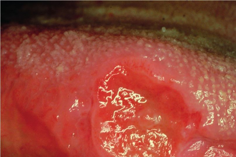
in addition to ulceration including swelling
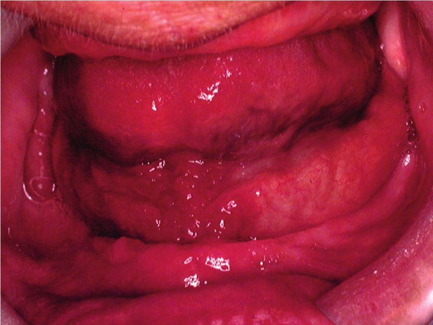
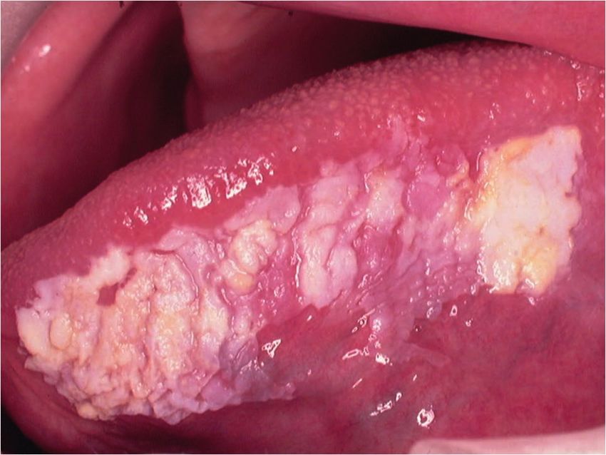
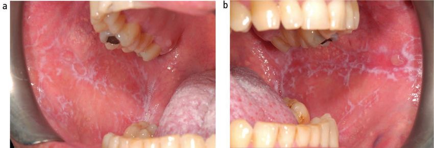
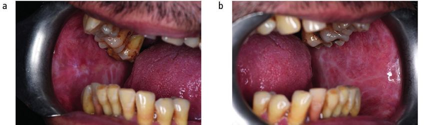
(Fig. 2), erythema (Fig. 3) or a speckled red/ Fig. 1 Squamous cell carcinoma (SCC) as a solitary deep ulcer with rolled margins with no
white patch (Fig. 4).4 obvious cause
Examination of the patient
A number of educational videos showing
methods of clinical examination of the mouth
and neck are freely available on the Internet
via providers such as YouTube or Google
video. However, the three minute ‘Oral Cancer
Recognition Toolkit’ video developed by
Cancer Research UK and the British Dental
Association is particularly useful for dental
professionals.5
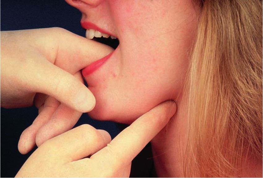
Extra-oral examination
The soft tissues of the neck should be palpated Fig. 2 SCC as mucosal swelling with speckled surface in left buccal mucosa
on both sides using the tips of the fingers to
detect any abnormal tissue swelling or enlarge-
ment of the lymph nodes. The patient’s neck
should be flexed and the examination start
in the submental region moving back to the
submandibular region, then down the jugular
chain to the supra-clavicular fossa.
Intra-oral examination
There is no correct order for examining the
oral soft tissues as long as that at the comple-
tion of the examination the entire mouth and
the oropharynx, including the tonsils has been
assessed. Good lighting is obviously essential.
Any mucosal abnormality noted should be Fig. 3 SCC as an erythematous mucosal patch in left oropharynx
palpated to determine consistency of the soft
tissue (Fig. 5). As a general rule, induration
reflects the presence of a benign or malignant detected. Small areas of mucosal change can areas of change, a clinical decision has to be
neoplasm while softness to palpation represents be removed with a surrounding margin of made as to what tissue to remove. As a rule
a non-neoplastic inflammatory condition. In clinically normal mucosa. There is divided the most suspicious looking area of mucosa
addition, mouth cancer is often painless until opinion on the complete removal of a small should be included in the biopsy material. The
well advanced while inflammatory conditions mucosal abnormality that is found to be cancer tissue specimen should therefore include any
are usually painful from the outset. since it may subsequently cause problems in aspects of ulceration, induration, erosion and
identifying the primary site. However, the vast erythema along with some apparently normal
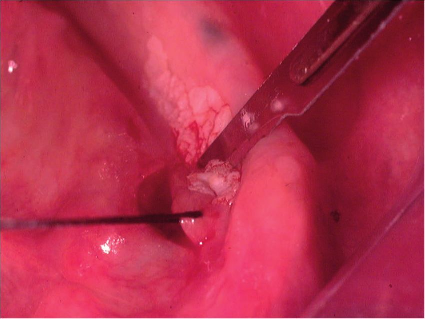
Mucosal biopsy majority of small localised mucosa lesions are adjacent mucosa.
A scalpel biopsy is the gold standard investiga- not squamous cell carcinoma and in the cases Following the placement of local anaesthesia,
tion for the diagnosis of any mucosal abnormal- where they are modern imaging techniques an incisional mucosal biopsy should involve the
ity. The biopsy may be excisional or incisional and clinical examination will indicate the taking of an ellipse of tissue from the affected
depending on the size of the abnormality diagnostic surgical site. In the case of larger site. It is helpful to firstly place a suture in the
834 BRITISH DENTAL JOURNAL | VOLUME 225 NO. 9 | NOVEMBER 9 2018
O
f
f
i
c
i
a
l
j
o
u
r
n
a
l
o
f
t
h
e
B
r
i
t
i
s
h
D
e
n
t
a
l
A
s
s
o
c
i
a
t
i
o
n
.
Mouth cancer CLINICAL
depends of the type of surgery being under-
taken. Although black silk, a non-resorbable
material, has historically been used for intra-
oral soft tissue closure, resorbable polyglactin
910 (a copolymer of 90% glycolide and 10%
L-lactide), is now the preferred material.
Sutures are available on a variety of needles
which differ in cross section, size and length.
A 19 mm curved reverse cutting needle, which
is triangular in cross section, is the needle of
choice for closure of the oral mucosa.
The biopsy tissue removed can be supported
on filter paper before placement in a pre-
labelled specimen pot containing 10% neutral
buffered formalin to minimise the impact
Fig. 4 SCC as a predominantly white mucosal patch on the left lateral margin of the tongue
of shrinkage and distortion during fixation
(Fig. 8). This step is important since curling
up of the tissue can cause cross sectioning
that may be mistaken for epithelial invasion in
tissue sections when examined microscopically
in two dimensions.
Punch biopsy
Punch biopsy, which removes a cylindrical
specimen of tissue of between 0.4–0.8 mm in
diameter, has been promoted as a simple and
quick method of sampling the oral mucosa.7
However, since the amount of tissue removed
is relatively small there is potential to not
obtain sufficient material when assessing the
epithelium for the presence of dysplasia or
Fig. 5 Bimanual palpation of soft tissues in the floor of mouth
invasion. On this basis some oral and maxillo-
facial pathologists have expressed a preference
for a scalpel biopsy rather than punch biopsy
(personal communication).
Potentially malignant conditions
A primary mouth cancer can either arise in
what was previously clinically normal tissue
or develop within a pre-existing mucosal
abnormally. Such pre-existing conditions that
are associated with an increased incidence
of undergoing malignant transformation are
referred to as being oral potentially malignant
disorders (OPMDs) (Box 1).
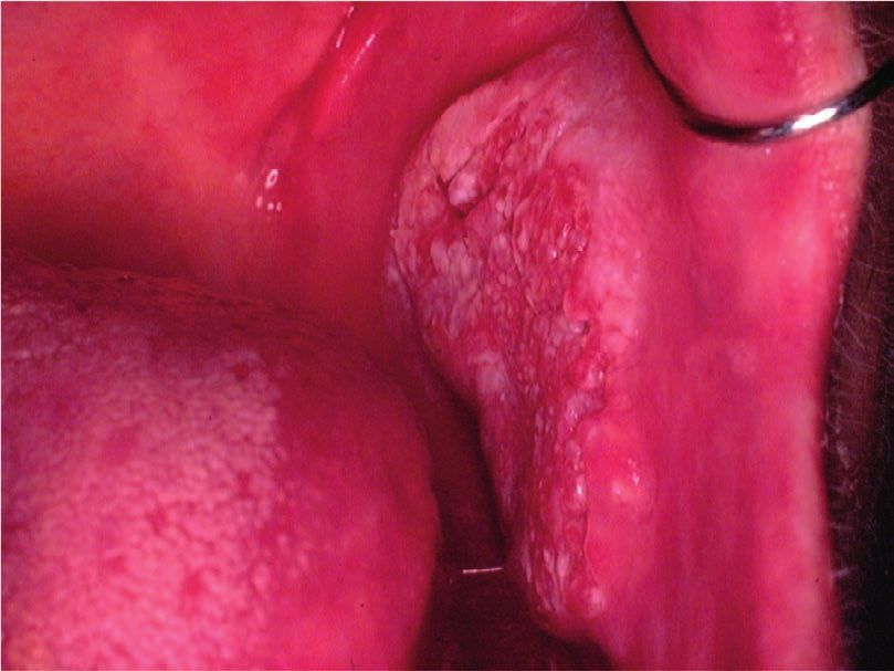
Leukoplakia
Fig. 6 Lower incision during an incisional biopsy of white patch in the floor of the mouth
The most frequently recognised OPMD is
leukoplakia (Fig. 9), which was first defined
tissue to be removed.6 This is preferable to the prevent bleeding potentially obscuring the site by the World Health Organisation (WHO) in
use of toothed-tissue tweezers since these may of the upper incision (Fig. 6). The tissue can 1978 as ‘a white patch or plaque that cannot
become dislodged and frequent re-application then be removed as a sheet of mucosa. Closure be characterised clinically or pathologically as
can cause crush damage to the biopsy material. of the surgical defect can be made with simple any other disease’.8 The term has subsequently
A number-15 blade with a rounded tip is the interrupted sutures using either a resorbable or been refined following various international
scalpel of choice for the majority forms of oral non-resorbable material (Fig. 7). A wide range workshops and is now used to describe ‘white
surgery. The lower incision is made first to of suture materials are available and their use plaques of questionable risk having excluded
BRITISH DENTAL JOURNAL | VOLUME 225 NO. 9 | NOVEMBER 9 2018835
O
f
f
i
c
i
a
l
j
o
u
r
n
a
l
o
f
t
h
e
B
r
i
t
i
s
h
D
e
n
t
a
l
A
s
s
o
c
i
a
t
i
o
n
.
CLINICAL Mouth cancer
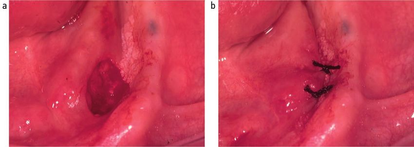
Fig. 7 Defect following removal of biopsy material (a). Closure of biopsy defect using simple Fig. 8 Biopsy tissue placed on filter paper
interrupted silk sutures (b) before placement into pathology specimen pot
Erythroplakia
Oral erythroplakia (Fig. 10) is defined as
‘a fiery red patch that cannot be character-
ised clinically or pathologically as any other
definable lesion’.15 Erythroplakia, in a similar
way to leukoplakia has no specific histopathol-
igical features. The term is used clinically to
record the presence of an erythematous
mucosal abnormality that does not have a
clinical appearance characteristic of known red
patches, such as denture-associated candidosis
or median rhomboid glossitis. Oral erythro-
plakia has the highest transformation rate of
all of the OPMDs, being reported as between
14–50%.16 Erythroleukoplakia is an alternative
Fig. 9 Leukoplakia on the left lateral margin of the tongue
clinical term that can be used when the mucosa
has a speckled red and white appearance.
(other) known diseases and disorders that and length of follow-up observation period. Importantly erythroleukoplakia is associated
carry no increased risk for cancer’.9 It is The global malignant transformation rate of with a high risk of malignant change.
essential to remember that leukoplakia is a leukoplakia is generally accepted to be 1.36%
clinical term and not a diagnosis. A biopsy is per year.13 Lichen planus
required to exclude known mucosal disorders. There is a rare form of leukoplakia, termed The aetiology of oral lichen planus (OLP) is
Although often associated with the presence proliferative verrucous carcinoma (PVL), unknown but does involve immune system
of epithelial dysplasia, leukoplakia itself has which has a reported transformation rate as since a primary histopathological feature is a
no specific histopathological features. The high as 70%.14 The presentation of PVL is char- sub-epithelial band of T-lymphocytes, indicat-
reported malignant transformation rate for acterised by an initial white patch that develops ing a cell-mediated reaction. OLP is one of the
oral leukoplakia has ranged from low levels of into multiple areas of exophytic/wart-like most frequently occurring mucosal conditions
0.13% in India10 and 2.6% in the UK11 to high changes within the mucosa. The aetiology of in the population, with a reported prevalence
levels of 17.5% in USA.12 These findings are PVL is unknown and treatment difficult due of between 0.5–2.2%.17 Different types of
undoubtedly influenced by the geographical to progressive recurrence following surgical OLP have been described, including reticular,
site, population studied, number of patients removal. atrophic, erosive, plaque-like and bullous,
based on the appearance of the mucosa.
However, actual typing in an individual patient
Box 1 Oral potentially malignant disorders of the oral mucosa9 is often difficult since different types may be
Leukoplakia present simultaneously and also change during
Erythroplakia the course of disease over months. Overall,
Palatal lesions in reverse smokers the most characteristic feature is the presence
Oral submucous fibrosis of bilateral white striations in the buccal
Actinic keratosis mucosa (Fig. 11). The reported malignant
Lichen planus transformation for OLP worldwide has varied
Discoid lupus erythematosus between 0.4–6.4% depending on population
studied and length of follow-up period. Two
836 BRITISH DENTAL JOURNAL | VOLUME 225 NO. 9 | NOVEMBER 9 2018
O
f
f
i
c
i
a
l
j
o
u
r
n
a
l
o
f
t
h
e
B
r
i
t
i
s
h
D
e
n
t
a
l
A
s
s
o
c
i
a
t
i
o
n
.
Mouth cancer                   CLINICAL

historical UK-based studies revealed yearly
transformation rates of 0.07% and 0.27%.18,19 A
recent systematic review revealed an increased
transformation risk in females with red clinical
forms on the tongue.20

Submucous fibrosis
Submucous fibrosis is a chronic disorder of the
upper alimentary tract, which presents most
obviously within the mouth as vertical fibrous
bands in the buccal mucosa that limit mouth
opening. The patient will also complain of an
overall burning sensation within the mouth,
and the buccal mucosa may appear white
or erythematous (Fig. 12). Oral submucous
fibrosis (OSF) has a strong association with          Fig. 10 Erythroplakia in the right side of the floor of mouth
the social habit of chewing areca (betel) nut,
which is popular in populations living in and
originating from the Indian subcontinent and
surrounding countries. Alkaloids within the
nut stimulate fibroblast proliferation. The
malignant transformation rate from a long-
term, follow-up study of OSF in an Indian
population was reported as 7.6%.21

Palatal keratosis in reverse smokers
Reverse smoking, in which the lit end of a cigar
or cigarette is placed in the mouth, is popular
in the rural populations of the Amazon, New
                                                      Fig. 11 Lichen planus presenting as bilateral and symmetrical white striations in the buccal
Guinea and Indian subcontinent. The physical          mucosa; (a) left and (b) right
irritation from heat and tobacco smoke
induces hyper-keratinisation and erythama-
tous changes within palatal mucosa. A high
incidence of cancer in the hard palate, which
is a relatively rare site in non-reverse smokers,
has been associated with this habit.22

Other OPMDs
Actinic keratosis is associated with exposure
to ultraviolet light and characteristically affects
the vermilion lower lip presenting as palpable
white plaques. Regular review is required             Fig. 12 Submucous fibrosis presenting as bilateral white patches in the buccal mucosa; (a)
and the development of palpable induration            left and (b) right
would indicate the need for biopsy to exclude
transformation into either a squamous cell               Although not included in the WHO list of          Detection aids
carcinoma or basal cell carcinoma (Fig. 13).          OPMDs, chronic hyperplastic candidosis (CHC)
The actual transformation rate of actinic             is recognised as having the potential to undergo     As mentioned above, mouth cancer has
keratosis is unknown. Discoid lupus ery-              malignant transformation.24 CHC characteristi-       a wide spectrum of clinical presentation
thematous, which can on occasion produce              cally presents as bilateral adherent white patches   with no pathognomonic feature. It has been
oral signs that resemble oral lichen planus or        in the buccal commissure regions of the mouth        demonstrated that dentists, dental hygienists
erythroplakia, has been reported to transform         (Fig. 15) or dorsum of tongue. This type of oral     and dental therapists are able to confidently
into squamous cell carcinoma. Dyskeratosis            candidosis is seen almost exclusively in smokers.    recognise mucosal abnormalities and have rel-
congenita, an example of inherited OMPD, is           Although the provision of systemic antifungal        atively high accuracy for the clinical detection
a bone marrow disorder that is associated with        therapy will produce a dramatic clinical improve-    of mouth cancer or OMPD.25 However, it
oral white patches (Fig. 14) which transform          ment, CHC will recur if the patient does not stop    is generally accepted that even while the
into mouth cancer and cause death in young            the tobacco habit. All patients with CHC should      most experienced clinician may have strong
adulthood.23                                          be provided with smoking cessation advice.           suspicion of the presence of squamous cell

BRITISH DENTAL JOURNAL | VOLUME 225 NO. 9 | NOVEMBER 9 2018837
                                                          O
                                                          f
                                                          f
                                                          i
                                                          c
                                                          i
                                                          a
                                                          l
                                                          j
                                                          o
                                                          u
                                                          r
                                                          n
                                                          a
                                                          l
                                                          o
                                                          f
                                                          t
                                                          h
                                                          e
                                                          B
                                                          r
                                                          i
                                                          t
                                                          i
                                                          s
                                                          h
                                                          D
                                                          e
                                                          n
                                                          t
                                                          a
                                                          l
                                                          A
                                                          s
                                                          s
                                                          o
                                                          c
                                                          i
                                                          a
                                                          t
                                                          i
                                                          o
                                                          n
                                                          .
CLINICAL Mouth cancer
of such diagnostic adjuncts is to increase the their light sources with ViziLite Plus using a
proportion of mouth cancers that are detected disposable stick (490–510 nm wavelengths) and
in primary care while at Stage I or Stage II. MicroLux DL using a battery operated device
The promotion of these diagnostic aids not (410–710 nm). Abnormal mucosa appears
only emphasises the importance within the ‘aceto-white’ when compared to normal tissues
dental profession to thoroughly assess the oral which are light blue. Both systems have been
mucosa but when used in the clinic also raises found to brighten areas of leukoplakia, which
awareness of the potential of mouth cancer in had already been detected using conventional
patients. However, the subjective interpreta- examination. Unfortunately, due to the lack
tion of the tests coupled with relatively good of surgical biopsy information in the studies
Fig. 13 Squamous cell carcinoma arising in
pre-existing actinic keratosis
sensitivity but poor specificity, makes their evaluating these two systems it is not possible
use problematic. There is limited evidence to to determine sensitivity and specificity figures.
support the use of light-based techniques in
primary care.27,28 At the present time further Fluorescence imaging
research is required to develop the technol- It has been discovered that cancer cells have
ogy to achieve a tool that could be used as different autofluorescence emission patterns
an effective screen for mouth cancer within when compared to normal tissues. The Visually
the general population. It is not possible to Enhanced Lesion Scope (VELscope, LED
present details of all the available diagnostic Dental Inc, White Rock, British Columbia,
adjuncts here (for reviews, see Macey et al.29 Canada) is a handheld device that shines high-
and Lingen et al.30). Some of the more widely intensity blue excitation light (400–460 nm
known systems are described below. wavelengths) onto the oral mucosa. Normal
tissues fluoresce green while malignant cells
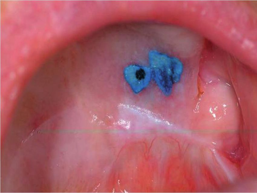
Vital tissue staining appear relatively dark. Unfortunately, some
The use of vital tissue staining in cancer normal tissue or benign conditions may also
detection is based on the assumption that a remain dark. A Cochrane review of twelve
nuclear dye will be taken up in the epithelial studies of the use of VELscope reported an
cells where there is a high density of nuclear overall sensitivity of 0.91 and specificity of
Fig. 14 White patch on dorsal surface of the
material, such as in malignancy, and therefore 0.58 for oral carcinoma or OPMDs.29
tongue in dyskeratosis congenital (Courtesy
of Professor Graham Ogden) have a different appearance to normal mucosa.
This methodology was originally used for Brush cytology
cervical screening in women. Toluidine blue Exfoliative cytology has been investigated as
carcinoma, there can never be 100% certainty (tolonium chloride) is a metachromatic dye a possible mouth cancer detection system.
until the diagnosis is confirmed on the basis has been used widely in this context. Retention OralCDx (OralCDx Laboratories, Inc) is an
of histopathological findings following gold of the dye, seen as dark blue, following applica- example of this technology that uses a special
standard mucosal biopsy. Occasionally a tion and de-staining with acetic acid is consid- designed brush to collect cells from an identi-
highly suspicious clinical area of mucosa ered positive (Fig. 16). However, it is not cancer fied mucosal abnormality within the mouth.
change is found to be an essentially benign specific and false positives due to uptake by The sample is then sent for computerised
abnormality and conversely a benign-looking benign inflammatory and ulcerative of the oral analysis for the presence of abnormal cells
mucosal abnormality can be found to contain mucosa are frequent. Equally, failure of the dye and reported as negative, atypical or positive.
a carcinoma. The lack of specific clinical signs to penetrate keratinised mucosa can lead to All atypical and positive results require a tissue
for mouth cancer probably in part accounts false negatives. A Cochrane review of the use biopsy. OralCDx has been found to have a
for the delay in a patient seeking an opinion of the toluidine blue test for detection of oral sensitivity of 0.92 and specificity of 0.94 to
of dentist or doctor in primary care and hence carcinoma or OPMDs in 14 studies reported detect dysplasia or squamous cell carcinoma.31
the high percentage of patients who are not an overall sensitivity of 0.84 and specificity of More recently cytological methods have been
diagnosed till the tumour has advanced to 0.70 for both conditions.29 combined with molecular markers in an
Stage III or Stage IV.1 attempt to improve their prognostic value.
A range of adjunctive clinical aids have Tissue reflectance light visualisation
been and are continuing to be developed The use of tissue reflectance or chemilumines- Salivary diagnostics
commercially to increase the detection of cence to detect cancer is based on the theory Molecular biology has enabled the ability to
potential mouth cancer at the chair side.26 that the increased nuclear to cytoplasmic ratio detect biomarkers (peptides, proteins, DNA,
The technology involved includes the use of of malignant epithelial cells results in increased mRNAs and miRNAs) in saliva and as such
nuclear dyes, tissue reflectance light visualisa- light reflectance when compared to normal provide a non-invasive diagnostic test and
tion and fluorescence imaging, all of which mucosa. ViziLite Plus (Panadent, Orpington, screening tool for human disease. The potential
aim to increase the clinician’s ability to detect UK) and MicroLux DL (Panadent, Orpington, to use saliva to detect the presence of cancer
an area of mucosal abnormality that may UK) are two cancer detection systems that has been researched in relation to tumours
represent cancer or OMPDs. The primary aim employ this approach. The systems differ in of the ovary, breast, pancreas and lung. In
838 BRITISH DENTAL JOURNAL | VOLUME 225 NO. 9 | NOVEMBER 9 2018
O
f
f
i
c
i
a
l
j
o
u
r
n
a
l
o
f
t
h
e
B
r
i
t
i
s
h
D
e
n
t
a
l
A
s
s
o
c
i
a
t
i
o
n
.
Mouth cancer                   CLINICAL

addition, a number of studies have been under-
taken to look at biomarkers for oral squamous
cell carcinoma.32–35 At the present time further
work is required to identify specific biomark-
ers within saliva that can be reliably used to
detect different forms of human cancer and
other forms of disease. Salivary diagnostic
technology has huge potential for the future
and could become a routine aspect of dental
primary care.
                                                   Fig. 15 Chronic hyperplastic candidosis presenting as bilateral white patches in the buccal
                                                   mucosa; (a) left and (b) right
Referral

As previously described, the outcome of mouth
cancer is significantly improved if the tumour
is detected and treated when it is less than
2 cm in diameter with no local metastasis. On
this basis any suspected cancer patient should
be referred for specialist assessment rapidly.
Terminology used in this situation is urgent
suspected cancer (USC) with an expectation,
in the UK, that a patient referred a USC will be
seen within 14 days (2 week wait, 2WW) or 10
working days(10 day rule).1 The 2WW system
for cancer, including head and neck cancer,
was introduced in the UK in 2000 and there is
little doubt that it has been valued by patients
since it speeds up the overall management.
An audit of 2WW rule in the oral and maxil-        Fig. 16 Toludine blue stain retained on palatal mucosal abnormality
lofacial surgery department at the Newcastle
General Hospital and then subsequently in
the oral surgery and oral medicine depart-         delay within a postal system. This has in part       Individual practitioners must be aware of their
ments at Newcastle Dental Hospital revealed        been overcome by the use of a fax machines           local referral system.
positive head and neck cancer detection rates      or a secure NHS email address. Personal                 In the UK, the National Institute for
of 11% and 7% respectively in the cohorts of       email accounts must not be used due to the           Health and Clinical Excellence (NICE)39 and
patients examined.36 A similar study at the        high risk of breach of patient confidentiality.      Healthcare Improvement Scotland (HIS)40
oral medicine department in King’s College,        However, free written communication via              have published referral guidelines to help clini-
London, reported that 8% of urgent referrals       any of these methods may be problematic in           cians decide which patients should be refereed
were found to have oral cancer.37 Both audits      that the clinical information provided by the        as USC. These guidelines focus on the present-
concluded that further education of referring      primary care practitioner may be insufficient        ing clinical symptoms and recommend the
practitioners and refinement of the referral       to enable the receiving specialist to identify the   need for a USC referral for any individual with:
guidelines were required to ensure a more          need for a 2WW appointment. The inclusion of         • Unexplained ulceration in the oral cavity
efficient service.                                 a question on a preformed referral forms ‘Do             lasting more than three weeks
   A systematic review of the impact of the        you think that this patient may have a cancer?’      • A persistent and unexplained lump in
2WW rule for head and neck cancer between          or a ‘Yes/No’ tick-box for USC is helpful but            the neck
2000–14 concluded that the conversion rate         again not foolproof. Details of risk factors for     • A lump on the lip or in the oral cavity con-
(proportion of 2WW referrals who were              the patient, in particular any tobacco, betel nut        sistent with oral cancer
diagnosed as having cancer) was falling, and       or alcohol habits, are important to include.         • A red or red and white patch in the
the detection rate (proportion of diagnosed           Electronic record management and referral             oral cavity consistent erythroplakia or
cancers referred under 2WW rule) was               systems within managed clinical networks                 erythroleukoplakia.
rising due to increased number of referrals.       (MCNs) are being introduced and these have a
In addition the impact of the 2WW rule on          range of advantages, including an assurance of         There has been discussion in relation to the
outcome, particularly survival, was not clear.38   rapid and safe delivery of the referral request.     recommendation that a doctor should refer
   Methods of referral from primary care           In addition, some electronic systems ensure          a patient to a dentist for assessment within
to specialist secondary care vary widely.          that all relevant information is provided by         two weeks if the doctor thinks that there is ‘a
Historically, referrals have been made in the      the referring practitioner since the referral will   lump on the lip or in the oral cavity consist-
form of a written letter with inevitable risk of   not be accepted until this is completed online.      ent with oral cancer or a red or red and white

BRITISH DENTAL JOURNAL | VOLUME 225 NO. 9 | NOVEMBER 9 2018839
                                                       O
                                                       f
                                                       f
                                                       i
                                                       c
                                                       i
                                                       a
                                                       l
                                                       j
                                                       o
                                                       u
                                                       r
                                                       n
                                                       a
                                                       l
                                                       o
                                                       f
                                                       t
                                                       h
                                                       e
                                                       B
                                                       r
                                                       i
                                                       t
                                                       i
                                                       s
                                                       h
                                                       D
                                                       e
                                                       n
                                                       t
                                                       a
                                                       l
                                                       A
                                                       s
                                                       s
                                                       o
                                                       c
                                                       i
                                                       a
                                                       t
                                                       i
                                                       o
                                                       n
                                                       .
CLINICAL Mouth cancer
patch in the oral cavity consistent erythro- 1. Cancer Research UK Website. Available at www.cancer- underwent malignant change. Gerodontology 2001; 18:
researchuk.org/health (accessed August 2018). 73–78.
plakia or erythroleukoplakia’. Concern has 2. Sciubba J J. Oral cancer. The importance of early 25. Brocklehurst P, Pemberton M N, Macey R, Cotton C,
been expressed that due to the lack of a clear diagnosis and treatment, Am J Clin Dermatol 2001; 2: Walsh T, Lewis M A O. Comparative accuracy of different
239–251. members of the dental team in detecting malignant
pathway between the doctor and the dentist
3. Ong T K, Murphy C, Smith A B, Kanatas A N, Mitchell and non-malignant oral lesions. Br Dent J 2015; 218:
this advice exposes the patient to unnecessary D A. Survival after surgery for oral cancer: a 30-year 525–529.
delay in diagnosis.41,42 experience. Br J Oral Maxillofac Surg 2017; 55: 911–916. 26. Warnakulasuriya S. Diagnostic adjuncts on oral cancer
4. Lewis M A O, Lamey P-J. A clinical guide to oral medicine. and precancer: an update for practitioners. Br Dent J
3rd ed. London: British Dental Journal, 2011. 2017; 223: 663–666.
5. Oral cancer recognition toolkit. Available at www. 27. Rashid A, Warnakulasuriya S. The use of light-based
Communication with the patient doctors.net.uk/eclientopen/cruk/oral_cancer_tool- (optical) detection systems as adjuncts in the detection
kit_2015_open/ (accessed 19 October 2018). of oral cancer and oral potentially malignant disorders: a
If a soft tissue examination reveals an abnor- 6. Oliver R J, Sloan P, Pemberton M N. Oral biopsies: meth- systematic review. J Oral Pathol Med 2015; 44: 307–328.
ods and applications. Br Dent J 2004; 196: 329–333. 28. Warnakulasuriya S. Translational research in oral
mality and it is felt that a referral is indicated 7. Lynch D P, Morris F M. The oral mucosa punch biopsy. oncology – A bridge between basic science and clinical
then what should the patient be told? It is J Am Dent Assoc 1990; 121: 145–149. application. Transl Res Oral Oncol 2016; 1: 1–2.
8. Kramer I R H, Lucas R B, Pindborg J J, World Health 29. Macey R, Walsh T, Brocklehurst P et al. Diagnos-
probably best to inform the patient that Organization Collaborating Centre for Oral Precancerous tic tests for oral cancer and potentially malignant
changes in the mouth are seen frequently and Lesions: Definition of leukoplakia and related lesions. An disorders in patients with clinically evident lesions.
aid to studies on oral precancer. Oral Surg Oral Med Oral Cochrane Database Sys Rev 2015; 5: Cd010276. DOI.
the vast majority are innocent but it is best to Pathol 1978; 46: 518–539. 10.1002/14651858.CD010276.pub2.
get it double checked by a specialist. Other 9. Warnakulasuriya S, Johnson N W, van der Waal I. 30. Lingen M W, Tampi M P, Urquhart O et al.Adjuncts for
Nomenclature and classification of potentially malignant the evaluation of potentially malignant disorders in the
information given to the patient and/or carer disorders of the oral mucosa. J Oral Pathol Med 2007; oral cavity: Diagnostic test accuracy systematic review
could include: 36: 575–580. and meta-analysisa report of the American Dental
10. Silverman S, Bhargava, K, Smith L W, Malaowalla A M. Association. J Am Dent Assoc 2017; 148: 797–813.e52.
• Where there are being referred Malignant transformation and natural history of oral 31. Scheifele C, Schmidt-Westhausen A M, Dietrich T, Reich-
• How long they may have to wait leukoplakia in 57: 518 industrial workers of Gujarat, art P A. The sensitivity and specificity of the OralCDx
India. Cancer 1976; 38: 1790–1795. technique: evaluation of 103 cases. Oral Oncol 2004;
• Who will see them
11. Warnakulasuriya S, Kovacevic T, Madden P et al. Factors 40: 824–828.
• What types of test may be done.43 predicting malignant transformation in oral potentially 32. Radhika T, Jeddy N, Nithya S, Muthumeenakshi R M.
malignant disorders among patients accrued over a Salivary biomarkers in oral squamous cell carcinoma –
10-year period in South East England. J Oral Pathol Med An insight. J Oral Biol Craniofac Res 2016; 6: S51–S54.
This communication has to be done sensitively 2011; 40: 677–683. DOI: 10.1016/j.jobcr.2016.07.003.
and obviously the amount of information given 12. Silverman S, Gorsky M, Lozada F. Oral Leukoplakia and 33. Bano S, David M P, Indira A P. Salivary biomarkers for
malignant transformation: a follow up study of 257 oral squamous cell carcinoma: An overview. IJSS Case
decided on an individual patient basis. patients. Cancer 1984; 53: 563–568. Rep Rev 2015; 1: 39–45.
Interestingly, it has been reported that 13. Petti S. Pooled estimate of world leukoplakia prevalence: 34. Spielmann N, Wong D T. Saliva: diagnostics and thera-
a systematic review. Oral Oncol 2003; 39: 770–780. peutic perspectives. Oral Dis 2011; 17: 345–354.
some dentists have a reluctance to discuss 14. Munde A, Karle R. Proliferative verrucous leukoplakia: 35. Cheng Y S, Rees T, Wright J. A review of research on
mouth cancer with their patients due to a lack An update. J Can Res Ther 2016; 12: 469–473. salivary biomarkers for oral cancer detection. Clin Transl
15. Pinborg J J, Reichart Smith C J, van der Waal I, Sobin Med 2014; 3: 3. DOI: 10.1186/2001-1326-3-3.
of confidence to answer patients’ questions.44 L H and pathologists in nine countries. World Health 36. Miller C C, Hierons R J. Two audits of the diagnosis of
There would appear to be a need for specific Organization international histological classification of oral cancer and the two-week rule following referrals
tumours, histological typing of cancer and precancer of from primary care practitioners in Newcastle. Prim Dent
training and guidance for the dental team on the oral mucosa. 2nd ed. Berlin: Springer, 1997. Care 2012; 19: 63–68.
how to raise the issue of mouth cancer during 16. Reichart P A, Philipsen H P. Oral erythroplakia – a 37. Singh P, Warnakulasuriya S. The two-week wait cancer
review. Oral Oncol 2005; 41: 551–561. initiative on oral cancer; the predictive value of urgent
routine examination and also how to commu- 17. De Rossi S S, Ciarrocca K. Oral lichen planus and referrals to an oral medicine unit. Br Dent J 2006; 201:
nicate any issues without causing unnecessary lichenoid mucositis. Dent Clin North Am 2014; 58: 717–720.
299–313. 38. Langton S, Siau D, Bankhead C. Two-week rule in head
anxiety. This aspect of mouth cancer could be 18. Rajentheran R, McLean N R, Kelly C G, Reed M F, Nolan and neck cancer 2000–2014: asystematic review. Br
a subject for future undergraduate and post- A. Malignant transformation of oral lichen planus. Eur J Oral Maxillofac Surg. 2016: 54: 120–131.
J Surg Oncol 1999; 25: 520–523. 39. National Institute for Health and Care Excellence.
graduate education.
19. Ingafou M, Leao J C, Porter S R, Scully C. Oral lichen Suspected cancer: recognition and referral. 22 June 2015.
planus: a retrospective study of 690 British patients. Oral Available at www.nice.org.uk/guidance/ng12 (accessed
Dis 2006; 12: 463–468. August 2018).
Conclusion 20. Giuliani M, Troiano, G, Cordaro M et al. Rate of malig- 40. Healthcare Improvement Scotland. Scottish referral
nant transformation of oral lichen planus: A systematic guidelines for suspected cancer. August 2014. Available
The clinical presentation of mouth cancer review. Oral Dis 2018; DOI: 1111/odi.12885 [Epub ahead at www.healthcareimprovementscotland.org/our_work/
of print]. cancer_care_improvement/programme_resources/scot-
is highly variable. Regular and thorough 21. Murti P R, Bhonsle R B, Pindborg J J, Daffary D K, Gupta tish_referral_guidelines.aspx (accessed August 2018).
examination of the soft tissues to detect any P C, Mehta F S. Malignant transformation rate in oral 41. Grimes D. Patel J, Avery C. New NICE referral guidance
submucous fibrosis over 17 period. Community Dent Oral for oral cancer: does it risk delay in diagnosis? Br J Oral
abnormality is an essential aspect of dental Epidemiol 1985; 13: 340–341. and Maxillofac Surg 2016; 55: 404–406.
primary care. If any mucosal change found is 22. Gupta P C, Mehta F S, Daftary D K et al. Incidence rates 42. Yeung C A. Referrals to dentists by GPs could delay diag-
of oral cancer and natural history of oral precancerous nosis of oral cancer. BMJ 2017; 356: i6784. Available at
thought to possibly represent cancer, then the lesions in a 10-year follow up study of Indian villagers. https://doi.org/10.1136/bmj.i6784 (accessed 19 October
patient needs to be referred appropriately for Community Dent Oral Epidemiol 1980; 8: 287–333. 2018).
23. Ogden G R, Connor E, Chisholm D M. Dyskeratosis 43. Kalavrezos N, Scully C. Mouth cancer for clinicians Part
a specialist evaluation. All dental profession- congenita: report of a case and review of the literature. 8: Referral. Dent Update 2016; 43: 176–185.
als should be aware of mouth cancer and feel Oral Surg Oral Med Oral Pathol 1988; 65: 586–591. 44. Awojobi O, Newton J T, Scott S E. Pilot study to train
24. Williams D W, Bartie K L, Potts A J C, Wilson M J, dentists to communicate about oral cancer: the impact
comfortable about discussing the subject with Fardy M J, Lewis M A O. Strain persistence of invasive on dentist’s self-confidence reported behaviour, confi-
their patients. Candida albicans in chronic hyperplastic candidosis that dence and beliefs. Br Dent J 2016; 220: 71–76.
840 BRITISH DENTAL JOURNAL | VOLUME 225 NO. 9 | NOVEMBER 9 2018
O
f
f
i
c
i
a
l
j
o
u
r
n
a
l
o
f
t
h
e
B
r
i
t
i
s
h
D
e
n
t
a
l
A
s
s
o
c
i
a
t
i
o
n
.
You can also read