Structure and evolutionary implications of the earliest (sinemurian, early Jurassic) dinosaur eggs and eggshells - Dinodata.de
←
→
Page content transcription
If your browser does not render page correctly, please read the page content below
www.nature.com/scientificreports

                        OPEN       Structure and evolutionary
                                   implications of the earliest
                                   (Sinemurian, Early Jurassic)
Received: 18 September 2018
Accepted: 14 February 2019         dinosaur eggs and eggshells
Published: xx xx xxxx
                                   Koen Stein 1,2, Edina Prondvai 3,4, Timothy Huang                   5,6
                                                                                                             , Jean-Marc Baele     7
                                                                                                                                       ,
                                   P. Martin Sander 8,9 & Robert Reisz 5,6,10

                                   One of the fossil record’s most puzzling features is the absence of preserved eggs or eggshell for the
                                   first third of the known 315 million year history of amniote evolution. Our meagre understanding of
                                   the origin and evolution of calcareous eggshell and amniotic eggs in general, is largely based on Middle
                                   Jurassic to Late Cretaceous fossils. For dinosaurs, the most parsimonious inference yields a thick,
                                   hard shelled egg, so richly represented in the Late Cretaceous fossil record. Here, we show that a thin
                                   calcareous layer (≤100 µm) with interlocking units of radiating crystals (mammillae) and a thick shell
                                   membrane already characterize the oldest known amniote eggs, belonging to three coeval, but widely
                                   distributed Early Jurassic basal sauropodomorph dinosaurs. This thin shell layer strongly contrasts
                                   with the considerably thicker calcareous shells of Late Jurassic dinosaurs. Phylogenetic analyses and
                                   their Sinemurian age indicate that the thin eggshell of basal sauropodomorphs represents a major
                                   evolutionary innovation at the base of Dinosauria and that the much thicker eggshell of sauropods,
                                   theropods, and ornithischian dinosaurs evolved independently. Advanced mineralization of amniote
                                   eggshell (≥150 µm in thickness) in general occurred not earlier than Middle Jurassic and may correspond
                                   with a global trend of increase in atmospheric oxygen.

                                   The origin of the amniote egg is a topic of great signifi ance because it represents one of the major evolutionary
                                   innovations in vertebrate evolution, allowing the group to complete their invasion of the terrestrial landscape
                                   and sever their reproductive cycle from the aquatic medium1. However, paleontological studies of this pivotal
                                   event have been greatly hampered by the poor early record of fossil eggs2,3. Recent attempts to fill the gaps in
                                   fossil eggshell phylogeny still leave at least 125 million years of amniote evolution between the appearance of
                                   amniotes in the fossil record and the fi st appearance of preserved terrestrial eggs or eggshells4–12. The oldest
                                   known eggs or eggshells have been reported7–12 from three Sinemurian (195-192 Ma) sauropodomorph dino-
                                   saurs, Massospondylus from the Elliot Formation of South Africa, Lufengosaurus from the Lufeng Formation of
                                   Yunnan, China, and Mussaurus from the Laguna Colorada Formation of Argentina (Fig. 1). Within the context of
                                   their respective localities, some of these materials have been examined to a limited extent. In a study on prenatal
                                   remains of Lufengosaurus, some of the authors of the current study previously provided a very brief description of
                                   its eggshell, and noted its extreme thinness12. Other authors working on Massospondylus initially discarded them

                                   1
                                     Earth System Science - AMGC, Vrije Universiteit Brussel, Pleinlaan 2, 1050, Brussels, Belgium. 2Royal Belgian
                                   Institute of Natural Sciences, Directorate ‘Earth and History of Life’, Rue Vautier 29, 1000, Brussels, Belgium.
                                   3
                                     Evolutionary Morphology of Vertebrates, Ghent University, K.L. Ledeganckstraat 35, 9000, Gent, Belgium.
                                   4
                                     MTA-ELTE Lendület Dinosaur Research Group, Eötvös Loránd University, Pázmány P. s. 1/C, 1117, Budapest,
                                   Hungary. 5International Center of Future Science, and Dinosaur Evolution Research Center of Jilin University,
                                   Changchun, Jilin Province, China. 6National Chung Hsing University, Taichung, 402, Taiwan. 7Department of Geology
                                   and Applied Geology, Faculty of Engineering, University of Mons, Place du Parc 20, 7000, Mons, Belgium. 8Steinmann
                                   Institute of Geology, Mineralogy, and Paleontology, Division of Paleontology, University of Bonn, Nussallee 8, 53115,
                                   Bonn, Germany. 9Natural History Museum of Los Angeles County, Dinosaur Institute, 900 Exposition Boulevard, Los
                                   Angeles, CA, 90007, USA. 10Department of Biology, University of Toronto Mississauga, Mississauga, Ontario, L5L
                                   1C6, Canada. Correspondence and requests for materials should be addressed to K.S. (email: kstein@vub.be) or R.R.
                                   (email: robert.reisz@utoronto.ca)

        Scientific Reports |   (2019) 9:4424 | https://doi.org/10.1038/s41598-019-40604-8                                                             1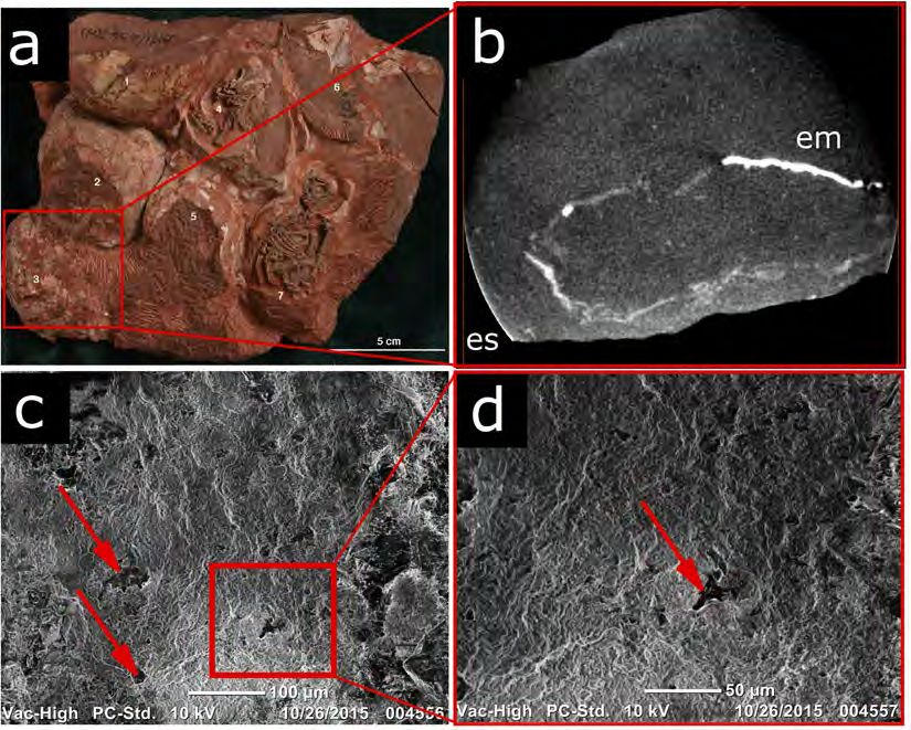
www.nature.com/scientificreports/ www.nature.com/scientificreports
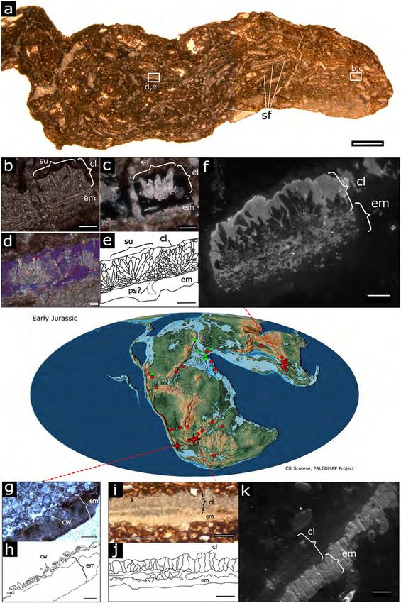
Figure 1. Basal sauropodomorph eggshell microstructure and their respective Sinemurian localities (crosses)
among the Rhaetian (green) to Sinemurian (red) global record of sauropodomorph fossil sites (circles). (a–f),
Lufengosaurus (Chuxiong Prefectural Museum, catalogue no. C2019 2A233), (g,h), Mussaurus (Instituto
‘Miguel Lillo”, Tucuman, catalogue no. PVL 5965), (i–k), Massospondylus (Bernard Price Institute of
Palaeontology, University of Witwatersrand, catalogue no. BP/1/5254). (a) Section through nugget containing
numerous Lufengosaurus eggshell fragments (plane polarized light, ppl). (b), close-up (ppl) of a Lufengosaurus
eggshell fragment, showing calcite crystals of the mammillary layer radiating from an organic core embedded
in the eggshell membrane. (c) As in (b) under cross polarized light (xpl), highlighting the calcite crystals of
a mammillary cone. (d) Different xpl view with lambda waveplate, e. line drawing of (d). (f) Lufengosaurus
cathodoluminescence view with 880 nm filter. (g) Mussaurus eggshell, showing thick eggshell membrane, and
distorted calcareous layer. (h) Line drawing of (g). (i) Massospondylus eggshell fragment (ppl), showing wedges
in the calcareous layer, and a homogenous eggshell membrane. (j), Line drawing of (i). (k) Massospondylus
Scientific Reports | (2019) 9:4424 | https://doi.org/10.1038/s41598-019-40604-8 2
www.nature.com/scientificreports/                                                              www.nature.com/scientificreports

                           cathodoluminescence view with 880 nm filter. Scale bars: in (a): 1 mm, (b–f,k): 50 µm, (g–j): 100 µm.
                           Abbreviations: cl, calcareous layer; cw, crystal wedges of calcareous layer; em, eggshell membrane; ps, pore
                           space; su, shell unit. See also Figs S1–S3. (Map from66 with permission).

                           as crocodile eggshells13, and later as having a diagenetically altered microstructure9. Mussaurus eggshell has to
                           our knowledge never been described in a formal publication. These remains are the earliest confi med amniote
                           eggshells recorded in the fossil record. Due to their rarity, fragmentary nature, and great geographic distance
                           from each other, they were never studied from the perspective of the evolution of amniote eggshell. Here we aim
                           to understand their microstructural features and try to elucidate when and how the earliest mineralized eggshells
                           could have evolved. To accomplish our goal, we utilised petrographic sections, analytical chemistry tools and
                           computational statistical methods (description in Materials and Methods section). Th s study contributes to our
                           understanding of the evolution of rigid shelled eggs; a key trait in the evolutionary success of archosaurs.

                           Results
                           Eggshell structure. The calcareous layer of Lufengosaurus eggshells (C2019 2A233) ranges 60–90 µm in
                           thickness. They consist of crocodile eggshell-like wedge- and crown-shaped shell units that are relatively wide
                           compared to the calcareous layer thickness (Fig. 1a–f). Polarized light microscopy suggest that the outer surface
                           of the eggshell is unaltered (Fig. 1f, Supplementary Information). The very thin crystalline layer (~10 µm) top-
                           ping the eggshell units is phosphatic in nature (Figs S1 and S2), and looks scalloped with shallow pits and low
                           ridges, not necessarily matching eggshell unit borders. These surface irregularities or tubercles are of such small
                           dimensions that the surface of the calcareous layer looks smooth (Fig. S2a), and it remains unclear if they match
                           the ornamentations seen in younger dinosaur eggshells. The bulk of the units, corresponding to the mammillary
                           cones, is formed by a calcite radial ultrastructure (sensu14) with interlocking crystalline units (Fig. 1c–f). The
                           patchy cathodoluminescence texture suggests some radial crystal wedges experienced recrystallization, but most
                           of the original microstructure is conserved (Fig. S1b. No tabular structures or horizontal accretion lines can be
                           observed. The growth centre of the units is embedded in a phosphorus-rich (Figs S1 and S2), thick fibrillar layer
                           (60–75 µm) representing the eggshell membrane (Fig. 1e). Pore spaces are rare and difficult to discern (Fig. 1e).
                           Due to the fragmentary nature of the materials, and because pores were not always unambiguously identifiable,
                           it was not possible to make an estimation of pore density. However, pore distribution does not appear to be
                           consistent with the presence of tubercles or depressions on the outer surface. A tangential section through the
                           membrane shows clusters of crystals with fl wer-like arrangements (Fig. S3). The lack of a thick palisade layer
                           and the overall thinness of the calcareous shell clearly distinguish Lufengosaurus eggshells from avian and other
                           younger dinosaurian eggshells.
                               The South African Massospondylus (BP/1/5254, BP/1/5347) calcareous eggshell layer is slightly thicker
                           (80–100 µm) than that of Lufengosaurus. The eggshell units are very difficult to discern (Fig. 1i,j). In the past,
                           these units have been interpreted as wedge-shaped8. Our cathodoluminescence analysis (Figs 1k and S1) shows
                           very high luminosity of calcite in the eggshell units, which supports the idea that these structures are the result
                           of diagenetic alteration of the original microstructure9 (Figs 1k and S1). Nonetheless, eggshell is present in
                           Massospondylus eggs from several different localities in South Africa, and of similar thickness as in Lufengosaurus,
                           and some features remain recognizable. The outer surface of the eggshell, as in Lufengosaurus, is rugged with
                           low tubercles and shallow depressions. Occasional pores are distributed unevenly throughout the shell surface
                           (Fig. 2c,d). Below the calcareous layer, a dark, isotropic layer (50–90 µm thick, cross polarized light) merges
                           with, or entirely obscures the mammillary cones (Fig. S3). We identify this layer as a remnant of the eggshell
                           membrane, given its position relative to the calcareous layer and its chemical similarity with the Lufengosaurus
                           shell membrane (rich in phosphate and calcite, Figs S1–S3). A shell membrane is also preserved in some of the
                           complete eggs with the embryos (Fig. 2a).
                               The Argentinian Mussaurus eggshell (PVL 5965) is severely affected by diagenesis. Only few sparse and
                           widely scattered calcite crystals, similar in size and shape to the radiating crystals in the mammillary cones of
                           Lufengosaurus eggshell units, remain of the calcareous layer (Fig. 1g,h). The eggshell membrane is preserved as a
                           thick (150–180 µm) phosphatic layer with little internal structure.
                               Calcareous layer to membrane thickness ratios may vary due to incomplete preservation of the membrane.
                           They range from ~1:1 in Lufengosaurus and ~1.5:1 Massospondylus, but remain uncertain in Mussaurus due to the
                           loss of an intact, coherent calcareous layer.
                               The identity of all three taxa is unquestionable based on the presence of embryonic remains10–12,15, contra13. We
                           thus reconstruct basal sauropodomorph eggshell as having a thin calcareous layer, composed of low, wide mammillary
                           cones (approximate width to height rations of 1:1) attached to a membrane of at least similar thickness (Fig. 3).

                           Taphonomic and evolutionary implications. The phylogenetically informed regression analysis of min-
                           eralized eggshell thickness versus egg mass in a wide taxonomic range of extant and extinct egg laying amniotes
                           (Fig. 4a) revealed a signifi ant positive relationship between egg size (mass) and shell thickness, with considerable
                           phylogenetic signal (λ = 0.86; p < 0.001; see SI). The regression function is largely determined by the taxa with
                           rigid-shelled eggs (non-avian dinosaurs, birds and crocodiles). Negative outliers, in which the size of the eggs and
                           their shell thickness are well below the regression line, are the extant and fossil groups with known or inferred
                           flexible shelled-eggs, such as marine turtles, squamates, and pterosaurs (Fig. S4). Interestingly, Lufengosaurus
                           and Massospondylus plot with these negative outliers emphasising the pronounced thinness of their calcareous
                           eggshell relative to their egg mass (Fig. 4). However, due to the interlocking nature of the crystal units, these
                           basal sauropodomorphs most likely had rigid eggshell. Th s interpretation is supported by the preservational

Scientific Reports |   (2019) 9:4424 | https://doi.org/10.1038/s41598-019-40604-8                                                             3
www.nature.com/scientificreports/ www.nature.com/scientificreports
Figure 2. Eggshell membrane and porosity in Massospondylus eggs (BP/1/5347). (a) Nest of Massospondylus
eggs with preserved embryos. Note the presence of numerous cracks in the eggs, likely caused by postmortem
crushing of the thin but hard eggshell. Eggshell membrane is exposed in egg number 4, just beneath the skull,
and in egg number 7, just beneath the right scapula. (b) CT scan of a complete egg in a, showing the eggshell
(es) and the detached preserved eggshell membrane (em). (c) Outer surface SEM image of a Massospondylus
eggshell fragment showing rare small and irregularly shaped pores occurring in random patterns (red arrows).
(d) Enlarged view of boxed area in (c). See also Figs S1–S3.
Figure 3. Reconstruction of a basal sauropodomorph egg showing detail of the eggshell. Eggshell units (esu)
form the calcareous layer (cl) and are embedded with organic cores in the eggshell membrane (em). See also
Figs S1–S3. Embryo reconstruction by R. David Mazierski with permission.
characteristics of all egg fragments recovered from the various sites. All retain their curvature, and even though
the eggs of Massospondylus are somewhat crushed, they show the typical cracking and fragmenting associated
with rigid structures (Fig. 1)16,17. Th s observation contrasts with the preservational characteristics of soft- helled
fossil material, now abundantly preserved for the pterosaur Hamipterus18.
Scientific Reports | (2019) 9:4424 | https://doi.org/10.1038/s41598-019-40604-8 4www.nature.com/scientificreports/ www.nature.com/scientificreports
Figure 4. Relationship between eggshell thickness and egg mass in different egg-laying archosauromorphs
and time calibrated maximum likelihood (ML) analysis of the ancestral states of relative eggshell thickness
evolution. (a) PGLS regression line and 95% confide ce band on the ln-transformed dataset. Massospondylus
and Lufengosaurus represent negative outliers (see SI) emphasizing the extreme thinness of the calcareous
layer compared to other dinosaurs. (b) ML ancestral state reconstruction of log-transformed calcareous layer
thickness (CL) to egg mass (EM) ratios. Note that the root was set to represent the hypothesized ancestral
flex ble shelled condition. Nodes represent (a), Archosauromorpha, (b), Archosauria (c), Ornithodira,
(d), Dinosauria, (e), birds. Note the independent acquisitions of thick eggshell in choristoderes (represented
by Hyphalosaurus), chelonians, crocodiles, pterosaurs and several dinosaur clades, as well as reversals in
chelonians. From the Sinemurian (199 Ma) onwards, eggshells (e.g. Testudofl xoolithus and Lourinhanosaurus)
show a signifi ant calcareous layer thickness increase corresponding with atmospheric oxygen increase. See also
Fig. S4.
A time-calibrated cladogram of archosauromorphs suggests the ancestral state for dinosaurs is the thin-shelled
condition (Fig. 4b, Supplementary Information). Maximum likelihoods (ML) of ancestral character states imply
low ratios of eggshell thickness to egg mass are plesiomorphic in Dinosauria. These calculations are based on
the logical assumption that the archosauromorph root node represents a poorly mineralized eggshell (cf.2,3),
the value of which is derived from the lowest observed value among the extant amniote taxa (Pelusios sinuatus)
(see Supplementary Information). Reconstructed relative eggshell thicknesses for the base of the dinosaur tree
are very close to those of the early sauropodomorphs described here. The ancestral state reconstruction suggests
independent eggshell thickening events in all major archosauromorph clades, but also within different dinosaur
clades. Evolutionary reversals are also demonstrated (cf. Pelusios sinuatus and Carretta carretta). Finally, it is
important to note that these thickening events generally occurred after the Sinemurian (~195 Ma).
Discussion
Our detailed examination of the eggs of these basal sauropodomorph dinosaurs shows that all have an extremely
thin mineralized eggshell layer. Different diagenetic settings of their respective localities affected the original
microstructure to different degrees, with Lufengosaurus having the best, and Mussaurus the least preserved details
(Fig. 1, Supplementary Information). The structural characteristics of these Early Jurassic dinosaur eggshells are
unlike those in any other known dinosaur. The extreme thinness could have resulted from decalcifi ation during
egg incubation, as seen in some Massospondylus eggs containing advanced stage embryos. However, this is unlikely
because there is no sign of resorption craters at the base of the crystal units, and the recent collection of a complete
Massospondylus nest with undeveloped embryos makes this unlikely11 (Fig. 2a). In addition, the similar thinness,
eggshell unit characteristics, and outer surface ornamentation suggest that the calcareous shell layers are similar
to their original thickness in both Lufengosaurus and Massospondylus. A low ratio of calcareous layer to eggshell
membrane thickness is usually associated with flex ble-shelled eggs of extant amniotes19, however, in line with
our other observations, we conclude that these early dinosaurs had thin, albeit rigid-shelled eggs (Fig. 3), a highly
unusual, unexpected condition. The semi-arid depositional conditions7,11,12 and relative thinness of the eggshell
suggest the eggs needed to be protected from dehydration17,20,21. Hence, as in many other dinosaurs22,23 and most
modern-day non-avian reptiles, the eggs were most likely buried in the nest, although this hypothesis needs further
support by more complete data on pore density and relative eggshell porosity. Previous studies have pointed to
a combination of nesting site fid lity, colonial nesting, and parental care in these early sauropodomorphs11,12.
It is thus possible that through their behavioural ecology, these sauropodomorphs created taphonomic
conditions that allowed the preservation of such delicate structures.
Eggshells with a comparatively thick membrane but thin calcareous layer are in sharp contrast with heavily
mineralized dinosaurian eggshells commonly found during the Cretaceous13,14. Ancestral state reconstruction
of this feature may be affected by lack of information from earlier reptilian clades and crucial basal taxa, such as
early ornithischians. Nonetheless, the lack of pre-Middle Jurassic rigid fossil eggshells3,5,24, the different mammil-
lary ultrastructure in crocodilian and dinosaurian eggshells, and the aragonitic nature of turtle eggshells, provide
strong support for the hypothesis of independent eggshell thickening events in these reptiles (see Supplementary
Scientific Reports | (2019) 9:4424 | https://doi.org/10.1038/s41598-019-40604-8 5www.nature.com/scientificreports/ www.nature.com/scientificreports
Information for further results and discussion of the ancestral state analysis). Th s scenario also favours the
independent origin of extended eggshell growth in the dinosaurian clades Ornithischia, Sauropodomorpha and
Theropoda. All known dinosaurian eggshells, including those described here, possess mammillae with radiating
calcite crystals14, therefore, mammillated eggshell with calcite radial ultrastructure can be considered a dinosau-
rian synapomorphy.
It seems straightforward to assume a flex ble non-mammillated → flex ble mammillated → rigid mammillated
succession of eggshell structural evolution. However, it does not have to be so strictly sequential or directional,
as there may be an intricate interplay between biological and environmental factors shaping eggshell structure
and composition19. Diversity in eggshell micro- and ultrastructure in different reptilian clades points to numer-
ous convergences, secondary losses and reversals. Turtles demonstrate this evolutionary complexity by revealing
conditional aragonite/calcite composition of the calcareous layer19,25 and multiple eggshell-softening events26
(Fig. 4b), even with complete loss of mammillae in the pleurodiran Pelusios sinuatus27.
Our ancestral state reconstruction shows an independent thickening of the calcareous layer in several archo-
sauromorph clades during the Jurassic. Interestingly, this does not seem to be directly related to increase in body
size, and hence egg size. However, the occurrence of the earliest strongly mineralized archosauromorph and turtle
eggshells in the Middle and Late Jurassic5,28 coincides with the recovery to modern day atmospheric oxygen val-
ues (Fig. 4b)29. The GEOCARBSULF model suggests atmospheric oxygen levels dropped during the Permian and
Triassic from an all-time high (32–33%) in the Late Carboniferous to an all-time low (15%) in the Early Jurassic29.
Such models calculate Phanerozoic atmospheric oxygen levels by representation of nutrient cycling and esti-
mation of productivity, or by isotope mass balance29–31. Estimated pO2 may vary depending on the used model,
nonetheless, a negative excursion in the Hettangian (201-199 Ma) clearly precedes a general trend of atmospheric
oxygen increase in the Sinemurian, 199-191 million years ago (Fig. 4b)29,31,32.
In modern reptiles, oxygen restriction is known to play an important inhibiting role on eggshell growth and
other aspects of embryonic development33,34. Furthermore, Plateosaurus, a Norian to Rhaetian basal sauropodo-
morph from Central Europe and Greenland (Fig. 1) is phylogenetically close to the materials presented here and
known from abundant remains (e.g.35), but hitherto no eggs have been found. Eggs predating the Early Jurassic
would likely be very difficult to fi d. Nonetheless, there is no evidence of any fossil eggs preserved during the 120
million years of amniote evolution that would predate the fi dings described here, anywhere around the globe
and in any type of depositional system. We suggest that egg physiology and low atmospheric oxygen levels may
have inhibited eggshell thickening before the end of the Early Jurassic, when atmospheric oxygen levels started to
rise again. However, it should be stated that this remains a hypothesis and further testing it is beyond the scope
of the current study.
Material and Methods
Thin sectioning. The eggshell from the Early Jurassic DaWa locality in the Lower Lufeng Formation of
Yunnan, China, is documented from a 3–4 cm long calcareous nodule containing numerous eggshell fragments
(but no bones) (Fig. 1). The material is housed in the Chuxiong Prefectural Museum under catalogue no. C2019
2A233. Uncut shell fragments can be identifi d from their high Ca and P content with µXRF (Fig. S2). Radial
and tangential petrographic sections were made from the sample (Figs 1a–e and S3). The eggshells were found
in a 10–20 cm thick monotaxic bonebed. The layer solely contains dislocated basal sauropodomorph embryonic
elements ascribed to Lufengosaurus12. In contrast to the DaWa locality, specimens from the Rooidraai locality in
South Africa are complete eggs with well-preserved embryos inside (Fig. 2)10,11,15. Thin sectioned Massospondylus
eggshell Figs 1i,j and S3) was not directly obtained from a nest with embryos, but sampled from an adjacent nest
in the same horizon containing embryonic remains ascribed to Massospondylus10. The Massospondylus material
is housed at the Bernard Price Institute of Palaeontology of the University of Witwatersrand under catalogue no.
BP/1/5254 and BP/1/5347. Despite the taphonomical difference between Lufeng and Rooidraai, the two locali-
ties are similar in geology, temporal range, environment, and faunal assemblages10–12. The Mussaurus eggshell
(Fig. 1g,h) fragment was sampled from a nest containing eggs with embryos (specimens stored at the Instituto
‘Miguel Lillo”, Tucuman, catalogue no. PVL 5965). The specimen was collected from the Early Jurassic of the
Laguna Colorada Formation of Patagonia, Argentina by researchers of the Museo Paleontológico Egidio Feruglio
in Trelew, near the original Mussaurus embryo discovery site7.
Light microscopy and SEM. Fossil eggshell specimens were thin sectioned in the Steinmann Institut
(University of Bonn) and studied under single plane polarizers (ppl) and cross-polarized light (xpl) under a Leica
DMLP and a Zeiss Axioskop compound microscope. Photos of sections were taken with a Leica 425 fi ecam and
Zeiss Axiocam. Scanning electron microscopy images were taken with a JEOL JSM 6300 (Tokyo, Japan).
Cathodoluminescence. Cathodoluminescence imaging (Figs 1 and S1) was performed using a Cambridge
Image Technology (CITL) Mark 5 cathodoluminescence system (Hatfi ld, UK) at University of Mons, Belgium.
Beam conditions were 15 kV acceleration voltage and 500 μA beam current. The cold cathode electron gun pro-
duced an unfocussed elliptical beam of ca. 60 mm2, which results in a current density of 8 µA/mm2. Helium
was used instead of air in order to improve beam stability. The cold cathode electron gun produced an unfo-
cused beam of a few mm in diameter. Spectral cathodoluminescence imaging was achieved by inserting narrow
bandpass optical filters within the lightpath. Filtering at 880 nm allowed observing the emission of Nd3+ which
substitutes Ca2+ in apatite. In this mode, the strong yellow-red cathodoluminescence of calcite is suppressed
and the infrared cathodoluminescence of apatite is enhanced. Filtering at 640 nm isolates the emission of Sm3+
but is also influenced by the strong cathodoluminescence of calcite, which is activated by Mn2+ at ca. 605 nm.
The cathodoluminescence images were captured with a high-sensitivity, Peltier-cooled digital color camera.
For spectral imaging, the camera was used in 2 × 2 binning mode in order to capture monochromatic images.
Scientific Reports | (2019) 9:4424 | https://doi.org/10.1038/s41598-019-40604-8 6www.nature.com/scientificreports/ www.nature.com/scientificreports
Cathodoluminescence spectra were recorded using a CITL OSA2 optical spectrometer with a Peltier-cooled CCD
detector and a spectral resolution of 4 nm. The spectra are corrected for background and ambiant light (dark
measurement) but not for system response.
µXRF and Raman spectroscopy. Identifying eggshell specimens with a thickness of only 100 to 200 µm
proved sometimes equivocal under the microscope. Moreover, the identity of the membrane and calcareous shell
was not always clear. Therefore, we employed spectroscopic methods to characterize chemical composition of the
eggshell components. First we used µX-ray fluorescence (µXRF, M4 Tornado, Bruker Nano Technologies, Berlin,
Germany) to identify major element distribution in fluorescence maps of fossil eggshell fragments (Fig. S2).
Element distribution maps show a relative counts signal after deconvolution. Only elements of interest (Ca, P,
Fe, Si) are highlighted. Line scans (Fig. S2c,e) show relative counts signal and were extracted from map data to
demonstrate gradients of element composition along a chosen transect in the samples. µXRF results were cross
referenced with Raman spectroscopy (Fig. S2f,g). We used a fully integrated confocal Raman microscope (LabRAM
HR Evolution, HORIBA Scientific, Kyoto, Japan) equipped with a high stability confocal microscope with
XYZ motorized stage and a multichannel air cooled CCD detector (spectral resolutionwww.nature.com/scientificreports/ www.nature.com/scientificreports
References
1. Reisz, R. R. The origin and early evolutionary history of amniotes. Trends Ecol. Evol. 12, 218–764 (1997).
2. Hirsch, K. F. The oldest vertebrate egg? J. Paleontol. 53, 1068–1084 (1979).
3. Sander, P. M. Reproduction in early amniotes. Science 337, 806–808 (2012).
4. Araújo, R. et al. Filling the gaps of dinosaur eggshell phylogeny: Late Jurassic Theropod clutch with embryos from Portugal. Sci.
Rep.-UK 3, 1924, https://doi.org/10.1038/srep01924 (2013).
5. Garcia, G., Marivaux, L., Pélissié, T. & Vianey−Liaud, M. Earliest Laurasian sauropod eggshells. Acta Palaeontol. Pol. 51, 99–104
(2006).
6. Fernandez, V. et al. Evidence of Egg Diversity in Squamate Evolution from Cretaceous Anguimorph Embryos. PLoS One 10,
e0128610 (2015).
7. Bonaparte, J. F. & Vince, M. El hallazgo del primer nido de dinosaurios triasicos, (Saurischia, Prosauropoda), Triasico Superior de
Patagonia, Argentina [The discovery of the fi st nest of Triassic dinosaurs (Saurischia, Prosauropoda,) from the Upper Triassic of
Patagonia, Argentina]. Ameghiniana 16, 173–182 (1979).
8. Grine, F. E. & Kitching, J. W. Scanning electron microscopy of early dinosaur eggshell structure: A comparison with other rigid
sauropsid eggs. Scanning Microscopy 1, 615 (1987).
9. Zelenitsky, D. K. & Modesto, S. P. Re-evaluation of the eggshell structure of eggs containing dinosaur embryos from the Lower
Jurassic of South Africa. S. Afr. J. Sci. 98, 407–408 (2002).
10. Reisz, R. R., Scott, D., Sues, H.-D., Evans, D. C. & Raath, M. A. Embryos of an early Jurassic prosauropod dinosaur and their evolutionary
signifi ance. Science 309, 761–764 (2005).
11. Reisz, R. R., Evans, D. C., Roberts, E. M., Sues, H. D. & Yates, A. M. Oldest known dinosaurian nesting site and reproductive biology
of the Early Jurassic sauropodomorph Massospondylus. P. Natl. Acad. Sci. USA 109, 2428–2433 (2012).
12. Reisz, R. R. et al. Embryology of Early Jurassic dinosaur from China with evidence of preserved organic remains. Nature 496,
210–214 (2013).
13. Carpenter, K. Eggs, nests, and baby dinosaurs: a look at dinosaur reproduction (Indiana Univ. Press, 1999).
14. Mikhailov, K. E. Fossil and recent eggshell in amniotic vertebrates: fi e structure, comparative morphology and classifi ation. Spec.
Pap. Palaeontol. 56, 1–77 (1997).
15. Reisz, R. R., Evans, D. C., Sues, H.-D. & Scott, D. Embryonic skeletal anatomy of the sauropodomorph dinosaur Massospondylus
from the Lower Jurassic of South Africa. J. Vertebr. Paleontol. 30, 1653–1665 (2010).
16. Hayward, J. L., Dickson, K. M., Gamble, S. R., Owen, A. W. & Owen, K. C. Eggshell taphonomy: environmental effects on fragment
orientation. Hist. Biol. 23, 5–13 (2011).
17. Marsola, J. C. et al. Palaeoenvironmental characterization of a crocodilian nesting site from the Late Cretaceous of Brazil and the
evolution of crocodyliform nesting strategies. Palaeogeogr., Palaeocl. 457, 221–232 (2016).
18. Wang, X., Kellner, A. W. A., Jiang, S., Cheng, X. & Wang, Q. Egg accumulation with 3D embryos provides insight into the life history
of a pterosaur. Science 358, 1197–1201 (2017).
19. Packard, M. J. & Demarco, V. G. In Egg Incubation (eds Deeming, D. C., Ferguson, M. W. J.) 53–70 (Cambridge Univ. Press, 1991).
20. Ar, A., Paganelli, C. V., Reeves, R. B., Greene, D. G. & Rahn, H. The avian egg: water vapor conductance, shell thickness and functional
pore area. Condor 76, 153–158 (1974).
21. Deeming, D. C. & Ferguson, M. W. J. Methods for the determination of the physical characteristics of eggs of Alligator mississippiensis:
a comparison with other crocodilian and avian eggs. Herp. J. 1, 458–462 (1990).
22. Sander, P. M., Peitz, C., Jackson, F. & Chiappe, L. Upper Cretaceous titanosaur nesting sites and their implications for sauropod
dinosaur reproductive biology. Palaeontogr. Abt. A 284, 69–107 (2008).
23. Tanaka, K., Zelenitsky, D. K. & Therrien, F. Eggshell porosity provides insight on evolution of nesting in dinosaurs. PLoS ONE 10,
e0142829–23, https://doi.org/10.1371/journal.pone.0142829 (2015).
24. Hirsch, K. F. Parataxonomic classifi ation of fossil chelonian and gecko eggs. J. Vertebr. Paleontol. 16, 752–762 (1996).
25. Baird, T. & Solomon, S. E. Calcite and aragonite in the egg shell of Chelonia mydas L. J. Exp. Mar. Biol. Ecol. 36, 295–303 (1979).
26. Zelenitsky, D., Therrien, F., Joyce, W. & Brinkman, D. B. First fossil gravid turtle provides insight into the evolution of reproductive
traits in turtles. Biol. Letters 4, 715–718 (2008).
27. Kusuda, S. et al. Diversity in the matrix structure of eggshells in the Testudines (Reptilia). Zool. Sci. 30, 366–374 (2013).
28. Lawver, D. R. & Jackson, F. D. A review of the fossil record of turtle reproduction: eggs, embryos, nests and copulating pairs. Bull.
Peabody Mus. Nat. Hist. 55, 215–236, https://doi.org/10.3374/014.055.0210 (2014).
29. Berner, R. A. Phanerozoic atmospheric oxygen: New results using the GEOCARBSULF model. Am. J. Sci. 309, 603–606 (2009).
30. Mills, B., Belcher, C. M., Lenton, T. M. & Newton, R. J. A modeling case for high atmospheric oxygen concentrations during the
Mesozoic and Cenozoic. Geology 44, 1023–1026, https://doi.org/10.1130/G38231.1 (2016).
31. Schachat, S. et al. Phanerozoic pO2 and the early evolution of terrestrial animals. P. Roy. Soc. B 285, 20172631, https://doi.org/10.1098/
rspb.2017.2631 (2018).
32. Royer, D. L., Donnadieu, Y., Park, J., Kowalczyk, J. & Goddéris, Y. Error analysis of CO2 and O2 estimates from the long-term
geochemical model geocarbsulf. Am. J. Sci. 314, 1259–1283 (2014).
33. Hempleman, S. C., Adamson, T. P. & Bebout, D. E. Oxygen and avian eggshell formation at high altitude. Resp. Physiol. 92, 1–12,
https://doi.org/10.1016/0034-5687(93)90115-Q (1993).
34. Owerkowicz, T., Elsey, R. M. & Hicks, J. W. Atmospheric oxygen level affects growth trajectory, cardiopulmonary allometry and
metabolic rate in the American alligator (Alligator mississippiensis). J. Exp. Biol. 212, 1237–1247, https://doi.org/10.1242/jeb.023945
(2009).
35. Galton, P. M. & Upchurch, P. In The Dinosauria, 2nd edition (eds Weishampel, D. B., Dodson, P., Osmolska, H.) 232–258 (University
of California Press, 2004).
36. Baele, J. M., Dreesen, R. & Dusar, M. Assessing apatite cathodoluminescence as a tool for sourcing oolitic ironstones. Anthropol.
Præhist. 126, 57–67 (2016).
37. Maddison, W. P. & Maddison, D. R. Mesquite: a modular system for evolutionary analysis. Version 3.10, http://mesquiteproject.org
(2016).
38. Deeming, D. C. Ultrastructural and functional morphology of eggshells supports the idea that dinosaur eggs were incubated buried
in a substrate. Palaeontology 49, 171–185 (2006).
39. Ferguson, M. W. J. The structure and composition of the eggshell and embryonic membranes of Alligator mississippiensis. Trans.
Zool. Soc. London 36, 99–152 (1982).
40. Grellet-Tinner, G., Wroe, S., Thompson, M. B. & Ji, Q. A note on pterosaur nesting behavior. Hist. Biol. 19, 273–277, https://doi.org/10.1080/
08912960701189800 (2007).
41. Hirsch, K. F. Contemporary and fossil chelonian eggshells. Copeia 382, 382–397, https://doi.org/10.2307/1444381 (1983).
42. Osborne, L. & Thompson, M. B. Chemical Composition and Structure of the Eggshell of Th ee Oviparous Lizards. Copeia 2005,
683–692, https://doi.org/10.1643/CH-04-280R1 (2005).
43. Wang, X. et al. Sexually Dimorphic tridimensionally preserved pterosaurs and their eggs from China. Curr. Biol. 24, 1–8, https://doi.
org/10.1016/j.cub.2014.04.054 (2014).
44. Unwin, D. M. & Deeming, D. C. Pterosaur eggshell structure and its implications for pterosaur reproductive biology. Zitteliana B28,
199–207 (2008).
Scientific Reports | (2019) 9:4424 | https://doi.org/10.1038/s41598-019-40604-8 8www.nature.com/scientificreports/ www.nature.com/scientificreports
45. Hoyt, D. F. Practical methods of estimating volume and fresh weight of bird eggs. Auk 96, 73–77 (1979).
46. Evans, S. E. & Jones, M. E. H. In New Aspects of Mesozoic Biodiversity (Vol. 132) 27–44 (Springer, 2010).
47. Chiari, Y., Cahais, V., Galtier, N. & Delsuc, F. Phylogenomic analyses support the position of turtles as the sister group of birds and
crocodiles (Archosauria). BMC Biol. 10, 65, https://doi.org/10.1186/1741-7007-10-65 (2012).
48. Fong, J. J., Brown, J. M., Fujita, M. K. & Boussau, B. A phylogenomic approach to vertebrate phylogeny supports a turtle-archosaur
affi ty and a possible paraphyletic Lissamphibia. PLoS ONE 7, e48990, https://doi.org/10.1371/journal.pone.0048990.t002 (2012).
49. Field, D. J. et al. Toward consilience in reptile phylogeny: miRNAs support an archosaur, not lepidosaur, affi ty for turtles. Evol. Dev.
16, 189–196, https://doi.org/10.1111/ede.12081 (2014).
50. Jarvis, E. D. et al. Whole-genome analyses resolve early branches in the tree of life of modern birds. Science 346, 1320–1331, https://
doi.org/10.1126/science.1253451 (2014).
51. Lloyd, G. T. et al. Dinosaurs and the Cretaceous TerrestrialRevolution. P. Roy. Soc. B 275, 2483–2490 (2008).
52. Nesbitt, S. J. The early evolution of archosaurs: relationships and the origin of major clades. Bull. Am. Mus. Nat. Hist. 352, 1–292
(2011).
53. Revell, L. J. Phytools: an R package for phylogenetic comparative biology (and other things). Methods Ecol. Evol. 3, 217–223, https://
doi.org/10.1111/j.2041-210X.2011.00169.x (2012).
54. Pinheiro, J. et al. Package ‘nlme’: Linear and Nonlinear Mixed Effects Models. CRAN repository, https://cran.r-project.org/web/
packages/nlme/nlme.pdf (2016).
55. Paradis, E. et al. Package ‘ape’: Analyses of Phylogenetics and Evolution. CRAN repository, http://ape-package.ird.fr/ (2015).
56. Wickham, H. & Chang, W. Package ‘ggplot2’: An Implementation of the Grammar of Graphics. CRAN repository, http://ggplot2.
org, https://github.com/hadley/ggplot2 (2016).
57. Hone, D. W. E. & Benton, M. J. An evaluation of the phylogenetic relationships of the pterosaurs among archosauromorph reptiles.
J. Syst. Palaeontol. 5, 465–469, https://doi.org/10.1017/S1477201907002064 (2007).
58. Bennett, S. C. The phylogenetic position of the Pterosauria within the Archosauromorpha. Zool. J. Linn. Soc. 118, 261–309 (1996).
59. Bennett, S. C. The phylogenetic position of the Pterosauria within the Archosauromorpha re-examined. Hist. Biol. 25, 545–563,
https://doi.org/10.1080/08912963.2012.725727 (2013).
60. Evans, S. E. The skull of Cteniogenys, a choristodere (Reptilia: Archosauromorpha) from the Middle Jurassic of Oxfordshire. Zool. J.
Linn. Soc. 99, 205–237 (1990).
61. Jalil, N. E. A new prolacertiform diapsid from the Triassic of North Africa and the interrelationships of the Prolacertiformes. J. Vertebr.
Paleontol. 17, 506–525 (1997).
62. Crawford, N. G. et al. More than 1000 ultraconserved elements provide evidence that turtles are the sister group of archosaurs. Biol.
Letters 8, 783–786, https://doi.org/10.1098/rsbl.2012.0331 (2012).
63. Lee, M. S. Turtle origins: insights from phylogenetic retrofitting and molecular scaffolds. J. Evol. Biol. 26, 2729–2738, https://doi.
org/10.1111/jeb.12268 (2013).
64. Lu, B., Yang, W., Dai, Q. & Fu, J. Using genes as characters and a parsimony analysis to explore the phylogenetic position of turtles.
PLoS ONE 8, e79348, https://doi.org/10.1371/journal.pone.0079348 (2013).
65. Schoch, R. R. & Sues, H.-D. A Middle Triassic stem-turtle and the evolution of the turtle body plan. Nature 523, 584–587, https://
doi.org/10.1038/nature14472 (2015).
66. Scotese, C. R. Atlas of Earth History, Volume 1, Paleogeography, PALEOMAP Project, Arlington, Texas (2001).
Acknowledgements
KS thanks the Fonds Wetenschappelijk Onderzoek Vlaanderen for funding and Ph. Claeys for helping improve
an earlier version of the MS. EP was funded by the Bijzonders Onderzoeksfonds – Universiteit Gent (grant
nr. 01P12815). Th s is contribution number 8 of DFG Research Unit 2685 “The Limits of the Fossil Record:
Analytical and Experimental Approaches to Fossilization”. RR was funded by Jilin University, National Chung
Hsing University and University of Toronto. TH was funded by Jilin University and National Chung Hsing
University. We thank Dr. Diego Pol for providing the Mussaurus eggshell materials.
Author Contributions
All authors contributed to the research. K.S. and E.P. wrote the manuscript. K.S., E.P., J.M.B., P.M.S. and R.R.
designed and performed experiments, contributed to writing. R.R. initiated and guided project, and together with
T.H. excavated and provided fossil materials.
Additional Information
Supplementary information accompanies this paper at https://doi.org/10.1038/s41598-019-40604-8.
Competing Interests: The authors declare no competing interests.
Publisher’s note: Springer Nature remains neutral with regard to jurisdictional claims in published maps and
institutional affiliations.
Open Access This article is licensed under a Creative Commons Attribution 4.0 International
License, which permits use, sharing, adaptation, distribution and reproduction in any medium or
format, as long as you give appropriate credit to the original author(s) and the source, provide a link to the Cre-
ative Commons license, and indicate if changes were made. The images or other third party material in this
article are included in the article’s Creative Commons license, unless indicated otherwise in a credit line to the
material. If material is not included in the article’s Creative Commons license and your intended use is not per-
mitted by statutory regulation or exceeds the permitted use, you will need to obtain permission directly from the
copyright holder. To view a copy of this license, visit http://creativecommons.org/licenses/by/4.0/.
© The Author(s) 2019
Scientific Reports | (2019) 9:4424 | https://doi.org/10.1038/s41598-019-40604-8 9You can also read