Effect of Aegle marmelos on the growth of brushite crystals
←
→
Page content transcription
If your browser does not render page correctly, please read the page content below
Anushya et al. Clinical Phytoscience   (2021) 7:43
https://doi.org/10.1186/s40816-021-00280-6

 ORIGINAL CONTRIBUTION                                                                                                                               Open Access

Effect of Aegle marmelos on the growth of
brushite crystals
G. Anushya1* , R. Mahesh2, T. H. Freeda1, R. Ramachandran3 and G. Raju4

  Abstract
  Background: The urinary stone formation is a very common health problem due to the prolonged medication.
  Calcium Hydrogen Phosphate Dihydrate (CaHPO4.2H2O, CHPD and Brushite) is one of the major constituent of
  urinary stone crystals. The formation and deposition of brushite crystals is the central cause of recurrent kidney
  stone disease among the global population. The present study aims to investigate the effect of Aegle marmelos leaves
  on the growth of brushite crystals to explicate the inhibitory effect of urinary stones from a different point of view.
  Methods: The CHPD crystals were grown by the Single Diffusion Gel growth technique. In order to inhibit the
  formation of urinary stones, CHPD crystals are grown in the laboratory in the pure form and by adding the prolonged
  medicine A. marmelos taken especially by diabetic patients. A comparative study of the pure and A. marmelos leaves
  added crystals has been carried out using the Growth factor, Powder X-ray diffraction, Microstructural parameters, FTIR
  and SEM-EDAX.
  Results: Total mass of the grown crystals is found to decrease with increasing concentrations of leaf extract of the A.
  marmelos. On comparing with undoped, crystalline size of the A. marmelos doped samples is reduced. Disappearance
  of absorption band and peak shift in the FTIR shows the incorporation of functional groups of A. marmelos. The
  morphology changes of the treated crystals are assessed in SEM.
  Conclusions: The result shows that herbal extracts prepared from A. marmelos have good inhibitory effect on the
  growth of the brushite urinary stone crystals considered.
  Keywords: Brushite, Aegle marmelos, Gel growth, X-ray diffraction, Microstructural parameters

Introduction                                                                            either oxalate or phosphate, or both in the form of
Nephrolithiasis is a common health issue that affects at                                apatite or brushite [7]. The first stage of kidney stone in-
least once in a lifetime between 2 and 5% of the popula-                                cludes the nucleation, growth and accumulation of crys-
tion worldwide. Because of this condition, the prevalence                               tals. In the second stage, the crystals are remained in the
of urinary stones is increasing and about 12,000 patients                               kidneys and/or renal tubules, and eventually they move
are hospitalized each year. Many risk factors have been                                 from the interior part of the kidney to the papillary sur-
associated with this debilitating and costly illness includ-                            face of the kidney to form a stone nidus. Minerals espe-
ing geographical [1] and weather conditions [2] physical                                cially calcium in the urine, then develop on the speck in
traits [3] nutritional issues [4] and more recently, meta-                              a similar manner to that in which a pearl grows in an
bolic syndrome [5] are some of the causes of an in-                                     oyster shell. The formation of the nidus may be analo-
creased risk of kidney stones. Approximately 85% of                                     gous to the first stage in the physiological calcification of
kidney stones are calcium stones [6] in combination with                                the bone in which the calcium phosphate nucleus de-
                                                                                        velops in the organic matrix [8, 9]. It is reported that cal-
* Correspondence: g.anushya7@gmail.com                                                  cium phosphate is the major solute component which
1
 Physics Research Centre, S.T. Hindu College, Nagercoil, Tamil Nadu 629 002,            crystallize rapidly in urine. Its presence is also reported
India
Full list of author information is available at the end of the article
                                                                                        in small quantities in renal and urethral stones [10].

                                        © The Author(s). 2021 Open Access This article is licensed under a Creative Commons Attribution 4.0 International License,
                                        which permits use, sharing, adaptation, distribution and reproduction in any medium or format, as long as you give
                                        appropriate credit to the original author(s) and the source, provide a link to the Creative Commons licence, and indicate if
                                        changes were made. The images or other third party material in this article are included in the article's Creative Commons
                                        licence, unless indicated otherwise in a credit line to the material. If material is not included in the article's Creative Commons
                                        licence and your intended use is not permitted by statutory regulation or exceeds the permitted use, you will need to obtain
                                        permission directly from the copyright holder. To view a copy of this licence, visit http://creativecommons.org/licenses/by/4.0/.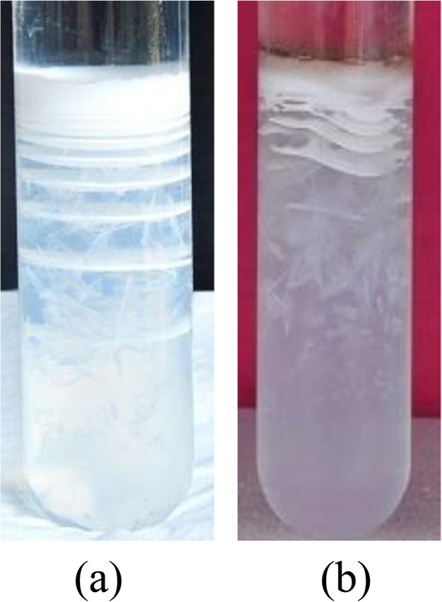
Anushya et al. Clinical Phytoscience       (2021) 7:43                                                          Page 2 of 14

Moreover, small amounts of calcium phosphate have              strontium, zinc and chromium [19]. Moreira et al. [20]
been found in the assumed binding portion of the stone         reported that the brushite stones were associated with
[11]. These observations suggest that other stones ori-        higher urinary calcium excretion and higher urinary
ginate from the precipitation of calcium phosphate and         supersaturation CaP when compared to apatite stones.
that the renal papilla is bound to the early stages of         Aggressive measures to reduce urinary calcium can be
these stones. Once a crystal nidus has been established,       especially helpful in preventing brushite stone formation.
it develops into a kidney stone by precipitation or by the        When any patient takes any drugs constantly and con-
process of crystal growth. Initially, kidney stones often      tinuously, it ends up with some side effects. In some cases,
have no symptoms. The first symptom of kidney stone is         the intake of drugs changes the biochemistry of the urine
extreme pain as stones block the flow of urine [12].           and it may induce the crystallization of metabolic com-
   Calcium Hydrogen Phosphate Dihydrate (CHPD,                 pounds of unusual morphology, which indicates the possi-
Brushite, and CaHPO4.2H2O) plays a major role in the           bility that there are peculiar conditions for the formation
urinary calculi formation. Calcium phosphate stone for-        of urinary stones. In order to find out the formation of
mation is related to conditions such as hyperparathyr-         urinary stones as being the unwanted effect of the intake
oidism [13] and renal tubular acidosis [14]. Brushite is a     of diabetic medicines, the present work is carried out. In
unique form of CaP that can form large symptomatic             this study, the A. marmelos is taken for consideration. A.
stones in certain patients. The molecular structure of         marmelos, known as Bael in India, is a famous Rutaceae
brushite is shown in Fig. 1. It can be difficult to treat      plant, one of Ayurveda’s most important plants [21]. Dif-
brushite stones as they are resistant to shock waves and       ferent parts of this plant, such as leaves, roots, seed, bark
ultrasonic lithotripsy and also require ballistic fragmen-     and fruit are used by the Ayurvedic practitioners due to
tation. Patients with brushite stone disease are less likely   its various medicinal properties [22, 23]. Traditionally,
to become stone free after surgery and often experience        fever, diabetes, diarrhea, abscesses and snake bites have
repetition in the stone despite maximum medical inter-         been treated with the plant [24, 25]. Moreover, extracts of
vention [16]. In order to evaluate the transportation of       A. marmelos have good anti-diabetic properties [26].
renal phosphate, Dhayat et al. [17] used the ratio of          Although several studies have been carried out with this
tubular maximum reabsorption of phosphate to glom-             plant, there is no documentation for the effect of diabetic
erular filtration rate (TmP/GFR) showing that low TmP/         drug A. marmelos on urinary stones. Hence, the present
GFR is correlated with increased excretion of calcium in       study is intended to investigate the effect of A. marmelos
the urine and increased prevalence of brushite stones.         leaves on urinary stone formation.
Xie et al. [18] showed that ACP plays a vital role in
nucleating calcium oxalate stones by promoting the ac-         Experimental
cumulation of amorphous calcium oxalate (ACO) pre-             Chemicals
cursors at early induction stages. As compared to other        Sodium Meta Silicate (SMS) – {Na2SiO3.9H2O} and
stones, phosphate stones were high in magnesium,               Calcium Acetate (Ca(C2H3O2)2) have been purchased
                                                               from Loboma chemicals Ltd. All other chemicals such as
                                                               Calcium Chloride (CaCl2) and Orthophosphoric acid
                                                               (H3PO4) have been purchased from Merck (Mumbai,
                                                               India). All the chemicals are of analytical grade and are
                                                               used without any further purification or post-treatment.
                                                               Double distilled water is used for cleaning the glass
                                                               wares.

                                                               Plant materials and identification
                                                               Fresh leaves of A. marmelos were collected from the nat-
                                                               ural surroundings and authenticated at the Department
                                                               of Botany, S.T. Hindu College, Nagercoil, Tamil Nadu,
                                                               India. The collected leaves were washed with double dis-
                                                               tilled water followed by shade drying. The dried leaves
                                                               were grounded into a fine powder and used for further
                                                               experiments.

                                                               Growth of brushite crystals
                                                               The single diffusion gel growth method is employed to
 Fig. 1 Molecular structure of brushite (Bindhu et al.) [15]
                                                               study the growth and inhibiting behavior of brushite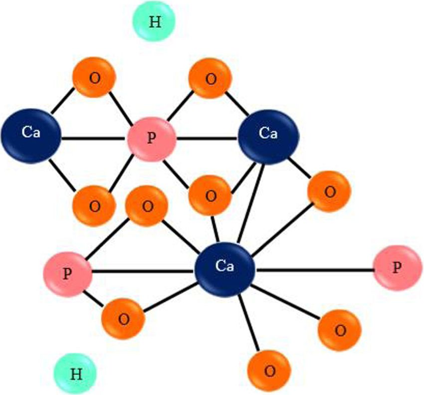
Anushya et al. Clinical Phytoscience   (2021) 7:43                                                                            Page 3 of 14

crystals using A. marmelos [27, 28]. For crystallization,             Fourier transform infrared spectroscopy
glass test tubes of size 140 mm length and 25 mm in                   The FTIR spectra were recorded in Shimadzu - FTIR
diameter are used in this method. Sodium meta-silicate                8400S with Spectral range: 4000–400 cm− 1 and Reso-
solution of specific gravity 1.03 g/cc has been impreg-               lution: 4 cm− 1 with KBr beam splitter and DLATGS
nated with orthophosphoric acid according to the de-                  Detector.
sired pH value. The solutions pH value is set to 6.5. The
time required to set the gel firmly was dependent on pH               Scanning Electron microscopy (SEM) and energy dispersive
value of the mixture. Within 48 h, good quality gels were             X-ray analysis (EDAX)
set in the test tubes for the pH value chosen above.                  The surface features of the crystals are studied using
Once when the solution undergoes a gelation phase, an                 Scanning Electron Microscope (SEM Model ZEISS EVO
aqueous solution of calcium chloride and calcium acet-                18). EDAX is recorded using dispersive spectrometer
ate of a specific molarity was carefully poured over the              (AMETEK EDAX) attached to the scanning electron
gel using pipette to prevent any breakage of the gel. The             microscope for carrying out elemental analysis of the
test tubes were capped with airtight stopples after pour-             crystals.
ing supernatant solution. The following reaction is ex-
pected to take place leading to the formation of calcium              Results and discussion
hydrogen phosphate dihydrate crystals.                                Growth kinetics
                                                                      The silica gel is set and 1.5 M concentration of calcium
                                                                      chloride and calcium acetate was poured on it, while we
                                                                      understand the growth mechanism, it is found that the
    CaCl2 :2H2 O þ CaC4 H6 O4 þ H3 PO4 →CaHPO4 :2H2 O þ CaCO3 þ HCl
                                                                      Ca2+ ions diffuse through the gel column and immedi-
                                                                      ately react with phosphate and precipitated forming Lie-
   To study the effect of A. marmelos leaf extract on the             segang rings in just 4–5 h. The number of Liesegang
growth of brushite crystals, the following method was                 rings gradually increase with time and a total of about 8
used. The finely powdered leaf extracts of different con-             rings are formed in the control test tubes (Fig. 2(a)). It is
centrations (10 mg to 50 mg) were separately added
along with calcium chloride and calcium acetate and the
crystals are grown as before. The Ca2+ ions and add-
itional ions available in the A. marmelos extract are dif-
fused into the gel and react with the phosphate ions in
the gel column. This reaction leads to the formation of
medicines added calcium hydrogen phosphate dihydrate
crystals thereby showing its promotery or inhibitory ef-
fect on these crystals. The above experiment was con-
ducted simultaneously with the control system to
compare the growth and morphology of the undoped
CHPD and A. marmelos doped CHPD crystals.
   The formation of Liesegang rings followed by subse-
quent crystallization and aggregation of CHPD crystals
were noted. In about 25 days, the crystal growth was
completed. The grown crystals removed and collected
carefully in a clean petri dish and then harvested using
distilled water to remove the gel. Then the harvested
crystals are dried by placing them at room temperature
in a filter paper. The dried crystals are finely powdered
and used for further characterization.

Characterization techniques
Powder X-ray diffraction
Powder X-ray diffraction (PXRD) is used to identify the
crystal structure and collected on a XPERT-PRO dif-
fractometer with CuKα radiation (λ = 1.54060 Å) over a
                                                                       Fig. 2 CHPD (Brushite) crystals grown in gel medium (a) Without
range of 10–80° in 0.05 step sizes with an integration
                                                                       Inhibitor, (b) 50 mg of A. marmelos added CHPD crystals
time of 10s.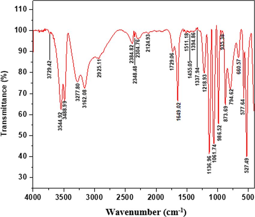
Anushya et al. Clinical Phytoscience (2021) 7:43 Page 4 of 14
found that the Liesegang rings of CHPD are formed be- negative charge of glutamate and asparate ions, for
cause of the diffusion of ions in the gel medium and instance, has little effect on the CHPD crystal faces,
shown that the diffusion kinetics is proved by studying although it can be enhanced by adding more
the distance [29, 30]. In due course of time, the crystals negative groups like OH−
are grown where the rings disappeared. The grown crys- 2. The amount of inhibition in a given crystalline face
tals of undoped CHPD are shown in Fig. 2(a). Figure 2(b) is specified by the structural fit between the organic
shows the grown crystals of CHPD by adding 50 mg of A. molecule and the ionic structure of the particular
marmelos. Comparing the undoped crystals, the number crystal face. This can have an effect on the
of crystals grown with A. marmelos is less. The results crystalline faces that are exposed to the solution.
infer the parameters such as the pH value, the concentra- The negatively charged small molecules such as
tion of reactants, and the column height of the solutions citrate ions, interact with the lateral surfaces of the
affect the formation of Liesegang rings [31, 32]. On the CHPD crystals and hence the crystallization is
other hand when we add herbal extracts to the super- slowed and the morphology of the crystals is
natant solution, we observe the less number of Liesegang modified.
rings and reduction in length of CHPD crystals grown. 3. The influence of the exposed hydration layer is seen
This may be due to the changes in the kinetics and diffu- on the surface of the crystal. In polyaspartic acid,
sion processes and inhibitory effectiveness [33]. such structural fit exist between the distances of
The Promotery/Inhibitory effect is judged on the basis carboxylic groups in the polyaspartic β and in the
of number of crystals grown and total mass of the grown case of CHPD, the distances of the calcium ion
crystals. The same condition is maintained for controlled across two adjacent layers make up a two-layer Ca-
and test samples. HPO4 below the hydrated layer parallel to the plane
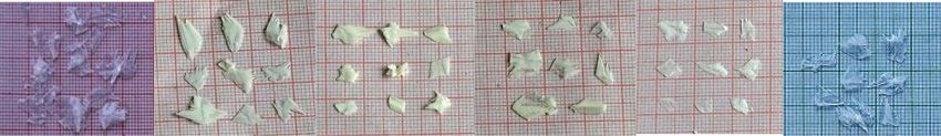
Figure 3 shows the harvested crystals. Figure 3 (a0) (010) [35].
shows undoped CHPD crystals. The crystals are in thin
platelet like structure with an average length of 1.2 cm.
The rest (a1-a5) show the CHPD crystals added with 10 Total mass of the grown CHPD crystals
mg, 20 mg, 30 mg, 40 mg and 50 mg of A .marmelos re- Figure 4 shows the histogram depicting total mass of
spectively. The average size of the A. marmelos doped CHPD crystals grown with A. marmelos at 10 mg, 20
CHPD crystals are less when compare to the undoped mg, 30 mg, 40 mg and 50 mg concentrations.
CHPD crystals. When the concentration of A. marmelos is For the cases of 10 mg to 50 mg doped samples, the
increased, the size of the crystals is decreased. At the same total mass is decreased than that of undoped. While
time it is reduced furthermore for 50 mg concentration. comparing with the prior concentrations, the total mass
Owing to its distinctive ionic structure, CHPD serves for 50 mg concentration is extremely low. The herbal
as a good model crystal for the study of interactions be- extracts (A. marmelos) contains phytochemicals such as
tween additives and crystals. Skiric et al. [34] have sum- alkaloids, terpenoids, amino acids, carbohydrates, flavo-
marized as follows: noids and phenols [36] etc. These phytochemicals inter-
act within themselves at low concentrations, and so the
1. The significance of the molecular structure and penetration decrease and so the total mass. At high con-
additive, i.e., tiny or very large molecule, number of centrations, the each constituent of phytochemicals ag-
chemical compounds in the molecule and its total glomerate forming an interface between the cations
charge in the growth of CHPD and other associated thereby decrease the formation of crystals.
crystals. This may be useful for selecting molecules The ANOVA statistical analysis is performed for total
that inhibit the growth of CHPD crystals. One mass and listed in Table 1.
Fig. 3 Harvested CHPD (Brushite) crystals grown without inhibitor (Undoped (a0)), with A. marmelos (a1-a5) respectively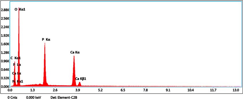
Anushya et al. Clinical Phytoscience       (2021) 7:43                                                                         Page 5 of 14

 Fig. 4 Promotery/Inhibitory effect of Brushite crystals

  The single factor analysis of variance (ANOVA)                               The XRD pattern of undoped CHPD crystals (Fig. 5)
followed by Tukeys test is carried out using origin 9                        show prominent peak at 2θ = 11.75° which corresponds
software. It is done to compare the total mass of                            to the (0 2 0) plane and the other peaks observed at
undoped with doped samples. ANOVA statistical ana-                           2θ = 21.00°, 23.47°, 29.32°, 30.53°, 34.19°, 35.60°, 37.10°,
lysis indicates that the variations in the total mass of                     39.69°, 41.47°, 42.27°, 45.42°, 47.90°, 48.53°, 50.24°,
CHPD with doped samples are highly significant at 0.05                       53.59°, 56.97°, 60.92° and 74.97° belongs to (1 2 1), (0 3
level.                                                                       1), (1 4–1), (1 2 1), (1 5 0), (0 6 0), (2 2–2), (1 6–1), (1
                                                                             5–2), (2 4 0), (1 2–3), (0 8 0), (0 6 2), (2 6 0), (3 4–1), (0
                                                                             9 1), (2 8–2) and (3 8–3) reflections, respectively, of
Powder X-ray diffraction                                                     monoclinic structure of CHPD (JCPDS No. 72–0713).
Powder X-ray diffraction study is performed on grown                         Compared to undoped CHPD crystals, the intensity of (0
crystals to identify the phase formation and degree of                       2 0) peak increases for the doped crystals. This results in
crystal perfection. Powder X-ray diffraction patterns are                    a decrease of peak intensity of all the remaining peaks.
recorded in 2θ steps of 0.05 between 10° - 80° using a                       The (0 2 0) peak appears with maximum intensity in
XPERT-PRO diffractometer with CuKα radiation at a                            undoped CHPD and medicines added CHPD crystals.
wavelength of 1.54060 Å operating at 30 mA and 40 kV.                        The higher intensities of (0 2 0) plane is a feature of typ-
The powder X-ray diffractogramfor both undoped and                           ical deviation from an isotropic orientation of crystallite
various medicines added CHPD crystals are shown in                           grains. The higher the number of plate like grains in the
Figs. 5 and 6 and the h k and l values in brackets are                       sample, the higher the fraction of the (0 2 0) planes
provided.                                                                    which are aligned along the specimen surface [34]. The
                                                                             highly resolved peaks at specific 2θ Bragg angles in the
                                                                             crystals indicate the crystalline nature of the grown crys-
Table 1 ANOVA statistical analysis of CHPD crystals
                                                                             tals. The software CellCalc is used to calculate lattice
Crystals     Group       Treatments                        Mean (gm) ± SD
                                                                             parameters and is tabulated in Table 2. The crystallo-
Brushite     A           Control (Undoped CHPD)            1.643 ± 0.41224
                                                                             graphic parameters obtained are well correlated with
             B           Control +A. marmelos              0.80 ± 0.08101    data available in the literature (JCPDS No [72–0713]).
                         10 mg                             0.70 ± 0.08101
                         20 mg                             0.68 ± 0.08101    The changes in the lattice parameters and unit cell
                         30 mg                             0.65 ± 0.08101    volume of doped samples may be attributed to the in-
                         40 mg                             0.58 ± 0.08101    corporation of added medicines in the host of CHPD
                         50 mg
                                                                             crystal.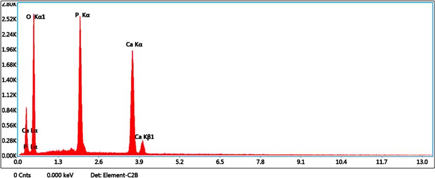
Anushya et al. Clinical Phytoscience   (2021) 7:43                    Page 6 of 14

 Fig. 5 Powder XRD spectrum of undoped CHPD crystal

 Fig. 6 Powder XRD spectrum of 50 mg A. marmelos added CHPD crystal
Anushya et al. Clinical Phytoscience        (2021) 7:43                                                                                    Page 7 of 14

Table 2 Unit cell parameters, Unit cell volume (V) and Angle (β) for undoped and A. marmelos added CHPD crystals
Samples                                     Unit cell parameters                                                Unit                        Angle
                                                                                                                cell                        (α = γ = 90°)
                                            a                      b                        c
                                                                                                                volume
                                            (Å)                    (Å)                      (Å)
                                                                                                                V
                                                                                                                (Å)3
Undoped CHPD                                5.87390                15.18049                 6.27126                 497.474                 β = 117.175
CHPD: 50 mg A. marmelos                     5.81243                15.09959                 6.23185                 489.266                 β = 116.549

Crystallite size                                                               with reference (JCPDS No: 72–0713 & 77–2303),
The crystallite size of the grown crystals is estimated                        using the relation between the stacking fault probabil-
from the Scherrer equation [37],                                               ity α and the peak shift Δ (2θ) [38].

           Kλ
    D¼                                                                                  2π 2
          βcosθ                                                                   α¼   pffiffiffi      Δ ð2θÞ
                                                                                     45 3 tanθ
  Where D is the crystallite size, K (=0.89) is the con-
stant, λ is the wavelength of the X-rays (λ = 0.54060 Å                          The Microstructural properties of the grown crystals
for CuKα radiation), β is the full width at half maximum                       calculated from the above formulas are given in Table 3.
value and θ is the Bragg diffraction angle.                                      The crystallite size, microstrain, Dislocation density
                                                                               and stacking fault probability of undoped CHPD and A.
Microstrain                                                                    marmelos added CHPD crystals are shown in Table 3.
The microstrain is calculated using the following rela-                        The crystallite size of the samples is determined by
tion [38].                                                                     employing Debye Scherrer’s equation. The crystallite size
                                                                               of undoped CHPD is around 51 nm. On doping with A.
                                                                               marmelos, the crystallite sizes are found to decrease.
          β cosθ                                                               This is probably due to the growth inhibition. The dis-
    ε¼
             4                                                                 location density of the doped crystals increases compar-
                                                                               ing with the undoped CHPD. This may be due to the
Dislocation density                                                            piling up of atoms one over the other. This in turn de-
A dislocation is an imperfection in a crystal associated                       forms the crystal structure and result into fracture due
with the misregistry of the lattice in one part of the crys-                   to brittleness. Thus the crystal formation is inhibited.
tal with that in another part [38, 39].                                        The stacking fault probability is found to increase on
  The dislocation density (δ) is evaluated by the formula                      doping, and no major changes are observed. Also it is
[40],                                                                          observed that the Microstructural parameters dislocation
                                                                               density and microstrain depend upon crystallite sizes.

          1                                                                    Fourier transform infrared spectroscopy
    δ¼
          D2                                                                   The structural studies of Calcium Hydrogen Phosphate
                                                                               Dihydrate crystals by IR spectra show various absorption
Stacking fault probability (α)                                                 bands which are characteristics of different vibrational
The stacking fault probability α is the fraction of                            modes. The FTIR spectra of undoped CHPD and 50 mg
layers undergoing stacking sequence faults in a given                          A. marmelos are shown in Fig. 7 and 8 and functional
crystal and hence one fault is expected to be found in                         groups are mentioned in Table 4.
  .                                                                              FTIR spectrum of undoped Calcium Hydrogen Phos-
1     layers. The stacking fault probabilities are calcu-
    α                                                                          phate Dihydrate (Fig. 7) show the absorption peaks at
lated from the shift of the X-ray line of the crystal                          wavenumbers range of 3162.08 cm− 1, 3277.8 cm− 1,

Table 3 Microstructural parameters of undoped and A. marmelos added CHPD crystals
Samples                                Crystallite size (nm)         Micro strain (ε)         Dislocation density             Stacking fault probability
                                                                                              δ × 1014 lines/m2               (α)
Undoped CHPD                           51                            1.69712                  3.84467                         0.03331
CHPD: 50 mg of A. marmelos             47                            0.85691                  4.52693                         0.09896
Anushya et al. Clinical Phytoscience (2021) 7:43 Page 8 of 14 Fig. 7 FTIR spectrum of undoped CHPD crystal Fig. 8 FTIR spectrum of 50 mg A. marmelos added CHPD crystal
Anushya et al. Clinical Phytoscience   (2021) 7:43                                                                       Page 9 of 14

Table 4 Band Assignments and absorption bands of undoped and A. marmelos added CHPD crystals
Sl. No              Observed IR frequencies wavenumbers (cm− 1)                   Band Assignments
                    Undoped CHPD                CHPD: 50 mg A. marmelos
1                   3729.42                     3721.99                           Weakly H bonded OH vibrations
2                   3544.92                     3544.92                           Weakly H bonded OH vibrations
3                   3488.99                     3488.02                           Weakly H bonded OH vibrations
4                   3277.8                      3278.76                           Weakly H bonded OH vibrations
5                   3162.08                     3162.08                           Weakly H bonded OH vibrations
6                   2925.11                     2928.41                           OH stretching vibrations
7                   2384.82                     2385.6                            Weak absorption HPO42−
8                   2348.48                     2346.83                           PO-H symmetric stretching vibrations
9                   2304.76                     2304.76                           O=P-OH Stretching vibrations
10                  2124.93                     2124.93                           O=P-OH Stretching vibrations
11                  1729.06                     1729.06                           Weak absorption HPO42−
12                  1649.02                     1648.06                           H-O-H Symmetric bending vibrations
13                  1511.18                     1509.53                           P=O stretching vibrations
14                  1455.05                     1454.26                           P=O stretching vibrations
15                  1394.86                     1364.34                           P=O asymmetric stretching vibrations
16                  1337.94                     1338.77                           P=O asymmetric stretching vibrations
17                  1218.93                     1215.07                           PO4 P=O associated Stretching vibrations
18                  1136.96                     1136.96                           PO4 P=O associated Stretching vibrations
19                  1061.74                     1062.7                            P=O Stretching vibrations
20                  986.52                      987.49                            P-O-P asymmetric stretching bond
21                  935.38                      –                                 P-OH stretching vibrations
22                  873.69                      873.69                            P-O-P asymmetric stretching bond
23                  794.62                      795.58                            P-O-P asymmetric stretching bond
24                  660.57                      656.72                            (H-O-) P=O bond (strong absorption) acid phosphates
25                  577.64                      577.64                            (H-O-) P=O bond (strong absorption) acid phosphates
26                  527.49                      527.49                            (H-O-) P=O bond (strong absorption) acid phosphates

3488.99 cm− 1, 3544.92 cm− 1 and 3729.42 cm− 1 are attrib-          wavenumbers 986.52 cm− 1, 873.69 cm− 1 and 794.62 cm− 1
uted to intermolecular and weakly H bonded OH because               are due to P-O-P asymmetric stretching vibrations [29,
of water of crystallization [41, 42]. The characteristic peak       42]. The strong absorption peaks of acid phosphates [29,
at 2925.11 cm− 1 is due to OH stretching vibrations of              48] lie at different wavenumbers of 660.57 cm− 1, 577.64
water [43] and the HPO42− weak absorption bands [30] is             cm− 1 and 527.49 cm− 1.
observed at 2384.82 cm− 1 and 1729.06 cm− 1. The band at              By comparing the FTIR spectra of undoped CHPD
2348.48 cm− 1 is attributed to PO-H symmetric stretching            and A. marmelos added CHPD, we determined that the
vibrations [44]. Two bands are observed due to O=P-OH               foremost distinction amongst them is the disappearance
stretching vibrations [45] of HPO42− near 2124.93 cm− 1,            of the peak encountered at 935.38 cm− 1 in the A. mar-
2304.76 cm− 1. An absorption band at 1649.02 cm− 1is oc-            melos added sample (Table 4) corresponding to P-OH
curred due to H-O-H symmetric bending vibrations [46]               stretching vibrations. The leaf extracts of A. marmelos
The absorption peak near 1511.18 cm− 1, 1455.08 cm− 1               contains phytochemicals like alkaloids, flavonoids, phe-
and 1061.74 cm− 1 are assigned due to P=O stretching vi-            nols, saponins, carbohydrates, protein, phytosterol, tan-
brations [29, 47]. The absorption takes place around                nins, glycosides, phenolic compounds, cardiac glycosides
1394.86 cm− 1 and 1337.94 cm− 1are due to P=O asymmet-              [36]. These phytochemicals are rich in polar groups
ric stretching vibrations [44]. The peaks about at 1218.93          which may interact with phosphate ion in CHPD leading
cm− 1 and 1136.96 cm− 1 are occurred due to P=O associ-             to the disappearance of P-O symmetric stretching vibra-
ated stretching vibrations of PO4, while peaks at                   tions respectively. Moreover, slight shifting in FTIR
Anushya et al. Clinical Phytoscience (2021) 7:43 Page 10 of 14 Fig. 9 SEM micrograph of undoped CHPD crystal Fig. 10 SEM micrograph of 50 mg A. marmelos added CHPD crystal
Anushya et al. Clinical Phytoscience   (2021) 7:43                                                               Page 11 of 14

 Fig. 11 EDAX spectrum of undoped CHPD crystal

spectra of doped samples suggests the encapsulation of           added CHPD crystals is shown in Fig. 10. Particles ag-
A. marmelos.                                                     glomerate and are in spherical shape. Particle size is in the
                                                                 range of 30–50 nm.
Scanning Electron microscopy                                       The SEM images of the undoped and A. marmelos
Particle size and Surface morphology of the grown crystals       added CHPD crystals show difference in their morpholo-
are revealed by scanning-electron microscopy (SEM                gies. The undoped CHPD crystals shows needle and
Model ZEISS EVO 18). The scanning electron micro-                platelets shaped morphology, whereas 50 mg A. marme-
graphs of the samples, undoped CHPD and 50 mg A. mar-            los added CHPD, we have the particles agglomerated
melos added CHPD crystals are shown in Figs. 9 and 10.           and are in spherical shape. The growth morphology of
  Figure 9 illustrates SEM photograph of undoped CHPD            CHPD crystals is typically in the form of thin plates with
crystals. Plate like structure is observed [15]. The particle    prominent (0 2 0) plane. The monoclinic unit cell of
size of undoped CHPD calculated using SEM picture is             CHPD consists alternating bi-layers of calcium, hydro-
around 94 nm. The SEM image of the 50 mg A. marmelos             gen phosphate ion and a layer of water molecules

 Fig. 12 EDAX spectrum of 50 mg A. marmelos added CHPD crystal
Anushya et al. Clinical Phytoscience (2021) 7:43 Page 12 of 14
oriented parallel to the (0 2 0) plane. As the bi-layers of Table 6 EDAX data for 50 mg A. marmelos added CHPD crystal
water molecules are exposed at the surface of (0 2 0) Elements Weight % Atomic %
face, the surface bound water can form bonds with mol- CK 17.1 24.8
ecules in the growth medium [49, 50]. The thin layer of NK 1.5 1.9
water molecules on the surface of the CHPD may form
OK 55.6 60.7
bonds with the phytochemicals of the A. marmelos ex-
tract, thus weakening the ionic nature of the CHPD and PK 11.0 6.2
leading to the structural modification of the doped crys- CaK 14.8 6.5
tals into tiny units. The reduction in platelet shape can
be attributed to the influence of added extracts onto the incorporation of A. marmelos caused a decrease in the
CHPD crystal formation altering the growth kinetics of number of grown brushite crystals and their average
the platelets [49, 51, 52]. The decrease in particle size of size. Powder X-ray diffraction exhibit monoclinic crystal
medicines added crystals may be due to the inhibition of structure and the lattice parameters are in good agree-
the grain growth. ment with the reported values. Also it confirms the in-
corporation of additional ions in the A. marmelos leaf
Energy dispersive X-ray analysis extracts into the host of CHPD. FTIR confirms the pres-
In order to study the elemental composition of undoped ence of functional groups. SEM shows the morpho-
and A. marmelos added CHPD crystals, qualitative and logical changes in the treated crystals. The SEM
quantitative analysis is performed by energy dispersive micrograph of crystals grown in the presence of A. mar-
X-ray analysis (EDAX) using dispersive spectrometer melos shows reduction in the grain size with addition of
(AMETEK EDAX) attached to the scanning electron A. marmelos as compared with pure CHPD crystals.
microscope. The spectrum obtained from EDAX analysis EDAX confirms the presence of required elements. From
of undoped CHPD and 50 mg A. marmelos added CHPD the above observed results, the intake of A. marmelos is
crystals is shown in Figs. 11 and 12 and the average good for diabetic patients as it reduces the growth of
atomic and weight percentage of individual elements is urolithiasis. However, further in vivo studies are needed
given in Tables 5 and 6. to evaluate its potential antiurolithiatic activity.
EDAX pattern of undoped CHPD (Fig. 11) shows the
presence of elements such as Calcium, Phosphorus and Abbreviations
CHPD: Calcium Hydrogen Phosphate Dihydrate; CaP: Calcium Phosphate;
Oxygen present in the grown crystal. This clearly indi- GFR: Glomerular Filtration Rate; ACP: Amorphous Calcium Phosphate;
cates that the crystals are of Calcium Hydrogen Phos- ANOVA: Analysis of Variance; JCPDS: Joint Committee on Powder Diffraction
phate Dihydrate indeed. The higher peak of Ca, P and O Standards
shows that the more concentrated the element are in the
Acknowledgements
specimen. The Ca/P ratio of undoped CHPD crystal if The authors are thankful to Department of Physics (DST-FIST), S.T. Hindu
found as 1.08, which is closely associated with the actual College, Nagercoil for providing facilities for the completion of this work.
value of 1 according to the chemical formula [53]. Simi-
Authors’ contributions
larly EDAX pattern of 50 mg A. marmelos added CHPD GA planned the study, designed the experiments and drafted the
crystal is shown in Fig. 12. The spectra corresponding to manuscript. RM participated in the preparation of herbal extracts and
50 mg A. marmelos added CHPD also show peaks corre- interpretation of results. THF supervised the project and reviewed the
manuscript. RR and GR involved in discussing the concepts. All authors read
sponding to all major elements Ca, P and O. The Ca/P and approved the final manuscript.
ratio of crystals grown with 50 mg A. marmelos is found
as 1.03. Funding
Not Applicable
Conclusion
Availability of data and materials
The effect of A. marmelos on the growth of brushite The datasets supporting the conclusions of this article are included within
crystals is examined in vitro. The present study reveals the article
that the herbal extracts of A. marmelos inhibit the
growth of brushite crystals. It is observed that the Declarations
Ethics approval and consent to participate
Table 5 EDAX data for undoped CHPD crystal Not Applicable
Elements Weight % Atomic %
Consent for publication
OK 55.5 73.6 Not Applicable
PK 18.5 12.7
Competing interests
Ca K 26.0 13.8
GA, RM, THF, RR and GR declare that they have no competing interests.Anushya et al. Clinical Phytoscience (2021) 7:43 Page 13 of 14
Author details 21. Balakumar S, Rajan S, Thirunalasundari T, Jeeva S. Antifungal activity of Aegle
1
Physics Research Centre, S.T. Hindu College, Nagercoil, Tamil Nadu 629 002, marmelos (L.) Correa (Rutaceae) leaf extract on dermatophytes. Asian Pac J
India. 2Department of Botany, S.T. Hindu College, Nagercoil, Tamil Nadu 629 Trop Biomed. 2011;1(4):309–12. https://doi.org/10.1016/S2221-1691(11)60049-X.
002, India. 3Department of Chemistry, The Madura College, Vidhya Nagar, 22. Chopra RN, Verma BS, Chopra IC. Supplement to Glossary of Indian
Madurai, Tamil Nadu 625 011, India. 4Department of Zoology, Pioneer Medicinal Plants. New Delhi: Publications & Information Directorate; 1969.
Kumaraswamy College, Nagercoil, Tamil Nadu 629 003, India. 23. Nirupama GS, Padmasri G, Vasanthi M. Comparative analysis of
phytochemical constituents present in various parts of Aegle marmelos.
Received: 22 July 2020 Accepted: 26 April 2021 Asian Pac J Trop Dis. 2012;2:S774–7. https://doi.org/10.1016/S2222-1808(12
)60263-1.
24. Narender T, Sweta S, Tiwari P, Pappi Reddy K, Khaliq T, Prathipati P, et al.
Antihyperglycemic and antidyslipidemic agent from Aegle marmelos. Bioorg
References Med Chem. 2007;17(6):1808–11. https://doi.org/10.1016/j.bmcl.2006.12.037.
1. Fakheriand RJ, Goldfard DS. Ambient temperature as a contributor to kidney 25. Karmase A, Birari R, Bhutani KK. Evaluation of anti-obesity effect of Aegle
stone formation: implications of global warming. Kid Int. 2011;79(11):1178– marmelos. Phytomedicine. 2013;20(10):805–12. https://doi.org/10.1016/j.
85. https://doi.org/10.1038/ki.2011.76. phymed.2013.03.014.
2. Eisner BH, Sheth S, Herrick B, Pais VM, Sawyer M, Miller N, et al. The effects 26. Phuwapraisirisan P, Puksasook T, Jong Aramruang J, Kokpol U.
of ambient temperature, humidity and season of year on urine composition Phenylethylcinnamides: a new series of a-glucosidase inhibitors from the
in patients with nephrolithiasis. BJU Int. 2012;110:1–4. leaves of Aegle marmelos. Bioorg Med Chem Lett. 2008;18(18):4956–8.
3. Rodgers AL. Race, ethnicity and urolithiasis: a critical review. Urolithiasis. https://doi.org/10.1016/j.bmcl.2008.08.024.
2013;41(2):99–103. https://doi.org/10.1007/s00240-012-0516-9. 27. Jethva H. Gel growth: a brief review. Mech Mater Sci Eng. 2017;9:1–7.
4. Trinchieri A. Epidemiological trends in urolithiasis: impact on our health care 28. Patel AR, Venkateswara RA. Crystal growth in gel media. Bull Mater Sci. 1982;
systems. Urol Res. 2006;34(2):151–6. https://doi.org/10.1007/s00240-005-002 4(5):527–48. https://doi.org/10.1007/BF02824961.
9-x. 29. Rajendran K, Dale KC. Growth and characterization of calcium hydrogen
5. Kohjimoto Y, Sasaki Y, Iguchi M, Matsumura N, Inagaki T, Hara I. Association phosphate dihydrate crystals from single diffusion gel technique. Cryst Res
of Metabolic Syndrome Traits and Severity of kidney stones: results from a Technol. 2010;45(9):939–45. https://doi.org/10.1002/crat.200900700.
Nationwide survey on Urolithiasis in Japan. Am J Kidney Dis. 2013;61(6):923– 30. Joshi VS, Joshi MJ. FTIR spectroscopic, thermal and growth morphological
9. https://doi.org/10.1053/j.ajkd.2012.12.028. studies of calcium hydrogen phosphate dihydrate crystals. Cryst Res
6. Coe FL, Evanand A, Worcester E. Kidney stone disease. J Clin Invest. 2005; Technol. 2003;38(9):817–21. https://doi.org/10.1002/crat.200310100.
115(10):2598–608. https://doi.org/10.1172/JCI26662. 31. Joseph KC, Joshi MJ. The study of the different parameters affecting
7. Parekh BB, Joshi MJ. Crystal growth and dissolution of brushite crystals by Liesegang rings formation during the growth of calcium hydrogen
different concentrations of citric acid solutions. Indian J Pure AP Phy. 2005; phosphate dihydrate crystals. Indian J Phys. 2002;76A(2):159–63.
43:675–8. 32. Joshi VS. Effect of supernatant solutions on the formation of Liesegang
8. Thomas WC, Howard JE. Studies on the mineralizing propensity of urine rings. Int J Innov. 2016;5(1):1027–31.
from patients with and without renal calculi. Trans Assoc Am Physicians. 33. Joshi VS, Parekh BB, Joshi MJ, Vaidya ADB. Inhibition of the growth of
1959;72:181. urinary calcium hydrogen phosphate dihydrate crystals with aqueous
9. Boyce WH, King JS. Present concepts concerning the origin of matrix and extracts of Tribulus terrestris and Bergenia Ligulata. Urol Res. 2005;33(2):80–6.
stones. Ann N Y Acad Sci. 1963;104(2):563–78. https://doi.org/10.1007/s00240-004-0450-6.
10. Tiselius HG. Stone incidence and prevention. Braz J Urol. 2000;26(5):452–62. 34. Sikiric M, Ivancic B, Milat O, Sarig S, Furedi MH. Factors influencing additive
11. Miller NL, Evan AP, Lingeman JE. Pathogenesis of renal calculi. Urol Clin N interactions with calcium hydrogen phosphate Dihydrate crystals. Langmuir.
Am. 2007;34(3):295–313. 2000;16(24):9261–6. https://doi.org/10.1021/la000704m.
12. Evan AP. Physiopathology and etiology of stone formation in the kidney 35. Joseph KC, Parekh BB, Joshi MJ. Inhibition of growth of urinary type calcium
and the urinary tract. Pediatr Nephrol. 2010;25(5):831–41. https://doi.org/10.1 hydrogen phosphate dihydrate crystals by tartaric acid and tamarind. Curr
007/s00467-009-1116-y. Sci. 2015;88(8):1–7.
13. National Endocrine and Metabolic Diseases Information Service. 36. Asaduzzaman M, Nahar L, Fazley Rabbi M, Hasan M, Khatun A, Tamannaa Z,
Hyperparathyroidism. (NIH Publication No.6–3425). 2006. https://www.niddk. et al. Phytochemicals, nutritional constituents, anti-bacterial and
nih.gov/-/media/Files/Endocrine- Diseases/Primary_Hyperparathyroidism_ hypoglycemic activity of Aegle Marmelos Lin. Leaf extract in Alloxan induced
508.pdf. Accessed 25 June 2018. diabetic rats. J Nutr Food Sci. 2016;6(4):1–7.
14. National Endocrine and Metabolic Diseases Information Service. Renal 37. Girase KD, Girase ND, Sawant DK, Patil HM, Bhavsar DS. Influence of Zn (II)
Tubular Acidosis. (NIH Publication No. 09–4696). 2008. https://www.niddk. doping on the structural and optical properties of gel grown Lead iodate
nih.gov/-/media/Files/Kidney-Disease/renaltubularacidosis_508.pdf. Accessed crystals. Adv Appl Sci Res. 2011;2(4):233–9.
25 June 2018. 38. Henry J, Mohanraj K, Sivakumar G. Electrical and optical properties of CZTS
15. Bindhu B, Veluraja K. Medical implications of Syzygium Cumini nut on the thin films prepared by SILAR method. J Asian Ceram Soc. 2016;4(1):81–4.
growth of Brushite crystals. Natl Acad Sci India Section A Physical Sci. 2018; https://doi.org/10.1016/j.jascer.2015.12.003.
89:587–92. 39. Thool GS, Singh AK, Singh RS, Gupta A, Susan MABH. Facile synthesis of flat
16. Krambeck AE, Handa SE, Evan AP, Lingeman JE. Brushite stone disease as a crystal ZnO thin films by solution growth method: a micro-structural
consequence of lithotripsy. Urol Res. 2010;38(4):293–9. https://doi.org/10.1 investigation. J Saudi Chem Soc. 2014;18(5):712–21. https://doi.org/10.1016/j.
007/s00240-010-0289-y. jscs.2014.02.005.
17. Dhayat NA, Luthi D, Schneider L, Mattmann C, Vogt B, Fuster GD. Distinct 40. Muthuselvi C, Dhavachitra M, Pandiarajan S. Growth and characterization of
phenotype of kidney stone formers with renal phosphate leak. Nephrol Dial aspirin crystal in the phosphoric acid medium. J Chem Pharm. 2016;8(5):
Transpl. 2019;34(1):129–37. https://doi.org/10.1093/ndt/gfy170. 804–14.
18. Xie B, Halter TJ, Borah BM, Nancollas GH. Aggregation of calcium phosphate 41. Madhurambal G, Subha R, Mojumdar SC. Crystallization and thermal
and oxalate phases in the formation of renal stones. Cryst Growth Des. characterization of calcium hydrogen phosphate dihydrate crystals. J Therm
2015;15(1):204–11. https://doi.org/10.1021/cg501209h. Anal Calorim. 2009;96(1):73–6. https://doi.org/10.1007/s10973-008-9841-1.
19. Abdel-Gawad M, Ali-El-Dein B, Mehta S, Al-Kohlany KM, Elsobky E. A 42. Yuvarani T, Manjula K, Ananda GP. Growth and characterization of calcium
correlation study between macro- and micro-analysis of pediatric urinary hydrogen phosphate Dihydrate crystals influenced by Costus igneus
calculi. J Pediatr Urol. 2014;10(6):1267–72. https://doi.org/10.1016/j.jpurol.2 aqueous extract. Int J Pharm Pharm Sci. 2017;9(5):173–8. https://doi.org/1
014.06.022. 0.22159/ijpps.2017v9i5.16838.
20. Moreira JM, Friedlander JI, Hartman C, Elsamra SE, Smith AD, Okeke Z. 43. Suryawanshi VB, Chaudhari RT. Spectroscopic studies of gel grown zinc
Differences in 24-hour urine composition between apatite and Brushite doped calcium hydrogen phosphate crystals. AIP Conference Proceedings
stone formers. Urology. 2013;82(4):768–72. https://doi.org/10.1016/j. of the 2nd International Conference on Condensed Matter and Applied
urology.2013.04.025. Physics. 2018;1953(1): 070025(1)-070025(4).Anushya et al. Clinical Phytoscience (2021) 7:43 Page 14 of 14
44. Trivedi MK, Branton A, Trivedi D, Nayak G, Bairwa K, Jana S. Spectroscopic
characterization of disodium hydrogen orthophosphate and sodium nitrate
after biofield treatment. J Chromatogr Sep Tech. 2015;6(5):1–5.
45. NIST Chemistry WebBook. http://webbook.nist.gov Accessed 5 July 2018.
46. Dalal PV, Saraf KB. Growth and study of barium oxalate single crystals in
agar gel. Bull Mater Sci. 2006;29(5):421–5. https://doi.org/10.1007/BF02914
071.
47. Hoffmann G, Veszpremi T, Nagy A. Properties of IR spectra of Phosphorus
doped SiO2 films; 1979. p. 176–84.
48. Borah BM, Lakshmi H, Das G. Biomimetic modulation of crystal morphology
using gel: from nano to micron-scale architectures. Mater Sci Eng C. 2008;
28(7):1173–82. https://doi.org/10.1016/j.msec.2007.10.059.
49. Curry NA, Jones DW. Crystal structure of brushite, calcium hydrogen
orthophosphate dihydrate: a neutron-diffraction investigation. J Chem Soc
A. 1971:3725–9. https://doi.org/10.1039/j19710003725.
50. Haninen D, Geiger A, Addadi L. Fibronectin adsorption to surfaces of
hydrated crystals. An analysis of the importance of bound water in protein
substrate interactions. Langmuir. 1993;9(4):1058–65. https://doi.org/10.1021/
la00028a030.
51. Abbona F, Christensson F, Franchini-Angela M, Ludager Madsen HE. Crystal
habit and growth conditions of brushite, CaHPO4.2H2O. J Cryst Growth.
1993;131(3–4):331–46. https://doi.org/10.1016/0022-0248(93)90183-W.
52. Diana KJ, George KV. Urinary stone formation: Efficacy of seed extract of
Ensete superbum (Roxb.) Cheesman on growth inhibition of calcium
hydrogen phosphate dihydrate crystals. J Cryst Growth. 2013;363:164–70.
53. Suguna K, Sekar C. Role of strontium on the crystallization of calcium
hydrogen phosphate Dihydrate (CHPD). J Minerals Materials
Characterization Eng. 2011;10(7):625–36. https://doi.org/10.4236/jmmce.2
011.107048.
Publisher’s Note
Springer Nature remains neutral with regard to jurisdictional claims in
published maps and institutional affiliations.You can also read