Short stature in two siblings heterozygous for a novel bioinactive GH mutant (GH-P59S) suggesting that the mutant also affects secretion of the ...
←
→
Page content transcription
If your browser does not render page correctly, please read the page content below
European Journal of Endocrinology (2013) 168 K35–K43                                                                                ISSN 0804-4643

CASE REPORT

Short stature in two siblings heterozygous for a novel bioinactive
GH mutant (GH-P59S) suggesting that the mutant also affects
secretion of the wild-type GH
Vibor Petkovic, Maria Consolata Miletta, Annemieke M Boot1, Monique Losekoot2, Christa E Flück, Amit V Pandey,
Andrée Eblé, Jan Maarten Wit3 and Primus E Mullis
Division of Pediatric Endocrinology, Diabetology and Metabolism, Department of Clinical Research, University Children’s Hospital Bern, Inselspital,
CH-3010 Bern, Switzerland, 1Division of Endocrinology, Department of Pediatrics, Beatrix Children’s Hospital, University Medical Center Groningen,
Groningen, The Netherlands, 2Center for Human and Clinical Genetics, Leiden University Medical Center, Leiden, The Netherlands and 3Department of
Pediatrics, Leiden University Medical Center, Leiden, The Netherlands
(Correspondence should be addressed to V Petkovic; Email: vibor.petkovic@dkf.unibe.ch)

                             Abstract
                             Objective: Short stature caused by biologically inactive GH is clinically characterized by lack of GH
                             action despite normal-high secretion of GH, pathologically low IGF1 concentrations and marked
                             catch-up growth on GH replacement therapy.
                             Design and methods: Adopted siblings (girl and a boy) of unknown family history were referred for
                             assessment of short stature (K4.5 and K5.6 SDS) at the age of 10 and 8.1 years respectively. They had
                             delayed bone ages (6.8 and 4.5 years), normal GH peaks at stimulation tests, and severely reduced
                             IGF1 concentrations (K3.5 and K4.0 SDS). Genetic analysis of the GH1 gene showed a heterozygous
                             P59S mutation at position involved in binding to GH receptor (GHR).
                             Results: Isoelectric focusing analysis of secreted GH in patient serum revealed the presence of higher
                             GH-P59S peak compared with that of wt-GH. Furthermore, computational simulation of GH-P59S
                             binding to GHR suggested problems in correct binding of the mutant to the GHR. In vitro GHR binding
                             studies revealed reduced binding affinity of GH-P59S for GHR (IC50, 30 ng/ml) when compared with
                             the wt-GH (IC50, 11.8 ng/ml) while a significantly decreased ability of the mutant to activate the
                             Jak2/Stat5 signaling pathway was observed at physiological concentrations of 25–100 ng/ml.
                             Conclusions: The clinical and biochemical data of our patients support the diagnosis of partial
                             bioinactive GH syndrome. The higher amount of GH-P59S secreted in their circulation combined with
                             its impact on the wt-GH function on GHR binding and signaling may alter GHR responsiveness to
                             wt-GH and could ultimately explain severe short stature found in our patients.

                             European Journal of Endocrinology 168 K35–K43

Introduction                                                                segments of GH that include the loop between residues
                                                                            54 and 74, the COOH-terminal half of helix 4, and
Human GH (hGH) plays a central role in many                                 the NH2-terminal region of helix 1 (3, 4). Binding site 2
physiological and metabolic processes (1). The actions                      consists of the residues near the NH2 terminus of the GH
of GH are important for regulating glucose, protein, and                    molecule and on the hydrophilic faces of helices 1 and 3
fat metabolism. GH is also needed for tissue mainten-                       (5). The binding of GH to the GHR recruits and induces
ance and repair throughout life and has a critical role                     signal transduction through cytosolic Jak2. Activation
for normal linear growth during childhood. It is mainly                     of the homodimeric GHR induces relative rotation of
synthesized, stored, and secreted by the somatotropes of                    two subunits, which brings the Jak2 in close proximity
the anterior pituitary gland as a protein that comprises                    allowing them to cross-phosphorylate themselves
a core of four helices in a parallel–antiparallel                           through tyrosine residues at the cytoplasmic tail of the
orientation with two disulfide bonds (2). The biological                    GHR (6, 7). These phosphotyrosines act as docking
actions of GH are mediated through activation of the GH                     points for cell signaling intermediates such as signal
receptor (GHR), which exists as an inactive dimer on the                    transducer and activator of transcription Stat5 (8).
surface of target cells. GH has two distinct domains                        Stat5 is recruited to the phosphorylated receptor tail,
(binding sites) with different affinities for binding to the                which results in its own phosphorylation by Jak2.
GHR. Binding site 1 (the high-affinity site) has been                       Phosphorylated-Stat5 forms a homodimer and trans-
identified as a patch composed of three discontinuous                       locates to the nucleus where it binds to a chromosomal

q 2013 European Society of Endocrinology                                                                                 DOI: 10.1530/EJE-12-0847
                                                                                                               Online version via www.eje-online.org

                                                                                                             Downloaded from Bioscientifica.com at 10/29/2020 05:19:34AM
                                                                                                                                                           via free access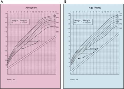
K36      V Petkovic and others                                                     EUROPEAN JOURNAL OF ENDOCRINOLOGY (2013) 168

GH-responsive element that drives transcriptional
regulation of multiple GH-responsive genes leading to
the biological effects of GH (8).
   One of the causes of growth failure is a disorder in the
GH–insulin-like growth factor 1 (IGF1) axis. In most cases
with sporadic isolated GH deficiency (GHD), the genetic
cause is unknown. The estimated incidence of GHD is
1/4000–10 000 live births (9, 10, 11), and much lower of
GH insensitivity and reduced bioactivity of the GH. The
diagnosis of ‘syndrome of bioinactive GH’ has often been
discussed and suggested in short children with the
phenotype resembling isolated GHD with normal or even
slightly elevated basal GH levels, low IGF1 concentration,
and normal catch-up growth on GH replacement therapy.
Short stature associated with bioinactive GH was first
described by Kowarski et al. (12) while additional cases
were reported in the 1980s on clinical basis (13, 14, 15,        Figure 1 Growth charts of the affected siblings. The charts of the girl
16, 17). Takahashi et al. (18) described a heterozygous          (A) and the boy (B) are shown together with percentiles (shown on
point mutation in the GH1 gene (D112G) found in a                the extreme right). The solid circles indicate the height measure-
                                                                 ments and the open circles corresponding to the bone ages. Full
Japanese patient with short stature. The D112G mutant            colour version of this figure available via http://dx.doi.org/10.1530/
involved a single nucleotide substitution within the GH          EJE-12-0847.
binding site 2 for the GHR, which interfered with a correct
binding to GHR/GH binding protein (GHBP) additionally            16.6 kg (K2.64 SDS), head circumference 49.7 cm
preventing dimerization of GHR (19). Further, six GH             (K1.75 SDS), and she was prepubertal. She had a
variants found in the heterozygous state were suggested to       strabismus of the left eye, had learning difficulties, and
be bioinactive by Millar et al. (20), but no clear correlation   was developmentally retarded by 3 years. The boy
between laboratory/clinical phenotype and patient geno-          presented with a height of 100.7 cm (K5.50 SDS) and
type was demonstrated. Moreover, in one of the more              a bone age of 4.5 years (Fig. 1B). Weight was 13.3 kg
convincing cases of bioinactive GH reported to date,             (K2.18 SDS), head circumference 50.4 cm (K1.25
a homozygous missense mutation C53S in the GH1 gene              SDS), and he was also prepubertal. He was born
led to disruption of the disulfide bridge between Cys-53         prematurely and had been admitted to hospital after
and Cys-165 in a short Serbian boy (21). Functional              birth but details about their medical history and their
studies demonstrated that both GHR binding and                   family history are missing. Furthermore, genetic analysis
Jak2/Stat5 signaling activity were significantly reduced         revealed a heterozygous P59S mutation in the GH1 gene
in the GH-C53S compared with wt-GH.                              in both patients (based on the HGVS nomenclature:
   Here, we describe two siblings with severe short              NM_000515.3, c.254COT; NP_000506.2, p.P85S).
stature carrying a heterozygous P59S mutation in GH1                At referral, laboratory assessment showed normal
gene. Clinical data of patients, which included normal           thyroid function, liver and renal function, screening for
GH peaks after stimulation, delayed bone ages and                celiac disease was negative, and no anemia or signs of
severely reduced IGF1 concentrations, suggested the              chronic infection were present. IGF1 of the girl was
diagnosis of bioinactive GH. The data of functional              70 ng/ml (K2.77 SDS) and that of the boy was
analysis combined with the clinical data of our patients         39 ng/ml (K3.01 SDS) while IGF-binding protein 3
support the diagnosis of partial bioinactive GH, which           (IGFBP3) of the girl was 1.69 mg/l (K1.5 SDS) and that
seems to be caused by the GH-P59S mutation also                  of the boy was 1.15 mg/l (K2.45 SDS). A GH
affecting the secretion of the endogenous wt-GH.                 stimulation test (clonidine) showed a GH peak of 17
                                                                 and 11 ng/ml respectively (23). Repeated analysis of
                                                                 IGF1 and IGFBP3 showed similar results. Further, IGF1
                                                                 generation tests were performed. After 2 weeks of rhGH
Subjects and methods                                             (0.7 mg/m2 per day), IGF1 increased in the girl and the
                                                                 boy from K2.61 and K3.16 SDS to K1.94 and C0.29
Patients                                                         SDS respectively. IGFBP3 increased from K1.22 and
Two siblings, a sister and her brother, adopted Roma             K2.09 SDS to K0.55 and C0.30 SDS respectively
children from Hungary, were referred to our clinic for           according to age and sex.
short stature at the age of 9.9 and 8.0 years respectively.
They had come to The Netherlands at the age of 6 and 4           Genetic analysis
years. At the first physical examination, the girl
presented with a height of 113.8 cm (K4.55 SDS)                  Genomic DNA was isolated from peripheral blood
and a bone age of 6.8 years (22) (Fig. 1A). Weight was           samples using the Autopure LS Instrument (Gentra

www.eje-online.org

                                                                                               Downloaded from Bioscientifica.com at 10/29/2020 05:19:34AM
                                                                                                                                             via free access
EUROPEAN JOURNAL OF ENDOCRINOLOGY (2013) 168 Severe short stature caused by a mutant GH (GH-P59S) K37
Systems). Direct sequencing of the GH1 and GHR genes (Sigma–Aldrich) for an additional 24 h when aliquots of
was carried out according to standard procedures culture medium were collected. Isoelectric focusing was
(primer sequences available upon request). Multiplex performed as described (25). Patient serum or culture
ligation-dependent probe amplification kits (P262 and media samples (200–300 ml) were electrofocused in a
P216, MRC Holland, The Netherlands) to detect buffer containing 1% hydroxypropyl methylcellulose
deletions and duplications were used according to the and 4% ampholine (pH gradient, 3.5–8.0) at 200 V
manufacturer’s instructions. for 12 h and then at 500 V for 12 h. The fractions
were collected and assayed for immunoreactive GH.
Cell culture and treatment
Hormonal measurement
Mouse pituitary (AtT-20/D16v-F2) cells were pur-
chased from American Type Culture Collection The serum GH was measured by the DSL-10-
(Manassas, VA, USA) and cultured in DMEM (4.5 g/l 1900 Active hGH ELISA assay kit (26) as described
glucose) supplemented with 10% heat-inactivated previously (21).
FCS (Life Technologies, Invitrogen AG) and 100 U/l
penicillin/streptomycin. This F2 subclone was developed 3D protein model and in silico mutagenesis
from the original AtT-20 cells, an ACTH-secreting cell of wt-GH
line established from a murine pituitary tumor.
Chinese hamster ovary (CHO-K1) cells were a gift The 3D structural model of wt-GH (NP_000506.2,
from Prof. U Wiesmann (Inselspital, Bern, Switzerland) P01241) was based on previously reported crystal
and were cultured in Ham’s F12 medium (Biochrom structure of hGH isoform 1 structure (PDB ID, 1HGU),
AG, Seromed, Berlin, Germany) supplemented with 10% which was chosen based on structure quality and
FCS, 100 U/l penicillin/streptomycin (Biochrom AG), and coverage of hGH sequence. Secondary structure features
2 mM L-glutamine (Gibco-BRL, Life Technologies). of the wt-GH structures were taken into account for the
Human embryonic kidney (HEK) 293 cells stably structure alignment. Amino acids 27–217 of human
expressing hGHR (293GHR) were a gift from Prof. R Ross wt-GH molecule were aligned with an X-ray crystal
(Northern General Hospital, Sheffield, UK) and were grown template structure to generate the structural alignments
in DMEM Nut F12 (Gibco), supplemented with 10% of full wt-GH sequence. We performed model building to
FCS, 100 U/l penicillin/streptomycin, 2 mM L-glutamine, repair the gaps in the X-ray crystal structure and enable
and 400 mg/ml geneticin G418 (Promega Corp.). the molecule to undergo molecular dynamic simulations
with the programs YASARA (27) and WHATIF (28).
First, a secondary structure prediction was performed
Production of GH peptides for building the missing parts in the crystal structure of
GH with the program DSC (29). The side chains in the
Wild-type GH was cloned in pcDNA3.1 (K) neo
newly built parts were optimized first by a steepest
(Invitrogen) vector as described previously (24). GH
descent and then a simulated annealing minimization.
mutant (GH-P59S) was made by site-directed mutagen-
At this stage, backbone atoms of aligned residues were
esis using the QuickChange Site-Directed Mutagenesis
kept fixed. The model was then subjected to 1000 ps
Kit from Stratagene AG (Basel, Switzerland). In order
refinement by molecular dynamic (30) simulation and
to produce GH variants, stable clones of wt-GH or then checked by the programs WHATCHECK (31),
GH-P59S were generated in CHO-K1 cells by trans- WHATIF (28), Verify3D (32, 33), ERRAT (34), and
fection with FuGene 6 (Roche Diagnostics AG). Ramachandran plot analysis (35, 36). In silico mutagen-
Concentrations of GH produced by CHO cells during esis was performed with YASARA and WHATIF and
3 days in serum-free Ham’s F12 medium were optimized by simulated annealing and a short 500 ps
measured by the DSL-GH ELISA Kit (DSL, Webster, TX, molecular dynamics (MD) simulation. Coordinates of
USA). To confirm that the mutation P59S does not affect two human hGH crystal structures (PDB IDs, 1HGU and
the affinity of the antibody used in DSL-ELISA, two 3HHR chain A) were used for comparative studies. The
different GH assays were performed on two samples of structural model of wt-GH and P59S mutant of GH in
CHO supernatant and the results were compared (21). complex with GHR was based on the structure of GHR
(amino acids N-49–254) in complex with GH (PDB ID,
Isoelectric focusing 3HHR) (2). Structure models were depicted with Pymol
(www.pymol.org) and rendered as ray-traced images
AtT-20 cells were cultured in DMEMC5% FCS in with POVRAY (www.povray.org). All numbering of
six-well plates and transfected using FuGene 6 (Roche amino acids in GHR is according to updated NCBI
Diagnostics AG). The cells were transiently transfected RefSeq of full-length GHR (NP_000154) containing 638
with 1 mg plasmid in total, containing either 1 mg amino acids, while numbering in older publications is
wt-GH or 0.5 mg of wt-GH and GH-P59S. After 24-h either N-49 (for 3HHR structure) (2)or N-18 (for 1A22
incubation, cells were stimulated with 50 mM forskolin structure) (37).
www.eje-online.org
Downloaded from Bioscientifica.com at 10/29/2020 05:19:34AM
via free accessK38 V Petkovic and others EUROPEAN JOURNAL OF ENDOCRINOLOGY (2013) 168
MD simulation for model refinement was then measured by the dual-luciferase reporter assay
(Promega) on a luminometer (Mediators PhL, Aureon
The MD simulations were performed using YASARA Biosystems, Vienna, Austria).
dynamics using AMBER03 force field (27). The
simulation cell was filled with water and the
AMBER03 (38) electrostatic potentials were evaluated Statistical analysis
for all water molecules; the one with the lowest or
The statistical significance of GH secretion was assessed
highest potential was turned into a sodium or chloride
using ANOVA one-way test plus Dunnett’s multiple
counter ion until the cell was neutral. We then ran
comparison test, comparing GH-P59S with wt-GH while
MD simulations with AMBER03 force field at 298K and
the statistical analysis for bioassay (testing bioactivity
0.9% NaCl in the simulation cell for 1000 ps to refine
through GHR binding and activation of Jak2/Stat5
the model. Simulation trajectories were analyzed with
pathway) was performed using the nonparametric
WHATIF functions and snapshots of simulation were
Mann–Whitney U test.
captured every 25 ps for further analysis. The best
model was selected for analysis and evaluation of
mutant amino acids.
Results
Receptor binding assay Analysis of GH in serum of the patients and
Receptor binding assays were performed using in culture medium of wt-GH and GH-P59S
293HEK cells stably expressing the hGHR (293GHR) expressing AtT-20 cells by isoelectric focusing
as described previously (21). Four independent experi- At stimulation tests, both patients had normal GH
ments were performed in triplicates and IC50 values for peaks. However, the ELISA-based method used to
the different GH peptides were determined by nonlinear measure GH concentration cannot distinguish the
regression analysis, using a single site competition secretion of wt-GH from that of GH-P59S. Therefore,
model (GraphPad Prism Software, version 5.0). to determine the specific concentration of each GH
variant, we performed isoelectric focusing of GH in
Luciferase reporter gene assay of Stat5 the serum of the affected patients (Fig. 2A), which
activation confirmed the presence of an increased GH-P59S peak
over the wt-form (area under the curve: P!0.05).
293GHR cells were used to assay Stat5 activation as In the control sample, only one peak (wt-GH) was detec-
described (39, 40). Briefly, cells were transfected with ted (Fig. 2B). The same analysis was also performed on
a Stat5-responsive luciferase reporter gene construct culture medium of forskolin-stimulated AtT-20 cells
(41, 42) and treated with increasing amounts of GH expressing wt-GH or both GH variants. The presence of
(wt-GH and GH-P59S) for 6 h. Luciferase expression two peaks was detected in medium of cells co-expressing
A 4 6.5 B 4 6.5
6.0 6.0
3 5.5 3 5.5
GH (ng/ml)
GH (ng/ml)
5.0 5.0
pH
pH
2 2
4.5 4.5
4.0 4.0
1 1
3.5 3.5
0 3.0 0 3.0
1 11 21 31 1 11 21 31
Fraction Fraction
C 4 6.5 D 4 6.5
6.0 6.0 Figure 2 Isoelectric focusing of GH in serum
3 5.5 3 5.5 from the male patient (A), of rhGH (B) and in
culture medium of AtT-20 cells co-expressing
GH (ng/ml)
GH (ng/ml)
5.0 5.0
wt-GH and GH-P59S (C) and expressing only
pH
pH
2 2
4.5 4.5 wt-GH (D). The serum (culture medium)
4.0 4.0 fractions were pooled separately and
1 1
assayed for GH immunoreactivity. The pH
3.5 3.5
gradient formed during isoelectric focusing is
0 3.0 0 3.0 indicated. The peaks for wt-GH and GH-P59S
1 11 21 31 1 11 21 31
are indicated by the open and solid arrows
Fraction Fraction respectively.
www.eje-online.org
Downloaded from Bioscientifica.com at 10/29/2020 05:19:34AM
via free accessEUROPEAN JOURNAL OF ENDOCRINOLOGY (2013) 168 Severe short stature caused by a mutant GH (GH-P59S) K39
may have an impact on its interaction with the acidic
E60, E62, and D182 residues on GHR (Fig. 3D). The
interaction of R64 in GH with E62 and D182 residues of
GHR has been reported to be important for GH–GHR
binding (reported as E44 and D164 based on N-18
numbering of amino acids in GHR) (37). The observed
changes affect multiple atomic contacts, but indivi-
dually, all can be considered to cause minor disturbance
in binding of GH-P59S to GHR and may rather lead to
moderate than to severe binding defects.
Functional analysis of the GH-P59S through
GHR binding and activation of the JAK/STAT
signaling pathway
As the computational analysis of wt-GH vs GH-P59S
binding to GHR performed through in silico mutagenesis
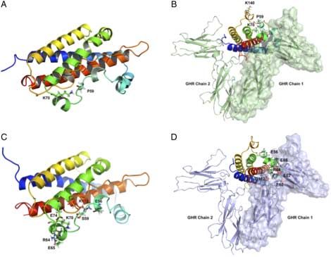
Figure 3 In silico mutagenesis and molecular dynamic simulations and MD simulation predicted lower binding of the
of GH-P59S binding to GHR. (A) The wt-GH shown as a ribbon
model with highlighted K70 and P59 residues. (B) Location of P59 in mutant variant, we performed GHR binding studies in
GH–GHR complex. (C) GH-P59S shown as a ribbon model 293GHR cells and compared the binding affinities of
with highlighted salt bridges between E56-K172, E65-R64, and wt-GH and GH-P59S (Fig. 4). The IC50 values for the
E74-K70. (D) GH-P59S superimposed on GH–GHR complex: the rhGH, wt-GH, and GH-P59S were found to be 13, 12,
positions of E56 and E65 in GH as well as the interaction between
R64 residue in GH and E60, E62, and D182 residues in GHR are and 30 ng/ml respectively. No significant difference was
highlighted. Full colour version of this figure available via http://dx. observed between the IC50 values from wt-GH and rhGH
doi.org/10.1530/EJE-12-0847. while a statistically significant difference was found
between the IC50 values from wt-GH and GH-P59S
wt-GH and GH-P59S (Fig. 2C; area under the curve of (ANOVA, P!0.05), confirming the reduced binding
GH-P59S vs wt-GH: P!0.05) while one peak corre- affinity of GH-P59S for the GHR.
sponding to wt-GH (Fig. 2D) was detected in culture Improper binding of GH variants to the GHR has an
medium of AtT-20 cells expressing only wt-GH. impact on the Jak2/Stat5 signaling cascade down-
stream of GHR. To investigate whether reduced binding
3D-structural model of wt-GH, generation of of GH-P59S variant consequently evoked abnormalities
GH-P59S by in silico mutagenesis, and MD in the activation of Jak2/Stat5 signaling pathway, we
simulation of GHR binding performed a bioassay using the combination of 293GHR
cells stably expressing GHR and Stat5-responsive
Based on its position in the GH molecule located within luciferase reporter gene assay system (23, 24). Using
the segment between residues 54–74 that are tightly in this in vitro system, which requires all steps of the
contact with GHR, P59 residue is considered to
contribute to the proper binding of GH to GHR through 125
rhGH
the GH binding site 1. Therefore, a mutation introduced
at this position might lead to defects in GHR binding. To wt-GH
100
Specific binding (%)
investigate this in more detail, the binding of GH-P59S GH-P59S
to GHR was analyzed by in silico mutagenesis following 75
the MD simulation and compared with wt-GH. In the
hGH crystal structure as well as in the repaired model,
50
the P59 is in contact with K70 to stabilize the small
helical turn containing R64 residue that interacts with
25
GHR (Fig. 3A and B). Energy analysis showed no drastic
changes, and P59S caused no change in protein
stability or changes in the core structure of the GH 0
molecule. However, multiple changes in local environ- 0.0 0.5 1.0 1.5 2.0 2.5 3.0 3.5
ment and bond order were observed, which may have Log (unlabeled GH; ng/ml)
an impact on the function of GH-P59S variant. A major
change was formation of additional salt bridges between Figure 4 Competitive displacement of 125I-GH binding to the GHR
by rhGH (positive control), wt-GH, and GH-P59S. Results are
E56-K172, E65-R64, and E74-K70 observed in the normalized to the amount of 125I-GH bound in the absence of any
GH-P59S (Fig. 3C). The R64 residue is crucial for unlabeled GH. Each point is the mean of three separate
binding to GHR and minor changes to its surroundings experiments performed in triplicateGS.D. (nZ3).
www.eje-online.org
Downloaded from Bioscientifica.com at 10/29/2020 05:19:34AM
via free accessK40 V Petkovic and others EUROPEAN JOURNAL OF ENDOCRINOLOGY (2013) 168
Jak2/Stat5 signaling pathway to be functional, we were (wt-GH 25). Moreover, the stimulation of the Jak2/Stat5
able to quantify the signal transduction activity of signaling pathway induced by both the wt-GH and
GH-P59S and to compare it with that of wt-GH. As the GH-P59S at 25 ng/ml (P59S 25/wt-GH 25) and at
expected and in line with the binding data, the GH-P59S 50 ng/ml (P59S 50/wt-GH 50) was significantly
mutant displayed reduced ability to activate the reduced (P!0.01) when compared with the stimu-
Jak2/Stat5 pathway when compared with wt-GH lation evoked only by wt-GH at 50 ng/ml (wt-GH 50)
(Fig. 5A). Significantly reduced bioactivity of GH-P59S and at 100 ng/ml (wt-GH 100). Three negative controls
was observed at the physiological dose of 25 ng/ml were used in this experiment: PSA, corresponding to
(P!0.05) as well as at 50 ng/ml (upper normal range) the supernatant of CHO cells transfected with pSecPSA;
and 100 ng/ml (P!0.01). No significant difference empty, representing the supernatant of CHO cells
in bioactivity between GH-P59S and wt-GH was found transfected with an empty pSec plasmid; and NT,
at the doses considered as sub-physiological (5 and which is the supernatant of non-transfected CHO cells.
10 ng/ml) and supra-physiological (200 and Furthermore, the size of 22 kDa for both wt-GH and
400 ng/ml) (Fig. 5A). GH-P59S was confirmed in protein extracts from CHO
As shown in Fig. 5B, co-stimulation of the 293GHR cells by western blot (data not shown).
cells with 12.5 ng/ml of wt-GH and GH-P59S (P59S
12.5/wt-GH 12.5) displayed a significantly reduced
activation of the Jak2/Stat5 pathway (P!0.05) when Discussion
compared with that evoked by the wt-GH at 25 ng/ml
A heterozygous missense mutation in GH1 gene
converting codon 59 from P (proline) to S (serine) was
A 300 identified in siblings (girl and a boy) presenting with the
clinical symptoms of severe growth retardation (K4.5
Percentage of wt-GH 50
250
and K5.6 SDS) and delayed bone ages (6.8 and 4.5
200
** years) at chronological ages of 10 and 8.1 years and
150
**
severely reduced IGF1 concentrations (K3.5 and K4.0
100 SDS). GH peaks of 17 and 11 ng/ml at stimulation tests
*
50
were found to be within normal range and a recently
performed IGF1 generation test revealed normal
0
increase in IGF1 following rhGH injections. Analysis
wt-GH 2
P59S 2
wt-Gh 10
P59S 10
wt-Gh 25
P59S 25
wt-Gh 50
P59S 50
wt-Gh 100
P59S 100
wt-Gh 200
P59S 200
wt-Gh 400
P59S 400
of GH in serum (isoelectric focusing) showed that the
abnormal peak (GH-P59S) was higher than the normal
B
one (wt-GH). The area under the peak corresponding to
300
the GH mutant variant was significantly higher when
Percentage of wt-GH 50
250 compared with that under wt-GH peak. Isoelectric
**
200 ** focusing of GH in culture medium from AtT-20 cells
150 ** co-expressing wt-GH and GH-P59S (mimicking hetero-
**
100
zygous conditions found in patients) detected both GH
*
peaks of the size comparable with that found in the
50
serum of patients. This proved our AtT-20-based
0 cellular model to be suitable and reliable to investigate
wt-GH 50
P59S 50
wt-GH 25
P59S 12.5/wt-GH 12.5
P59S 25/wt-GH 25
P59S 50/wt-GH 50
wt-GH 100
PSA
Empty
NT
these events in vitro.
Based on the clinical data, the diagnosis of partial
bioinactive GH syndrome was suggested. As the patients
were adopted siblings of unknown clinical family
history, no DNA from biological parents or other family
members was available for genetic analysis to check
Figure 5 (A) Jak2/Stat5 signaling capacity of wt-GH and GH-P59S. other individuals for the same mutation.
Results of wt-GH (black bars) and GH-P59S (white bars) The bioinactive GH syndrome is characterized by
stimulation are expressed as x-fold induction relative to the basic genetic defects in the GH1 gene giving rise to GH
activity of unstimulated cells and represent the meanGS.D. of three
separate experiments performed in duplicate (nZ3). (B) Effect of
variants (mutants) that are secreted from the pituitary
the co-stimulation of the 293GHR cells with wt-GH and GH-P59S. gland in the ‘classical’ pulsatile manner, reaching
Results of stimulation with wt-GH (black bars), GH-P59S (white normal or slightly high concentrations in circulation.
bars), co-stimulation with both (dark gray bars), and with negative These GH mutants often display either overall structural
controls (light gray) are expressed as percentage of wt-GH 50 aberrances or defects that are localized to the binding
activity and represent the meanGS.D. of three separate experi-
ments performed in duplicate (nZ3). **P!0.01, *P!0.05. sites for GHR, each of which affects their correct binding
Statistical significance was assessed using the nonparametric to GHR, impacting the Jak2/Stat5 signaling pathway.
Mann–Whitney U test. Comparative analysis of the protein sequence of hGH
www.eje-online.org
Downloaded from Bioscientifica.com at 10/29/2020 05:19:34AM
via free accessEUROPEAN JOURNAL OF ENDOCRINOLOGY (2013) 168 Severe short stature caused by a mutant GH (GH-P59S) K41
with orthologs from various vertebrate species clearly when compared with wt-GH, which was comparable to
demonstrated that the P59 residue has been conserved that of GH-P59L mutation in the previous report (43).
throughout evolution. Substitution of proline (P) by The Jak2/Stat5 pathway is thought to be the most
leucine (L) at position 59 in the GH1 gene has been important signaling pathway attributed to the growth-
recently reported to cause partial GHD combined with promoting effects of GH (45), and improper binding of
features of bioinactive GH syndrome in a patient GH to GHR may lead to subsequent Jak2/Stat5 signaling
presenting with modest growth retardation (43). abnormalities. The data we obtained in the bioassay
Comparative analysis of 3D structural models of clearly demonstrated significantly lower capability of
wt-GH and GH-P59L revealed no difference in protein GH-P59S to activate Jak2/Stat5 pathway when
stability or in core structure between these GH variants. compared with wt-GH and the absence of a synergistic
Moreover, MD simulation of wt-GH and GH-P59L effect between these GH variants in Jak2/Stat5 acti-
binding to GHR suggested reduced binding interactions vation. In addition, these data also revealed that
of GH-P59L with GHR, which was confirmed through GH-P59S was slightly more potent in Jak2/Stat5
competition-binding and signaling studies (bioassay). pathway activation than GH-P59L, as 25 ng/ml
In this study, our experimental data obtained in vitro GH-P59S was the lowest concentration at which
(analysis of GH secreted in serum, GHR binding, and statistically significant reduction in bioassay was
signaling data) complement the clinical data of our observed as opposed to 50 ng/ml in the case of
patients making them well in line with the suggested GH-P59L (43). Taken together, the results generated in
diagnosis of partial bioinactive GH syndrome. The the functional analysis of GH-P59S mutant are quite
mutation identified in these patients was at the same similar to those obtained through characterization of
position (P59) in GH1 gene, as in the study mentioned GH-P59L mutation previously reported. However, of
earlier, but with the difference that serine (S) was the importance and new is that GH-P59S is able to have an
substituting amino acid. Moreover, the patients
impact on secretion of the wt-GH as demonstrated by the
carrying GH-P59S mutation presented with severe
isoelectric focusing data. This fact may explain the
growth failure (based on SDS for age and height) as
dramatic influence on height and height velocity when
opposed to a patient with GH-P59L mutation reported
compared with GH-P59L.
with modest growth retardation. In order to explain
such a broad difference in phenotype (short stature) In conclusion, the biochemical data of GH-P59S
evoked by S vs L substitution at position 59, we decided functional analysis are in line with clinical data
to investigate in more detail the impact of GH-P59S supporting the diagnosis of partial bioinactive GH
mutation at the molecular and cellular level. The syndrome. GH secretion (based on GH stimulation
crystallographic model of the hGH/GHBP complex tests) seems to be normal in these patients while the
(44) revealed that the P59 is located in the long analysis of GH in serum from the patients revealed a
crossover loop between helices 1 and 2 of the GH significantly higher amount of the mutant variant
molecule. Furthermore, the studies of homolog- and compared with wt-GH. Hence, the secretion data
alanine-scanning mutagenesis identified the segment combined with effects of GH-P59S on GHR binding
between residues 54 and 74 to be tightly in contact with and signaling may explain severe short stature found in
GHR, in which the residues are strictly close to P59 these patients. Alternatively, the mutant variant might
(namely F54, E56, I58, and R64) were confirmed to be alter responsiveness of GHR to either wt-GH and/or
important in in vitro receptor binding (3, 4). Similar to rhGH treatment ultimately having an impact on normal
the case of GH-P59L, comparative analysis of 3D growth. Finally, as reported in this study, even a slight
structural models of wt-GH and GH-P59S showed that alteration at a same position (like amino acid replace-
protein stability and the core structure of GH molecule ment) in GH has an impact on its function and
were not affected by P59S mutation and that the overall highlights that not only genetic studies but also
3D structure of the mutant variant does not differ from functional analysis is of importance in defining the
that of the wt-GH. Analysis of GH binding to GHR mechanism of action of any novel GH mutations also
performed by MD simulation takes into consideration heterozygously inherited.
the influence of local factors (pH, temperature, and
water), the nature of amino acid substitution intro- Declaration of interest
duced (serine for proline), and its position in GH/GHR
complex. This approach proved effective to predict the The authors declare that there is no conflict of interest that could be
behavior and interaction of amino acid replacement perceived as prejudicing the impartiality of the research reported.
with other residues helping us to anticipate GHR
binding abnormalities. Our analysis of wt-GH and Funding
GH-P59S binding to GHR yielded reduced binding This study was supported by a grant of Swiss National Science
interactions of GH-P59S with GHR. Competition- Foundation 320000-121998 to P E Mullis and grants from
binding studies confirmed these predictions revealing Schweizerischen Mobiliar Genossenschaft Jubiläumsstiftung and
significantly lower affinity of GH-P59S for the GHR Novartis foundation for Biomedical Research to A V Pandey.
www.eje-online.org
Downloaded from Bioscientifica.com at 10/29/2020 05:19:34AM
via free accessK42 V Petkovic and others EUROPEAN JOURNAL OF ENDOCRINOLOGY (2013) 168
References 19 Chihara K, Takahashi K, Kaji H, Goji K, Okimura Y & Abe H. Short
stature caused by natural growth hormone antagonist. Hormone
1 Rosenfeld RG & Hwa V. The growth hormone cascade and its Research 1998 49 41–45. (doi:10.1159/000053067)
role in mammalian growth. Hormone Research 2009 71 (Suppl 2) 20 Millar DS, Lewis MD, Horan M, Newsway V, Easter TE, Gregory JW,
36–40. (doi:10.1159/000192434) Fryklund L, Norin M, Crowne EC, Davies SJ et al. Novel mutations
2 De Vos AM, Ultsch M & Kossiakoff AA. Human growth hormone of the growth hormone 1 (GH1) gene disclosed by modulation of
and extracellular domain of its receptor: crystal structure of the clinical selection criteria for individuals with short stature.
the complex. Science 1992 255 306–312. (doi:10.1126/science. Human Mutation 2003 21 424–440. (doi:10.1002/humu.10168)
1549776) 21 Besson A, Salemi S, Deladoey J, Vuissoz JM, Eble A,
3 Cunningham BC, Jhurani P, Ng P & Wells JA. Receptor and Bidlingmaier M, Burgi S, Honegger U, Fluck C & Mullis PE. Short
antibody epitopes in human growth hormone identified by stature caused by a biologically inactive mutant growth hormone
homolog-scanning mutagenesis. Science 1989 243 1330–1336. (GH-C53S). Journal of Clinical Endocrinology and Metabolism 2005
(doi:10.1126/science.2466339) 90 2493–2499. (doi:10.1210/jc.2004-1838)
4 Cunningham BC & Wells JA. High-resolution epitope mapping 22 Tanner JM, Whitehouse RH, Marshall WA, Healy MJR & Goldstein H.
of hGH-receptor interactions by alanine-scanning mutagenesis. In Assessment of Skeletal Maturity and Prediction of Adult Height (TW2
Science 1989 244 1081–1085. (doi:10.1126/science.2471267) Method). London: Academic Press, Inc. Ltd., 1975.
5 Cunningham BC, Ultsch M, De Vos AM, Mulkerrin MG, Clauser KR 23 Mullis P & Ranke, MB. In Diagnostics of Endocrine Function in
& Wells JA. Dimerization of the extracellular domain of the human Children and Adolescents (4th revised and extended edition). Basel:
growth hormone receptor by a single hormone molecule. Science Karger Publishers, 2011.
1991 254 821–825. (doi:10.1126/science.1948064) 24 Deladoëy J, Stocker P & Mullis PE. Autosomal dominant GH
6 Brown RJ, Adams JJ, Pelekanos RA, Wan Y, McKinstry WJ, deficiency due to an Arg183His GH-1 gene mutation: clinical and
Palethorpe K, Seeber RM, Monks TA, Eidne KA, Parker MW et al. molecular evidence of impaired regulated GH secretion. Journal of
Model for growth hormone receptor activation based on subunit Clinical Endocrinology and Metabolism 2001 86 3941–3947.
rotation within a receptor dimer. Nature Structural and Molecular (doi:10.1210/jc.86.8.3941)
Biology 2005 12 814–821. (doi:10.1038/nsmb977) 25 Tsvetnitsky V, Auchi L, Nicolaou A & Gibbons WA. Character-
7 Gent J, Van Den Eijnden M, Van Kerkhof P & Strous GJ. Dimerization ization of phospholipid methylation in rat brain myelin.
and signal transduction of the growth hormone receptor. Molecular Biochemical Journal 1995 307 239–244.
Endocrinology 2003 17 967–975. (doi:10.1210/me.2002-0261) 26 Tildsley GJ & Dilly SA. Audit of general surgical pathology
8 Herrington J & Carter-Su C. Signaling pathways activated by the experience of histopathology trainees. Journal of Clinical Pathology
growth hormone receptor. Trends in Endocrinology and Metabolism 1991 44 424–427. (doi:10.1136/jcp.44.5.424)
2001 12 252–257. (doi:10.1016/S1043-2760(01)00423-4) 27 Krieger E, Darden T, Nabuurs SB, Finkelstein A & Vriend G.
9 Lacey KA & Parkin JM. Causes of short stature. Lancet 1974 303 Making optimal use of empirical energy functions: force-field
42–45. (doi:10.1016/S0140-6736(74)93041-4) parameterization in crystal space. Proteins 2004 57 678–683.
10 Rona RJ & Tanner JM. Aetiology of idiopathic growth hormone (doi:10.1002/prot.20251)
deficiency in England and Wales. Archives of Disease in Childhood 28 Vriend G. WHAT IF: a molecular modeling and drug
1977 52 197–208. (doi:10.1136/adc.52.3.197) design program. Journal of Molecular Graphics 1990 8 52–56.
11 Vimpani GV, Vimpani AF, Lidgard GP, Cameron EHD & (doi:10.1016/0263-7855(90)80070-V)
Farquhar JW. Prevalence of severe growth hormone deficiency. 29 King RD, Saqi M, Sayle R & Sternberg MJ. DSC: public domain
BMJ 1977 2 427–430. (doi:10.1136/bmj.2.6084.427) protein secondary structure predication. Computer Applications in
12 Kowarski AA, Schneider J, Ben-Galim E, Weldon VV & the Biosciences 1997 13 473–474.
Daughaday WH. Growth failure with normal serum RIA–GH 30 Hamdan M, Bordini E, Galvani M & Righetti PG. Protein alkylation
and low somatomedin activity: somatomedin restoration and by acrylamide, its N-substituted derivatives and cross-linkers and
growth acceleration after exogenous GH. Journal of Clinical its relevance to proteomics: a matrix assisted laser desorption/
Endocrinology and Metabolism 1978 72 461–464. (doi:10.1210/ ionization-time of flight-mass spectrometry study. Electrophoresis
jcem-47-2-461) 2001 22 1633–1644. (doi:10.1002/1522-2683(200105)22:9
13 Bright GM, Rogol AD, Johanson AJ & Blizzard RM. Short stature !1633::AID-ELPS1633O3.0.CO;2-C)
associated with normal growth hormone and decreased somato- 31 Hooft RW, Vriend G, Sander C & Abola EE. Errors in protein
medin-C concentrations: response to exogenous growth hormone. structures. Nature 1996 381 272. (doi:10.1038/381272a0)
Pediatrics 1983 71 576–580. 32 Bowie JU, Luthy R & Eisenberg D. A method to identify protein
14 Frazer TE, Gavin JR, Daughaday WH, Hillman RE & Weldon VV. sequences that fold into a known three-dimensional structure.
Growth hormone dependent growth failure. Journal of Pediatrics Science 1991 253 164–170. (doi:10.1126/science.1853201)
1982 101 12–15. (doi:10.1016/S0022-3476(82)80171-6) 33 Luthy R, Bowie JU & Eisenberg D. Assessment of protein
15 Hayek A & Peake GH. A new syndrome of short stature due models with three-dimensional profiles. Nature 1992 356
to biologically inactive but immunoreactive growth hormone. 83–85. (doi:10.1038/356083a0)
Pediatric Research 1978 12 413. (doi:10.1203/00006450-197 34 Colovos C & Yeates TO. Verification of protein structures: patterns
804001-00304) of nonbonded atomic interactions. Protein Science 1993 2
16 Plotnick LP, Van Meter QL & Kowarski AA. Human growth 1511–1519. (doi:10.1002/pro.5560020916)
hormone treatment of children with growth failure and normal 35 Ramachandran GN, Ramakrishnan C & Sasisekharan V. Stereo-
growth hormone levels by immunoassay: lack of correlation with chemistry of polypeptide chain configurations. Journal of Molecular
somatomedin generation. Pediatrics 1983 71 324–327. Biology 1963 7 95–99. (doi:10.1016/S0022-2836(63)80023-6)
17 Rudman D, Kutner MH, Blackston RD, Cushman RA, Bain RP & 36 Hooft RW, Sander C & Vriend G. Objectively judging the quality
Patterson JH. Children with normal-variant short-stature: of a protein structure from a Ramachandran plot. Computer
treatment with human growth hormone for six months. New Applications in the Biosciences 1997 13 425–430.
England Journal of Medicine 1981 305 123–131. (doi:10.1056/ 37 Clackson T, Ultsch MH, Wells JA & de Vos AM. Structural and
NEJM198107163050302) functional analysis of the 1:1 growth hormone:receptor complex
18 Takahashi Y, Shirono H, Arisaka O, Takahashi K, Yagi T, Koga J, reveals the molecular basis for receptor affinity. Journal of Molecular
Kaji H, Okimura Y, Abe H, Tanaka T et al. Biologically inactive Biology 1998 277 1111–1128. (doi:10.1006/jmbi.1998.1669)
growth hormone caused by an amino acid substitution. 38 Liu H, Elstner M, Kaxiras E, Frauenheim T, Hermans J & Yang W.
Journal of Clinical Investigation 1997 100 1159–1165. (doi:10. Quantum mechanics simulation of protein dynamics on long
1172/JCI119627) timescale. Proteins 2001 44 484–489. (doi:10.1002/prot.1114)
www.eje-online.org
Downloaded from Bioscientifica.com at 10/29/2020 05:19:34AM
via free accessEUROPEAN JOURNAL OF ENDOCRINOLOGY (2013) 168 Severe short stature caused by a mutant GH (GH-P59S) K43
39 Von Laue S, Finidori J, Maamra M, Shen X-Y, Justice S, Dobson PRM & 43 Petkovic V, Eble A, Pandey AV, Betta M, Mella P, Fluck CE, Buzi F &
Ross RJM. Stimulation of endogenous GH and interleukin-6 Mullis PE. A novel GH-1 gene mutation (GH-P59L) causes
receptors selectively activates different Jaks and Stats, with a Stat5 partial GH deficiency type II combined with bioinactive GH
specific synergistic effect of dexamethasone. Journal of Endocrinology syndrome. Growth Hormone and IGF Research 2011 21 160–166.
2000 165 301–311. (doi:10.1677/joe.0.1650301) (doi:10.1016/j.ghir.2011.04.002)
40 Ross RJM, Esposito N, Shen X-Y, von Laue S, Chew SL, 44 Ultsch MH, Somers W, Kossiakoff AA & de Vos AM. The crystal
Dobson PRM, Postel-Vinay MC & Finidori J. A short isoform of structure of affinity-matured human growth hormone at 2 Å
the human growth hormone receptor functions as a dominant resolution. Journal of Molecular Biology 1994 236 286–299.
negative inhibitor of the full-length receptor and generates large (doi:10.1006/jmbi.1994.1135)
amounts of binding protein. Molecular Endocrinology 1997 11 45 Rosenfeld RG & Hwa V. Editorial: toward a molecular basis
265–273. (doi:10.1210/me.11.3.265) for idiopathic short stature. Journal of Clinical Endocrinology
41 Sotiropoulos A, Moutoussamy S, Renaudie F, Clauss M, Kayser C, and Metabolism 2004 89 1066–1067. (doi:10.1210/jc.2004-
Gouilleux F, Kelly PA & Finidori J. Differential activation of Stat3 and 0092)
Stat5 by distinct regions of the growth hormone receptor. Molecular
Endocrinology 1996 10 998–1009. (doi:10.1210/me.10.8.998)
42 Moutoussamy S, Kelly PA & Finidori J. Growth-hormone-receptor
and cytokine-receptor-family signaling. European Journal of Received 26 September 2012
Biochemistry 1998 255 1–11. (doi:10.1046/j.1432-1327.1998. Revised version received 22 November 2012
2550001.x) Accepted 4 December 2012
www.eje-online.org
Downloaded from Bioscientifica.com at 10/29/2020 05:19:34AM
via free accessYou can also read