Comparative Biology of the Pollination Mechanisms in Acmopyle pancheri
←
→
Page content transcription
If your browser does not render page correctly, please read the page content below
Annals of Botany 86: 149±158, 2000
doi:10.1006/anbo.2000.1167, available online at http://www.idealibrary.com on
Comparative Biology of the Pollination Mechanisms in Acmopyle pancheri and
Phyllocladus hypophyllus (Podocarpaceae s. l.)
M . MOÈ L L E R *{, R . R . M IL L {, S . M . GL I D E W E L L {, D. M A S S O N {, B . W I L L I A M S O N {
and R . M . B AT E M A N }
{Royal Botanic Garden Edinburgh, 20A Inverleith Row, Edinburgh EH3 5LR, UK, {Scottish Crop Research Institute,
Invergowrie, Dundee DD2 5DA, UK and }Department of Botany, Natural History Museum, London SW7 5BD, UK
Received: 25 January 2000 Returned for revision: 10 March 2000 Accepted: 27 March 2000
The pollination mechanisms of Acmopyle pancheri (Brongn. & Gris) Pilg. and Phyllocladus hypophyllus Hook.f. were
investigated by conventional microscopical techniques and by nuclear magnetic resonance (NMR) imaging.
Dissimilarities include the orientation of the ovule and type of pollen; Phyllocladus has erect ovules and wettable
pollen with vestigial sacci, whereas Acmopyle has more-or-less erect ovules and non-wettable, functionally saccate
pollen. Similarities include the mode of formation of the pollination drop and its response upon pollination. In both
genera, pollination triggers pollination drop retraction and drop secretion ceases. Neither NMR imaging nor con-
ventional histology of Phyllocladus ovules revealed any speci®c tissue beneath the ovule which could be responsible
for pollination drop retraction. It is more likely, therefore, that the drop is channelled into the vascular supply or the
apoplast. These ®ndings invalidate the taxonomic value of the pollination mechanism as a suite of characters
traditionally used to separate Phyllocladaceae from Podocarpaceae. # 2000 Annals of Botany Company
Key words: Acmopyle pancheri, gymnosperms, NMR imaging, nuclear magnetic resonance imaging, Phyllocladaceae,
Phyllocladus hypophyllus, Podocarpaceae, pollination drop, pollination mechanism.
I N T RO D U C T I O N the only recognized example yielding non-saccate pollen is
Saxegothaea Lindl. All other members of the family
The pollination mechanisms of conifers have recently
reputedly have saccate pollen, though the sacci are rather
received much attention (see review by Owens et al.,
rudimentary in some species of Dacrydium Sol. ex Forst.
1998). Many gymnosperms are characterized by a pollina-
and are supposedly vestigial in Phyllocladus Rich. ex Mirb.
tion drop mechanism whereby a viscous liquid is secreted,
Doyle's initial ®ndings were substantiated on a larger
usually overnight, by the ovule at receptivity. Air-borne
sample of Podocarpaceae taxa by Tomlinson et al. (1991)
pollen is captured on this drop, and triggers drop retraction
and Tomlinson (1994). Tomlinson et al. (1991) argued that
whereby the pollen in the drop is carried up the micropyle
the pollination mechanism of most Podocarpaceae diers
to eect fertilization of the ovule. Whether or not pollen
from that found in most extant gymnosperms. Podocarp
capture in a particular conifer taxon is by a drop mechan-
drops can `scavenge' pollen that has fallen near the ovule
ism is correlated with other characters, chie¯y presence or
before drop secretion. Tomlinson et al. (1991) considered
absence of sacci on the pollen grains and orientation of the
that this facilitated eective pollination and was correlated
ovule at the time of pollination (usually categorized as
with the reduction in ovule number per cone (usually to one
erect, inverted or intermediate). Doyle (1945) ®rst pointed
or two only) that is characteristic of most Podocarpaceae.
to the fact that the distribution of saccate and non-saccate
They found that, with the exception of Phyllocladus, a
pollen in modern conifers is correlated with the method
generalized pollen-scavenging mechanism was common to
of pollen capture in the micropyle. His study, however,
all podocarps studied, although the genera varied in the
concentrated mainly on the north-temperate conifer
details of the process. At that time, pollination drops had
families and the range of pollination mechanisms in the
not been observed in several genera, including Acmopyle
southern hemisphere family Podocarpaceae was, at that
Pilg. (studied here), Dacrydium, Falcatifolium de Laub. and
time, poorly known. Saccate pollen (as found in Pinaceae
Lagarostrobos Quinn (Tomlinson et al., 1991). Later,
and most Podocarpaceae) is associated with production of
Tomlinson et al. (1997) developed their thesis further, con-
an inverted pollination drop, and is non-wettable, ¯oating
trasting the pollination mechanism in Phyllocladus and
upwards on the meniscus of the drop. Non-saccate,
other podocarps possessing a drop mechanism (i.e. exclud-
wettable pollen was associated with the absence of a
ing Saxegothaea). Although Tomlinson et al. (1997) used
drop. In the Podocarpaceae as traditionally circumscribed,
Phyllocladaceae and Podocarpaceae as accepted names for
* For correspondence: Fax 44 (0) 131 248 2901, e-mail M.moeller@ separate families, these authors were careful to note that,
rbge.org.uk because of the small number of podocarp species sampled,
0305-7364/00/070149+10 $35.00/00 # 2000 Annals of Botany Company
150 MoÈller et al.ÐPollination Mechanisms in Podocarpaceae
`it might be dangerous to generalise about the whole family, under a Zeiss Stemi 2000-C stereomicroscope, at
but the features of ovule orientation, pollen structure and 45 magni®cation. P. hypophyllus plants produced an
presence of a pollination drop, so far as they are known, abundance of ovules and pollination drops. As it was
strongly suggest that the features here described (a suite of believed that drop size would have an obvious eect on
16 contrasted characters: see Table 2 of Tomlinson et al., retraction time, care was taken to use drops of approxi-
1997) are likely to occur in other members of the family that mately equal size for the pollination experiments (approx.
possess saccate pollen' (Tomlinson et al., 1997: 221). 1 mm diameter).
In this paper we document, compare and contrast the
pollination mechanisms of Acmopyle pancheri (Brongn. &
Gris) Pilg. and Phyllocladus hypophyllus Hook.f. and use Photography
the new information to test the hypothesis put forward by In order to document pollination drop retraction, indi-
Tomlinson et al. (1997). Acmopyle was, for lack of available vidual shoots (P. hypophyllus) or rooted lateral shoot
material, not studied by Tomlinson's research team so that cuttings (A. pancheri) were taken from glasshouse cultiva-
its pollination mechanism has only recently been elucidated tion and photographed under laboratory conditions, using
(MoÈller et al., 1999). Although the mechanism is known for Fujichrome Velvia (ISO 50) colour slide ®lm in a Canon
at least three other species of Phyllocladus, all of them are AT-1 SLR camera ®tted with a Canon FD 50 mm macro
Australasian and the research reported here extends our lens and bellows. The camera was ®tted with a Centon
knowledge to the only tropical member of the genus, MR 20 ring ¯ash. Stereomicroscope (Zeiss Stemi 2000-C)
P. hypophyllus. In order to investigate in greater depth the photographs were taken on Fujichrome 64T (ISO 64)
underlying mechanism in pollination drop retraction, tungsten balanced slide ®lm.
nuclear magnetic resonance (NMR) imaging was applied
as a non-invasive, non-destructive method (Chudek and
Hunter, 1997). Principally mapping water distribution, Scanning electron microscopy
NMR imaging has the ability to monitor water movement Pollen grains, air dried for 3 d, were mounted on alumin-
and thus potentially to observe the fate of the pollination ium stubs, sputter coated with gold/palladium using an
drop within the ovule and phylloclade tissue. Emscope SC500 sputter coater, and examined under a Zeiss
DSM 962 scanning electron microscope at 5 kV. Micro-
graphs were taken on Kodak Technical Pan ®lm and
M AT E R I A L S A N D M E T H O D S developed on Ilford Multigrade paper.
Plant material
Living material from breeding populations of Acmopyle Nuclear magnetic resonance imaging
pancheri and Phyllocladus hypophyllus, cultivated under
Segments of compound phylloclades of P. hypophyllus
glasshouse conditions at the Royal Botanic Garden
were supported vertically with their bases in water in
Edinburgh (RBGE), was used for this study. The sources
10 mm open glass tubes. NMR imaging was carried out in a
were of known wild origin; the A. pancheri material (RBGE
Bruker AMX300 SWB spectrometer at a ®eld of 7.1T in a
accession 19842681) originated from New Caledonia
10 mm diameter coil. The sample was rotated about the
(Province Sud, Mont Mou) and the P. hypophyllus material
vertical so that it was coplanar with one of the vertical
(RBGE accession 19672556) originated from Malaysia
gradient planes. This allowed images to be acquired as
(Sarawak, Gunong Murud District). Voucher specimens
single 2 mm thick slices using a spin-echo soft-hard imaging
were prepared and deposited in the herbarium at E.
pulse sequence: 2000 ms since selective 908 soft, sinc-shaped
RF pulse followed by a 32 ms 1808 hard pulse. The pulse
Pollination experiments delays were selected to give the best image contrast and gave
rise to an echo time (TE) of 50 ms and a repeat time (TR) of
Phyllocladus hypophyllus ovules are erect with a readily 500 ms resulting in images with both T1 and T2 weighting
accessible pollination drop (Fig. 1D). Although receptive (Glidewell et al., 1997). The data were acquired as 2562
A. pancheri cones are topographically erect and the ovules matrices and fast Fourier transformed by the Bruker
obliquely erect (Fig. 1B) with a drop slightly concealed by UXNMR software package. A given phylloclade segment
the uppermost sterile bract (Fig. 2A), no dissection was was imaged, then removed from the magnet, pollinated and
necessary to facilitate pollination experiments. In order to replaced in the spectrometer for the remainder of the
provide uniform environments, drop retraction after con- experiment. Image acquisition time was approx. 4 min and
trolled pollination was observed under laboratory condi- images were acquired every 5 min until no further changes
tions. Pollination was eected using each species' `own' were observed, a period of around 2 h.
pollen, harvested from glasshouse-cultivated male plants,
except for one experiment where female P. hypophyllus cones
were pollinated with A. pancheri pollen. Pollination was R E S U LT S
achieved by dusting pollen on to newly formed pollination
Cone morphology in relation to pollination drop formation
drops, carefully attempting to apply similar amounts of
pollen to each drop. Observations were carried out on Each female A. pancheri cone comprises usually four to six
shoots (P. hypophyllus) or rooted cuttings (A. pancheri) sterile bracts, which gradually fuse to form a `receptacle'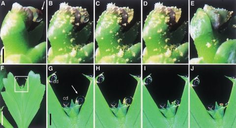
MoÈller et al.ÐPollination Mechanisms in Podocarpaceae 151
F I G . 1. Morphology of pollen and female cones. Acmopyle pancheri: A, SEM image of saccate pollen; B, microphotograph of a longitudinal
section through a female cone showing the obliquely erect ovule and the hook-like micropylar opening. Phyllocladus hypophyllus: C, SEM image
of pollen with vestigial sacci (vs); D, microphotograph of a female cone showing an individual erect ovule with attached pollination drop;
E, pollination drop after pollination showing sinking pollen (same scale as D). Bars 10 mm (A and C); 0.5 mm (D); 1 mm (B). e, Epimatium; fb,
fertile bract; i, integument; lsb, last sterile bract; mf, micropylar fork; mh, micropylar hook; mo, micropylar ori®ce; o, ovule; pd, pollination drop;
rc, `resin' canal; s, sacci; vb, vascular bundle.
after pollination, and a single (rarely two) fertile bract(s) more-or-less downwards at the stage of receptivity
at the apex, bearing the ovule(s). The cones are topo- (Figs 1B, 2A).
graphically erect at the time of receptivity. The uppermost Pollination drops were observed from early January until
sterile bract is often positioned opposite the micropylar the middle of February. Due to the morphology of the
hook (Figs 1B, 2A). The ovule is obliquely erect to micropylar hook, the exuded pollination drop is often
horizontal. The seed is invested in its lower half by the attached to the sterile bract that is positioned in front of the
epimatium, whose distal boundary is marked by a ridge micropyle. However, within these parameters, great varia-
partially encircling the seed. The tip of the sterile bracts, the tion was observed in cone morphology at the receptive stage
epimatium, the outer integument area and the outer surface (Mill et al., unpubl. res.); this frequently allowed pollination
of the micropyle are all covered with a waxy deposit that drops to form that were not attached to the last sterile bract.
renders the surface non-wettable. The integumental out- These were ideal for observations of the pollination mech-
growth surrounding the micropylar ori®ce is hook-like, anism (Fig. 2A). Their size ranged from 600±800 mm in
extended in an approx. 908 curve and ®nally points diameter. Under the cultivation conditions at RBGE,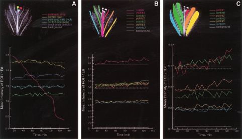
152 MoÈller et al.ÐPollination Mechanisms in Podocarpaceae
F I G . 2. Time sequences illustrating the dynamics of pollination drop resorption after experimental pollination. Acmopyle pancheri: shape and size
of pollination drop (arrow) prior to pollination (A), immediately after pollen application (B), 30 min (C), 1 h (D) and 1 d (E) after pollination
(E, dierent cone). Phyllocladus hypophyllus: F, a single segment of a phylloclade illustrating dimensions and area displayed in G±J (open box).
G, Shape and size of pollinated drop (arrow), unpollinated control drop (cd) and detached `evaporation control' drop (dd) prior pollination.
H±J, Pollination drop immediately after (H), 15 min after (I) and 30 min after (J) pollination. Bars 1 mm (A and G); 5 mm (F).
pollination drops could be observed on receptive cones at Pollination experiments
all times of the day, apart from on sunny days, when the
A. pancheri pollen has a spherical body with a collapsed
consequent resorption/evaporation was followed by over-
centre in the non-hydrated state and is 40±45 mm across,
night re-formation of the drop.
excluding the two large lateral sacci (Fig. 1A). The grains
In P. hypophyllus, conventional leaves and shoots are are non-wettable; they ¯oat when placed on water drops.
replaced by ¯attened phylloclades (modi®ed shoot com- Upon experimental pollination, the saccate pollen ¯oated
plexes according to Tomlinson et al., 1989), which can be upwards and became concentrated at the micropylar ori®ce
simple or compound ( pinnate). Both simple and compound (Fig. 2B). After pollination, drops were resorbed within
phylloclades can occur on the same plant; compound phyl- 30±60 min (Fig. 2A±D) and the pollen was drawn into the
loclades consist of alternately arranged segments (Keng, micropyle. After drop retraction, secretion ceased and no
1978). Each simple phylloclade, or each segment of a further pollination drop formation occurred (Fig. 2E).
pinnate phylloclade, is bilobed; the apical notch is ¯anked Untreated control drops, however, remained unchanged
by small bract-like structures representing the true leaves over the same period of observation, with re-formation
(Fig. 3C). Female cones of P. hypophyllus consist of clusters after any sun-induced daytime drop dissipation.
of two to ®ve (occasionally six) ovules in terminal positions Non-hydrated P. hypophyllus pollen is 30±33 mm in
in the notches of bilobed simple phylloclades, or in the diameter and displays lateral circular depressions, with
notches of the bilobed segments of pinnate phylloclades. two vestigial sacci (Fig. 1C). The pollen was shown to be
The ovules are slightly bilaterally ¯attened and each is wettable and sank upon exposure to pollination drops
subtended by a scaly bract (Fig. 1D). Except for the rim of (Fig. 1E). Although the qualitative response of pollination
the integumental outgrowth and the micropylar ori®ce, the drops upon pollination was identical throughout all
outer surface of the entire cone is covered with a waxy layer, experiments, quantitative dierences were observed. In all
rendering it non-wettable (Fig. 1D). cases active pollination drop resorption was observed, but
Pollination drops were observed from early January until the time for retraction ranged from 10 min to approx. 2 h.
the end of February. The ovules reformed drops repeatedly The main factors causing variation in retraction time
after they had been experimentally removed. They varied appeared to be the size of the detached segment/phylloclade
greatly in size from 250 mm to 41 mm in diameter, and the experimental conditions. Drops pollinated on ovules
depending on age and ovule size. of a whole compound phylloclade disappeared most quickly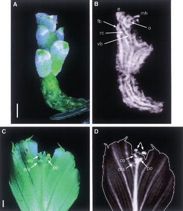
MoÈller et al.ÐPollination Mechanisms in Podocarpaceae 153
F I G . 3. Morphology of female cones. Acmopyle pancheri: A, photograph of a single cone. B, Single slice NMR image in LS plane, labelling as in
Fig. 1B. Phyllocladus hypophyllus: C, photograph of a single segment of the compound phylloclade used in NMR pollination experiment prior to
exudation of pollination drops. D, Single slice NMR image with control (left) and pollinated (right) pollination drop (arrows), outline drawn
manually. Bars 1 mm. co, Control ovule; o/s, additional ovule/scale complex; po, pollinated ovule.
(within 10 min) when observed under stereomicroscope projection of all structures to be seen in single slice (2 mm
illumination. Without the heat from the microscope lamp, thick) selective images. These could be acquired in a
ovules on compound phylloclades withdrew their drops relatively short time allowing a time lapse of 5 min between
within 45±60 min, whereas ovules attached to single seg- the acquisition of successive images. The results of these
ments required up to 120 min for complete drop resorption. investigations on the fate of the pollination drop within
The same result was observed for individual segments in plant tissue after pollination are displayed in Figs 4 and 5.
NMR glass sample vials, either in the light under laboratory Although the signal from the pollinated drop disappeared
conditions or in the dark inside the NMR magnet. gradually (Fig. 4A±H), the absence of a concomitant local-
P. hypophyllus cones borne on compound phylloclades ized increase in the area of the ovule or beneath indicates
and pollinated with A. pancheri pollen showed a slower that no specialized tissue exists into which the pollination
response, requiring approx. 90 min for full drop retraction. drop is `pumped'. The subtraction images in false colour
(Fig. 4J±P) illustrate the loss of signal intensity from the
pollinated drop while the control drop shows no change.
NMR imaging
Changes in the mean signal intensities of a number of
P. hypophyllus is an ideal subject for NMR imaging, as `regions of interest' (ROIs) as a function of time are
the laminar shape of the phylloclade segment allows a depicted in Fig. 5 which shows the steady decrease in154 MoÈller et al.ÐPollination Mechanisms in Podocarpaceae F I G . 4. Time course of NMR images of a phylloclade segment of Phyllocladus hypophyllus during pollination drop retraction. A, Before pollination; B±H, 15, 35, 55, 75, 95, 115, 130 min after pollination; J±P, false colour dierence images of B-B, C-B, D-B, E-B, F-B, G-B, H-B respectively; I, false colour intensity scale for images 4J to 4P; O, outlines of principle vascular bundles and pollination drops superimposed. pollinated drop signal (Fig. 5A) and slight increase in the the sum of all ROIs, there was a linear overall increase in other ROIs, particularly in distal intercostal areas close to signal with time (data not shown). The increase in total the ovules (Fig. 5C). Plots of the background-corrected signal from the phylloclade segment thus exceeded the loss integral signal intensity over dierent ROIs showed that, for in signal from the drop. The signal from the control drop
MoÈller et al.ÐPollination Mechanisms in Podocarpaceae 155
F I G . 5. Plot of mean intensities of indicated `regions of interest' (ROIs) on Phyllocladus hypophyllus segment as a function of time since
pollination. The inset shows the position of the ROIs on the phylloclade segment. A, Red, pollinated drop; yellow, control drop; cyan, pollinated-
side ovule; green, control-side ovule; blue, bract-scale complex; grey, background. B, Magenta, midrib (2nd order axis); red, prib1 ( ®rst 3rd order
axis on pollinated drop side of midrib); orange, prib2 (second 3rd order axis on pollinated drop side of midrib); gold, prib3 (third 3rd order axis on
pollinated drop side of midrib); bright green, crib1 ( ®rst 3rd order axis on control drop side of midrib); dark green, crib2 (second 3rd order axis on
control drop side of midrib); dark cyan, crib3 (third 3rd order axis on control drop side of midrib); grey, background. C, Red, poll1 (area between
midrib and prib1); magenta, poll2 (area between prib1 and prib2); violet, poll3 (area between prib2 and prib3); bright green, cont1 (area between
midrib and crib1); dark green, cont2 (area between crib1 and crib2); dark cyan, cont3 (area between crib2 and crib3); grey, background.
(Terminology adapted from Tomlinson et al., 1989.) The dashed lines are parallel to the time axis to allow easier visualization of changes in
intensity as a function of time while displaying several plots on common axes.
showed an initial slight increase before decreasing to result cone axis in Phyllocladus (Fig. 1D). Acmopyle diers from
in a ®nal small net decrease in signal integral. most other Podocarpaceae with respect to ovule orien-
Several attempts to image receptive female A. pancheri tation, and its hook-like micropyle can best be compared
cones by NMR failed because the size of most pollination with that of Lepidothamnus Phil. (Tomlinson et al., 1991).
drops relative to the rest of the cone was at the minimum Characters shared by A. pancheri and Lepidothamnus inter-
limit of NMR resolution and the irregular 3-dimensional medius (Kirk) Quinn are the morphologically obliquely
shape of the cone meant that a full picture could only be erect ovule and the topographically inverted orientation of
acquired by 3D imaging which takes longer than single slice the micropyle. In Lepidothamnus the micropyle is trumpet-
acquisition at the same spatial resolution. shaped and ¯ared at the mouth (Tomlinson, 1992), while in
A. pancheri it is tube-like, bent downwards with two
fork-like prongs (Mill et al., unpubl. res.). The shared
DISCUSSION similarity in cone morphology, however, is counterbalanced
Cone morphology by many other morphological dissimilarities and the genera
are not considered closely related within the family (Kelch,
Acmopyle and Phyllocladus contrast signi®cantly in the 1998).
morphology of their female cones. In Acmopyle, as in the
majority of members of the Podocarpaceae, the number of
fertile bracts is reduced to one (two are formed only rarely),
Pollination mechanism
in a topographically erect receptive cone. In contrast,
female cones of Phyllocladus are arranged in a spiral In Podocarpaceae, the orientation of the pollination drop
phyllotaxis (Tomlinson, 1992; Tomlinson et al., 1997) in is correlated with physical properties of the pollen.
clusters of two to ®ve ovules with random orientation. Previously investigated genera with more or less inverted
Although the orientation of ovules within the cone is drops have saccate pollen that allows the pollen grain to
obliquely erect to more-or-less horizontal in Acmopyle ¯oat to the mouth of the micropyle (Tomlinson et al., 1991;
(Fig. 1B), they are more-or-less erect with respect to the Tomlinson, 1994). This close correlation has also been156 MoÈller et al.ÐPollination Mechanisms in Podocarpaceae
demonstrated for Acmopyle in the present study (Fig. 2B), a physiological aspects of the pollination process in both
genus not previously investigated in this respect (Doyle, genera require more detailed study.
1945; Tomlinson, 1994). In terms of pollen ¯otation,
A. pancheri behaves like most other Podocarpaceae invest-
NMR imaging
igated (Tomlinson, 1994), having a more-or-less inverted
pollination drop and saccate pollen. However, major dier- A slight linear (regression factor 0.955) increase in overall
ences were observed in pollination response. Most Podo- intensity of the specimen was observed over the 2 h period
carpaceae have a prolonged period of pollination drop of the experiment. This contrasts with an observed overall
production and repeatedly exude pollination drops over decrease in another shorter experiment in which none of the
several days irrespective of the presence of pollen. They also drops were pollinated (data not shown). If any change in
have an expanded wettable area beneath the ovule for pollen overall intensity were expected, it would be a decrease, due
capture via the pollination drop, further increasing their either to dehydration of the sample or to instrumental drift
capability to scavenge pollen (Tomlinson et al., 1991, 1997). in the form of deviation from optimal tuning and shim-
In Acmopyle, this area is provided by the presence of a distal ming. The lack of any change in the intensity of the back-
sterile bract whose basal portion is not waxy, and thus is ground indicates the increase is not electronic in origin.
wettable (Mill et al., unpubl. res.). This creates a potential There appears to be no obvious `receiving chamber' for the
pollen-scavenging area into which the micropyles of the retracted pollinated drop (Figs 4, 5) but the slight increase
ovule(s) protrude and on to whose surface the pollination observable in the signal from the remainder of the
drop often becomes attached. Inversion of the pollination phylloclade segment suggests that the drop has not just
drop in Acmopyle is eected by the downwards inclination evaporated or fallen o. The mean intensities as depicted in
of the micropylar hook (Fig. 1B). However, the topography Fig. 4 are of a `slice' whose thickness exceeds the maximum
of the receptive Acmopyle ovule, whose nucellar canal (as diameter of the drops at all times, so assuming the cross-
opposed to the micropylar ori®ce) is directed slightly section remains circular throughout the experiment (reason-
upwards at receptivity, does not allow pollen to ¯oat on to able as the drop is subtended by a circular ori®ce and from
the nucellus. This is contrary to the interpretations of visual observation of other specimens), much or all of the
Acmopyle by Doyle (1945), which were based on the observed decrease in intensity in the pollinated drop can be
assumption (since proved by us to be wrong: Mill et al., accounted for by its reduction in size. Both pollinated and
control drops have the same mean intensity relative to
unpubl. res.) that the receptive ovule was inverted.
maximum diameter, indicating that there is no dierence in
Pollination drop retraction in A. pancheri was shown,
relaxation time between them and thus suggesting no gross
however, to be an active process, commencing immediately
change in chemical composition or viscosity of the
upon pollination. Once pollen had activated drop retraction,
pollinated drop over the 2 h duration of the experiment.
further drop secretion was irreversibly stopped, a response
The base of the phylloclade segment was immersed in
identical to that observed by us in P. hypophyllus. Here, too,
water, so it is reasonable to assume that the vascular system
secretion of further pollination drops ceased once pollina-
was saturated and of ®xed volume. Hence any increase in
tion had occurred and the drop had been retracted. Our intensity observed in the vasculature must come from
results thus largely con®rm earlier work on two other species dilution eects as the contents of the pollinated drop are
of Phyllocladus, P. trichomanoides D. Don, and P. toatoa redistributed in the remainder of the sample. Estimated
Molloy (Tomlinson et al., 1997; P. toatoa listed as changes in the intensity of the vascular axes, as a result of
`P. glaucus'): pollination drop retraction was triggered by the distribution of the pollinated drop within them, lead to
pollen, either conspeci®c or foreign (Acmopyle pollen had a greater increases in intensity than those observed. Increases
slightly less stimulative eect than conspeci®c pollen); in intensity were also observed, however, in the intercostal
pollination drop disappearance was not due simply to regions between the vascular axes. Since, in addition to
termination of secretion and net evaporation (see detached vascular traces emanating from the central 2nd order axis as
pollination drop in Fig. 2G±J). The more rapid disappear- well as lateral `veins' from the 3rd order axes, these regions
ance of pollinated drops under illumination compared with comprise non-vascular parenchymal tissue, there is the
unilluminated samples can simply be explained by an possibility of diusion of the pollinated drop contents in
increased evaporation as the lamp generated considerable the apoplast or into intercellular gas spaces (axis-terminol-
heat. The fact that shoot/phylloclade size also aected the ogy follows Tomlinson et al., 1989). Such diusion would
rapidity of drop retraction indicates either a link to lead to an increase in signal as air spaces became ®lled with
photosynthetic processes that diminishes with reduced liquid; other changes would be the result of dilution as
photosynthetic area, or an eect of the vascular system, a above. A diminution in the percentage increase in signal
larger system being more ecient in relocating the drop intensity with increased distance from the ovule is observed
volume. However, the ®rst hypothesis is unlikely, as the and it is interesting to note that the intercostal areas on the
response is similar whether the cone is placed in the dark or same side of the 2nd order axis as the pollinated drop show
in the light. The mechanism of pollen-triggered drop greater increases than those on the control drop side
retraction is presumably biochemically based in both (Fig. 5). Together with the rise and subsequent fall in the
genera, as Tomlinson et al. (1997) showed that, in Phyl- total integrated signal from the control drop, these changes
locladus, foreign pollen initiates drop retraction whereas suggest that the pollinated drop may be gradually assim-
inorganic material and physical disturbance do not. The ilated into the tissues of the phylloclade segment includingMoÈller et al.ÐPollination Mechanisms in Podocarpaceae 157
the vascular axes. The dierences between the two halves of Tomlinson et al., 1989; Bobrov et al., 1999). However,
the phylloclade segment, which are not observed in the 3rd Farjon (1998) followed recent taxonomic tendencies and
order axes suggest that there may also be some apoplastic treated Phyllocladus as the only genus of Phyllocladaceae.
water movement. Acmopyleaceae have not yet received acceptance by any
NMR imaging shows great potential for the more workers outside Bobrov's team. These segregations, and
detailed investigation of the fate of the pollinated drop in those of other genera within the family Podocarpaceae sensu
this and other species. lato, have generally been on the basis of characters of the
female cones; authors who have segregated genera on this
Taxonomic implications basis have given little consideration to other suites of
characters that might unite them into a more coherent
Both genera used in this study have been segregated from whole. One such suite is leaf anatomy, which was used by
Podocarpaceae as separate families: Acmopyle as Acmopy- Buchholz and Gray (1948) to de®ne sections within
leaceae Melikian & Bobrov (Melikian and Bobrov, 1997; Podocarpus L'HeÂr. ex Pers., many of which, however, have
Bobrov et al., 1999) and Phyllocladus as Phyllocladaceae since been raised to generic rank.
Bessey (Bessey, 1907; Bobrov et al., 1999, as Phyllocladaceae To separate Phyllocladaceae from Podocarpaceae, Tom-
(Pilg.) Bessey; Keng, 1973, as Phyllocladaceae E.L. Core ex linson et al. (1997) listed a suite of characters related to the
Keng). Irrespective of whether Phyllocladus and/or Acmo- pollination mechanism including cone orientation, pollen
pyle are included, the family Podocarpaceae is rather
hydrodynamics, pollination drop shape, pollen capture and
heterogeneous. This may partly re¯ect its long recorded
drop retraction mechanisms. The pollination mechanism
history; fossils are known from the Triassic (Rissikia
appears to be uniform across the genus Phyllocladus, since
Townrow: Townrow, 1967) and the genera recognized
identical results were obtained in studies by Tomlinson et al.
today are disparate in their morphology, possibly as a result
(1997) on two New Zealand species and in the present study
of extinctions of intermediates (Kelch, 1998). This has
on the only tropical member of the genus, P. hypophyllus.
resulted in the segregation of some genera as individual
families. Acmopyleaceae was de®ned principally on the We found that the pollination mechanism of Acmopyle
basis of fruit anatomical characters, as well as the dimorphic appears to be intermediate between other previously invest-
leaves (Melikian and Bobrov, 1997; Bobrov et al., 1999); igated Podocarpaceae and Phyllocladaceae (Table 1).
however, the leaf character is also found in Dacrycarpus and Although some morphological features characteristic of
Falcatifolium. Phyllocladaceae were de®ned by Bessey Podocarpaceae are present in Acmopyle (e.g. saccate pollen,
(1907) by their megasporophylls not arranged in strobili, non-wettable pollen), the pollination drop secretion and
while Keng (1973) used, among other characters, the retraction characteristics are identical to those of Phyllo-
phylloclades, the erect ovules, and the arillate seeds seated cladus. Pollination mechanisms appear to be more diverse
on a scaly ¯eshy cup. Of these characters, only the within this family than was hitherto appreciated, although
phylloclades are truly diagnostic for Phyllocladus; all others we remain ignorant of the presence or absence of a
are expressed in at least one genus of Podocarpaceae s. s. pollination drop in several genera listed by Tomlinson
Many authors have disagreed with Keng's perception of (1994). In the case of Falcatifolium taxoides (Brongn. &
Phyllocladus and include it in Podocarpaceae (e.g. de Gris) de Laub., absence of proof was not proof of absence;
Laubenfels, 1969, 1978, 1988); some regard it as a derived the presence of a pollination drop has since been discovered
member of that family (e.g. Hart, 1987; Quinn, 1987; in the species (data not shown).
T A B L E 1. Comparison of pollination mechanisms among Acmopyle, Podocarpaceae (excluding Acmopyle) and
Phyllocladus (data for Podocarpaceae and Phyllocladus modi®ed after Tomlinson et al., 1997)
Phyllocladaceae Podocarpaceae
Phyllocladus Acmopyle Saxegothaea Other genera
Cone orientation Random Erect Random Erect
Pollen
Form Non-saccate or sacci Saccate Non-saccate Saccate
vestigial
Hydrodynamics Wettable Non-wettable Wettable Non-wettable
Pollination drop
Orientation with respect to cone axis +Erect +Inverted No drop formed +Inverted
Repeated secretion Stops upon pollen- Stops upon pollen- n.a. Continues after pollen
capture capture capture
Drop retraction
Stimulus Requires pollen Requires pollen n.a. Does not require pollen
Mechanism Metabolic? Metabolic? n.a. Physical (evaporation)
n.a., Not applicable.158 MoÈller et al.ÐPollination Mechanisms in Podocarpaceae
The taxonomic value of the pollination mechanism as a de Laubenfels DJ. 1969. A revision of the Malesian and Paci®c rain-
suite of characters for the reliable separation of Phyllocla- forest conifers, I. Podocarpaceae, in part. Journal of the Arnold
Arboretum 50: 274±369.
daceae from Podocarpaceae is invalidated by our ®ndings. de Laubenfels DJ. 1978. The taxonomy of Philippine Coniferae and
In the present state of knowledge, however, the similarities Taxaceae. Kalikasan 7: 117±152.
in pollination mechanisms between Acmopyle and Phyllo- de Laubenfels DJ. 1988. Coniferales. In: van Steenis CGGJ, de Wilde
cladus are not a re¯ection of an evolutionary relationship WJJO, eds. 1986, Flora Malesiana, vol. 10. Dordrecht, Boston &
London: Kluwer Academic Publishers.
but represent a convergence, as indicated by molecular Doyle J. 1945. Developmental lines in pollination mechanisms in the
phylogenetic analyses of the group (Kelch, 1998; Sinclair Coniferales. Scienti®c Proceedings of the Royal Dublin Society 24:
et al., unpubl. res.). Like fruit or leaf anatomical characters, 43±62.
the pollination mechanism represents a single character suite Farjon A. 1998. World checklist and bibliography of conifers. Kew:
and taxonomic conclusions reached using only single- Royal Botanic Gardens.
Glidewell SM, Williamson B, Goodman BA, Chudek JA, Hunter G.
character-suite datasets are sometimes unsound and do 1997. An NMR microscopic study of grape (Vitis vinifera L.).
not stand the test of time. Thus, the pollination mechanism Protoplasma 198: 27±35.
character suite, on its own, can neither lend support to the Hart JA. 1987. A cladistic analysis of conifers: preliminary results.
separation of Phyllocladaceae from Podocarpaceae Journal of the Arnold Arboretum 68: 269±307.
Kelch DG. 1998. Phylogeny of Podocarpaceae: comparison of evidence
(although the mechanism in Phyllocladus still appears
from morphology and 18S rDNA. American Journal of Botany 85:
unique to that genus), nor be used to support the recogni- 986±996.
tion of Acmopyleaceae. A ®nal conclusion can only be Keng H. 1973. On the family Phyllocladaceae. Taiwania 18: 142±145.
drawn after the examination of all genera of Podocarpaceae Keng H. 1978. The genus Phyllocladus. Journal of the Arnold
sensu lato and when data from many datasets have been Arboretum 59: 249±275.
Melikian AP, Bobrov AVFCh. 1997. Sistematicheskoe polozhenie
compared. roda Acmopyle Pilg. (Podocarpaceae s.l.) po dannym sravnitel'noj
morphologii, anatomii i ul'trastruktury semyan. Proceedings of the
International Conference on Plant Anatomy and Morphology:
92±93. Mezhdunarodn. konf. po anat. i morph. rast. (Sankt-
AC K N OW L E D G E M E N T S Peterburg, iyun' 1997). Tezisy dokladov: 92±93.
We thank Frieda Christie (RBGE) for the pollen SEM MoÈller M, Mill RR, Bateman RM, Glidewell SM, Williamson B,
Masson D. 1999. Pollination drop mechanism and cone develop-
micrographs and the horticultural sta of RBGE for main- ment of Acmopyle pancheri (Podocarpaceae): present state of
taining the living material used in this study, and an knowledge. In: Farjon A, ed. 4th International Conifer Conference,
anonymous reviewer for constructive comments on the 23±26 August 1999, Wye College, EnglandÐProgramme &
manuscript. The Royal Botanic Garden Edinburgh and Abstracts, 35±36.
Owens JN, Takaso T, Runions CJ. 1998. Pollination in conifers. Trends
the Scottish Crop Research Institute are supported by the in Plant Science 3: 479±485.
Scottish Executive Rural Aairs Department (SERAD). Quinn CJ. 1987. The Phyllocladaceae KengÐa critique. Taxon 36:
The NMR imager at SCRI was purchased by Mylne®eld 559±565.
Research Services Ltd. This research was primarily funded Tomlinson PB. 1992. Aspects of cone morphology and development in
by SERAD (Flexible Fund project RBG-003-98). Podocarpaceae (Coniferales). International Journal of Plant
Science 153: 572±588.
Tomlinson PB. 1994. Functional morphology of saccate pollen in
conifers with special reference to Podocarpaceae. International
L I T E R AT U R E C I T E D Journal of Plant Science 155: 699±715.
Tomlinson PB, Braggins JE, Rattenbury JA. 1991. Pollination drop
Bessey CE. 1907. A synopsis of plant phyla. University Studies in relation to cone morphology in Podocarpaceae: a novel
(Nebraska) 7: 275±373 (reprinted as 1±99). Lincoln, Nebraska: reproductive mechanism. American Journal of Botany 78:
University of Nebraska. 1289±1303.
Bobrov AVFCh, Melikian AP, Yembaturova EY. 1999. Seed morpho- Tomlinson PB, Braggins JE, Rattenbury JA. 1997. Contrasted
logy, anatomy and ultrastructure of Phyllocladus L.C. & A.Rich. pollen capture mechanisms in Phyllocladaceae and certain
ex Mirb. (Phyllocladaceae (Pilg.) Bessey) in connection with the Podocarpaceae (Coniferales). American Journal of Botany 84:
generic system and phylogeny. Annals of Botany 83: 601±618. 214±223.
Buchholz JT, Gray NE. 1948. A taxonomic revision of Podocarpus I. Tomlinson PB, Takaso T, Rattenbury JA. 1989. Cone and ovule
The sections of the genus and their subdivisions with special ontogeny in Phyllocladus (Podocarpaceae). Botanical Journal of
reference to leaf anatomy. Journal of the Arnold Arboretum 29: the Linnean Society 99: 209±221.
49±63. Townrow JA. 1967. On Rissikia and Mataia, podocarpaceous conifers
Chudek JA, Hunter G. 1997. Magnetic resonance imaging of plants. from the Lower Mesozoic of southern lands. Papers and Proceed-
Progress in Nuclear Magnetic Resonance Spectroscopy 31: 43±62. ings of the Royal Society of Tasmania 101: 103±136.You can also read