Directional control of cell motility through focal
←
→
Page content transcription
If your browser does not render page correctly, please read the page content below
The FASEB Journal • Research Communication
Directional control of cell motility through focal
adhesion positioning and spatial control of Rac
activation
Nan Xia,* Charles K. Thodeti,* Tom P. Hunt,† Qiaobing Xu,‡ Madelyn Ho,*
George M. Whitesides,‡ Robert Westervelt,† and Donald E. Ingber*,1
*Vascular Biology Program, Departments of Pathology and Surgery, Children’s Hospital and Harvard
Medical School; †Department of Physics and Division of Engineering and Applied Sciences; and
‡
Department of Chemistry and Chemical Biology, Harvard University, Boston, Massachusetts, USA
ABSTRACT Local physical interactions between cells angiogenesis, and cancer metastasis. Most work on
and extracellular matrix (ECM) influence directional cell directional control of cell movement has focused on
motility that is critical for tissue development, wound gradients of soluble chemokines (1) that, in turn,
repair, and cancer metastasis. Here we test the possibility generate intracellular gradients of signal transduction
that the precise spatial positioning of focal adhesions (1– 4). However, cell movement also can be influenced
governs the direction in which cells spread and move. by physical interactions between cells and their extra-
NIH 3T3 cells were cultured on circular or linear ECM cellular matrix (ECM) adhesions. For example, cells
islands, which were created using a microcontact printing move from regions of high ECM compliance to more
technique and were 1 m wide and of various lengths stiff regions, a process as known as durotaxis (5–7).
(1 to 8 m) and separated by 1 to 4.5 m wide nonadhe- When plated on single-cell sized ECM islands of differ-
sive barrier regions. Cells could be driven proactively to ent geometric shapes created with microfabrication
spread and move in particular directions by altering either techniques, various cells preferentially extend new mo-
the interisland spacing or the shape of similar-sized ECM tile processes (e.g., lamellipodia, filopodia, micro-
islands. Immunofluorescence microscopy confirmed that spikes) from their corners rather than along their
focal adhesions assembled preferentially above the ECM edges (8, 9). Cell migration direction also can be
islands, with the greatest staining intensity being observed controlled by constraining cell geometry using similar
at adhesion sites along the cell periphery. Rac-FRET microengineered ECM islands and then releasing the
analysis of living cells revealed that Rac became activated physical restrictions to movement using electrochemi-
within 2 min after peripheral membrane extensions ad- cal techniques (10). Importantly, these findings are
hered to new ECM islands, and this activation wave relevant because cells also preferentially extend and
propagated outward in an oriented manner as the cells migrate along ECMs with specialized shapes (e.g.,
spread from island to island. A computational model, fibrils) during tissue development (11) and tumor
which incorporates that cells preferentially protrude angiogenesis (12). However, the mechanism by which
membrane processes from regions near newly formed physical interactions between cells and ECM govern the
focal adhesion contacts, could predict with high accuracy direction in which they move remains unclear.
the effects of six different arrangements of micropat- The effects of ECM on directed cell migration are
terned ECM islands on directional cell spreading. Taken mediated by transmembrane integrin receptors that
together, these results suggest that physical properties of mechanically couple to submembranous focal adhesion
the ECM may influence directional cell movement by proteins, such as vinculin, that physically link the ECM
dictating where cells will form new focal adhesions and to the intracellular cytoskeleton (1, 13). Integrin bind-
activate Rac and, hence, govern where new membrane ing influences cell movement by stimulating the small
protrusions will form.—Xia, N., Thodeti, C. K., Hunt, GTPase Rac (14 –16), which activates the Arp2/3 com-
T. P., Xu, Q., Ho, M., Whitesides, G. M., Westervelt, R., plex (17) and thereby nucleates actin polymerization
Ingber, D. E. Directional control of cell motility through (18 –20), which drives lamellipodial protrusion (21,
focal adhesion positioning and spatial control of Rac 22). Arp 2/3 also transiently associates with vinculin
activation. FASEB J. 22, 1649 –1659 (2008) and colocalizes with it in nascent focal adhesions
known as focal complexes (23). Interestingly, activated
Key Words: migration 䡠 extracellular matrix 䡠 microcontact Rac fails to promote lamellipodia extension in vinculin
printing 䡠 cytoskeleton 䡠 mechanical 䡠 traction
1
Correspondence: Vascular Biology Program, KFRL
11.127, Children’s Hospital, Boston, MA 02115, USA. E-mail:
Directional cell motility is critical for normal donald.ingber@childrens.harvard.edu
tissue development as well as the immune response, doi: 10.1096/fj.07-090571
0892-6638/08/0022-1649 © FASEB 1649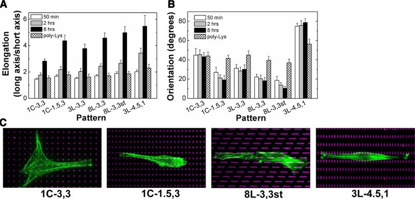
null cells (24), suggesting that focal adhesion assembly recorded with a CCD camera (Hamamatsu, Tokyo, Japan) on
may play an important role in directional cell motility. a Diaphot 300 inverted microscope (Nikon, Tokyo, Japan)
This possibility is supported by the observation that equipped with phase contrast optics and epifluorescence
illumination and processed using the computerized image
single cells anchored to polygonal ECM islands prefer- acquisition and analysis tools of IP Lab Spectrum and Ratio-
entially form focal adhesions in their corners near sites Plus software (Scanalytics, Fairfax, VA, USA). The microscope
where new lamellipodia will form when the cells are was also equipped with an on-stage heater that maintained
stimulated with soluble chemokines, and cells on circu- the temperature at 37°C at all times, and the culture medium
lar islands (i.e., without corners) do not exhibit any was covered with a thin layer of mineral oil to prevent
directional bias (9). Focal adhesions also appear to evaporation. Immunofluorescence microscopy was per-
formed as described (8). Fluorescence images were acquired
form preferentially, directly behind the leading edge of
on a Leica TCS SP2 confocal laser scanning microscope with
cells migrating in the direction of increasing ECM an ⫻63/1.4 NA oil immersion objective (Leica Microsystems,
stiffness (25, 26), and changes in mechanical force Bannockburn, IL, USA) and processed using Leica software
transfer across integrins can modulate focal adhesion or Adobe Photoshop (Adobe Systems, San Jose, CA, USA).
assembly (27). Taken together, these findings imply Morphometric analysis of cell shape, orientation, and migra-
that the spatial positioning of focal adhesions may tion was performed by outlining the borders of individual
determine the location in which new motile processes cells within differential interference contrast images using the
computerized image acquisition and analysis tools described
form, and hence, govern the direction in which cells above; 25 to 50 cells were analyzed for each experimental
move. condition. Cell elongation was defined as the ratio of the
If these findings are true, then it should be possible maximal to minimal cell length. In the migration assays, cells
to proactively dictate the direction in which cells will were stimulated with PDGF (50 ng/ml), and the centroids of
move by controlling the location where cells can form the migrating cell recorded at 20 min intervals over 8 h were
new focal adhesions. In the present study, we test this plotted and connected to generate the migration path, which
hypothesis directly by using microcontact printing was then used to calculate the speed and direction of cell
migration.
methods to create culture substrates containing ECM
islands on the size scale of individual focal adhesions
Rac FRET analysis
surrounded by nonadhesive regions, and then deter-
mining whether altering island shape or position influ-
ences the direction of cell spreading and movement. NIH 3T3 cells were transfected with the FRET reporter of Rac
activity, Raichu-Rac1 (30), using effectene transfection re-
agent (Qiagen, Valencia, CA, USA) and cultured for 24 h on
the different micropatterned substrates in serum-free me-
MATERIALS AND METHODS dium. We studied cells cultured on the 3L-4.5,1 pattern in
studies analyzing spatial control of Rac activation dynamics at
Experimental system high resolution because this pattern has the largest difference
in island spacing between the x and y directions, which greatly
increases our ability to perform this difficult visualization
The micropatterned substrates containing micrometer-sized technique. For FRET analysis, fluorescence images were
ECM islands separated by nonadhesive barrier areas were acquired every minute on a Leica TCS SP2 confocal laser-
created using microcontact printing and coated with rhoda- scanning microscope with an ⫻63/1.4 NA oil immersion
mine conjugated fibronectin (Cytoskeleton, Denver, CO, objective using Leica FRET sensitization software (CFP and
USA), as described (28, 29). Mouse NIH 3T3 fibroblasts were FRET excitation at 458 nm; YFP excitation at 514 nm).
maintained at 37°C under 10% CO2 in high-glucose Dulbec- Calibration images were acquired from the samples contain-
co’s modified Eagle’s medium (DMEM), supplemented with ing only Donor (CFP), Acceptor (YFP), and FRET (Raichu-
10% fetal bovine serum, glutamine (0.3 mg/mL), penicillin Rac1), respectively, and the apparent FRET efficiency was
(100 U/ml), streptomycin (100 g/mL), and 20 mM HEPES. automatically calculated by the software. We also validated the
Subconfluent cell monolayers were serum-starved for 1 day, efficiency of the Raichu-Rac1 construct by acceptor photo-
trypsinized, and plated on micropatterned substrates in bleaching method, which confirmed that donor (CFP) fluo-
DMEM containing high-density lipoprotein (10 g/mL), rescence and associated FRET increases when the acceptor
transferrin (5 g/mL), and 1% bovine serum albumin. The (YFP) is bleached.
plating density was low to minimize cell-cell contacts.
Computational modeling
Reagents
In the computer simulation program that was performed
Mouse antivinculin antibody, fluorophore-conjugated sec-
using MATLAB, “cells” represented by N circles with a spher-
ondary antibodies, fluoresceinated phalloidin, ML-7 and
ical diameter D were generated at random locations above a
FITC-labeled-poly-l-lysine were purchased from Sigma (St.
microarray of discrete “virtual” ECM islands separated by
Louis, MO, USA). The motility factor, human PDGF-BB was
nonadhesive regions. The initial contact area between the
purchased from BioVision (Mountain View, CA, USA). The
adhering cell and the substrate was assumed to be circular
FRET probe, Raichu-Rac1, was kindly provided by Dr.
with a diameter of D⬘; both D and D⬘ were set at 15 m. Every
Michiyuki Matsuda (Osaka University, Osaka, Japan) (30).
point (point resolution was set at 0.5 m) along the cell’s
boundary where it contacted an ECM island had an equal
Microscopy and imaging opportunity to extend membrane protrusions in a circular
sweep outside of the cell body with a radius r. The size
We used a serum-free cell culture medium for live cell distribution of radius r of new membrane extensions was set
imaging, as described previously (8). Live cell images were so that large extensions decreased exponentially with r based
1650 Vol. 22 June 2008 The FASEB Journal XIA ET AL.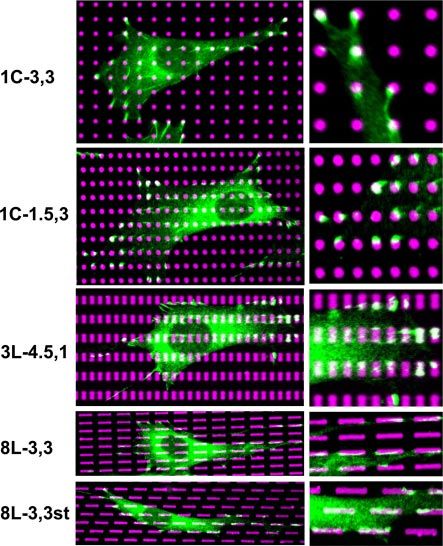
on experimental observations of living cells. Any protrusion (1C-3,3) they spread more slowly and to a lesser degree
that contacted a neighboring ECM island formed a new focal over the first 2 h after plating (Fig. 2, middle). Al-
adhesion and remained anchored; other protrusions re- though directional extension initiated about the same
tracted back onto the cell. The cell boundary was then
modified to incorporate the newly formed adhesive contacts time, the cells on the 1C-3,3 micropattern also elon-
to form a new polygonal map of the extended cell border as gated more gradually and to a lesser degree than
it spread over the cell-substrate interface. This completed one control cells on the unpatterned substrates. In contrast,
cycle of simulated cell spreading, and then the process was when cells were cultured on similarly shaped FN islands
reiterated multiple times. that were more closely spaced in the x direction (1C-
1.5,3), cell spreading and directional elongation initi-
ated more rapidly and increased in parallel during the
RESULTS same time course (Fig. 2, right). We have previously
shown that cells can attach and spread on these micro-
Control of cell spreading by varying ECM island fabricated substrates (31) as well as standard ECM-
shape and position coated substrates (32) in the absence of de novo protein
synthesis, and we observed similar differences in
Six different planar microarrays of focal adhesion-sized spreading rates when protein synthesis was inhibited
circular or linear FN islands, separated by nonadhesive using cycloheximide (data not shown). Comparison of
regions, were created using microcontact printing. As cells on all six different FN island arrangements re-
displayed in Fig. 1, these included patterns of 1 m vealed that the isotropic arrangement of FN circles
diameter circular FN islands that were either separated (1C-3,3) and the anisotropic array of linear islands with
by 3 m in both the x and y directions (1C-3,3) or by 1.5 the smallest interisland spacing (3L-4.5,1) produced
m in the x direction and 3 m in the y direction the lowest and greatest cell elongation, respectively,
(1C-1.5,3); linear FN islands 1 m high (y direction) when analyzed at 50 min, 2 h or 8 h after cell plating
and 3 or 8 m wide (x direction) separated by 3 m in (Fig. 3A). However, virtually all of the linear substrates
both directions (3L-3,3 and 8L-3,3 respectively); linear and the anisotropic array of circular FN islands exhib-
FN islands 3 m wide ⫻ 1 m high separated by 4.5 and ited a similar high level of elongation after 8 h of
1 m in x and y directions, respectively (3L-4.5,1); and culture. Interestingly, differences in cell elongation
linear 8 m wide ⫻ 1 m high FN lines equally spaced could not be detected until 2 h after plating, and they
by 3 m in both directions, but staggered along a 60° became significant (P⬍0.05) only after 8 h (Fig. 3A).
offset (8L-3,3st). Analysis of elongated cells on the different micropat-
When NIH 3T3 cells were plated on these substrates terned substrates at similar times confirmed that, al-
in serum-free medium, cell spreading proceeded in a though cells did not display a preferred orientation on
manner different from that on standard FN-coated the isotropic array of FN circles (1C-3,3), cells on the
plastic substrates. For example, cell spreading reached anisotropic array of similar shaped FN islands (1C-
maximal levels within ⬃30 min after plating on control 1.5,3) elongated in a highly oriented manner (Fig. 3B).
FN-coated dishes, and only after this time did the cells In our analysis, we determined the average angular
begin to elongate in an anisotropic manner and exhibit orientation of cells by measuring the direction of their
a bipolar form (Fig. 2 left). In contrast, when cells were longest axis over a range from 0 to 90°, and, thus,
cultured on the isotropic microarray of 1 m FN circles randomly oriented cells will display an average orienta-
tion of 45° with a very wide distribution of values, and
this was what we observed for cells on the 1C-3,3 pattern
(Supplemental Fig. S1). The cells also spread and
aligned preferentially along the main axis of the linear
FN islands even when they were distributed isotropi-
cally (3L-3,3 and 8L-3,3) or in a staggered pattern
(8L-3,3st), and greater cell alignment was observed on
the longer islands (Fig. 3B). Decreasing the spacing
between FN circles to 1.5 m in the x direction
(1C-1.5,3) similarly promoted greater cell orientation
in this direction, and decreasing the spacing between
linear islands in the y direction to 1 m while simulta-
neously increasing the interisland separation in the x
direction to 4.5 m (3L-4.5,1) produced the most
potent effect causing almost all of the cells to orient in
the y direction (90°) (Fig. 3B).
The efficiency of cell alignment also correlated with
Figure 1. Schematic designs of a small region from six
different planar microarrays of FN islands used in this study,
the degree of alignment of F-actin filament bundles in
and fluorescence micrographs showing corresponding sub- the cytoskeleton within cells on these various substrates
strates created with the microcontact printing technique that (Fig. 3C). Moreover, when we examined cells cultured
were imprinted with fluorescent fibronectin. on FN island microarrays, bright vinculin staining
PHYSICAL CONTROL OF CELL MIGRATION 1651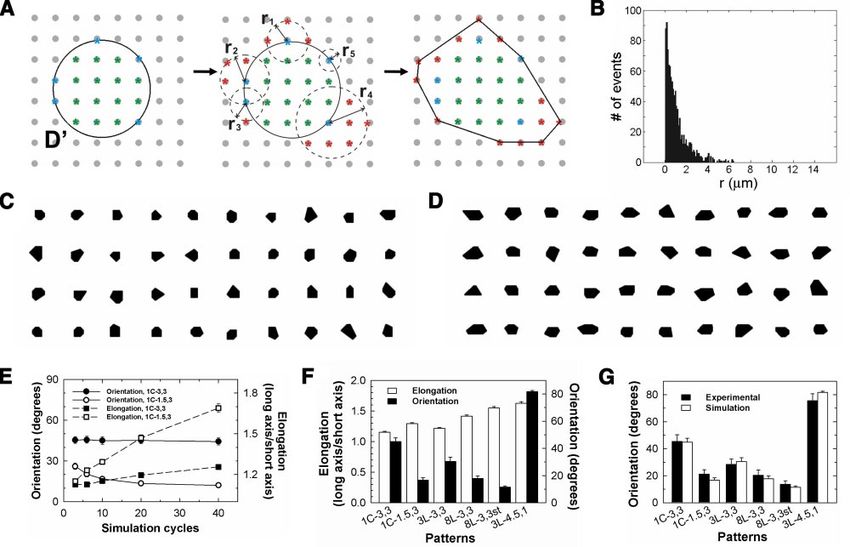
Figure 2. Effects on the dynamics of cell spreading. Top: time-lapse phase contrast microscopic images recorded at 5, 20, 40, 60,
and 80 min (top to bottom) after plating on standard FN-coated plastic (left) vs. micropatterned substrates with two different
patterns: 1C-3,3 (middle) and 1C-1.5,3 (right). Bottom: graphs showing the effects of the respective substrates on projected cell
area and elongation during the progress of spreading. Scale bar ⫽ 40 m.
could be detected in regular shapes and patterns that substrates on cell orientation and elongation were
corresponded directly to the form and position of each greatly reduced (Fig. 3A, B), indicating that specific
microfabricated FN island (Fig. 4), as observed in binding interactions between cells and ECM are critical
previous studies (28, 38). Interestingly, in contrast to for these observed directional responses.
effects on elongation that were only observed after 8 h,
significant micropattern-dependent differences in cell
Initial cell orientation and elongation govern the
alignment were detected within 50 min after plating
direction of cell movement
(Fig. 3B). Thus, the position of these focal adhesion-
sized islands influenced cell orientation from the ear-
liest times after cell adhesion, and once alignment was We next examined whether the effects of ECM island
established it appeared to govern the direction in shape and distribution on cell orientation and elonga-
which cells would preferentially reorient their actin tion that we observed at early times influence the
cytoskeleton and elongate their shape at later times. In direction of cell motility when stimulated by soluble
contrast, when these same adhesive islands were coated chemokines. When the motility factor, PDGF, was
with polylysine rather than FN, the effects of these added to the cells that had spread for 3 h in the
1652 Vol. 22 June 2008 The FASEB Journal XIA ET AL.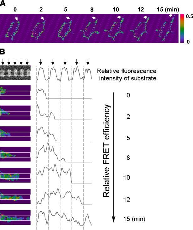
Figure 3. Effects of different FN microarrays on cell elongation, orientation, and cytoskeletal organization. A) Cell elongation
(ratio of the longest axis of the cell to its shortest) is presented for cells cultured for 50 min (white bars), 2 h (gray bars), or
8 h (black bars) on different micropatterned FN substrates labeled as indicated in Fig. 1, or for 6 h on similar island microarrays
coated with polylysine (cross-hatched bars). B) Cell orientation (degrees of the longest axis deflected from horizontal)
measured under similar conditions and at the same times as in A. C) Overlay of fluorescence micrographs showing the arrays
of representative FN microisland patterns (magenta) and the actin cytoskeleton (green) of adherent cells cultured for 8 h.
serum-free medium, cell migration speeds were similar
on all six micropatterned substrates; however, the di-
rection of movement differed significantly (Fig. 5A).
Again, cell migration direction was essentially random
(45° with a wide distribution) on the isotropic FN
circles and more oriented on the anisotropic patterns
and linear FN islands (Fig. 5A). But most importantly,
the direction of motility and the efficiency of orienta-
tion scaled directly with the effects of these same
micropatterned substrates on cell orientation at earlier
times (Figs. 3B vs. 5A). For example, whereas cells
migrated in a random Brownian walk on the isotropic
array of FN circles (Fig. 5B), cells could be made to
preferentially migrate in either the x or y direction with
great efficiency by decreasing interisland spacing in the
x or y direction using the 1C-1.5,3 and 3L-4.5,1 sub-
strates, respectively (Fig. 5B). Thus, the early effects of
FN island shape and position on cell orientation govern
both the orientation in which cells will elongate and the
direction in which they will move when stimulated with
soluble motility factors.
Cells protrude new membrane processes near nascent
focal adhesions
Figure 4. The shape and distribution of vinculin-containing
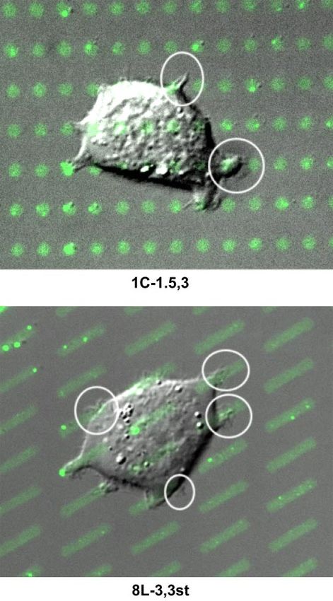
Protrusion of new membrane processes, such as lamel- focal adhesions corresponds to those of the micropatterned
lipodia and filopodia, is fundamental to cell spreading FN islands. Immunofluorescence microscopic overlay images
and migration (37). We, therefore, monitored forma- of cells cultured for 8 h on different micropatterned distri-
butions (indicated at left) of rhodamine-FN-coated islands
tion of new lamellipodia during cell spreading on the (magenta) and stained with antivinculin antibodies (green).
micropatterned substrates. During this process, cells Insets: higher magnification views of the codistribution of
appeared to preferentially form new membrane protru- vinculin and rhodamine FN (white) within the corresponding
sions near regions close to the micropatterned FN images at left.
PHYSICAL CONTROL OF CELL MIGRATION 1653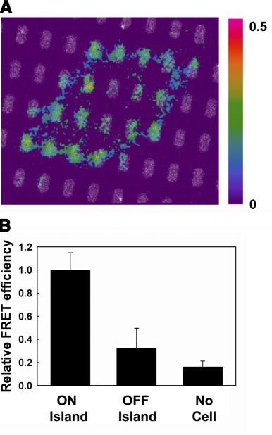
Figure 5. Control of cell migration by altering the shape, size, and position of FN adhesive islands. A) Graph showing the effects
of the various FN microarrays on cell migration speed (white bars; m/h) and direction (black bars; degrees deflected from
horizontal) in NIH3T3 cells stimulated with PDGF (50 ng/ml). B) Representative examples of migration paths of the
PDGF-stimulated cells migrating on three different FN microarray patterns: 1C-3,3 (left), 1C-1.5,3 (middle) and 3L-4.5,3 (right).
The position of cell centroids was measured over 8 h at 20 min intervals. Scale bar⫽100 m.
islands and to extend these processes over both adja- observed was likely primarily due to signaling elicited in
cent FN-coated islands and nearby nonadhesive areas response to new integrin binding to these micropat-
(Fig. 6). These small lamellipodial protrusions that terned FN islands. This point was made even clearer
extend over nonadhesive regions during early phases of when we monitored dynamic changes of Rac activity as
cell spreading are actin-rich, as previously demon- cells progressively spread across multiple FN islands of
strated (9), and confirmed by fluorescent phalloidin pattern 3L-4.5,1. Lamellipodia extended centripetally
staining (data not shown). Analysis of 60 protrusions from existing focal adhesions near the cell periphery
within randomly chosen cells spreading on all six and transiently crossed over adjacent nonadhesive ar-
micropatterned substrates revealed that ⬃88% of pro- eas to contact adjacent FN islands (Fig. 8). When the
trusions originated from regions of the cell directly same cell was followed over a period of 15 min, the cell
above or adjacent to FN islands along the cell periph- could be observed to pull its body forward to cover the
ery. Dynamic microscopic analysis of living cells re- newly contacted FN island and then to extend another
vealed that these extending processes stabilized when new process in the same direction from this nascent
the protrusions contacted adjacent FN islands and adhesion site; reiteration of this process resulted in
formed new cell-ECM adhesions, whereas those that progressive spreading and movement of the cell in a
extended over nonadhesive regions of the substrate consistent direction, as dictated by the smaller interis-
retracted back onto the cell surface. Adhesion of the land spacing in the one direction relative to the other
new processes to the nearby FN islands also was accom- in the micropattern (3L-4.5,1) used in this study (Fig.
panied by an overall change of cell shape as the cell 8A). Importantly, the Rac-FRET analysis revealed that
body was progressively pulled forward to cover the Rac1 became locally activated within 2–3 min after the
newly occupied adhesive area. cell’s surface membrane contacted each new FN island,
and this process of Rac activation reiterated with a
Spatial control of Rac activation spatiotemporal pattern that closely paralleled the direc-
tional elongation process (Fig. 8B). Thus, control of
The small GTPase, Rac1 controls lamellipodia forma- oriented cell spreading by focal adhesion positioning
tion, and it can be activated by cell-ECM adhesion in an appeared to be spatially and temporally coupled to the
integrin-dependent manner (14 –16). Analysis of the pattern of Rac activation and associated lamellipodia
spatial distribution of Rac activity in cells cultured on extension.
the different FN microarrays (Figs. 7 and 8) using an
intramolecular Rac1-FRET reporter (30) revealed Computer simulation of cell spreading on microarray
higher FRET efficiency (and hence Rac GTPase activa- of FN islands
tion) in regions directly above FN islands compared
with the intervening nonadhesive areas beneath a cell To test whether spatial control of membrane process
(Fig. 7A). Quantification of the results of FRET analysis extension by ECM island location is sufficient to ex-
of multiple cells grown on pattern 3L-3,3 demonstrated plain how the oriented cell spreading occurs that
that Rac activation levels were ⬃3⫻ higher in regions determines the subsequent cell migration direction, we
overlying FN islands than in the nonadhesive areas created a computer simulation program to model the
beneath spreading cells, and ⬎6⫻ higher than that process of cell spreading on substrates with a discontin-
observed in regions of the substrate that were not uous spatial distribution of ECM proteins. As described
covered by cell membrane (Fig. 7B). in Materials and Methods, this simulation program
Because these cells were cultured in serum-free me- mimics the progressive events of cell adhesion to ECM
dium without added chemokines, the Rac activity we islands, local extension of new membrane protrusions,
1654 Vol. 22 June 2008 The FASEB Journal XIA ET AL.compared to those on the isotropic circular islands
(Fig. 9D, E). Interestingly, cell orientation reached
optimal levels by early times (10 to 20 cycle iterations);
whereas cells continued to elongate with repeated
iterations (20 to 40 cycles) (Fig. 9E), much as we
observed in studies with living cells (Fig. 3A, B).
When we simulated the effects of the six different FN
island arrangements used in our experimental studies,
we found that some patterns were more effective at
aligning cells than the others, and cell elongation also
proceeded at different paces on different patterns (Fig.
9F). Cell orientation was most dramatically influenced
by altering the interisland spacing or the alignment of
the islands, whereas changing island shape (i.e., increas-
ing the length of the islands) was relatively less effec-
tive. Importantly, when the simulation results describ-
ing the effects of all six island patterns on cell
orientation were compared with experimental data,
excellent correspondence between predictions and re-
sults were obtained (Fig. 9G). Thus, this simulation
method could predict the effects of ECM island shape
and position on both the spreading and orientation of
cells that govern the direction in which they move.
Figure 6. Overlays of Normarski and fluorescence micro-
scopic images showing single cells spreading over multiple FN
islands (green) on two different micropatterned substrates:
1C-1.5,3 and 8L-3,3st. Note that the cells preferentially extend
new motile processes (white circles) from regions directly
above where new adhesive contacts form with islands at the
cell periphery. Images shown were acquired within 2 h after
plating cells on micropatterns.
and formation of new adhesions and extension that we
observed in our experimental studies (Fig. 9A). The
size distribution of the length of new membrane pro-
trusions (Fig. 9B) was also based on experimental Figure 7. Rac-FRET analysis of Rac1 activity in cells adherent
observations. The simulation results revealed that on to micropatterned FN substrates. A) An overlay of fluores-
the isotropic microarray of FN circles (1C-3,3), spread- cence images showing Rac FRET efficiency, as indicated by
ing cells develop heterogeneous shapes and random the color bar (red is maximum), and the distribution of the
orientations (Fig. 9C, E). In contrast, on the anisotropic FN islands (blue) underlying the same cell. Images shown
pattern of similar sized FN circles (1C-1.5,3), cells were acquired within 2 h after plating cells on micropatterns.
B) Comparison of FRET efficiencies measured in regions of
started from the very beginning of the spreading pro- cells directly above FN islands (ON Islands), over intervening
cess to elongate preferentially in the x direction where noncoated regions of the substrate (OFF Islands), and in
the interisland spacing distances are smaller, and thus regions of the substrate over FN islands that are not covered
these cells gradually developed more elongated shapes by cells (No Cell).
PHYSICAL CONTROL OF CELL MIGRATION 1655Figure 8. Time-lapse analysis of Rac-FRET effi-
ciency reveals local control of Rac activation. A)
Low-magnification view of Rac1 activity mea-
sured by FRET analysis within a whole cell
spreading over multiple FN islands on a 3L-4.5,1
substrate during a period of 15 min. White
arrowheads indicate increased Rac activity
(FRET efficiency) concentrated in a dot-like pat-
tern directly above FN islands at the top right
portion of the cell, which is accompanied by cell
extension in this direction over time. B) Higher-
magnification view of 5 FN islands at the leading
edge of the cell shown in A. Note that Rac activity
is high above the most peripheral FN island the
cell contacts at time 0 (first island at the left) and
that it is elevated again when the cell touches the
second island 2 min later; however, Rac FRET
efficiency is low (blue) in the region of the cell
membrane that overlies the intervening region
of the substrate that is free of FN. This process
reiterates as regions of low and high Rac activa-
tion are observed as the cell progressively ex-
tends new membrane and forms new FN con-
tacts, respectively, as it spreads to the right over
time. The black curved line indicates relative
FRET efficiency quantitated over a line draw
horizontally across these images; small arrows
correspond to the position of each FN island;
note the spatial correspondence between FN
islands and higher Rac FRET efficiency. Images
were recorded within 2 h after cell plating.
As a control, we modified the simulation program to remains: How are these physical environmental cues
permit membrane protrusions to be extended any- translated into changes in cell migration direction?
where along the cell periphery (i.e., rather than be Past studies have suggested that focal adhesion posi-
restricted to regions in contact with the ECM islands) tion may govern this response based on the finding that
and found that cells spread with random orientation on focal adhesions often localize just behind the leading
the anisotropic patterns from the beginning of the edge of migrating cells (25, 26, 33, 34) and that these
spreading process (data not shown). Thus the spatial cell anchoring complexes also contain multiple signal-
correspondence between the location of the ECM ing molecules (e.g., vinculin, Arp2/3) that are involved
island and the position where new membrane processes in the actin polymerization response that drives lamel-
protrude was critical to computer program’s ability to lipodia extension (35). Moreover, when individual cells
successfully predict cell spreading and orientation be- are cultured on polygonal ECM islands, focal adhesions
havior on these six different ECM substrates. preferentially form in the same corner regions of these
polygon-shaped cells where new lamellipodia extend
(8, 9). However, these are only correlations: a causal
DISCUSSION role for focal adhesion position in control of direc-
Control of directional cell motility is critical for embry- tional cell movement has never been demonstrated.
ogenesis, angiogenesis, inflammation, and tissue re- Here we addressed this challenge directly by using a
pair. Although most work in this field has assumed that microfabrication technique to present ECM molecules
oriented cell movement is controlled by gradients of in a predefined spatial distribution at the micrometer
soluble motility factors, recent results from a variety of scale in order to exert spatial control over focal adhe-
experimental systems has shown that directional cell sion positioning in living cells. Using this approach, we
migration also can be regulated by local variations of found that lamellipodia preferentially form in close
ECM adhesion or mechanics (6, 8 –10). For example, proximity to nascent focal adhesions that form where
cells move up gradients of substrate stiffness and pref- cells form new contacts to ECM islands along the cell
erentially extend lamellipodia from their corners when periphery. Moreover, by changing the shape, size, and
physically constrained by being cultured on single spacing of the islands, we could promote formation of
cell-sized polygonal ECM islands (6, 9). The question focal adhesions and associated lamellipodia as well as
1656 Vol. 22 June 2008 The FASEB Journal XIA ET AL.Figure 9. Computer simulation of cell spreading on FN microarrays. A) Schematic illustration of the simulated cell spreading process. Solid circle indicates contact area of diameter D⬘ between the simulated cell and the culture substrate; smaller stippled circles indicate range of extension of individual membrane protrusions with radius r that extend from peripheral contacts with underlying FN islands. B) Histogram of the distribution of lengths (radii) of cellular protrusions used in the simulation that is based on experimental observations. C) Simulated cell shapes obtained after 20 cycles of spreading on the 1C-3,3 substrate (100 cells were generated at each time point; 40 representative final cell shapes are shown here). D) Similar results obtained with simulated cells on the 1C-1.5,3 substrate. E) Graph showing the effects of simulation cycle number on cell elongation and orientation on the 1C-3,3 and 1C-1.5,3 substrates. F) Comparison of the simulation predictions generated with 40 cycles of cell spreading for the effects of all six types of FN microarrays on cell elongation and orientation. G) A similar comparison of the simulation predictions (40 cycles) with experimental results (measured 2 h after plating) describing the effects of the different FN subtrates on cell orientation. associated cell elongation in an oriented manner. Most our studies. This makes sense given that the formation of importantly, this oriented response also governed the membrane protrusions requires energy as well as the direction in which the cells moved when subsequently ability to concentrate multiple molecules involved in the stimulated by soluble motility factors. Thus, physical cues membrane protrusion machinery within a small region of from the ECM appear to control directional cell motility the cell surface; thus, small protrusions would be ex- by governing where cells will form new focal adhesions. pected to be more likely and more easy to form than large These experimental findings were supported by the ones. development of a computational simulation that pre- In the computational model, we also assumed that dicted the effects of all six different ECM micropatterns each intersection between the cell periphery and an with great precision. In the simulation, the cell diameter D adhesive island had an equal capability to form a and the initial diameter of the cell-substrate contact area membrane protrusion. This assumption was derived D⬘ were both set as 15 m. In reality, D⬘ is often smaller from our experimental observation that, regardless of than D; however, when we reduced the value of D⬘ to D/3 cell shape, membrane protrusions formed stochasti- or D/2, the aforementioned trends observed with the cally in regions of FN islands distributed all along the simulation results remained unchanged (not shown). The cell periphery with no perceptible directional bias (Fig. radius r of radial extension in the simulation was com- 4), and membrane protrusion has been similarly been puter generated each time an extension was made follow- shown to be a random, nonoriented process in cells ing a preset size distribution rule for r in our simulation. cultured on standard tissue culture substrates (i.e., Although it was technically possible to incorporate any coated with a homogenous layer of ECM components) size distribution rule for r into the program, we assumed (36). Thus, this assumption seems reasonable, espe- in our simulation that large extensions decreased expo- cially given the ability of our model to predict initial nentially with r based on observations with living cells in spreading and orientation behavior with great precision. PHYSICAL CONTROL OF CELL MIGRATION 1657
The spreading process in the simulation is a probability- substrate. However, if these evenly distributed adhesions driven process, and the only constraint is spatial restric- are all linear in form and oriented in the same direction, tion to protrusion formation in regions near focal adhe- the structural bias of the focal adhesions also will generate sions. In the absence of this single constraint, cells spread an oriented tension field when the cell pulls against them. with random orientation even on the anisotropic mi- This will promote realignment of the actin cytoskeleton cropatterns. This is important because our experimental until the stress fibers orient along this same axis, and all findings confirmed that membrane protrusive activity is stresses will be brought into balance. Similarly, because activated by cell adhesion to ECM and spatially con- the likelihood that cells will form new adhesive contact strained to regions near newly formed focal adhesions. increases as the spacing between neighboring islands Furthermore, cells started to extend anisotropically from decreases, cells will preferentially spread in the direction the very beginning of the spreading process on the that crosses the minimum interisland spacing distance on anisotropic micropatterns. Taken together with our past each substrate. In fact, interisland spacing had a dramatic observations that increasing the distance between adja- effect on cell orientation and directional migration com- cent ECM islands greatly decreases the likelihood of a cell pared to the shape and size of the focal adhesions. spanning two islands (28), these results suggest that the Moreover, when these cells exert tractional forces on delay in spreading we observed here on ECM islands multiple fixed contacts oriented in a single direction, the compared to unpatterned substrates is due to the large actin cytoskeleton will again progressively reorient itself nonadhesive regions that separate the islands. Essentially, along this same axis in order to minimize tensional the cells have to extend longer lamellipodia over much stresses. A related response may occur during durotaxis larger distances than on unpatterned substrates to form because cells will form stronger focal adhesions and new matrix contacts. But the simulation could also predict greater mechanical resistance sites at one end of the cell that ECM islands of different shape (e.g., 1 m circles vs. and thereby create a similar tension axis (in this case, 8 m lines) will promote different degrees of elongation. extending from regions of low to high ECM rigidity) that This result may be due to the fact that longer lines again predicts the direction of cell spreading, actin align- produce continuous sites for new adhesions in the same ment, and movement. direction as the cell progressively spreads (and D⬘ in- Thus, our results suggest that directional cell move- creases) and extends new protrusions. ment may be controlled through a mechanochemical Focal adhesion formation has been shown to be mechanism that involves both local and global physical sensitive to the level of mechanical forces that cells interactions between the cell and the ECM substrate. generate and exert on their ECM adhesions. Focal Importantly, however, the chemical composition of focal adhesion assembly increases with higher levels of cy- adhesions also varies across the bottom surface of the cell. toskeletal tension controlled by the Rho-ROCK path- For example, we found that while vinculin appeared way, whereas tension dissipation leads to focal adhesion relatively evenly distributed in focal adhesions throughout disassembly (27). The adhesive islands studied in the the basal cell surface, other molecules that are critical for present study were microprinted with the same FN motility signaling, such as activated Rac, only appeared coating concentration, and they all were similarly rigid. along with vinculin in focal adhesions located directly However, they differed in their ability to resist and above ECM islands along the cell periphery. Our real-time support cell tractional forces exerted in different direc- imaging studies confirmed that these represent the most tions because of variations of their size, shape, and newly formed (nascent) focal adhesions, or focal com- position. For example, a linear adhesive island aligned plexes, that have previously shown to be enriched for with the long axis of a cell can resist higher traction these signaling molecules (23). More importantly, using forces in that direction (due to exposure of more ECM Rac-FRET analysis we could demonstrate directly that Rac contact area along the applied tension field lines) and becomes activated locally within these nascent focal adhe- hence promote linear assembly of focal adhesions and sions within minutes after ECM binding and that it greater actin stress fiber formation and alignment in triggers lamellipodia extension from adjacent sites. This this direction relative to a circular island. Thus, the finding might be mediated by phosphorylation of the shape and orientation of the focal adhesions that focal adhesion protein paxillin, which has been shown to formed closely matched that of the micropatterned FN activate Rac and thereby up-regulate focal adhesion turn- islands. This is also analogous to what happens when over and protrusion formation by recruiting the RacGEF cells are cultured on ECM substrates that exhibit gra- complex, GIT1-PIX-PAK to new adhesion sites (33, 34). In dients of mechanical stiffness where larger focal adhe- separate studies, we also showed that the initial direction sions form on the pole of the cell that contacts the of lamellipodia extension governs the orientation in more rigid ECM that can better resist cell tensional which the cell will subsequently elongate, and the direc- forces (6). tion in which it will move when stimulated by soluble Cells continually realign their internal cytoskeletal chemokines. Moreover, using computer simulation, this filaments (e.g., stress fibers) along the applied tension spatial coupling between focal adhesion formation and lines that are dictated by their unique pattern of fixed new membrane protrusion was shown to be sufficient to adhesions. As we demonstrated experimentally, if focal predict the cell elongation and orientation results we adhesions are evenly distributed and isotropic in form, obtained in our experimental studies. Taken together, the cell and cytoskeleton orient randomly relative to the these results show that physical cues from the ECM govern 1658 Vol. 22 June 2008 The FASEB Journal XIA ET AL.
the direction in which the cell will move by directing                      19.   Mullins, R. D., Heuser, J. A., and Pollard, T. D. (1998) The
                                                                                  interaction of Arp2/3 complex with actin: nucleation, high
where the cell will form new focal adhesions, which, in                           affinity pointed end capping, and formation of branching
turn, determines where motility signal responses will be                          networks of filaments. Proc. Natl. Acad. Sci. 95, 6181– 6186
activated inside the cell.                                                  20.   Svitkina, T. M., and Borisy, G. G. (1999) Arp2/3 complex and
                                                                                  actin depolymerizing factor cofilin in dendritic organization
  These studies were supported by U.S. National Institutes of                     and treadmilling of actin filament array in lamellipodia. J. Cell
                                                                                  Biol. 145, 1009 –1026
Health grant CA45548 and National Science Foundation
                                                                            21.   Nobes, C. D., and Hall, A. (1995) Rho, Rac, and Cdc42 Gtpases
grant DMR-0213805 in support of the Materials Research                            regulate the assembly of multimolecular focal complexes associ-
Science and Engineering Center of Harvard University.                             ated with actin stress fibers, lamellipodia, and filopodia. Cell 81,
                                                                                  53– 62
                                                                            22.   Ridley, A. J., Paterson, H. F., Johnston, C. L., Diekmann, D., and
                                                                                  Hall, A. (1992) The small Gtp-binding protein Rac regulates
REFERENCES                                                                        growth-factor induced membrane ruffling. Cell 70, 401– 410
                                                                            23.   DeMali, K. A., Barlow, C. A., and Burridge, K. (2002) Recruit-
 1.   Ridley, A. J., Schwartz, M. A., Burridge, K., Firtel, R. A.,                ment of the Arp2/3 complex to vinculin: coupling membrane
      Ginsberg, M. H., Borisy, G., Parsons, J. T., and Horwitz, A. R.             protrusion to matrix adhesion. J. Cell Biol. 159, 881– 891
      (2003) Cell migration: integrating signals from front to back.        24.   Goldmann, W. H., and Ingber, D. E. (2002) Intact vinculin
      Science 302, 1704 –1709                                                     protein is required for control of cell shape, cell mechanics, and
 2.   Ueda, M., Sako, Y., Tanaka, T., Devreotes, P., and Yanagida, T.             rac-dependent lamellipodia formation. Biochem. Biophys. Res.
      (2001) Single-molecule analysis of chemotactic signaling in                 Commun. 290, 749 –755
      Dictyostelium cells. Science 294, 864 – 867                           25.   Beningo, K. A., Dembo, M., Kaverina, I., Small, J. V., and Wang,
 3.   Van Haastert, P. J. M., and Devreotes, P. N. (2004) Chemotaxis:             Y.-l. (2001) Nascent focal adhesions are responsible for the
      signalling the way forward. Nat. Rev. Mol. Cell. Biol. 5, 626 – 634         generation of strong propulsive forces in migrating fibroblasts.
 4.   Xiao, Z., Zhang, N., Murphy, D. B., and Devreotes, P. N. (1997)             J. Cell Biol. 153, 881– 888
      Dynamic distribution of chemoattractant receptors in living           26.   Pelham, R. J., Jr., and Wang, Y.-l. (1997) Cell locomotion and
      cells during chemotaxis and persistent stimulation. J. Cell Biol.           focal adhesions are regulated by substrate flexibility. Proc. Natl.
      139, 365–374                                                                Acad. Sci. 94, 13661–13665
 5.   Discher, D. E., Janmey, P., and Wang, Y. L. (2005) Tissue cells       27.   Riveline, D., Zamir, E., Balaban, N. Q., Schwarz, U. S., Ishizaki, T.,
      feel and respond to the stiffness of their substrate. Science 310,          Narumiya, S., Kam, Z., Geiger, B., and Bershadsky, A. D. (2001)
      1139 –1143                                                                  Focal contacts as mechanosensors: externally applied local me-
 6.   Lo, C. M., Wang, H. B., Dembo, M., and Wang, Y. L. (2000) Cell              chanical force induces growth of focal contacts by an mDia1-
      movement is guided by the rigidity of the substrate. Biophys. J.            dependent and ROCK-independent mechanism. J. Cell Biol. 153,
      79, 144 –152                                                                1175–1186
 7.   Peyton, S. R., and Putnam, A. J. (2005) Extracellular matrix          28.   Chen, C. S., Alonso, J. L., Ostuni, E., Whitesides, G. M., and
      rigidity governs smooth muscle cell motility in a biphasic                  Ingber, D. E. (2003) Cell shape provides global control of focal
      fashion. J. Cell. Physiol. 204, 198 –209                                    adhesion assembly. Biochem. Biophys. Res. Commun. 307, 355–361
 8.   Brock, A., Chang, E., Ho, C. C., LeDuc, P., Jiang, X. Y.,
                                                                            29.   Tan, J. L., Liu, W., Nelson, C. M., Raghavan, S., and Chen, C. S.
      Whitesides, G. M., and Ingber, D. E. (2003) Geometric deter-
                                                                                  (2004) Simple approach to micropattern cells on common culture
      minants of directional cell motility revealed using microcontact
                                                                                  substrates by tuning substrate wettability. Tissue Eng. 10, 865– 872
      printing. Langmuir 19, 1611–1617
                                                                            30.   Itoh, R. E., Kurokawa, K., Ohba, Y., Yoshizaki, H., Mochizuki, N.,
 9.   Parker, K. K., Brock, A. L., Brangwynne, C., Mannix, R. J., Wang,
                                                                                  and Matsuda, M. (2002) Activation of Rac and Cdc42 video
      N., Ostuni, E., Geisse, N. A., Adams, J. C., Whitesides, G. M., and
                                                                                  imaged by fluorescent resonance energy transfer-based single-
      Ingber, D. E. (2002) Directional control of lamellipodia exten-
                                                                                  molecule probes in the membrane of living cells. Mol. Cell. Biol.
      sion by constraining cell shape and orienting cell tractional
                                                                                  22, 6582– 6591
      forces. FASEB J. 16, 1194 –1204
10.   Jiang, X. Y., Bruzewicz, D. A., Wong, A. P., Piel, M., and            31.   Roberts, C., Mrksich, M., Martichonok, V., Ingber, D. E., and
      Whitesides, G. M. (2005) Directing cell migration with asymmet-             Whitesides, G. M. (1998) Using mixed self assembled monolay-
      ric micropatterns. Proc. Natl. Acad. Sci. 102, 975–978                      ers presenting GRGD and EG3OH groups to characterize
11.   Nakatsuji, N., and Johnson, K. E. (1984) Experimental manip-                long-term attachment of bovine capillary endothelial cells to
      ulation of a contact guidance system in amphibian gastrulation              surfaces. J. Am. Chem. Soc. 120, 6548 – 6555
      by mechanical tension. Nature 307, 453– 455                           32.   Mooney, D. J., Langer, R., and Ingber, D. E. (1995) Cytoskeletal
12.   Folkman, J., Watson, K., Ingber, D., and Hanahan, D. (1989)                 filament assembly and the control of cell spreading and func-
      Induction of angiogenesis during the transition from hyperpla-              tion by extracellular matrix. J. Cell Sci. 108, 2311–2320
      sia to neoplasia. Nature 339, 58 – 61                                 33.   Nayal, A., Webb, D. J., Brown, C. M., Schaefer, E. M., Vicente-
13.   Geiger, B., Bershadsky, A., Pankov, R., and Yamada, K. M.                   Manzanares, M., and Horwitz, A. R. (2006) Paxillin phosphoryla-
      (2001) Transmembrane extracellular matrix-cytoskeleton                      tion at Ser273 localizes a GIT1-PIX-PAK complex and regulates
      crosstalk. Nat. Rev. Mol. Cell. Biol. 2, 793– 805                           adhesion and protrusion dynamics. J. Cell Biol. 173, 587–589
14.   Clark, E. A., King, W. G., Brugge, J. S., Symons, M., and Hynes,      34.   Webb, D. J., Donais, K., Whitmore, L. A., Thomas, S. M., Turner,
      R. O. (1998) Integrin-mediated signals regulated by members of              C. E., Parsons, J. T., and Horwitz, A. F. (2004) FAK-Src signalling
      the Rho family of GTPases. J. Cell Biol. 142, 573–586                       through paxillin, ERK and MLCK regulates adhesion disassem-
15.   Del Pozo, M. A., Price, L. S., Alderson, N. B., Ren, X. D., and             bly. Nat. Cell Biol. 6, 154 –161
      Schwartz, M. A. (2000) Adhesion to the extracellular matrix           35.   DeMali, K. A., and Burridge, K. (2003) Coupling membrane
      regulates the coupling of the small GTPase Rac to its effector              protrusion and cell adhesion. J. Cell Sci. 116, 2389 –2397
      PAK. EMBO J. 19, 2008 –2014                                           36.   Dubin-Thaler, B. J., Giannone, G., Dobereiner, H. G., and
16.   Price, L. S., Leng, J., Schwartz, M. A., and Bokoch, G. M. (1998)           Sheetz, M. P. (2004) Nanometer analysis of cell spreading on
      Activation of Rac and Cdc42 by integrins mediates cell spread-              matrix-coated surfaces reveals two distinct cell states and STEPs.
      ing. Mol. Biol. Cell 9, 1863–1871                                           Biophys. J. 86, 1794 –1806
17.   Higgs, H. N., and Pollard, T. D. (2001) Regulation of actin           37.   Condeelis, J. (1993) Life at the leading-edge - the formation of
      filament network formation through Arp2/3 complex: Activation               cell protrusions. Annu. Rev. Cell Biol. 9, 411– 444
      by a diverse array of proteins. Annu. Rev. Biochem. 70, 649 – 676     38.   Chen, C. S., Mrksich, M., Huang, S., Whitesides, G. M., and
18.   Machesky, L. M., Atkinson, S. J., Ampe, C., Vandekerckhove, J.,             Ingber, D. E. (1997) Geometric control of cell life and death.
      and Pollard, T. D. (1994) Purification of a cortical complex                Science 276, 1425–1428
      containing 2 unconventional actins from acanthamoeba by
      affinity-chromatography on profilin-agarose. J. Cell Biol. 127,                                           Received for publication June 6, 2007.
      107–115                                                                                               Accepted for publication December 6, 2007.

PHYSICAL CONTROL OF CELL MIGRATION                                                                                                                1659
You can also read