A cancer-unique glycan: de-N-acetyl polysialic acid (dPSA) linked to cell surface nucleolin depends on re-expression of the fetal ...
←
→
Page content transcription
If your browser does not render page correctly, please read the page content below
A cancer-unique glycan: de-N-acetyl polysialic acid
(dPSA) linked to cell surface nucleolin depends on
re-expression of the fetal polysialyltransferase
ST8SIA2 gene
Gregory Moe ( gmoe@saccharo.com )
Saccharo, Inc. https://orcid.org/0000-0003-3410-5065
Lindsay M. Steirer
UCSF Benioff Children's Hospital Oakland
Joshua A. Lee
UCSF Benioff Children's Hospital Oakland
Adarsha Shivakumar
UCSF Benioff Children's Hospital Oakland
Alejandro D. Bolanos
UCSF Benioff Children's Hospital Oakland
Research Article
Keywords: glycans, de-N-acetyl polysialic acid, nucleolin, ST8SIA2, immune shielding, cancer
Posted Date: February 26th, 2021
DOI: https://doi.org/10.21203/rs.3.rs-246416/v1
License: This work is licensed under a Creative Commons Attribution 4.0 International License.
Read Full License
Page 1/22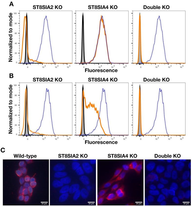
Abstract
Background: Polysialic acid (PSA) modifies a few cell surface proteins in humans mainly during fetal
development and some blood cells in adults. Two genes in humans, ST8SIA2 and ST8SIA4, code for
polysialyltransferases that synthesize PSA. The product of ST8SIA2, STX, is highly expressed during fetal
development and in many cancers but not in adult normal human cells. The product of ST8SIA4, PST1, is
expressed in fetal and some adult tissues and also in many cancers. We identified a derivative of PSA
containing de-N-acetyl neuraminic acid residues (dPSA), which is expressed on the cell surface of human
cancer cell lines and tumors but not normal cells.

Methods: dPSA-modified proteins in several human cancer cell lines and normal blood cells were
identified using co-immunoprecipitation with anti-dPSA antibodies and mass spectroscopy. RNAi and
CRISPR were used to knockdown and knockout, respectively, the polysialyltransferase genes in two
different cell lines to determine effect on production of cell surface dPSA measured by flow cytometry
and fluorescence microscopy.

Results: We found that dPSA is linked to nucleolin, a nuclear protein reported to be on the cell surface of
many cancers but not normal cells. Knocking down expression of ST8SIA2 with RNAi or knocking out
each gene individually and in combination using CRISPR showed that cell surface dPSA depended on
expression of ST8SIA2 and not ST8SIA4.

Conclusions: The presence of dPSA specifically in a broad range of human cancers offers novel
possibilities for targeting the dPSA antigen and synthetic pathway for detection, treatment, and
prevention of cancer.

Background
Human polysialic acid (PSA or polySia), is a developmentally regulated homopolymer of a2-8-linked 5-N
acetyl neuraminic acid residues that can be greater than 100 residues in length. Humans have two genes,
ST8SIA2 and ST8SIA4, that code for enzymes that synthesize PSA (polysialyltransferases STX and
PST1, respectively). While both genes are highly expressed in humans during fetal development [1], PST1
is expressed mainly in lymphoid tissues and lymphocytes [2], while STX appears not to be present at
significant levels in any adult normal tissues based on Northern blot [1] and publicly available RNA-seq
and protein databases (as summarized, for example, by GeneCards: the human gene database [3]). Of the
five proteins confirmed to be polysialylated in humans [4-8], neural cell adhesion molecule (NCAM) is the
most abundant, particularly during fetal development, and is the most thoroughly investigated [9]. A
number of human cancers are reported to express PSA-NCAM abnormally [10-14] where its role in
mediating interactions among cells and between cells and the extracellular matrix is associated with
metastasis and poor clinical prognosis [10, 14]. Although both polysialyltransferases STX and PST1
appear to synthesize the same polysaccharide, there may be differences in substrate specificity [15, 16] or
functional activity. STX was reported to produce shorter polymers compared to PST1 and, in some

                                                  Page 2/22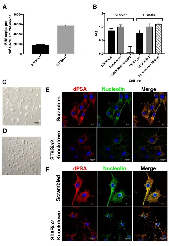
circumstances, the two enzymes may work synergistically [17]. Recently, mutations in the promoter region
of ST8SIA2 gene were associated with schizophrenia [18]. However, to date, there are no data suggesting
that the two polysialyltransferases have a preference for modifying a specific protein, and functional
distinctions between the two are unclear.

Previously, we reported the discovery of a de-N-acetylated form of PSA (dPSA) and of anti-dPSA
antibodies that were reactive with dPSA antigens [19, 20] on placental trophoblasts, the surface of cancer
cells, and inside some normal cells within the perinuclear space [21, 22]. Also, human microbial
pathogens that produce PSA, including Neisseria meningitidis serogroup B [19] and Leishmania major
[23], display cell surface dPSA under conditions corresponding to encountering a human host [23, 24].
Although de-N-acetyl sialic acid-containing derivatives of gangliosides have been described previously in
human melanoma cell lines [25, 26] and de-N-acetylation of polysaccharides occurs in many species [27],
proteins modified with dPSA and the function of dPSA in human cell biology, particularly cancer, are
unknown.

In this study, we identified nucleolin as the protein modified with dPSA. We investigated the role of
STX/ST8SIA2 and PST1/ST8SIA4 related to the production of dPSA and found that STX/ST8SIA2 was
responsible for production of dPSA on the surface of cancer cells, and we characterized the effect of
interfering with ST8SIA2 and ST8SIA4 gene expression on cell surface dPSA and cancer cell morphology.
Importantly, we show that cell surface dPSA is unique to cancer cells and widely expressed among
different cancers. The unique presence of dPSA on the surface of human trophoblasts, cancer cells, and
microbial pathogens raises the possibility of a role for dPSA in immune shielding.

Methods
Antibody reagents

Anti-nucleolin monoclonal mouse antibody MS-3 was acquired from Santa Cruz Biotechnology (Santa
Cruz, CA), anti-a-tubulin was from Invitrogen and anti-b-tubulin Type III was from Sigma-Aldrich (St. Louis,
MO). Irrelevant murine and human subclass control antibodies were obtained from Southern Biotech
(Birmingham, AL) and BioXCell (Lebanon, NH). Anti-dPSA monoclonal antibodies (mAbs) SEAM 2 and
SEAM 3 and anti-PSA mAb SEAM 12 were produced as described previously [28]. Irrelevant mAb, 14C7 (
murine IgG3), was produced as described previously [29]. All anti-mouse secondary antibodies
conjugated with Alexa Fluor fluorochromes were obtained from Thermo Fisher Scientific (Waltham, MA).
SEAM 2, SEAM 3, SEAM 12 and 14C7 used in this study were purified by Protein A affinity
chromatography. Antibody concentrations were determined by absorbance at 280nm.

Cell culture

SK-MEL-28 human melanoma and panels of gastric, ovarian, and pancreatic cancer cell lines (HTB-72,
TCP-1008, TCP-1021, and TCP-1026, respectively) were obtained from American Type Culture Collection
(Manassas, VA) and cultured in medium recommended by ATCC. CHP-134 cells were obtained from
                                                  Page 3/22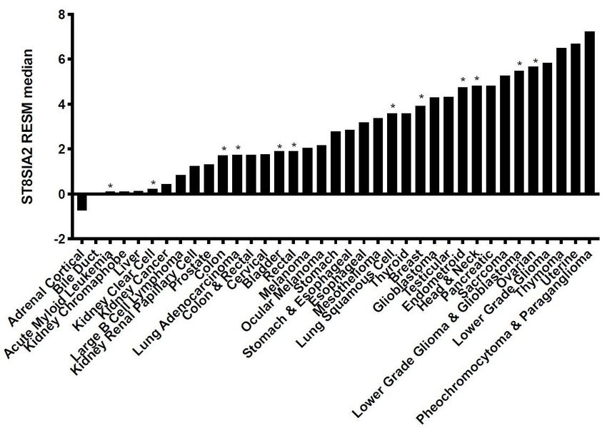
MilliporeSigma (Burlington, MA). Kelly cells were a gift from J. Saba at UCSF Benioff Children’s Hospital
Oakland. Kelly, SK-MEL-28, RNAi mutant SK-MEL-28, CHP-134, and CRISPR knockout cells were grown
routinely in flasks containing RPMI 1640 medium, penicillin/streptomycin, and 10% fetal bovine serum
(FBS) at 37 °C in 5% CO2. Confluent cells were sub-cultured (1∶3 to 1∶8) by treating with 0.25%
(weight/volume) trypsin/0.53 mM EDTA or AccutaseÒ solution (Thermo Fisher Scientific) and washing in
media before re-seeding into new growth medium. CHP-134 and CHP-134 CRISPR knockout clone cells
were suspended by pipetting. Medium for the SK-MEL-28 mutant cell lines also contained 10 µg/mL
blasticidin. Adherent cells used for immunoprecipitation, fluorescence activated cell sorting (FACS)
binding, and fluorescence labeling experiments were treated with AccutaseÒ and washed with medium
before use. Human normal peripheral blood mononuclear cells (PBMCs) were purchased from AllCells
(Alameda, CA).

Protein extraction from cells

Cells from each cell culture were extracted using the ProteoExtract® Subcellular Proteome Extraction Kit
(MilliporeSigma). In brief, the differential detergent extraction procedure used four extraction buffers
sequentially, along with a protease inhibitor cocktail to prevent protein degradation during the extraction
and Benzonase® nuclease (Sigma-Aldrich) to degrade contaminating nucleic acids. The manufacturer’s
instructions for extraction were followed, and the cell extracts were separated into four fractions: F1
(cytosolic fraction), F2 (cell membrane fraction), F3 (nucleic protein fraction), and F4 (cytoskeletal
fraction).

Co-immunoprecipitation

Dynabeads M-270 epoxy magnetic beads (Thermo Fisher Scientific) covalently linked to SEAM 2 or the
irrelevant murine IgG3 mAb 14C7 were prepared following the manufacturer’s protocol. As F1, F2, and F3
were the only fractions to react in an immuno-dot blot (see, for example, Additional file 1, Supplementary
Fig. S1), they were the only fractions that were further purified through co-immunoprecipitation. Each
fraction was incubated separately with SEAM 2- or 14C7-linked magnetic beads. The beads were
separated using a magnet, washed with the respective extraction buffer alone, and then with buffer
containing PSA (50 µg/mL; colominic acid from Sigma-Aldrich) to remove nonspecific binding antigens.
Finally, buffer containing 50 µg/mL N-propionyl polysialic acid [22] with 36% de-N-acetyl polysialic acid
(N-Pr dPSA) was used to elute the antigens binding specifically to SEAM 2 from each fraction.

LC/MS/MS protein identification

The eluted proteins from each cell fraction were resolved on 4–12% gradient SDS-PAGE gels (NuPAGE,
Thermo Fisher Scientific). The gels were stained with SimplyBlue™ Coomassie stain (Thermo Fisher
Scientific). The excised gel slices were extracted overnight in 50 mM ammonium bicarbonate containing
50% acetonitrile. The disulfide bonds were reduced and modified with iodoacetamide per University of
California San Francisco In-Gel Digestion Protocol [30] prior to trypsin digest (Promega Corp., Madison,

                                                  Page 4/22
WI). LC/MS/MS protein identification of tryptic peptides was performed by the University of California
Davis Proteomics Core facility.
Laser scanning confocal microscopy
SK-MEL-28, ST8SIA2 knockdown mutant, and scrambled RNA negative control cells (∼105 cells/mL)
were cultured on glass coverslips coated with human placental Type IV collagen (Sigma-Aldrich). After an
overnight incubation, cells were fixed with 4% formaldehyde for 1 h in phosphate buffered saline (PBS).
For staining internal antigens, coverslips were treated with ice-cold 0.25% Triton X-100 in PBS for 10 min.
The coverslips were blocked in blocking buffer (2% goat serum in PBS/0.25% Tween). Primary antibodies
(5µg/mL) in blocking buffer were added to the coverslips and incubated at ambient temperature. After
PBS/Tween washes, goat anti-mouse isotype-specific secondary antibodies conjugated to Alexa Fluor
488 or Alexa Fluor 594 (Thermo Fisher Scientific) were added (1:200 dilution) in blocking buffer at room
temperature in the dark. Subsequently, the cells were washed with PBS/Tween, then PBS. Finally, DNA
was stained with 4′,6-diamidino-2-phenylindole (DAPI) in PBS for 10 min before mounting with mounting
medium (Electron Microscopy Sciences, Hatfield, PA). Confocal images were obtained using a Zeiss
LSM710 laser scanning confocal microscope and analyzed using ImageJ Software [31] and JACoP [32].
Creating knockdown cell lines targeting ST8SIA2 and ST8SIA4 by RNA interference
Knockdown cell lines were produced in SK-MEL-28 human melanoma cells by vector-based interfering
RNA using a BLOCK-iTTM Pol II miRNA RNA vector system (Thermo Fisher Scientific). SK-MEL-28 cells
were transfected with one of four vector constructs, two targeting each gene, called pcDNA 6.2-GW/+
EmGFP – PolyST, where PolyST represents ST8SIA2-1, ST8SIA2-2, ST8SIA4-1, and ST8SIA4-2. An
additional scrambled control construct which does not target any known vertebrate gene was made, for a
total of five vector constructs.
For each construct, multiple clonal cells lines were established. Following the Thermo Fisher Scientific
TurboFect Transfection Reagent general protocol (up to step 7), approximately 5 × 10⁴ cells were seeded
in each 24-well plates with growth medium 24 h prior to transfection. Cells were trypsinized and replated
after transfection into 6-well plates (continuing with step 3 of the BLOCK-iTTM Pol II miR RNAi Expression
Vector Kits - Generating Stable Cell Line protocol), then selected for survival in medium with 5 µg/mL
blasticidin. Blasticidin-resistant cell lines were cloned by limiting dilution and a subset of 4 scrambled
control constructs, 7 ST8SIA2-1 constructs, 2 ST8SIA2-2 constructs, 4 ST8SIA4-1 constructs, and 3
ST8SIA4-2 constructs were screened by fluorescence microscopy. Cells with EmGFP fluorescence were
identified as cells containing the integrated plasmid.
Integration of the vector sequence was confirmed by isolating the genomic DNA (5 Prime PerfectPure
DNA Cultured Cell Kit, Thermo Fisher Scientific) from 20 cell lines, amplifying the DNA target sequence
with conventional PCR (MJ Research, Inc., South San Francisco, CA) using a T7 primer and running the
products on a 0.6% agarose gel containing 0.01% ethidium bromide. After cutting out the PCR products
Page 5/22from the gel (Qiagen QiaQuick Gel Extraction kit, Thermo Fisher Scientific), the PCR product was
sequenced (Sequetech, Mountain View, CA).
Quantifying expression of polysialyltransferase mRNA using real-time qPCR
RNA from the cells was isolated (Fermentas GeneJet RNA purification kit, Thermo Fisher Scientific), and
cDNA was produced (Fermentas Maxima First Strand cDNA Synthesis Kit) for use in a Taqman Gene
Expression real-time qPCR assay (Thermo Fisher Scientific). The relative mRNA copy numbers of
ST8SIA2 or ST8SIA4 was determined using glyceraldehyde-3-phosphate dehydrogenase (GAPDH) as an
internal control for each of the clonal cell lines (in triplicate samples). Cell lines with decreased mRNA
copy number compared to a control cell line with integrated plasmid and cultured in the presence of
blasticidin were propagated for further study.
CRISPR knockout of ST8SIA2 and ST8SIA4 genes in CHP-134 cells
CHP-134 cell lines with ST8SIA2 and ST8SIA4 genes knocked out individually and in combination were
constructed by WuXi AppTec Co. Ltd. (Shanghai, China) The sequences ataaccagacgctctctctg and
actatgtgcttgacaggcgc were targeted for knocking out ST8SIA2 and ST8SIA4, respectively. After
confirming biallelic gene knockouts by reverse transcribing mRNA to cDNA and cloning and sequencing
the cDNA, the cell lines were further subcloned a second time as described above for SK-MEL-28 RNAi
mutants to ensure clonality.
Antibody binding to cells
FACS binding experiments were performed with CHP-134 wild-type and CRISPR knockout clone cells as
described previously [22]. Cells were suspended in fresh RPMI 10% FBS medium. The cell count was
adjusted to 106 live cells/mL. Test Abs and controls were added (10µg/mL) to cells in tubes and
incubated at ambient temperature for 45 min while mixing by mechanically rotating the tubes. The cells
were washed with RPMI 10% FBS, then resuspended in the same medium containing secondary goat anti-
mouse IgG AlexFluor 647 (Thermo Fisher Scientific). After 30 min incubation with mixing, the cells were
washed once with Dulbecco’s phosphate buffered saline (DPBS) without Mg2+ or Ca2+ salts and
suspended in DPBS containing 0.5% (volume/volume) formaldehyde for 10 min at room temperature.
Binding was analyzed using a LSR Fortessa Flow Cytometer (BD Biosciences, San Jose, CA). FlowJo
(TreeStar, Woodburn, OR) was used for data analysis.
Results
Anti-dPSA co-immunoprecipitates dPSA and nucleolin from human cancer cells
To determine the identity of antigens potentially modified with dPSA, we prepared cytoplasmic, cell
membrane, nuclear, and cytoskeletal fractions (F1–F4, respectively) from human SK-MEL-28 melanoma
cells. The fractions were combined with anti-dPSA mAb SEAM 2 or an irrelevant control murine IgG3 mAb
Page 6/22(14C7) covalently linked to magnetic beads. The cytoskeletal fraction was not subjected to co-
immunoprecipitation since this fraction, which contains SDS, was not reactive with SEAM 2 in an
immunodot blot (Additional file 1, Supplementary Fig. S1). The proteins bound to the beads were
specifically eluted with N-Pr PSA containing 36% de-N-acetyl sialic acid in extraction buffer after a wash
with buffer containing PSA. SEAM 2 has more than 100 times greater avidity for the N-Pr PSA antigen
than dPSA containing N-acetyl sialic acid residues based on an inhibition ELISA [19]. The eluted proteins
were resolved on SDS-PAGE gels. As shown in Fig. 1, only the sample from the SEAM 2 co-
immunoprecipitated membrane fraction (Fig. 1B, lane F2) contained stained proteins. The proteins
appear largely as a “smear” rather than distinct bands. Typically, proteins modified with PSA run in SDS-
PAGE gels over a relatively wide range of apparent mass because of variable PSA length. Variable
amounts of PSA de-N-acetylation may result in additional heterogeneity. Four relatively dark-staining
regions of the gel spanning the range of eluted dPSA antigens were excised from the gel and processed
to generate tryptic peptides for LC/MS/MS mass fingerprinting (indicated by arrows in Fig. 1B, lane F2).
As controls, gel slices were taken from the same relative positions in the sample from the membrane
fraction control irrelevant IgG3 mAb co-immunoprecipitation (Fig. 1A, lane F2).
The tryptic peptide LC/MS/MS analysis of the bands excised from the SEAM 2-F2 immuno-precipitate
identified nucleolin (14 exclusive peptides, 16 exclusive unique spectra, 167/710 amino acids identified in
the segment including residues 139–624) as the predominant protein in slices marked P1 (~145 kDa)
and P2 (~115 kDa). In addition, there were trace amounts of several ribosome-associated proteins in both
fractions. One function of nucleolin is to promote assembly of ribosomes in nucleoli. Slices marked P3
(~71 kDa) contained ribosome-associated proteins and a-tubulin in addition to nucleolin, and P4 (~48
kDa) contained ribosome-associated proteins and actin. There were no cell-derived proteins identified in
the control slices. The calculated molecular mass of nucleolin is 77 kDa, but nucleolin can have several
apparent masses ranging from ~50 kDa to ~100 kDa because of multiple covalent modifications
including sialylation, the presence of highly acidic regions near the N-terminal region, and self-cleaving
activity [33, 34]. The experiment was repeated and included an additional control of co-
immunoprecipitation with the anti-PSA mAb SEAM 12 (Additional file 1, Supplementary Fig. S2). Again,
nucleolin was the predominant protein co-immunoprecipitated by SEAM 2 from the membrane fraction in
gel slices corresponding to lane F2 bands P1 and P2 in Fig. 1B (38 exclusive unique peptides, 53
exclusive unique spectra, 274/710 amino acids identified in the segment including residues 73–629),
with smaller amounts of nucleolin also present in the SEAM 12 co-immuno-precipitate, and trace
amounts in the irrelevant control. To determine whether dPSA was unique to SK-MEL-28 cells or common
to all dPSA containing cells, the same co-immunoprecipitation procedure of subcellular-enriched cell
fraction F2 was repeated for Kelly neuroblastoma, SNU-1 gastric cancer cells, and normal PBMCs
(Additional file 1, Supplementary Figs. S3 and S4, respectively). All displayed anti-dPSA reactivity inside
and outside cells by FACS and fluorescence microscopy except for PBMCs, which had anti-dPSA
reactivity with both SEAM 2 and SEAM 3 inside cells (i.e., detergent-treated) but not on the surface of cells
not treated with detergent to permeabilize them (Additional file 2, Supplementary Fig. S1. The cells tested
represent a range of STX and PST1 expression based on RNA-seq data from publicly available databases
Page 7/22[3, 35]: Kelly having high STX and low PST1; PBMCs having no STX and high PST1; and SNU-1 having
low STX and PST1. Nucleolin was co-immunoprecipitated by SEAM 2 from the membrane fraction (F2)
of Kelly (19 exclusive unique peptides, 22 exclusive unique spectra, 190/710 amino acids identified in the
segment including residues 73–649), SNU-1 (20 exclusive unique peptides, 32 exclusive unique spectra,
314/710 amino acids identified in the segment including residues 73–648), and PBMCs (10 exclusive
unique peptides, 14 exclusive unique spectra, 214/710 amino acids identified in the segment including
residues 80–648) that was not present in corresponding gel slices of the irrelevant negative control
(Additional file 1, Supplementary Figs. S2-S4). Importantly, there were no peptides observed in any of the
anti-dPSA immunoprecipitation experiments identifying proteins known to be modified with PSA [36].
Therefore, none of the known PSA-modified proteins appear to be modified with dPSA.
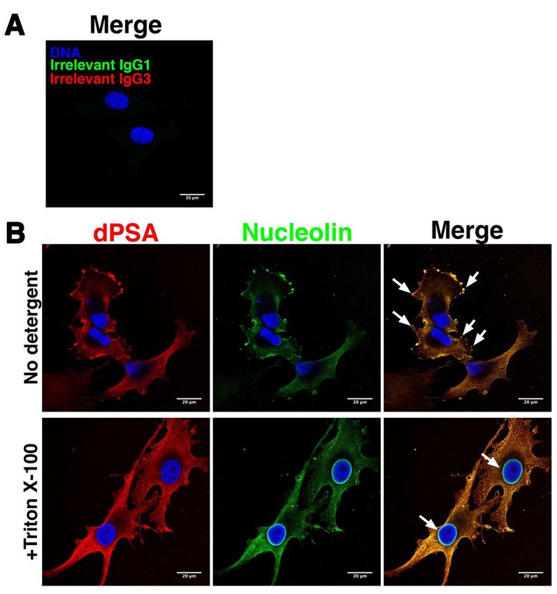
Anti-dPSA is co-localized with nucleolin on the surface of SK-MEL-28 melanoma cells
Nucleolin is a nuclear protein, but it is also found on cilia of airway epithelial cells, where it serves as a
receptor for various pathogens [37] and has been reported to be present on the cell surface of cancer cells
[38]. Since anti-dPSA mAb SEAM 2 co-immunoprecipitated nucleolin, we asked whether nucleolin is co-
localized with dPSA in SK-MEL-28 cells. Anti-nucleolin mAb MS-3 and anti-dPSA mAb SEAM 2 were used
to detect each antigen combined with subclass-specific fluorescently labeled secondary antibodies by
laser scanning confocal fluorescence microscopy. As a control, the cells were treated with an irrelevant
murine IgG3 mAb and the secondary antibodies. As shown in Fig. 2A, there was no staining with
irrelevant mAbs or secondary antibodies alone. In wild-type SK-MEL-28 cells, dPSA (Fig. 2B, red
fluorescence) was present on the surface of cells and was concentrated in lamellipodia, podosomes, and
filopodia (examples indicated by arrows in Fig. 2B, merge image). Anti-nucleolin staining (Fig. 2B, green
fluorescence) was identical with respect to location and intensity of staining to anti-dPSA staining (Fig.
2B, merge).
dPSA-nucleolin is present on the surface of primary and metastatic pancreatic, gastric, and ovarian
cancer cell lines
To determine whether dPSA co-localization with nucleolin was unique to SK-MEL-28 cells or common to
cancer cells, we performed similar confocal fluorescence microscopy experiments labeling dPSA with
SEAM 2 and nucleolin with anti-nucleolin mAb MS-3 on sets of seven pancreatic, five gastric, four ovarian
primary and metastatic cell lines (Additional file 2, Supplementary Figs. S6-S8, respectivley). The panels
of cancer cell lines are representative of different genetic alterations of signaling mechanisms for each
cancer [39]. Control antibodies included irrelevant murine IgG1 and IgG3 mAbs and Alexa Fluor-
conjugated secondary antibodies (Additional file 2, Supplementary Figs. S5-S8). All the cancer cell lines
were positive for both dPSA and cell surface nucleolin, which were co-localized as determined by analysis
of Z-stack confocal micrographs using JACoP [32], except for the ovarian teratocarcinoma cell line PA-1
(Additional file 2, Supplementary Fig. S8). dPSA staining was much stronger and consistent for all cell
lines that were positive for dPSA, whereas anti-nucleolin staining was less consistent with anti-nucleolin
being less reactive for cell lines having high polysialyltransferase expression. There was no surface anti-
Page 8/22dPSA or nucleolin staining on human normal PBMCs (Additional file 2, Supplementary Fig. S5), which
include subsets of cells with high levels of PST1 and PSA-NCAM expression.
Membrane-associated dPSA-nucleolin is distinct from nuclear nucleolin
Next, we looked at staining patterns inside cells by treating fixed SK-MEL-28 cells with the detergent
Triton X-100 to permeabilize them and allow entry of primary and secondary antibodies. dPSA inside cells
exhibited a web-like pattern typical of smooth endoplasmic reticulum and trans golgi network (Fig. 2B,
+Triton X-100). Again, anti-nucleolin staining matched that of anti-dPSA inside cells with respect to
location and intensity of staining. The exception was the strong anti-nucleolin staining around the
nuclear membrane, which was not reactive with anti-dPSA mAbs (Fig. 2B, +Triton X-100).
Effect on dPSA and cell surface nucleolin of knocking down expression of ST8SIA2 with siRNA in human
SK-MEL-28 cells
SK-MEL-28 cells express both ST8SIA2 and ST8SIA4 (Fig. 3A). Previously, we showed that transient
ST8SIA4 RNAi knockdown in SK-MEL-28 cells decreased production of PSA and dPSA, demonstrating
that dPSA was derived from PSA [22]. In this study, we constructed mutant SK-MEL-28 cell lines by
chromosomal integration of plasmids expressing siRNA targeting ST8SIA2. The relative expression of
both genes in total mRNA was determined by quantitative real-time PCR and normalized using GAPDH
mRNA as in internal control (Fig. 3A). There was a significant reduction in the amount of ST8SIA2 mRNA
in the ST8SIA2 knockdown compared to the wild-type and the scrambled control constructs (Fig. 3B).
There was no effect in the ST8SIA2 knockdown mutant on expression of ST8SIA4 mRNA (Fig. 3B), which
was ~3-fold higher than the wild-type level of ST8SIA2 mRNA (Fig. 3A).
RNAi knockdown of ST8SIA2 resulted in altered cell morphology (Fig. 3D) compared to the negative
control cell line expressing a scrambled RNA (Fig. 3C). The cells with ST8SIA2 RNAi knockdown had a
distinct rounded appearance with relatively short lamellipodia circularly arrayed around the cell (Fig. 3D).
Cell surface dPSA and nucleolin were decreased in the ST8SIA2 RNAi knockdown mutant compared to
the control SK-MEL-28 mutant expressing a scrambled RNA (Fig. 3E). Similarly, the cytoplasmic amounts
of dPSA and nucleolin in Triton X-100-treated cells (Fig. 3F) were reduced in the ST8SIA2 knockdown
mutant. siRNA had no effect on anti-nucleolin staining of the nuclear membrane (Fig. 3F) showing dPSA-
modified and nuclear nucleolin were distinct forms of the protein.
CRISPR knockout of ST8SIA2 but not ST8SIA4 eliminated cell surface and intracellular dPSA in human
CHP-134 neuroblastoma cells
To determine whether cell surface dPSA depends on the activity of one or both polysialyltransferases, we
used CRISPR to knock out each gene individually and in combination in human neuroblastoma CHP-134
cells. CHP-134 cells were chosen since both genes are relatively highly expressed [22] and we wanted to
determine whether the effects of interfering with expression of the polysialyltransferases in SK-MEL-28
cells could be replicated in another cancer cell line by a different method. Cell lines transfected with
Page 9/22plasmids coding for Cas9 and RNAs targeting the respective polysialyltransferase gene(s) were cloned,
and the targeted gene knockouts were confirmed. When ST8SIA2 alone or in combination with ST8SIA4
was knocked out, cell surface dPSA, as measured by FACS and fluorescence microscopy with SEAM 3,
was eliminated (Fig. 4A). Interestingly, knocking out ST8SIA2 also eliminated cell surface PSA (Fig. 4B)
suggesting that, in CHP-134 cells, polysialyltransferases may work synergistically as suggested by
Angata et al. [17]. PSA in CHP-134 cells mainly modifies NCAM [40]. Knocking out ST8SIA4 had no effect
on cell surface dPSA but reduced cell surface PSA (Fig. 4A and B, respectively). Therefore, cell surface
dPSA in CHP-134 cells depended entirely on expression of ST8SIA2.
Unlike SK-MEL-28 cells, there were no apparent differences in the morphology of CHP-134 mutants.
However, nuclei in cell lines with ST8SIA2 and ST8SIA2+ST8SIA4 knocked out were larger (on average
1.45 ±0.24 times greater based on length times width measurements of 20 nuclei each) compared to
wild-type or the ST8SIA4 knockout cell lines (Fig. 4C). The differences in the size of the nuclei depended
on cell attachment to a surface, as there were no differences in the size of the nuclei of cells in
suspension.
Intracellular antigens reactive with both SEAM 2 and SEAM 3 were reduced but still detected in detergent-
treated cells after knocking out ST8SIA2, ST8SIA4 or both genes in CHP-134 cells and in normal PBMCs
that do not express ST8SIA2 suggesting that the internal staining described previously [21] may result
from cross-reactivity with an antigen that does not depend on either STX or PST1. For example, an
intracellular sialylated glycan produced by another sialyltransferase that is subsequently de-N-acetylated
[26, 41, 42]. However, the protein identified by immunoprecipitation and mass spectroscopy in both
cancer cells and normal PBMCs was nucleolin. Taken together, the data suggest nucleolin modified with
de-N-acetyl sialic acid may be an anchor glycan that, when polysialylated by STX, results in transfer of
dPSA-nucleolin to the cell surface.
ST8SIA2 is abnormally expressed in many cancers
Based on recent data from publicly available RNA-seq and proteomics databases, ST8SIA2 expression in
cells of adult normal tissues is either very low or non-existent (see, for example, GeneCards: the human
gene database [3]). However, based on RNA-seq data from The Cancer Genome Atlas (TCGA) Program
[35] for 37 cancers, most express some level of ST8SIA2 that does not occur in corresponding cells of
human normal tissues (Fig. 5). The ST8SIA2 gene expression data are consistent with the reported
prevalence of dPSA-linked cell surface nucleolin in human cancers [43]. Based on combined gene
expression and proteomics data, the polysialyltransferase does not appear to be produced at measurable
levels in any adult normal tissue. However, as shown in Fig. 5, higher expression is observed in most
cancers where it is associated with invasiveness, metastasis, and poor prognosis for many cancers [44].
Discussion
Altered glycosylation patterns of cell surface proteins occur in nearly all types of cancer. Excessive
sialylation of glycoproteins and glycolipids is central to the aberrant regulation of cell adhesion in
Page 10/22metastatic cancer, which in turn can result from re-expression and/or overexpression of genes normally
expressed during development but not in cells of adult normal tissues. In this study, we gathered further
evidence that dPSA is a novel tumor-associated carbohydrate antigen. We have shown that dPSA is
linked to nucleolin, which is normally a nuclear protein but is reported to be on the cell surface of many
cancers [43]. We have also shown that the presence of dPSA-nucleolin on the surface of cancer cells is
dependent on re-expression of a fetal polysialyltransferase gene, ST8SIA2, which may indicate a novel
cancer pathway to further investigate for additional points of therapeutic intervention.
Protein polysialylation is highly restricted. To date, only five proteins in humans are reported to be
modified by polysialylation [21]: NCAM [5], E-selectin ligand-1 [7], the scavenger receptor CD36 [8],
neuropilin-2 [4], and STX itself [6]. In this study, none of the previously known polysialylated proteins were
co-immunoprecipitated from any of the cancer cell lines tested or human normal PBMCs with anti-dPSA
antibodies suggesting that dPSA modification is exclusive to nucleolin. The large size and electrostatic
repulsion of polyanionic PSA is thought to block homophilic interactions between adhesion molecules
such as NCAM [9] during nervous system development. However, partial de-N-acetylation, which is
common to many polysaccharides [27], alters the charge of PSA as de-N-acetylation produces
zwitterionic polysaccharides (ZPS) with different chemical and biological properties. For example,
microbial ZPS are frequently involved in immune shielding such as formation of biofilms [45], modulating
T cell function [46], and have increased immunogenicity compared to N-acetylated polysaccharides [47].
Nucleolin is an RNA binding protein found throughout the cell where it has been detected in the
cytoplasm, nucleus, nucleoli, cell membrane and on cilia [48]. The reason for the difference in nucleolin
subcellular localization is thought to be the result of post-translational modifications, which include
sialylation—although polysialylation of nucleolin was not known to occur previously [49]. However, in
cancerous cells but not normal cells, nucleolin is reported to be expressed consistently and abundantly on
the cell surface [43]. The functional purpose of cell surface nucleolin in cancer is unknown but, based on
the data presented here, may be to direct intracellular dPSA to the cell surface.
Cell surface dPSA depended on ST8SIA2 gene expression only. Knocking down or knocking out ST8SIA2
in two different cell lines also reduced or eliminated cell surface nucleolin, respectively. In addition to the
cancer cell lines, further evidence for the link between STX and cell surface dPSA-nucleolin was shown
with human normal PBMCs, which do not express ST8SIA2 and do not have cell surface dPSA or
nucleolin (Additional file 1, Supplementary Fig. S1). Although cumulative data from RNA-seq suggest a
low level of ST8SIA2 gene expression in human heart and brain, the presence of STX protein has not
been confirmed and we did not find cell surface dPSA in either tissue during a immunohistochemical
study of 18 major human organs [21]. Overall, the data presented here show that there are distinct
functions of each polysialyltransferase, at least with respect to the subcellular distribution of dPSA and
its presence on nucleolin.
Conclusions
Page 11/22Expression of genes coding for polysialyltransferases, in particular, are restricted in adult human normal
tissues but commonly expressed in most, if not all, cancers. This study suggests that ST8SIA2
expression and cell surface dPSA-nucleolin are limited to cancer cells and the developing embryo [1, 21].
Since anti-dPSA mAbs have direct cytotoxic effects on multiple cancer cell lines [22], approaches that
target ST8SIA2 and dPSA to prevent and treat cancers without adverse effect on normal cells may be
possible. The presence of dPSA on human trophoblasts [21], microbial pathogens [23, 24, 47], and cancer
cells [21, 22] raises the possibility that the function of dPSA may be related to an unrecognized
mechanism of immune shielding, since all of the above have in common a need to avoid being targeted
by immunological mechanisms of protection.
List Of Abbreviations
CRISPR, clustered regularly interspaced short palindromic repeats; DPBS, Dulbecco’s phosphate buffered
saline; dPSA, de-N-acetyl neuraminic acid-containing poly alpha2-8 N-acetyl neuraminic acid; N-Pr dPSA,
N-propionyl polysialic acid with 36% de-N-acetyl polysialic acid; FACS, fluorescence activated cell sorting;
FBS, fetal bovine serum; GAPDH, glyceraldehyde-3-phosphate dehydrogenase; mAb, monoclonal
antibody; NCAM, neural cell adhesion molecule; PBMCs, peripheral blood mononuclear cells; PSA, poly
alpha2-8 N-acetyl neuraminic acid; RNAi, RNA interference; RNA-seq, mRNA sequencing; siRNA, small
interfering RNA; ZPS, zwitterionic polysaccharide.
Declarations
Ethics approval and consent to participate: Not applicable
Consent for publication: Not applicable
Availability of data and materials: The datasets supporting the conclusions of this article are included
within the article and its additional files.
Competing interests: G.R.M. is an inventor on several patent applications and issued patents related to
therapies and diagnostic applications targeting dPSA. Rights to these inventions have been assigned to
UCSF Benioff Children’s Hospital Oakland. G.R.M. is also a founder and stockholder of, and consultant to,
Saccharo, Inc., a company focused on developing the therapeutic and diagnostic potential of dPSA.
Funding: This work was supported by grants from Saccharo, Inc., Children’s Hospital Oakland Research
Institute, ImpactAssets, and the Family of Jennifer Leigh Wells.
Authors’ contributions: G.R.M and L.M.S. conceived, planned, and performed experiments and wrote the
manuscript. A.D.B., J.A.L., and A.S. performed experiments and wrote portions of the manuscript.
Acknowledgements: We thank Eileen Y. Ivasauskas of AccuWrit, Inc. for technical writing and editing the
manuscript.
Page 12/22References
1. Angata K, Nakayama J, Fredette B, Chong K, Ranscht B, Fukuda M: Human STX polysialyltransferase
forms the embryonic form of the neural cell adhesion molecule. Tissue-specific expression, neurite
outgrowth, and chromosomal localization in comparison with another polysialyltransferase, PST. J
Biol Chem 1997, 272:7182-7190.
2. Drake PM, Stock CM, Nathan JK, Gip P, Golden KP, Weinhold B, Gerardy-Schahn R, Bertozzi CR:
Polysialic acid governs T-cell development by regulating progenitor access to the thymus. Proc Natl
Acad Sci USA 2009, 106:11995-12000.
3. GeneCards: The Human Gene Database [https://www.genecards.org]
4. Curreli S, Arany Z, Gerardy-Schahn R, Mann D, Stamatos NM: Polysialylated neuropilin-2 is expressed
on the surface of human dendritic cells and modulates dendritic cell-T lymphocyte interactions. J
Biol Chem 2007, 282:30346-30356.
5. Finne J, Finne U, Deagostini-Bazin H, Goridis C: Occurrence of alpha 2-8 linked polysialosyl units in a
neural cell adhesion molecule. Biochem Biophys Res Commun 1983, 112:482-487.
6. Simon P, Baumner S, Busch O, Rohrich R, Kaese M, Richterich P, Wehrend A, Muller K, Gerardy-Schahn
R, Muhlenhoff M, et al: Polysialic acid is present in mammalian semen as a post-translational
modification of the neural cell adhesion molecule NCAM and the polysialyltransferase ST8SiaII. J
Biol Chem 2013, 288:18825-18833.
7. Werneburg S, Buettner FF, Erben L, Mathews M, Neumann H, Muhlenhoff M, Hildebrandt H:
Polysialylation and lipopolysaccharide-induced shedding of E-selectin ligand-1 and neuropilin-2 by
microglia and THP-1 macrophages. Glia 2016, 64:1314-1330.
8. Yabe U, Sato C, Matsuda T, Kitajima K: Polysialic acid in human milk. CD36 is a new member of
mammalian polysialic acid-containing glycoprotein. J Biol Chem 2003, 278:13875-13880.
9. Rutishauser U: Polysialic acid in the plasticity of the developing and adult vertebrate nervous system.
Nat Rev Neurosci 2008, 9:26-35.
10. Amoureux MC, Coulibaly B, Chinot O, Loundou A, Metellus P, Rougon G, Figarella-Branger D:
Polysialic acid neural cell adhesion molecule (PSA-NCAM) is an adverse prognosis factor in
glioblastoma, and regulates olig2 expression in glioma cell lines. BMC Cancer 2010, 10:91.
11. Gluer S, Schelp C, von Schweinitz D, Gerardy-Schahn R: Polysialylated neural cell adhesion molecule
in childhood rhabdomyosarcoma. Pediatr Res 1998, 43:145-147.
12. Moolenaar CE, Muller EJ, Schol DJ, Figdor CG, Bock E, Bitter-Suermann D, Michalides RJ: Expression
of neural cell adhesion molecule-related sialoglycoprotein in small cell lung cancer and
neuroblastoma cell lines H69 and CHP-212. Cancer Res 1990, 50:1102-1106.
13. Roth J, Zuber C, Wagner P, Blaha I, Bitter-Suermann D, Heitz PU: Presence of the long chain form of
polysialic acid of the neural cell adhesion molecule in Wilms' tumor. Identification of a cell adhesion
molecule as an oncodevelopmental antigen and implications for tumor histogenesis. Am J Pathol
1988, 133:227-240.
Page 13/2214. Tanaka F, Otake Y, Nakagawa T, Kawano Y, Miyahara R, Li M, Yanagihara K, Nakayama J, Fujimoto I,
Ikenaka K, Wada H: Expression of polysialic acid and STX, a human polysialyltransferase, is
correlated with tumor progression in non-small cell lung cancer. Cancer Res 2000, 60:3072-3080.
15. Galuska SP, Rollenhagen M, Kaup M, Eggers K, Oltmann-Norden I, Schiff M, Hartmann M, Weinhold B,
Hildebrandt H, Geyer R, et al: Synaptic cell adhesion molecule SynCAM 1 is a target for
polysialylation in postnatal mouse brain. Proc Natl Acad Sci U S A 2010, 107:10250-10255.
16. Rollenhagen M, Kuckuck S, Ulm C, Hartmann M, Galuska SP, Geyer R, Geyer H, Muhlenhoff M:
Polysialylation of the synaptic cell adhesion molecule 1 (SynCAM 1) depends exclusively on the
polysialyltransferase ST8SiaII in vivo. J Biol Chem 2012, 287:35170-35180.
17. Angata K, Suzuki M, Fukuda M: ST8Sia II and ST8Sia IV polysialyltransferases exhibit marked
differences in utilizing various acceptors containing oligosialic acid and short polysialic acid. The
basis for cooperative polysialylation by two enzymes. J Biol Chem 2002, 277:36808-36817.
18. Arai M, Yamada K, Toyota T, Obata N, Haga S, Yoshida Y, Nakamura K, Minabe Y, Ujike H, Sora I, et al:
Association between polymorphisms in the promoter region of the sialyltransferase 8B (SIAT8B)
gene and schizophrenia. Biol Psychiatry 2006, 59:652-659.
19. Moe GR, Dave A, Granoff DM: Epitopes recognized by a nonautoreactive murine anti-N-propionyl
meningococcal group B polysaccharide monoclonal antibody. Infect Immun 2005, 73:2123-2128.
20. Moe GR, Dave A, Granoff DM: Molecular analysis of anti-N-propionyl Neisseria meningitidis group B
polysaccharide monoclonal antibodies. Mol Immunol 2006, 43:1424-1431.
21. Nakano TA, Steirer LM, Moe GR: The expression profile of de-N-acetyl polysialic acid (NeuPSA) in
normal and diseased human tissue. J Biol Chem 2011, 286:40343.
22. Steirer LM, Moe GR: An Antibody to de-N-Acetyl Sialic Acid Containing-Polysialic Acid Identifies an
Intracellular Antigen and Induces Apoptosis in Human Cancer Cell Lines. PLoS ONE 2011, 6:e27249.
23. Iovannisci DM, Plested CP, Moe GR: Evidence for rosettes as an unrecognized stage in the life cycle
of Leishmania parasites. J Eukaryot Microbiol 2010, 57:405-414.
24. Flitter BA, Ing JY, Moe GR: Effect of human serum on de-N-acetyl sialic acid epitope expression and
antibody activity against N. meningitidis group B. Vaccine 2010, 28:5967-5972.
25. Hanai N, Dohi T, Nores GA, Hakomori S: A novel ganglioside, de-N-acetyl-GM3 (II3NeuNH2LacCer),
acting as a strong promoter for epidermal growth factor receptor kinase and as a stimulator for cell
growth. J Biol Chem 1988, 263:6296-6301.
26. Sjoberg ER, Chammas R, Ozawa H, Kawashima I, Khoo KH, Morris HR, Dell A, Tai T, Varki A:
Expression of de-N-acetyl-gangliosides in human melanoma cells is induced by genistein or
nocodazole. J Biol Chem 1995, 270:2921-2930.
27. Pascual S, Planas A: Carbohydrate de-N-acetylases acting on structural polysaccharides and
glycoconjugates. Curr Opin Chem Biol 2020, 61:9-18.
28. Granoff DM, Bartoloni A, Ricci S, Gallo E, Rosa D, Ravenscroft N, Guarnieri V, Seid RC, Shan A, Usinger
WR, et al: Bactericidal monoclonal antibodies that define unique meningococcal B polysaccharide
epitopes that do not cross-react with human polysialic acid. J Immunol 1998, 160:5028-5036.
Page 14/2229. Moe GR, Zuno-Mitchell P, Hammond SN, Granoff DM: Sequential immunization with vesicles
prepared from heterologous Neisseria meningitidis strains elicits broadly protective serum antibodies
to group B strains. Infect Immun 2002, 70:6021-6031.
30. UCSF In-Gel Digestion Protocol [http://msf.ucsf.edu/protocols.html]
31. Abramoff MD, Magelhaes, P.J., Ram, S.J.: Image Processing with ImageJ. Biophotonics Internat
2004, 11:36-42.
32. Bolte S, Cordelieres FP: A guided tour into subcellular colocalization analysis in light microscopy.
Journal of microscopy 2006, 224:213-232.
33. Fang SH, Yeh NH: The self-cleaving activity of nucleolin determines its molecular dynamics in
relation to cell proliferation. Exp Cell Res 1993, 208:48-53.
34. Ginisty H, Sicard H, Roger B, Bouvet P: Structure and functions of nucleolin. J Cell Sci 1999, 112(Pt
6):761-772.
35. The Cancer Genome Atlas Program [www.cancer.gov/tcga]
36. Bhide GP, Colley KJ: Sialylation of N-glycans: mechanism, cellular compartmentalization and
function. Histochem Cell Biol 2017, 147:149-174.
37. Jeong KI, Piepenhagen PA, Kishko M, DiNapoli JM, Groppo RP, Zhang L, Almond J, Kleanthous H,
Delagrave S, Parrington M: CX3CR1 Is Expressed in Differentiated Human Ciliated Airway Cells and
Co-Localizes with Respiratory Syncytial Virus on Cilia in a G Protein-Dependent Manner. PLoS One
2015, 10:e0130517.
38. Hovanessian AG, Puvion-Dutilleul F, Nisole S, Svab J, Perret E, Deng JS, Krust B: The cell-surface-
expressed nucleolin is associated with the actin cytoskeleton. Exp Cell Res 2000, 261:312-328.
39. COSMIC. Catalogue of Somatic Mutations in Cancer [https://www.sanger.ac.uk/tool/cosmic]
40. Livingston BD, Jacobs JL, Glick MC, Troy FA: Extended polysialic acid chains (n greater than 55) in
glycoproteins from human neuroblastoma cells. J Biol Chem 1988, 263:9443-9448.
41. Popa I, Pons A, Mariller C, Tai T, Zanetta JP, Thomas L, Portoukalian J: Purification and structural
characterization of de-N-acetylated form of GD3 ganglioside present in human melanoma tumors.
Glycobiology 2007, 17:367-373.
42. Song WX, Vacca MF, Welti R, Rintoul DA: Effects of gangliosides GM3 and De-N-acetyl GM3 on
epidermal growth factor receptor kinase activity and cell growth. J Biol Chem 1991, 266:10174-
10181.
43. Hovanessian AG, Soundaramourty C, El Khoury D, Nondier I, Svab J, Krust B: Surface expressed
nucleolin is constantly induced in tumor cells to mediate calcium-dependent ligand internalization.
PLoS One 2010, 5:e15787.
44. Falconer RA, Errington RJ, Shnyder SD, Smith PJ, Patterson LH: Polysialyltransferase: a new target in
metastatic cancer. Curr Cancer Drug Targets 2012, 12:925-939.
45. Lee MJ, Geller AM, Bamford NC, Liu H, Gravelat FN, Snarr BD, Le Mauff F, Chabot J, Ralph B,
Ostapska H, et al: Deacetylation of Fungal Exopolysaccharide Mediates Adhesion and Biofilm
Page 15/22Formation. mBio 2016, 7:e00252-00216.
46. Cobb BA, Kasper DL: Zwitterionic capsular polysaccharides: the new MHCII-dependent antigens. Cell
Microbiol 2005, 7:1398-1403.
47. Moe GR, Bhandari TS, Flitter BA: Vaccines containing de-N-acetyl sialic acid elicit antibodies
protective against neisseria meningitidis groups B and C. Journal of immunology 2009, 182:6610-
6617.
48. Tayyari F, Marchant D, Moraes TJ, Duan W, Mastrangelo P, Hegele RG: Identification of nucleolin as a
cellular receptor for human respiratory syncytial virus. Nat Med 2011, 17:1132-1135.
49. Carpentier M, Morelle W, Coddeville B, Pons A, Masson M, Mazurier J, Legrand D: Nucleolin
undergoes partial N- and O-glycosylations in the extranuclear cell compartment. Biochemistry 2005,
44:5804-5815.
50. Hoadley KA, Yau C, Wolf DM, Cherniack AD, Tamborero D, Ng S, Leiserson MDM, Niu B, McLellan MD,
Uzunangelov V, et al: Multiplatform analysis of 12 cancer types reveals molecular classification
within and across tissues of origin. Cell 2014, 158:929-944.
Figures
Figure 1
Page 16/22Anti-dPSA mAb co-immunoprecipitates nucleolin from the SK-MEL-28 membrane fraction. Differential
detergent extraction was used to separate human melanoma SK-MEL-28 cells into subcellular fractions
for co-immunoprecipitation with a control murine IgG3 mAb (14C7) or anti-dPSA mAb SEAM 2 (both are
subtype IgG3) linked to magnetic beads. Fractions F1, F2, and F3 are cytoplasmic, cell membrane, and
nuclear fractions, respectively. Sections of the gels in lanes F2 indicated by arrows and labeled P1–P4 of
both gels were excised and proteins contained in them identified by in-gel trypsin digestion and
LC/MS/MS peptide mass fingerprinting. (A) Proteins co-immunoprecipitated by the irrelevant IgG3 mAb.
(B) Proteins co-immunoprecipitated by SEAM 2.
Page 17/22Figure 2
dPSA is co-localized with nucleolin on the cell surface and inside SK-MEL-28 human melanoma cells.
dPSA and nucleolin were identified on the surface (no detergent) or inside (+Triton X-100) SK-MEL-28
cells with anti-dPSA mAb SEAM 2 or anti-nucleolin mAb MS-3, respectively, and detected by fluorescently
labeling with Alexa Fluor 594 (red fluorescence) or Alexa Fluor 488 (green fluorescence)-conjugated anti-
mouse subtype-specific antibodies using confocal laser scanning microscopy. Blue fluorescence is DAPI
Page 18/22DNA staining. (A) Cells with irrelevant murine IgG1 and IgG3 and secondary antibodies and (B) cells with
SEAM 2 and MS-3; arrows in merge image indicate dPSA concentrated in lamellipodia, podosomes, and
filopodia in No detergent micrographs. Arrows in merge image of +Triton X-100-treated cells show nuclear
nucleolin, which is not marked by mAb SEAM 2. Scale bars, 20 µm.
Figure 3
Page 19/22RNAi knockdown of ST8SIA2 decreases expression of membrane-associated dPSA and nucleolin but not
nuclear nucleolin. RNAi was used to knockdown expression of ST8SIA2 in SK-MEL-28 human melanoma
cells and the effect on dPSA and nucleolin production was determined by confocal laser scanning
microscopy. (A) The absolute ST8SIA2 and ST8SIA4 mRNA copy number relative to that of GAPDH in
parent SK-MEL-28 cells. N=3. (B) Relative expression of mRNA coding for ST8SIA2 and ST8SIA4
polysialyltransferases in parent, a scrambled RNA negative control and ST8SIA2 RNAi knockdown SK-
MEL-28 mutants measured by RT-PCR. N=3. Brightfield micrographs of (C) scrambled control compared
to (D) ST8SIA2 knockdown mutant show morphological differences. Confocal laser scanning
micrographs of the scrambled and ST8SIA2 knockdown mutant show dPSA and nucleolin are co-
localized on (E) the surface and (F) inside cells but are both decreased when ST8SIA2 is knocked down
except for nucleolin in the nuclear membrane. dPSA and nucleolin were identified on the surface (E–F,
upper panels) or inside (treated with Triton X-100; E–F, lower panels) SK-MEl-28 cells with anti-dPSA mAb
SEAM 2 or anti-nucleolin mAb MS-3, respectively, and detected by fluorescently labeling with Alexa Fluor
594 (red fluorescence) or Alexa Fluor 488 (green fluorescence)-conjugated anti-mouse subtype-specific
antibodies using confocal laser scanning microscopy. Blue fluorescence is DAPI DNA staining. Scale
bars, 20 µm.
Page 20/22Figure 4
Knocking out (KO) ST8SIA2 eliminates cell surface dPSA and PSA from CHP-134 cells. Cell surface dPSA
(A) and PSA (B) on live wild-type (dotted blue line) and the indicated mutants (solid orange lines)
detected by FACS using anti-dPSA mAb SEAM 3 and anti-PSA mAb SEAM 12 as primary antibodies.
Controls included no primary antibody (gray-filled histograms) and irrelevant IgG2b or IgG2a mAbs,
respectively (solid black lines). Fluorescence micrographs (C) of wild-type CHP-134 transfected with an
Page 21/22empty plasmid compared to the CRISPR knockout cell lines of the indicated genes. Red fluorescence is
anti-dPSA (SEAM 3); blue fluorescence is DAPI DNA staining. Scale bars, 10 µm.
Figure 5
ST8SIA2 is re-expressed in most of the 37 human cancers included in TCGA. mRNA expression from
RNA-seq data is shown relative to the Pan-Cancer [50] median (Pan-Cancer median expression is
determined from cancers indicated by asterisks over the bars).
Supplementary Files
This is a list of supplementary files associated with this preprint. Click to download.
Additionalfile1.pdf
AdditionalFile2.pdf
Page 22/22You can also read


