NAPPO DIAGNOSTIC PROTOCOLS DP 01 - Citrus Tristeza Virus (CTV) The Secretariat of the North American Plant Protection Organization 1431 Merivale ...
←
→
Page content transcription
If your browser does not render page correctly, please read the page content below
NAPPO DIAGNOSTIC PROTOCOLS

DP 01
Citrus Tristeza Virus (CTV)

The Secretariat of the North American Plant Protection Organization
1431 Merivale Road, 3rd Floor, Room 140
Ottawa, Ontario, Canada, K1A 0Y9
March 19, 2013
Contents
                                                                                                                            Page

Review.............................................................................................................................3
Approval ..........................................................................................................................3
Implementation ................................................................................................................3
Amendment Record.........................................................................................................3
Distribution.......................................................................................................................3
1.     Pest information ....................................................................................................4
1.1    Transmission.........................................................................................................4
2.     Taxonomic information..........................................................................................5
3.     Detection and identification ...................................................................................5
3.1    Host plants ............................................................................................................5
3.2    Signs and symptoms.............................................................................................5
4.     Risk of pest spread ...............................................................................................6
5.     Methods for detection and identification................................................................6
5.1    ELISA (Enzyme Linked Inmunosorbent Assay......................................................7
5.2. Immunoimpression- ELISA .................................................................................14
5.3    Biological techniques ..........................................................................................15
6.     Records...............................................................................................................17
7.     Contacts for additional information......................................................................17
8.     References..........................................................................................................18

DP 01                                                                                                                             2
Citrus Tristeza Virus (CTV)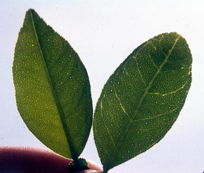
1.     Pest information

Citrus tristeza closterovirus (CTV) is the cause of one of many economically serious
diseases of citrus. CTV probably originated in Asia and was disseminated to many
citrus producing countries by infected propagative material. Subsequent spread by
aphid vectors caused epidemics in the 20th century. Millions of trees planted on sour
rootstocks have been lost to tristeza decline in many countries, including the United
States, Spain, and Brazil. Trees on tolerant or resistant rootstocks may be affected by
stem pitting strains that can reduce yields and quality (Bar-Joseph et al. 1989; Garnsey
et al. 1998; Lee and Bar-Joseph 2000).

CTV probably occurs in all citrus growing areas worldwide, although its incidence
varies. CTV is usually detected in isolated citrus trees and still is not considered
epidemic in many countries such as Albania, Algeria, Cyprus, Egypt, France, Greece,
Italy, Jordan, Lebanon, Libya, Morocco, Palestine, Portugal, Syria, Tunisia and Turkey
and in some Central and South America countries such as Belize, Chile, Ecuador, El
Salvador, Honduras, Mexico and Nicaragua. CTV is considered to cause the most
damaging viral disease of citrus. In Argentina, with its occurrence in 1945, it caused the
death of approximately 10 million trees, and in 1959 this number increased to 18 million.
Brazil reported over 10 million of trees lost in 1958. In the sixties in Spain and in the
eighties in Venezuela, CTV destroyed approximately 16 million of trees (Rocha-Peña et
al. 1995).

In Mexico, CTV was first detected in 1983 in the State of Tamaulipas, and in 1986 in
Veracruz; both outbreaks were quickly eliminated. However, the virus was subsequently
reported in other Mexican States.

Since 1939, the disease has also been observed in California, United States.
Observations made by Halma, Smoyer and Schwalm (1944, 1945) indicated that quick
decline reported in California was similar to the "tristeza" disease reported in Brazil.
Fawcett and Wallace (1946) at the same time as Meneghini (1946), reported that
tristeza in Brazil was caused by a virus. It can be concluded that this virulent strain of
tristeza caused the death of at least 3 million citrus trees in California. Grant and
Schneider (1951) reported for the first time the occurrence of CTV in Florida, although
no doubt its presence in the state goes back many years before its identification. The
tristeza strains identified in the state of Florida are different than those present in
California. In Texas, Olson and Sleeth (1954) and Olson and McDonald (1954) found
‘Meyer’ lemon trees infected with tristeza. The ‘Meyer’ lemon was the first host reported
in the State of Arizona (Carpenter 1956).

1.1 Transmission

CTV can be transmitted by grafting and semi-persistently by one of several different
species of aphid including Toxoptera citricida (Kirkaldy), T. aurantii (Boyer de
Fonscolombe), Aphis gossypii (Glover), and A. spiraecola (Patch) (Moreno et al. 2008).
Efficiency of aphid transmission can be affected by the donor and receptor host, by the
virus isolate and its ability to replicate in a given host, by the vector species and its
biology, and by environmental and horticultural factors. The most efficient aphid vector
is T. citricida (the Brown Citrus Aphid). However, aphid transmission by other species is
efficient enough to cause economically significant spread of CTV (Rocha-Peña et al.
1995). T. citricida was detected in February 2000 in the northern part of the states of
Quintana Roo and Yucatán, México. It has now been detected in the states of Yucatan,
DP 01                                                                                   4
Citrus Tristeza Virus (CTV)
Quintana Roo, Veracruz, Tabasco, Chiapas, Oaxaca, Puebla, and Campeche in
Mexico. In the United States, T. citricida has been reported from Florida but not from
California, Arizona, or Texas. Extensive research has been done on the epidemiology of
CTV in different citrus growing areas and how it is influenced by aphid, host and virus
isolate factors (Gottwald et al. 1999). Techniques have been developed and tested for
monitoring and predicting rates and patterns of spread (Hughes et al. 2001). In general,
once natural infection rates reach 0.1% to 0.5%, infection rates increase rapidly and
suppression by tree removal becomes difficult. In areas with active natural spread, the
level of actual infection will exceed that determined by testing since some recent
infection will not be detected. CTV has not been demonstrated to be seed-transmitted.
Many species of citrus and specific related genera of Rutaceae (Aurantioideae sub-
family) have been reported to be hosts of CTV. However, trifoliates (Poncirus trifoliata)
and many of their hybrids are resistant to CTV.

2.     Taxonomic information

CTV is a member of the family Closteroviridae and has the binomial Closterovirus citrus
tristeza virus. The pathogen itself is a flexible virion of about 2,000 X 11 nm and has a
non-segmented, positive-sense, single stranded ribonucleic acid (RNA) genome of
approximately 20 Kb. Studies on the molecular properties of CTV have revealed that it
is a complex virus group. Three major genotype groups have been recognized along
with some variations among these. Recombinants between different genotypes and the
fact that single “pure” isolates are actually populations of RNA variants further increase
the complexity (Rubio et al. 2001).

Associations have been established between biological reactions and various genetic
markers, but at this point no specific molecular pathogenicity determinants for decline or
stem pitting have been established (Nikolaeva et al., 1998, Hilf and Garnsey 2000,
Moreno et al. 2008).

3.     Detection and identification

3.1       Host plants

Natural hosts for CTV include mainly species of the genera Citrus and Fortunella, but
infection has been detected in genera related to the genus Citrus, that have been
experimentally inoculated such as Aegle, Aeglopsis, Afraegle, Atalantia, Citropsis,
Clausena, Eremocitrus, Hesperthusa, Merrillia, Microcitrus, Pamburus, Pleiospermium
and Swinglea. Experimental inoculations have also been achieved with non-citrus
species such as Passiflora gracilis y Passiflora coerulea using the aphid vector (Moreno
et al. 2008)

3.2 Symptoms

Different isolates of CTV vary greatly in the symptoms they produce in different citrus
hosts. Some are extremely mild and cause little injury, others cause medium damage
even in sensitive hosts. Some are severe in specific hosts and benign in others, and
some can cause very severe reactions in a wide range of hosts. Different isolates can
have a wide range of severity in some hosts and, conversely, the same isolate can vary
in symptoms expressed in different hosts. Graft inoculation of a range of different
indicator plants is commonly used to develop a profile of the biological properties of
CTV and to predict probable severity in commercial hosts (Garnsey et al. 2005).
DP 01                                                                                   5
Citrus Tristeza Virus (CTV)
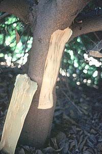
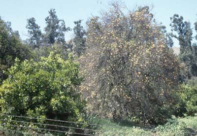
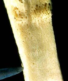
Common CTV symptoms include vein clearing, stunting, chlorosis, several types of
stem pitting, and seedling chlorosis (Moreno et al. 2008).

CTV isolates and symptom phenologies can be categorized as follows: i) tristeza
decline or quick decline strains which induce a collapse of citrus grown on sour orange
rootstock; ii) mild (weak) strains that are symptomless in citrus grown on CTV-tolerant
or resistant rootstocks; iii) seedling yellows strains which produce stunting and chlorosis
in seedlings of orange, grapefruit, or Eureka lemon in a greenhouse virus index
(generally considered to indicate severe strains); iv) stem pitting strains which induce
moderate to severe stem pitting in bark and adjacent wood in branches and trunks of
sweet orange, grapefruit, or mandarins regardless of rootstock.

There are essentially two commercially important CTV diseases of citrus. One is the
classic tristeza or decline that is associated with a decline of trees grafted on sour
orange rootstocks caused by a virus-induced phloem necrosis at the bud union (Figures
1a, 1b). The classic decline disease has caused severe losses in many citrus growing
areas, including California, and still threatens citrus industries having sour orange as the
main rootstock. The CTV-induced decline can be avoided by use of decline-tolerant or –
resistant rootstocks in lieu of sour orange; however, the use of these rootstocks may
result in other diseases and horticultural problems (Moreno et al. 2008).

The second major CTV-induced disease is stem pitting. In contrast to decline, stem
pitting can reduce the vigour and production of scions regardless of rootstock. Some
rootstocks can be affected by stem pitting even when the scion is not. CTV stem pitting
isolates has caused serious losses in many other citrus growing areas (Moreno and
Garnsey 2005; Rocha-Peña et al. 1995).

4.     Risk of pest spread

CTV may be spread to various citrus growing areas within the same country or to many
countries as a result of propagation by grafting and the subsquent transmission of the
virus in this fashion, added to spread by the vector.

5.     Methods for detection and identification

CTV can be detected by biological indexing and various non-biological methods
including light and electron microscopy, serology and a variety of molecular-based
techniques. The latter include several types of reverse transcription polymerase chain
reaction (RT-PCR), including an immunocapture PCR with multiple molecular markers
(MMM) (Hilf et al. 2005) and real time PCR (Ruiz-Ruiz et al. 2009), SSCP analysis
(Rubio et al. 1996), oligoprobes (Narvaez et al. 2000) and RFLP analysis (Gillings et al.
1993). The following techniques are approved by NAPPO: biological indexing, ELISA,
and immunoprint ELISA.

It is important to consider that sampling to detect CTV infected propagative material
must be done during the citrus flushing season, coinciding with less hot months (when
the monthly average temperature is around 27 °C and with little fluctuation during the
year). When moving plants within Mexico, sampling is done regardless of temperature.
Each sample is made up of 10-15 cm shoots, of five trees, taken from the four tree
quadrants. If plants are too small and have no shoots, petioles can be used.

DP 01                                                                                     6
Citrus Tristeza Virus (CTV)
In California, where temperatures vary more than in Mexico, CTV incidence is very low
during the winter and summer months (when temperatures are extreme) in most of the
regions of California. The ideal season to detect CTV is between April and June or in
October/November. This allows testing to be performed when incidence is high,
providing more reliable diagnostics (Dodds et al. 1987).

5.1 ELISA (Enzyme Linked Inmunosorbent Assay

To detect CTV, the Enzyme Linked Inmunosorbent Assay (ELISA) serological technique
is used. This technique is based in the capability of certain proteins, known as
antibodies, to recognize and link a specific antigen associated to the pathogen. This is a
reliable and quick technique to detect a plant pathogen virus. Some variants have been
developed, but so far the DAS-ELISA (Double Antibody Sandwich) direct method is the
most commonly used (Salazar 1990). This technique consist of specific antibodies
absorption to a polyestirene plate which later on the antigen is added to and then reacts
to the antibodies adhered to the plate. Then, an enzymatic conjugate is added (second
antibody) to form the complex antibody-antigen-antibody.

The specificity of monoclonal antibodies and the ease to trade them (since they are
homogenous and stable), in addition to the advantages offered by the technique such
as sensibility, low cost, speed, reliability and capability for mass usage, have favoured
the application of the ELISA technique in all citrus growing countries to detect CTV,
using monoclonal antibodies capable to recognize any virus isolate without any reaction
with components of the host plant.

5.1.1. Procedure

The antibodies developed by Nikolaeva et al. (1995) and/or Lee et al. (2005) (primary
antibodies CREC 1052, CREC 27, CREC 28, CREC 29, CREC 30, or CREC 31 and the
secondary antibody G604) are commonly used in California. Agdia and BioReba
commercial kits for CTV are presently used in Mexico.

The United States uses two different techniques, one by the Citrus Clonal Protection
Program (CCPP) using Gumpf antibodies and the other used by the Germplasm
Repository (NCGRCD) which uses Lee et al. (2005) antibodies.

5.1.1.1 Procedure of the Citrus Clonal Protection Program (CCPP) (Clark and Adams
1977, modified for tristeza by J.M. Díaz of CCPP).

A. Sample preparation
1. Collect young branches from the four tree quadrants (or eight quadrants for trees
     at risk of field infection).
2. Remove the bark from the branch, calculate approximately 1.0 g and cut in pieces
     of 1-2 mm.
3. Prepare the samples to 1:10 (p/v) in extraction buffer, using the KLECO tissue
     pulverizer or the Ultra-Turaz T25 Tissumizer 1 .

1   Manufacturing: Kleco Tissue Pulverizer, 14097 Ave 272,Visalia, CA 93292, 559-732-3785 or Tissuemizer,Ultra Turraz T25, Janke & Kunkel

DP 01                                                                                                                                       7
Citrus Tristeza Virus (CTV)
Buffer PBS
NaCl 8.0 g
KH2PO4 0.2 g
Na2HPO4.7H2O 2.17 g
KCl 0.2 g
NaN3 0.2 g
Adjustd the pH to 7.4, 1 L of H2O.
PBS-Tween Buffer: PBS Buffer + Tween20 2 ml/L
Extraction Buffer: Buffer PBSTween + 20 g/L PVP40 polyvinylpyrrolidone (Sigma)
4. Samples can be frozen and thaw before preparing the plate. This helps lower the
bottom of the optical density (recommended but not required).
B. Preparation of ELISA 2 plates
1. Place 200 µl of γ-globulin in buffer coat in each well of the plate. Incubate for 4
hours at 37 ºC. Normal concentration is 1-2 µg/ml. Ideal concentration must be
determined by limiting dilution.
Buffer coat
Na2CO3 1.59 g
NaHCO3 2.93 g
NaN3 0.2 g
Adjust the pH at 9,6, in 1 L of H2O.
2. Wash the plate with PBS-Tween. Let it settle for 3 minutes. Repeat washing 3
times, emptying the plate each time.
C. Sample addition
1. Place proportionally 200 µl of samples in each of two wells. Incubate at 6 ºC
overnight or at 37 ºC for 4 hours.
2. Wash the plate 3 times as in step B.2 and wash the plate 3 times with distilled
water, which helps lower the bottom of optical density.
D. Addition of secondary and conjugated antibodies
1. Place proportionally 200 µl of secondary antibody [according to Drs. A and O
Karasev (Nikolaeva et al. 1995)] in each well to the ideal pre-determined dilution.
Incubate at 37 ºC for 4 hours. For this step, use the conjugated buffer.
Enzyme conjugate buffer: Buffer PBS-Tween + 20 g/L PVP40 polivinylpirrolidona +
2 g/L ovalbumin (Sigma A-7030)
2. Wash the plate 3 times as in step B.2.
3. Place proportionally 200 µL of commercial conjugate (Sigma A-8025 Anti-Rabbit
IgI Alkaline Phosphatase Antibody, developed on goat), which usually has a
dilution of 1:8.000 to 1:10.000. Incubate at 37 ºC for 4 hours.
E. Substratum addition (P-nitrophenyl phosphate disodium, Sigma S-0942, 5 mg
tablets)
1. Wash the plate 3 times as indicated in step B.2.
2. Place proportionally 200 µl of recently prepared substrate at a concentration of 0,6
-- 1,0 mg/ml of p-nitrophenyl phosphate in substrate buffer.
2
Microtiter plates: Immulon IV Flat plates, Fisher #14245153 (50 plates/pack)
DP 01 8
Citrus Tristeza Virus (CTV)Substrate Buffer
Diethanolamine 97.0 ml
NaN3 0.2 g
Adjust the pH to 9.8, in 1 L of H2O.
F. Evaluation of the plate
1. Incubate at room temperature until you can see the reaction. Reaction time is
usually ~ 30 minutes.
2. Evaluate the results to measure absorbency at 405 nm in the plate reader 3 .
Positive samples are considered those that are two times greater than the average
value of the negative control (healthy witness).
5.1.1.2 Procedure of the Germplasm Repository (NCGRCD)
A. Selection and sample preparation
A1. Bark samples
1. Collect twigs that have just matured (fresh and dark green) from four sides of the
tree. Bark should be easily stripped from this type of tissue.
2. Place the flushes from each tree in a labelled plastic bag. Store the samples in an
ice chest if it will be more than a few minutes before the samples are processed.
3. In the laboratory, strip the bark from each of the collected flushes.
4. Estimate about 0.25 g of bark, and chop into 1 – 2 mm long pieces.
5. Add to 5 ml of coating buffer in a 50 ml test tube.
6. Homogenize for 15 seconds using a tissue miser or Polytron type homogenizer.
7. Store at 4 °C overnight before loading into the ELISA plate for maximum
extraction.
Alternative method:
1. Estimate about 0,20 g of bark and chop into 1-2 mm long pieces.
2. Place in a 2 ml centrifuge tube with screw cap, add 2 bbs and fill the tube (1.5 ml)
with coating buffer.
3. Homogenize for 5 minutes using a mini-bead beater.
4. Store at 4 °C overnight before loading into the ELISA plate for maximum
extraction.
Chopped midribs or leaf petioles can also be used in these procedures.
Coating Buffer
Na2CO3 1.59 g
NaHCO3 2.93 g
adjust to pH 9.6 with HCl in 1 L H2O.
A2. Leaf samples
1. Collect two leaves from each of four sides of the tree. They must be fully expanded
leaves from the last growth flush..
2. Roll one leaf from each side of the tree using a leaf roller and collect the sap into a
1.5 ml microcentrifuge tube. Keep the other four leaves as a backup sample.
3. Centrifuge for 5 minutes.
3
ELISA plate reader: Molecular Devices EMax
DP 01 9
Citrus Tristeza Virus (CTV)4. Pipet 200 μl of the supernatant into a dilution tube filled with 800 μl of coating
buffer. Mix by pipetting the solution up and down a few times.
Direct extraction using stem from new, tender flush tissue
1. After step 4 (wash procedure), fill all sample wells of the plate proportionally with
200 μl Aliquots of coating buffer containing 0.1 % Tween-20.
2. Chop the stem in 1-2 mm long pieces using pruning shears.
3. Drop the pieces of tissue directly into the wells filled with the buffer.
4. Incubate overnight at 4 ºC.
B. Preparation of control
For BSD-ELISA usage of glycerol-prepared controls stored at –20oC is preferred for
standardization purposes (Lee et al. 2005). A healthy (CTV -) and a positive (CTV +)
control are included with the detection kit. These controls are ready to load into the
ELISA plate. 100 ul aliquots are loaded into wells that are pre-filled with 100 ul of
coating buffer with 0.2% Tween 20.
Alternatively, lyophilized, dried or fresh tissue can be used for negative and positive
controls. In this case, use about 50 mg of tissue in 5 ml of coating buffer. Let the dried
tissue hydrate as long as possible up to 24 hours before homogenizing. A good strategy
is to hydrate the controls early in the morning, grind late in the afternoon, and extract
overnight at 4oC with the candidate samples.
C. Preparation of ELISA plates
1. Coat the ELISA plates with polyclonal antiserum made against purified CTV
(preparation of IgG CREC27, CREC28, CREC31, CREC1051, or CREC 1052).
Dilute the “CREC29 coating buffer” with the coating buffer to the concentration
indicated on the coating buffer tube (usually 1:5,000). Add 200 μl of the diluted
antiserum to each of the wells on the plate except the uncoated control well; two
wells may be left as an uncoated control, usually wells A11 & A12. Incubate 1 to 3
hours at room temperature, or overnight at 4 oC. (Note that when using overnight
incubations, higher dilutions can be used for the coating antiserum. For example,
if running the step at room temperature using a 1/5,000 dilution, or if running an
overnight incubation in the cold, use a 1/10,000 dilution.)
D. Wash
1. Shake the solution from the plate and wash 3X under a stream of deionized water.
2. Fill the plate with PBST, and let set for 5 minutes or longer (let set for at least 30
minutes on the first wash).
PBST
NaCl 8.0 g
KH2PO4 0.2 g
Na2HPO4 1.15 g (anhydrous)
KCl 0.2 g
Adjust to pH 7,4 using HCl , 1 L.
Add 1 ml of Tween 20
3. Repeat steps 1 and 2 twice.
4. Repeat step 1.
5. Invert and tap on a clean paper towel to drain excess water immediately before the
next step. Make sure the plate does not dry out.
DP 01 10
Citrus Tristeza Virus (CTV)E. Samples
1. Fill all wells of the ELISA plate with 100 μl of coating buffer containing 0.2%
Tween-20.
2. Add 100 μl aliquot of the sample per well, use two wells per sample. Mix the
samples with the buffer already in the well by pipetting up and down in the well a
few times.
3. Incubate overnight at 4 ºC.
4. Wash the plate following the wash procedure.
F. Secondary antibody
1. Dilute the G-604 secondary antibody to a 1:20,000 dilution in conjugate buffer 4 .
Conjugate buffer (500 ml)
PBST 500 ml
PVP-40 10.0 g
(Bovine serum albumin, Fraction V). 1.0 g
2. Add 100 μl of this solution to all wells in the plate.
3. Incubate for 1 hour at 37 ºC, or overnight at 4 ºC.
4. Wash the plate following the wash procedure.
G. Conjugate
1. Dilute Rabbit Anti-goat antibody conjugate with alkaline phosphatase to the
recommended dilution on the tube (usually 1:30.000) in conjugate buffer.
2. Add 100 μl aliquots of this solution to all wells in the plate.
3. Incubate for 3 hours at 37 oC or overnight at 4ºC.
4. Wash the plate following the wash procedure.
H. Reaction
1. Add 0.6 mg/ml of substrate (4-nitrophenyl phosphate hexahydrate) to coating
buffer. Make sure the coating buffer is fresh, unless it was prepared using NaN3 or
it is not older than a few days and stored at 4ºC.
2. Add 200 μl aliquots of this solution to each well.
3. Incubate at room temperature.
4. Read the plate after one or two hours, and/or on the next morning. Colour usually
develops very slowly. To calibrate it, set the plate reader to blank on the uncoated
wells. This will subtract the absorption of light caused by the plate and the buffer.
No significant background reaction should develop during an overnight
development of the plate. If this is the case, the secondary antibody should be
diluted further.
I. Evaluation
1. Evaluate the controls first. The uncoated control should not give any reaction at all
since the reader is adjusted to zero on these wells,. Reaction in the uncoated wells
indicates improper washing of the plates after the conjugate step, improper
preparation of one of the buffers, or loss of the specificity of the antibodies against
the antigen.
4
Each lot of G-604 must be calibrated making a series of dilutions.
DP 01 11
Citrus Tristeza Virus (CTV)2. It is not unusual for the buffer only wells to show some reaction, sometimes even
more than that of the healthy control (no antibodies).
3. The healthy (negative) control wells should show none or very low reaction.
4. A good plate will show OD405 values under 0.050 for the healthy and buffer only
wells, and values over 1.000 for the positive controls. In that case, a sample is
considered positive if the average OD value is more than 0.100. Antibody dilutions
need to be adjusted when higher values are obtained for the negative controls, or
lower values obtained for the positive controls.
Note: For all protocols, the final reaction volume in each step is the same. For this case, it is100 μl.
5.1.1.3 Procedure used with the commercial Agdia kit
1. Preparation of the sample
a. Remove the bark from each shoot 5 of the sample collected. Weigh 0,2 g of bark
and cut into pieces of 1-2 mm.
b. Empty the tissue in a 50 ml tube and add 2 ml of extraction buffer.
Extraction buffer solution (Macerated plant samples)
Sodium sulfite (anhydrous) 1.3 g
Polyvinylpyrrolidone (PVP) Molecular weight of 24 - 20.0 g
40,000
Sodium Azide 0.2 g
Ground Egg (chicken) albumin, Grade II 2.0 g
Tween-20 20.0 g
Dissolve in 1Lt of Phosphate buffer solution 1X (PBST)
Note: Adjust the pH to 7,4 (+/- 0,2). Store at 4 °C on refrigeration
c. Blend for 15 seconds using a tissue homogenizer.
d. For better extraction, store at 4 °C overnight before placing the samples on the
ELISA plate.
2. Preparation of ELISA plates
a. Sensitize the polystyrene plate. Place 100 μl of the specific antibody dilution for
CTV (Dilution 1:200 6 ) in each well plus the coating buffer solution taking into
account the sample distribution pattern, considering the samples and negative and
positive controls with their replicates.
b. Incubate in wet chamber for 2 hours at 37 +/- 2 °C or overnight at 4 °C.
Coating buffer
Sodium carbonate (anhydrous) 1.59 g
Sodium bicarbonate 2.93 g
Sodium azide 0.2 g
Adjust the pH at 9,6 (+/- 0,2). Store at 4 °C
Dissolve in 1 L of distilled water
5
Leaves main veins or petioles can also be used.
6 Dilution commonly used. Follow the manufacturer’s instructions.
DP 01 12
Citrus Tristeza Virus (CTV)3. Plate washing
a. Wash the plate 3 to 5 times 7 with PSTB-T 1X
Phosphate (PBST) buffer solution (wash)
Sodium chloride 8.0 g
Dibasic sodium phosphate (anhydrous) 1.15 g
Monobasic potassium phosphate (anhydrous) 0.2 g
Potassium chloride 0.2 g
Tween 20 0.5 g/ml
Dissolve in 1L of distilled water
Adjust the pH to 7,4 (+/- 0,2), washing solution can be prepared as a
concentrate first and then diluted. Prepare a concentration of 20X (20
times concentrated).
4. Sample addition
a. Plant samples of interest are crushed or homogenized with extraction buffer. Add
100 μl of the homogenized tissue to each well, with its replicates, in the previously
selected wells.
5. Control preparation
a. In each of the wells associated with the negative controls, place either a 100 μl of
the extraction solution by itself or commercial negative control. It is recommended
not to use the wells close to the edges since they can develop non-specific
reactions (Salazar 1990).
b. Dilute the positive control and place 100 μl of this in the wells.
c. Incubate in wet chamber for 2 hours at 37 +/- 2°C or overnight at 4 °C.
d. Wash the plate 6 to 10 times with PSTB-T 1X.
6. Add the antibody + conjugate
a. Mix the monoclonal antibody (Bottle A) with the enzymatic conjugate (Bottle B) in
ECI buffer in a 1:200 8 proportion for both antibodies. From this mix, 100 μl is
poured in each well and the plate is kept in a wet chamber for two hours 37+/- 2°C.
b. Dissolve in 1000 ml of phosphate buffer solution 1X (PBST):
Conjugate solution (ECI)
Bovine serum albumin (BSA) 2.0 g
Poly vinyl pyrrolidinone (PVP) 10.0 g
Molecual weight 24-40,000
Sodium azide 0.2 g
Adjust the pH to 7,4 (+/- 0,2), store at 4°C.
e. Wash the plate 6 to 10 times.
7. Addition of substrate.
a. Prepare the substrate solution from the enzyme diluting the para-nitrophenyl
phosphate in extraction buffer in a proportion of 1mg/ml (usually one tablet/5 ml).
7
Discard the plate contents in a sink, shake hard several times on paper towel. Fill each of the sensitized wells with the wash buffer, starting on
the first line, then the second line and so on, to cover all the needed wells, let stand for 1 minute and discard again the plate contents, shake well
over the paper towel.
8
Dilution commonly used. Follow the manufacturer’s instructions.
DP 01 13
Citrus Tristeza Virus (CTV)b. Place 100 μl in all the working wells, including the target and incubate under dark
conditions from 15 to 45 minutes.
Buffer solution for PNP (developing)
Diethanolamine 97.0 ml
Sodium azide 0.2 g
Adjust the final volume to 1 L. with sterile distilled water. Adjust the
pH to 9,8 with hydrochloric acid, store at 4°C in refrigeration in amber
bottle or wrap in aluminium to protect from light.
8. Plate evaluation
a. Turn on and program the ELISA plate reader.
b. Note if there is colour in the wells. Change in colour is indicative of positive results.
c. Read in the plate reader and print the readings.
d. Analyze the results and record them in the record book for laboratory samples,
writing the date of the response. Record the results in the database.
Considerations
For a diagnostic with reliable readings, the following controls must be included in the
plate:
• Uncovered wells
• Extraction buffer
• Healthy plant (negative control)
• Sick plant (positive control)
Infected plants with different CTV variants can be included 9 as positive controls. A
negative control corresponding to a healthy plant of the same species should also be
included. In both cases (positive and negative controls) replicates should be included.
Positive readings must be twice the value of the negative control. Negative results of
unknown samples must present absorbency values similar to the negative controls.
If the samples are different than their replicate or have readings 1.5 times the value of
the negative control, the diagnostic must be repeated. If the positive controls do not
react properly, the diagnostic must also be repeated.
5.2. Immunoimpression- ELISA
The preparation of the plant extracts to be diagnosed is the most important limiting
factor to detect CTV with conventional ELISA. Therefore, some variants have been
developed such as immunoimpression-ELISA, which is a technique that uses capture
membranes that do not require sample crushing or homogenization, thus allowing the
analysis of thousands of samples in a simple and fast way (Cambra et al. 2000).
The process consists of four basic and sequential stages:
1. Collection and impression of membrane samples.
2. Blocking the pressed membrane and reaction.
3. Washing.
4. Developing and reading the results.
9 Two mild and two severe.
DP 01 14
Citrus Tristeza Virus (CTV)5.2.1 Procedure
1. Make transversal or oblique cuts to young shoots 10 , leaves, pedicels, or peduncles
of newly ripe fruit with very sharp instruments.
2. Firmly press the cross sectional tissue against the nitrocellulose membrane of 0.45
mm pore, which is used as an immunoadsorbent. Make two impressions per shoot,
cutting the ends from its base and apex. This will allow making 10 impressions per
mature tree. Positive and negative controls should be included.
3. Let the membranes dry for a few minutes and keep them in a dry place and
protected from light until they are analyzed.
4. Block the pores from the membrane (and the rest of the surface) with a bovine
serum albumin (BSA) solution with a concentration of 1 % in distilled water. The
membranes must stay in the blocking solution for 1 hour at room temperature, or for
16 hours if the operation is done at 4 ºC. Slightly shake the membrane to soak it
and cover it completely with the blocking solution.
5. Add a solution of specific monoclonal antibodies for CTV, marked with the alkaline
phosphatase enzyme (0.1 mg/ml) to the membrane soaked with the albumine
solution.
6. Incubate for 2 or 3 hours at room temperature with the solution completely covering
the membrane. Once the reaction time has elapsed, discard the solution of
conjugated antibodies.
7. Rinse the membrane 11 with washing buffer (PBS + 0.05% Tween 20)
8. Add the substrate (BCIP-NBT Sigma Fast Tablets) 12 specific for the enzyme (10
tablets in 100 ml of distilled water). Slightly cover the membranes and incubate at
room temperature until violet-purple precipitation start to show.
9. When positive controls have shown some colour, stop the reaction by washing the
membranes with running water. Precipitations will show after 3-7 minutes of
incubation at room temperature.
10. Take the reading from the membranes once they are dry. In many cases, a reading
at first sight is enough, but they should be observed with a magnifying glass or
dissecting microscope.
5.3 Biological techniques
Biological indexing is a technique that, under proper conditions, guarantees an accurate
diagnosis. Biological indexing (bioindexing) for citrus pathogens is based on the use of
citrus indicator plants free of pathogens, which react to the virus infection by expressing
diagnostic symptoms depending on the pathogen and isolate (Roistacher 1991). Each
sample (budwood to be tested) is inoculated into replicated indicator plants.
Uninoculated indicators are used as healthy (negative) controls and indicators
inoculated with tissue known to be infected with mild to severe isolates of the pathogen
are used as positive controls.
Biological indexing requires appropriate environmental conditions in a greenhouse
environment and one month to a year to evaluate the material tested, depending upon
the pathogen.
10
It is preferable to collect shoots of 10-15 cm from the youngest material available, from various areas around the tree, preferably from the
medium-high section of the canopy, which is the area most likely to be visited by aphids.
11
The washing stage is essential since it eliminates all the antibodies marked with alkaline phosphatase which have not reacted. That iway there
would only be traces of enzyme on the selections of printed plant material infested with CTV.
12
Nitro blue tetrazolium and bromo chlorine indolyl phosphate (NBT+BCIP)
DP 01 15
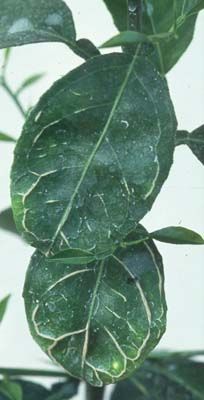
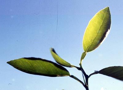
Citrus Tristeza Virus (CTV)Bioindexing has been validated as reliably identifying citrus pathogens in mixed infections, which are common in the case of citrus (Vidalakis et al. 2004). 5.3.1. Procedure The procedures used for biological indexing for CTV are well established (Roistacher 1991). For the biological indexing of CTV the preferred indicator plant is Mexican lime (Citrus aurantiifolia), the small-fruited acid lime. The Mexican lime seedlings must be healthy (i.e, no infections) with stems of 4-10 mm diameter and 100 cm height. The seedlings of Mexican lime should be planted 3 per pot. Each pot will have 2 inoculated seedlings and 1 non-inoculated. The uninoculated plant will serve as a healthy control. There should be between 4 and 8 replicates from each tree diagnosed and at least 2 positive controls (one strong, one weak) for each test. From the plants to be tested, take budsticks from a minimum of four quadrants of each tree. In the case of field trees where there is a risk of infection, take budsticks from eight quadrants. This is very important in the case of foundation trees maintained in the field. “Buds” are grafted two per indicator plant. It is important that the inoculum include phloem tissue and that the phloem of the inoculum and indicator are in good contact since CTV is phloem-limited. If leaf disks are used, a minimum of five or six should be used per plant. The blades used to do the graft should be disinfected with a solution of 10 % of commercial sodium hypochloride between samples. Immediately after the inoculation the plants should be cut back to a height of 20-25 cm. Once the plants are inoculated, symptom expression is favoured by maintaining temperatures of 24 – 28 ºC (day) and 17 – 21 ºC (night) for 2 – 4 months. After two weeks, the grafting tape should be removed and survival recorded. The side branches of the seedlings should not be pruned for the first three flushes (more or less 8 weeks) in order to obtain the maximum quantity of leaves to observe for symptoms. The majority of inoculated plants will show symptoms within five weeks and almost all within eight weeks. The primary symptom of tristeza is vein clearing in young and mature leaves. This symptom is observed best in leaves held above so that sunlight shines through the leaf. The best time to observe these symptoms is when the leaf just stops increasing in size. In the case of severe strains of tristeza (seedling yellows), the vein clearing can develop into vein corking. Another characteristic symptom of tristeza in Mexican lime is leaf cupping. This is seen when conditions are very good for indexing. It can persist in mature leaves. On the other hand, leaf cupping is also a symptom of vein enation in Mexican lime and so leaf cupping by itself is not diagnostic for tristeza. After eight weeks, the bark can be removed and stem pitting observed with certain strains of tristeza. However, stem pitting in Mexican lime is better evaluated by removing the side shoots after the third flush (about eight weeks) and training the plants to a single leader. Stem pitting can then be evaluated after four to six months after the initial inoculations. Indicator plants can also be used to biocharacterize isolates of tristeza (Garnsey et al. 1987; Roistacher 1991). In addition to the intensity of the reaction in Mexican lime, the isolates can be evaluated for stem pitting (Figure 1g), seedling yellows (Figure 1h), or quick decline. All these symptoms indicate a severe strain that can produce economic consequences. DP 01 16 Citrus Tristeza Virus (CTV)
Although various isolates of tristeza can cause stem pitting or seedling yellows in various indicator plants, 'Duncan' grapefruit is preferred for this use. Sour orange can serve as an additional indicator for stem pitting and 'Madame Vinous' or 'Pineapple' sweet orange can show whether a strain causes stem pitting in sweet orange varieties. Thus, five indicators are needed to biocharacterize different isolates of tristeza: Mexican lime (basic symptoms, stem pitting); sour orange (seedling yellows, stunting); sweet orange (stem pitting, stunting); 'Duncan' grapefruit (seedling yellows, stem pitting, stunting); and sweet orange budded on sour orange (phloem necrosis, stunting). The grapefruit and sour orange are grown three per pot and the sweet orange one per pot for these uses. Observation of decline on sour orange rootstocks has taken 2 – 3 years in the past. Recently, a more rapid method has been developed (Pina et al. 2005) where a reduction of growth of sour orange buds grafted onto ‘Madam Vinous’ sweet orange inoculated with a decline-inducing strain has been correlated with decline- inducing isolates. 6. Records A record of tested samples must be kept containing the following: Code for the reference number of the sample. Variety and origin of the sample. Symptoms description (including pictures, as appropriate) or no symptoms. Methods used in the diagnostic and the results obtained with each method, including controls (pictures of the diagnostic gel o records of results for the ELISA testing, on which the diagnostic was based) Name of the laboratory, and when appropriate, the names of the persons responsible for the diagnostic. Records and evidences of the diagnostic results should be kept for at least one year. 7. Contacts for additional information Centro Nacional de Referencia Fitosanitaria. (CNRF). Dirección General de Sanidad Vegetal. Guillermo Pérez Valenzuela No. 127 Col. Del Carmen, Coyoacán, Del. Coyoacán, México, DF 04100. Instituto de Fitosanidad. Colegio de Postgraduados. Campus Montecillo Km. 35.5 Carr. México- Texcoco CP. 56230 Montecillo, Edo. de México. México. Citrus Clonal Protection Program, Department of Plant Pathology, University of California, Riverside, CA 92521 USA. USDA-ARS. National Clonal Germplasm Repository for Citrus & Dates, 1060 Martin Luther King Blvd., Riverside, CA 92507, USA. DP 01 17 Citrus Tristeza Virus (CTV)
8.     References

Bar-Joseph, M., R. Marcus and R.F. Lee. 1989. The continuous challenge of citrus
tristeza virus control. Annual Review of Phytopathology 27:291-316.

Cambra, M., M.T. Gorris, M. P. Román, E. Terrada, E. Camarasa, S. M. Garnsey, E.
Camarasa, A. Olmos and M. Colomer. 2000. Routine detection of Citrus Tristeza Virus
by Direct Immunoprinting-ELISA Method Using Specific Monoclonal and Recombinant
Antibodies. Fourteenth OICV Conference. Pp. 34-41.

Carpenter, J.B. 1956. Identification of tristeza in Meyer lemon in Arizona. Plant Disease
Reporter 40:8.

Clark, M.F. and A.N. Adams. 1977. Characteristics of the micro-plate method of
enzyme-linked immunosorbent assay for the detection of plant viruses. Journal of
General Virology 34:475-483.

Fawcett, H.S. and J.M. Wallace. 1946. Evidence of virus nature of citrus quick decline.
California Citrograph 32:88-89.

Garnsey, S. M., T.R. Gottwald and R.K. Yokomi. 1998. Control Strategies for Citrus
Tristeza Virus (CTV). pp 639-658. In: Plant Virus Disease Control. A. Hadidi, R. K.
Khetarpal and H. Koganezawa (eds.) APS Press, St. Paul, MN.

Grant, T.J. and H. Schneider. 1951. Initial evidence of the presence of tristeza or quick
decline of citrus in Florida. Phytopathology 43:51-52.

Gillings M.P., Broadbent, J. Indsto and R.F. Lee. 1993. Characterization of isolates and
strains of citrus tristeza closterovirus using restriction analysis of the coat protein gene
amplified by the polymerase chain reaction. J Virol Methods 44:305–317

Gottwald, T.R., G. Gibson, S.M. Garnsey and M. Irey. 1999. Examination of the effect
of aphid vector population composition on the spatial dynamics of citrus tristeza virus
spread by stochastic modeling. Phytopathology 89:603-608.

Halma, F.F., K.M. Smoyer and H.W. Schwalm. 1944. Quick decline associated with
sour rootstocks. California Citrograph 29:245.

Halma, F.F., K.M. Smoyer and H.W. Schwalm. 1945. Rootstock in relation to quick
decline of citrus. California Citrograph 30:150-151.

Hilf, M.E. and S.M. Garnsey. 2000. Characterization and classification of Citrus tristeza
virus isolates by amplification of multiple molecular markers, Pp. 18-27. In: Proc. 14th
Conf. IOCV, IOCV, Riverside, CA.

Hilf, M.E., V.A. Mavrodieva and S.M. Garnsey. 2005. Genetic marker analysis of a
global collection of isolates of Citrus tristeza virus: Characterization and distribution of
CTV genotypes and association with symptoms. Phytopathology 95: 909-917.

Hughes, G., T.R. Gottwald and S.M. Garnsey. 2001. Development of methods and
models and their application to disease problems in the perennial citrus crop system. In:

DP 01                                                                                     18
Citrus Tristeza Virus (CTV)
Jeger M.J. and Spence N.J. (eds). Biotic interactions in plant pathogen associations.
CABI Publishing Wallingford, UK.

Lee, R.F. and M. Bar-Joseph. 2000. Tristeza. Pp 61-63. En: Compendium of Citrus
Diseases. 2nd edition. Timmer, L.W., S.M. Garnsey, and J.H. Graham (editors). St Paul
(MN): American Phyotpathological Association.

Lee, R.F., M. G. H. Dekkers and M. Bar-Joseph. 2005. Development of stable, uniform
antigen controls for use in ELISA assays for Citrus tristeza virus. Pp 127-136 In: Proc.
16th Conf. IOCV. IOCV, Riverside.

Meneghini, M. 1946. Sôbre a naturaza e transmissibilidade do doencia “tristeza” do
citrus. O Biologico 15:115-118.

Moreno P.S and S.M. Garnsey. 2010. Citrus Tristeza Disease -A Worldwide
Perspective. Pp.27-49 In: Karasev, A.V. y Hilf, M.E. (eds.). Citrus tristeza virus complex
and tristeza diseases. American Phytopathological Society Press.

Moreno P.S., Ambros, M.R. Albiach-Marti, J. Guerri and L. Pena. 2008. Citrus tristeza
virus: a pathogen that changed the course of the citrus industry. Molecular Plant
Pathology 9: 251-268.

Narvaez, G., B.S. Skander, M.A. Ayllon, L. Rubio, J. Guerri and P. Moreno. 2000. A
new procedure to differentiate citrus tristeza virus isolates by hybridization with
digoxigenin-labelled cDNA probes. J. Virol. Methods 85:83-92.

Nikolaeva O.V., A.V. Karasev, S.M. Garnsey and R.F. Lee 1998. Serological
differentiation of the citrus tristeza virus isolates causing stem pitting in sweet orange.
Plant Dis. 82:1276-1280.

Olson, E.O. and J.R. McDonald. 1954. Tristeza in satsuma varieties in Texas. Plant
Disease Reporter 38:439-441.

Olson, E.O. and B. Sleeth. 1954. Tristeza virus carried by some Meyer lemon trees in
Texas. Proceedings of the Rio Grande Valley Horticultural Institute 8:84-88.

Rocha-Peña, M.A. 1995. Citrus tristeza virus and its aphid vector Toxoptera citricida.
Plant Disease 79:437-445.

Roistacher, C.N. 1991. Graft-Transmissible Diseases of Citrus: Handbook For Detection
And Diagnosis. Rome: Food and Agricultural Organization of the United Nations.

Rubio, L., M.A. Ayllón, J. Guerri, H. Pappu, C.L. Niblett and P. Moreno. 1996.
Differentiation of citrus tristeza closterovirus (CTV) isolates by single-strand
conformation polymorphism analysis of the coat protein gene. Ann. Appl. Biol. 129: 479-
489.

Rubio, L., M.A. Ayllón, P. Kong, A. Fernandez, M. Polek, J. Guerri, P. Moreno and B.W.
Falk. 2001. Genetic variation of Citrus tristeza virus isolates from California and Spain:
evidence for mixed infections and recombination. J. Virology 75:8054-8062

DP 01                                                                                   19
Citrus Tristeza Virus (CTV)
Ruiz-Ruiz, S., P. Moreno, J. Guerri and S. Ambrós. 2009. Discrimination between mild
and severe Citrus tristeza virus isolates with a rapid and highly specific real-time
reverse transcription-polymerase chain reaction method using TaqMan LNA probes.
Phytopathology 99:307-315.

Salazar, L. F. 1990. Metodología Para La Detección De Virus De Papa: Pasado,
Presente Y Futuro. Revista Latinoamericana de la Papa. 3(1):1-12

Vidalakis, G., S.M. Garnsey, J.A. Bash, G.D. Greer and D.J. Gumpf. 2004. Efficacy of
bioindexing for graft-transmissible citrus pathogens in mixed infections. Plant Disease
88:1328- 1334.

DP 01                                                                               20
Citrus Tristeza Virus (CTV)
a                                            b                        c

  d                                   e                             f

  g                                                                                           h

Figure 1. Symptoms of Citrus Tristeza Closterovirus, a) Tristeza decline of sweet orange grown on sour
orange rootstock, b) and c) phloem necrosis of bud union of sweet orange – sour orange infected with
decline strain of CTV, d) vein clearing in leaves of Mexican lime indicator, e) vein corking in severe
(seedling yellows) strain of CTV in Mexican lime indicator, f) Leaf cupping in leaves of Mexican lime
indicator, g) stem pitting in Eureka lemon indicator, h) seedling yellows reaction in grapefruit indicator. (All
photos by CN Roistacher)

DP 01                                                                                                       21
Citrus Tristeza Virus (CTV)
You can also read