Method for obtaining high-resolution proteomic analysis from pericarps of guarana
←
→
Page content transcription
If your browser does not render page correctly, please read the page content below
Method for obtaining high-resolution proteomic
analysis from pericarps of guarana
                         A.L. Souza1, P.C.S. Angelo2, P.P.O. Nogueira3, J.F.C. Gonçalves4,
                         A.M. Franco4, S. Astolfi-Filho1, J.L. López-Lozano5 and E.V. Andrade1

                         1
                           Centro de Apoio Multidisciplinar, Laboratório de Biologia Molecular,
                         Universidade Federal do Amazonas, Manaus, AM, Brasil
                         2
                           EMBRAPA Amazônia Ocidental, Manaus, AM, Brasil
                         3
                           Fundação Osvaldo Cruz, Manaus, AM, Brasil
                         4
                           Instituto Nacional de Pesquisas da Amazônia, Manaus, AM, Brasil
                         5
                           Fundação de Medicina Tropical do Amazonas, Manaus, AM, Brasil

                         Corresponding author: E.V. Andrade
                         E-mail: edandrade@ufam.edu.br / edandrade2003@yahoo.com.br

                         Genet. Mol. Res. 13 (3): 8014-8024 (2014)
                         Received September 12, 2013
                         Accepted December 17, 2013
                         Published September 29, 2014
                         DOI http://dx.doi.org/10.4238/2014.September.29.14

                         ABSTRACT. Guarana has great agricultural potential and is largely
                         used therapeutically and in the production of non-alcoholic energy
                         drinks. Genomic and proteomic studies are crucial to identify proteins
                         that play central roles in the maintenance and viability of fruits, as
                         well as to identify proteins related to the main metabolic pathways.
                         However, the success of any protein analysis starts with the protein
                         extract preparation, which needs to offer an extract that is free of
                         contaminants. This study aimed to evaluate different extraction
                         methods to obtain high-quantity and high-quality extracts that are
                         compatible with analysis by 2-dimensional electrophoresis and tandem
                         mass spectrometry protein identification. Three different methods were
                         tested: trichloroacetic acid (TCA)/acetone, sodium dodecyl sulfate
                         (SDS)/phenol, and polyvinylpolypyrrolidone (PVPP)/SDS/phenol.
                         The extract obtained from the TCA/acetone precipitation presented
                         low solubility and contamination with lipids and carbohydrates. On the

Genetics and Molecular Research 13 (3): 8014-8024 (2014)            ©FUNPEC-RP www.funpecrp.com.br
Protein extraction from Paullinia cupana 8015
other hand, the quality of the extract gradually improved after using
phenol and PVPP/phenol, enabling a yield up to 2 mg/g macerated
tissues and the detection of 457 spots by 2-dimensional electrophoresis.
The effectiveness of the procedure used was validated by identification
of 10 randomly selected proteins by mass spectrometry. The procedure
described here can be a starting point for applications using tissues of
other organs of guarana or tissues of species that are similar to guarana.
Key words: Plant proteomics; Two-dimensional electrophoresis;
Polyphenols
INTRODUCTION
Guarana, Paullinia cupana (Kunth) var. sorbilis (Mart.) Ducke, is a rainforest vine
that is widely used in traditional (Mendes and Carlini, 2007) and clinical medicine (de
Oliveira Campos et al., 2011; Rodrigues et al., 2012b; Portella et al., 2013). Besides, the
extracts from roasted guarana seeds are used for the production of non-alcoholic energy
drinks (McLellan and Lieberman, 2012). Because of its great economical relevance, a better
understanding of its physiology and secondary metabolism has been sought from different
approaches. Previous studies of the guarana fruits with seeds identified several transcripts
encoding enzymes and proteins that participate in biosynthetic routes, such as caffeine
synthesis and important physiological processes including those related to plant-pathogen
interactions (Ângelo et al., 2008; Figueirêdo et al., 2011). The increased knowledge about
these sequences and their proteins can facilitate obtaining products from guarana for health
applications (Costa et al., 2011) and developing plants with valuable agronomic traits.
However, transcriptome analysis is not enough to obtain a complete picture of all the cel-
lular processes that occur in complex organisms like eukaryotes. Proteomic studies offer
a good approach to follow those results (Quiala et al., 2012) and to identify proteins that
play central roles in the maintenance and viability of fruits when facing diverse environ-
mental conditions. However, the success of any protein analysis starts with the protein
extract preparation, and the ideal method must capture the greatest amount of protein while
minimizing degradation and contamination by non-protein content (Isaacson et al., 2006;
Jorrín-Novo et al., 2009).
Vegetal tissues have biochemical properties that can impair the use of general methods
for protein extraction. Because of the biochemical heterogeneity, large amounts of proteases
and oxidative enzymes, and the presence of non-protein contaminants can disturb the subse-
quent steps in proteomic approaches (Boonmee et al., 2011; Witzel et al., 2011). Therefore,
there is not a universal protocol for extraction or a solvent that can capture an entire proteome,
and adjustments to previously described protocols are required (Prinsi et al., 2011; Chatterjee
et al., 2012; Rodrigues et al., 2012a; Zhang et al., 2012).
Guarana has high levels of oxidative enzymes, phenol, nitrogen, and terpene com-
pounds, which hinder the application of methods for protein extraction that were developed
in other species. Similar difficulties were reported by Wang et al. (2003), who observed that
when proteins of the olive leaf were extracted by direct homogenization in aqueous buf-
fer and precipitated in organic solvent, polyphenols and other contaminants were adsorbed
Genetics and Molecular Research 13 (3): 8014-8024 (2014) ©FUNPEC-RP www.funpecrp.com.brA.L. Souza et al. 8016
to the proteins, which caused the pellet to darken due to oxidation. This contamination
hampers subsequent dissolution, making the protein extract unsuitable for 2-dimensional
electrophoresis (2-DE) analysis. A very efficient method involves the dissolution of pro-
teins in phenol and precipitation in ammonium acetate in methanol (Hurkman and Tanaka,
1986). This method was successfully used for coffee (Franco et al., 2009), olive leaves
(Wang et al., 2003), and other recalcitrant tissues such as those from tomato, avocado, and
orange peel (Saravanan and Rose, 2004), as well as banana, apple, and potato (Carpentier
et al., 2005). Despite the description of various biological properties of guarana and signifi-
cant amounts of information that were obtained from its transcriptome, proteomic profiles
from the different fruit tissues in the various developmental phases have not been reported.
Therefore, the establishment of a reproducible protocol to prepare protein extracts from
guarana is expected to be useful. In this study, we present the results from trials of already
reported methods and a modified method, which ensured the achievement of high-quality
guarana total protein extracts for use in 2-DE and the identification of spots by mass spec-
trometry (ESI Q-TOF-MS).
MATERIAL AND METHODS
Plant material
The BRS-Amazonas cultivar of guarana [(P. cupana (Kunth) var. sorbilis (Mart.)
Ducke] was developed and is maintained at the Germplasm Bank of Embrapa Western Ama-
zon (3°S, 60°W, 50 m, Manaus, Amazonas, Brazil). Open-pollinated fruits from healthy plants
were collected 15 ± 7 days after pollination, dissected in pericarps, arils and seeds, conditioned
on dry ice, and transported for storage at -80°C. For total protein extraction, 1 g green pericarp
was crushed in the presence of liquid nitrogen using a pre-cooled pestle and mortar until a fine
powder was formed. Three procedures were tested in order to obtain suitable protein extracts
from guarana pericarps and are schematically described in Figure 1.
Protein extraction methods
Trichloroacetic acid (TCA)/acetone
The TCA/acetone precipitation was carried out as described by Isaacson et al. (2006).
Briefly, the powder was resuspended in 10 mL solution I [10% (v/v) TCA; 2% (v/v) 2-mer-
captoethanol in cold acetone]. The mixture was incubated overnight at -20°C and centrifuged
(30 min at 5000 g and 4°C). The pellet was washed with 10 mL cold acetone and centrifuged
(10 min at 4000 g and 4°C). The supernatant was discarded, and the wash step was repeated
twice. During the wash steps, the sample was divided into 2 fractions: a heavy fraction that
sedimented spontaneously and a light fraction (supernatant) that was less dense and required
centrifugation to precipitate. At the last wash step the light fraction was transferred to micro-
tubes (1-mL aliquots), centrifuged (10 min at 4000 g and 4°C), dried at room temperature for
20 min, and stored at -20°C. Likewise, the heavy fraction was submitted to centrifugation
and dried as described above and submitted to the second protein extraction method, sodium
dodecyl sulfate (SDS)/phenol, which is described below.
Genetics and Molecular Research 13 (3): 8014-8024 (2014) ©FUNPEC-RP www.funpecrp.com.brProtein extraction from Paullinia cupana 8017
SDS/phenol
The SDS/phenol method was conducted as reported by Wang et al. (2003), with
modifications. The heavy fraction that was obtained from the TCA/acetone precipitation was
homogenized in 10 mL cold solution II [10% (v/v) TCA in acetone]. The mixture was soni-
cated at a frequency of 22.5 kHz (3 pulses of 20 s at 1-min intervals) in an ice bath using the
ultrasound Fisher 100 Sonic Dismembrator. The re-precipitation of proteins was obtained by
incubating the solution at -20°C for 40 min and centrifugation (30 min at 4000 g and 4°C). The
pellet was washed twice with 10 mL ice-cold acetone, centrifuged (10 min at 4000 g and 4°C),
dried at room temperature, and separated into aliquots of 0.15 g. The aliquots were solubilized
in 0.6 mL solution III [30% (w/v) sucrose; 2% (w/v) SDS; 0.1 M Tris-HCl, pH 7.5; and 2%
(v/v) 2-mercaptoethanol]. The mixture was homogenized with a vortex for 30 s and left at
room temperature for 15 min. Next, 0.6 mL saturated phenol in Tris-HCl, pH 7.5, was added,
and the samples were agitated in a vortex for 20 s for protein solubilization, incubated for 5
min at room temperature, and centrifuged (5 min at 10,000 g and 4°C). The phenol phase was
recovered and added to 3 volumes solution IV [0.1 M ammonium acetate in methanol] and
precipitated for 1 h at -20°C. The proteins were recovered by centrifugation (10 min at 10,000
g and 4°C) and washed 3 times with 2 volumes cold methanol. The pellet was dried at room
temperature for 15 min and stored at -20°C.
Polyvinylpolypyrrolidone (PVPP)/SDS/phenol
The PVPP/SDS/phenol procedure was carried out as a combination of steps described
by Isaacson et al. (2006) and by Wang et al. (2003). The powder of guarana green pericarp was
obtained as described above except for the addition of 5% (w/w) PVPP during maceration to
trap phenolic compounds, and it was homogenized in solution I. The samples were sonicated
in an ice bath (as described above) and incubated at -20°C for 40 min for protein precipitation.
Proteins were recovered by centrifugation (30 min at 4000 g and 4°C), washed twice with 10
mL ice-cold acetone, and centrifuged (10 min at 4000 g and 4°C). The pellet was dried at room
temperature for 15 min and separated into aliquots of 0.15 g. The aliquots were solubilized in
0.6 mL solution III and treated as described for the SDS/phenol method.
Protein quantification and 2-DE
One aliquot of each extract was solubilized in 0.5 mL 3 M urea, and the protein con-
centration was estimated using the bicinchoninic acid protein assay kit (BioAgency, Brazil)
according to manufacturer instructions. The concentration of the proteins was determined by
comparison with a standard curve (y = 0.002x + 0.0209; R2 = 0.9956) for bovine serum albu-
min. 2-DE was performed using a standard procedure (O’Farrell, 1975), with slight modifica-
tions. The aliquots (250 µg) of proteins that were obtained from each extraction procedure
described above were resuspended in 250 µL DeStreak Rehydration Solution (GE Healthcare,
Sweden) and 1% immobilized pH gradient (IPG) buffer, pH 3-10 or 3-11 NL (GE Healthcare)
and loaded onto an immobilized pH gradient strip (GE Healthcare, pH 3-10 NL or 3-11 NL)
for overnight passive in-gel rehydration (10 h). Isoelectric focusing was performed at 20°C
in an Ettan IPGPhor3 (GE Healthcare) with a continuous current of 50 mA/strip under the
Genetics and Molecular Research 13 (3): 8014-8024 (2014) ©FUNPEC-RP www.funpecrp.com.brA.L. Souza et al. 8018
following conditions: 150 V for 2 h, 300 V for 2 h, 1000 V for 4 h (gradient), 8000 V for 2 h
(gradient), and 8000 V for 2 h. The proteins on the IPG strips were reduced and alkylated for
the separation in the second dimension, which was performed on 12% SDS polyacrylamide
gels in the SE600Ruby System (18 x 16 cm, GE Healthcare) at 50 mA and 250 V for 4 h. Poly-
acrylamide gels were fixed using 10% (v/v) acetic acid and 40% (v/v) methanol for 30 min,
followed by staining with colloidal Coomassie blue (8% ammonium sulfate, 0.8% phosphoric
acid, 0.08% Coomassie blue G-250, and 20% methanol) overnight and destaining with deion-
ized water. Gels were scanned (Image Scanner, GE Healthcare), and the images were analyzed
with the Image Master Platinum software (Version 6). Spot counts were obtained according to
the saliency and smooth parameters, which were equal to 100 and 2, respectively. Three repro-
ducible gels (over 70% similarity) were run corresponding to at least 2 independent extraction
procedures from the PVPP/SDS/phenol extraction method. The gel with the highest number
of spots was regarded as the reference gel.
Protein identification by ESI Q-TOF-MS
Ten protein spots that were taken randomly from the map obtained from the PVPP/
SDS/phenol extraction were subjected to tryptic in-gel digestion (sequencing grade-modi-
fied trypsin, lot: 24247001; Promega, USA), according to Hanna et al. (2000), and analyzed
by MS. An aliquot (4.5 μL) of the resulting peptide mixture was separated by C18 (75 μm
x 100 mm) nanoACQUITY UltraPerformance LC (UPLC) System (Waters, UK) coupled
with a Q-TOF Ultima mass spectrometer (Waters) with nano-electrospray source at a flow
rate of 0.6 mL/min. The gradient was 2-90% acetonitrile in 0.1% formic acid over 20 min.
The instrument was operated in the “top three” mode, in which one MS is acquired followed
by tandem MS of the top three most intense peaks that were detected. The resulting spec-
tra were processed using Mascot Distiller 2.2.1.0, 2008, Matrix Science (MassLynx V4.1)
and searched against guarana’s expressed sequence tags database (available at http://www.
ncbi.nih.gov) using Mascot, with carbamidomethylation as a fixed modification, oxidation of
methionine and 1 trypsin missed cleavage, and a tolerance of 0.1 Da for both precursor and
fragment ions.
RESULTS AND DISCUSSION
Protein yield and evaluation of 2-DE gels
In an attempt to obtain guarana protein extracts that were free of interfering compounds
for applications in proteomic analysis, 3 different procedures were evaluated in terms of protein
yield and number and resolution of spots (Figure 1). The protein yield of the extracts obtained
by the TCA/acetone, SDS/phenol, and PVPP/SDS/phenol methods was 1.81, 0.29 and 2.45
mg/g macerated tissues, respectively. The sum of protein content obtained from the TCA/ac-
etone and the SDS/phenol methods was 2.1 mg protein/g tissue, which was comparable to the
protein content that was extracted with the PVPP/SDS/phenol method. These results indicate
that the modifications that we introduced and the addition of PVPP during tissue maceration to
trap phenolic compounds improved the protein yields from guarana pericarps by 1.4- and 8.4-
fold compared to the TCA/acetone and SDS/phenol methods, respectively.
Genetics and Molecular Research 13 (3): 8014-8024 (2014) ©FUNPEC-RP www.funpecrp.com.brProtein extraction from Paullinia cupana 8019
Figure 1. Schematic representation of 3 methods used to extract protein from guarana pericarps. Steps that were
used for at least 2 procedures are represented in solid-lined boxes. Steps that were specific to a single procedure are
represented by different types of dotted lines. PVPP = polyvinylpolypyrrolidone; TCA = trichloroacetic acid; SDS
= sodium dodecyl sulfate.
Wang et al. (2003) reported that when proteins were extracted from olive leaves by
direct homogenization in aqueous buffer and precipitation in organic solvent, polyphenols
and other contaminants were adsorbed to the proteins, which caused the pellet to darken due
to oxidation. Similar results were observed for guarana pericarps. The TCA/acetone method
resulted in pellets with low solubility in aqueous buffer and a slightly yellowish color (data
not shown), possibly due to the presence of pigments. The low solubility in water is likely
caused by precipitation in the presence of TCA and acetone, as described by Nandakumar et al.
(2003). On the other hand, the pellets that were obtained by the SDS/phenol and PVPP/SDS/
phenol methods were easily dissolved in inorganic buffer and were whitish in color (data not
Genetics and Molecular Research 13 (3): 8014-8024 (2014) ©FUNPEC-RP www.funpecrp.com.brA.L. Souza et al. 8020
shown), which was expected for minor oxidation. For some species, such as papaya, however,
the TCA method resulted in the highest protein yield compared with the phenol-based methods
(Rodrigues et al., 2009). These data reinforce that there is not a common procedure for protein
extract preparation from plant tissues.
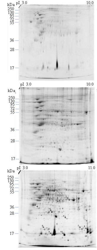
The total number of spots that were detected with the TCA/acetone, SDS/phenol, and
PVPP/SDS/phenol methods was 135, 271, and 457, respectively (Figure 2). Treating the mac-
erated tissue with TCA in acetone is important to inhibit proteases and efficiently remove salts
and polyphenols that are soluble in organic solvents (Nandakumar et al., 2003) because they
interfere with the isoelectric focusing. However, as can be observed in Figure 2A, this treat-
ment was not sufficient to eliminate carbohydrates and lipids from green pericarps of guarana,
which blocked gel pores and caused the spots to smear. Besides the presence of non-protein
contents in the sample, the low solubility probably contributed to the lower number of spots
after the TCA/acetone precipitation. The contaminants were only removed after dissolution
in phenol, which resulted in better solubility of the sample and an improved migration profile
following 2-DE. However, the resolution was still compromised by the presence of vertical
stripes, mainly in the alkaline region of the gel (Figure 2B). This was probably a consequence
of the presence of phenolic compounds in the protein extract, which reversibly (by hydrogen
bonds) or irreversibly (by oxidation) bind to proteins, leading to heterogeneity of the sample
charges and smearing on the gel (Carpentier et al., 2005). These phenolic compounds were
completely removed only by the use of PVPP during the maceration of tissues, preventing the
oxidation of proteins (Isaacson et al., 2006). The improved quality of the protein extract was
defined by the striking reduction in the horizontal smears and vertical stripes, and by the num-
ber of detected spots (Figure 2C), which was approximately 3- and 1.6-fold higher than those
from the TCA/acetone precipitation and SDS/phenol methods, respectively. Because one of
the main bottlenecks for a successful proteomic approach is the richness and resolution of the
spots, these results indicate that PVPP/SDS/phenol is the best method to obtain high-yield and
high-quality protein extracts from guarana pericarps.
Protein identification by ESI Q-TOF-MS
Proteins that were identified by ESI Q-TOF-MS are indicated in Figure 2C (arrows)
and listed in Table 1. For some identified proteins, there were differences between the ob-
served and the theoretical molecular weight and isoelectric point, which could be due to post-
translational modifications. Proteins that were identified in this study included the following:
enolase (spot 1), glyceraldehyde-3-phosphate dehydrogenase C subunit (spot 2), photosystem
II oxygen-evolving complex 33-kDa subunit (spot 3), ascorbate peroxidase (APX, spot 4),
23-kDa oxygen-evolving protein of photosystem II (spot 5), major latex-like protein (spot 6),
cyclophilin (spot 7), histone 2b (spot 8), superoxide dismutase (spot 9), and lectin (spot 10).
Reactive oxygen species could be generated from different cellular processes in plants
such photosynthetic metabolism and oxidative stress (Shine and Guruprasad, 2012). Superoxide
dismutase contributes during protection against oxidative stress (Waters et al., 2012), promoting
the dismutation of superoxide radicals that produce hydrogen peroxide, which is mainly removed
by APX in plant cells (Klein et al., 2012; Suzuki et al., 2013). In this reaction, APX catalyzes
the reduction of H2O2 to H2O through the oxidation of ascorbate (ASC) to monohydroascorbate,
which will be reduced again to ASC in the glutathione-ASC cycle (Damanik et al., 2010). APX
contributes to the response to abiotic stress factors, such as heat and light (Suzuki et al., 2013).
Genetics and Molecular Research 13 (3): 8014-8024 (2014) ©FUNPEC-RP www.funpecrp.com.brProtein extraction from Paullinia cupana 8021
A
B
C
Figure 2. Proteomic map from the green pericarp of guarana. Protein extracts were prepared using A. trichloroacetic
acid (TCA)/acetone; B. sodium dodecyl sulfate (SDS)/phenol; and C. polyvinylpolypyrrolidone (PVPP)/SDS/
phenol methods. The isoelectric focusing was performed on a 13-cm strip followed by SDS-polyacrylamide gel
electrophoresis on a 12% gel. Visualization was performed by staining with colloidal Coomassie blue. For all gels,
250 µg protein was used. Arrows in C indicate the spots that were selected for mass spectrometry identification.
Genetics and Molecular Research 13 (3): 8014-8024 (2014) ©FUNPEC-RP www.funpecrp.com.brA.L. Souza et al. 8022
Table 1. Proteins identified by mass spectrometry.
Spot No. pI/MW (kDa) Exp pI/MW (kDa) Theo Score Protein ID Database ESTs
1 5.48/26.5 5.56/47.9 132 Enolase 3,310
2 9.37/20.2 7.7/36.5 84 Glyceraldehyde-3-phosphate dehydrogenase C subunit 3,105
3 5.23/22.3 5.85/35.2 637 Photosystem II oxygen-evolving complex 33-kDa subunit 10,697
4 5.77/22.7 5.64/27.4 168 Ascorbate peroxidase 401
5 8.42/22.9 8.27/28.0 51 23-kDa oxygen-evolving protein of photosystem II 2,730
6 6.2/15.4 5.46/17.1 34 Major latex-like protein 13,126
7 8.9/19.5 8.69/18.1 138 Cyclophilin 1,157
8 10.3/20.5 10.1/15.8 195 Histone 2b 14,467
9 7.3/25.0 5.77/15.1 185 Superoxide dismutase 3,922
10 8.8/19.4 6.0/47.5 218 Lectin 9,625
Names, theoretical (Theo) and experimental (Exp) values for the isoelectric points (pIs) and molecular weights
(MW), and expressed sequence tags (ESTs) of each identified protein are indicated. The scores were obtained from
the National Center for Biotechnology Information.
Enolase (2-phospho-D-glycerate hydrolase) is an essential glycolytic enzyme that
catalyzes the interconversion of 2-phosphoglycerate and phosphoenolpyruvate. This enzyme
was differentially expressed in seeds of Lolium rigidum, and the authors discussed its role in
thermotolerance during mechanisms of dormancy induction and release (Goggin et al., 2011).
The glyceraldehyde-3-phosphate dehydrogenase C subunit plays an important role in glycoly-
sis by providing ATP and NADH to cells, and it contributed to non-glycolytic processes in
Arabidopsis thaliana and was modulated by posttranslational modifications (Bedhomme et
al., 2012; Wojtera-Kwiczor et al., 2013).
Photosystem II is an important multiprotein oxygen-evolving complex that is located
in the thylakoid membranes of chloroplasts. The contribution of different proteins including
the photosystem II oxygen-evolving complex 33-kDa subunit on this photosynthetic electron
transport chain, which can produce oxygen from water and light, has been vastly demonstrated
(Millaleo et al., 2013). In young plants of 2 guarana clones that were grown in conditions of
light stress, the greater activity involving photochemical processes (high-electron transport
rate) in the tolerant clone contributed to a more significant decrease in the degree of photoin-
hibition compared to the sensitive clone.
The major latex-like protein, which is found in the aqueous matrix of latex, has an
unknown function (Sun et al., 2010). However, its similarity to proteins that are related to
pathogenesis suggests its involvement in defense against phytopathogens. Cyclophilin, which
is found in plants, fungi, and bacteria, presents cis-trans-prolyl peptide isomerase activity
that catalyzes the rotation of peptide bonds at the amino end of proline residues (Trandinh
et al., 1992). Additionally, the action of cyclophilin as an ATP-independent molecular chap-
erone has been described (Ellis and van der Vies, 1991). The lectins are a specialized group
of proteins that can bind and precipitate carbohydrates. They are ubiquitously distributed in
plants, animals, and fungi, and they are involved in diverse biological processes including the
defense system in plants (Peumans and Van Damme, 1995). This group also includes a class
of carbohydrate-binding proteins of non-immune origin that was identified in mistletoe, ricin
and concavalin A, and a lectin from Polygonatum cyrtonema that acts in cancer treatment by
inducing apoptosis in cancer cells (Fu et al., 2011).
In conclusion, we developed an efficient procedure to extract total protein from gua-
rana pericarps that uses PVPP and phenol. This procedure allows the production of high-
quality protein extracts that are suitable for proteomic analysis and identification of spots by
Genetics and Molecular Research 13 (3): 8014-8024 (2014) ©FUNPEC-RP www.funpecrp.com.brProtein extraction from Paullinia cupana 8023
MS. This procedure also efficiently prepares protein extracts from seeds (data not shown) and
contributed to proteomic analyses of guarana seeds and pericarps at different stages of devel-
opment that are in progress in our laboratory. According to the results presented in this study,
we propose the procedure based on PVPP and phenol as the method to obtain protein extracts
from plants that present biochemical characteristics that are similar to those of guarana, such
as high levels of polyphenol, carbohydrate, and lipid contamination.
ACKNOWLEDGMENTS
Research supported by CNPq, FAPEAM, CAPES, and FINEP. This study was carried
out by the Amazon Proteomic Network (PROTEAM). We thank the Mass Spectrometry Lab-
oratory at Brazilian Biosciences National Laboratory, CNPEM-ABTLuS, Campinas, Brazil,
for its support with the mass spectrometry analysis.
REFERENCES
Ângelo PC, Nunes-Silva CG, Brigido MM, Azevedo JS, et al. (2008). Guarana (Paullinia cupana var. sorbilis), an anciently
consumed stimulant from the Amazon rain forest: the seeded-fruit transcriptome. Plant Cell Rep. 27: 117-124.
Bedhomme M, Adamo M, Marchand CH, Couturier J, et al. (2012). Glutathionylation of cytosolic glyceraldehyde-
3-phosphate dehydrogenase from the model plant Arabidopsis thaliana is reversed by both glutaredoxins and
thioredoxins in vitro. Biochem. J. 445: 337-347.
Boonmee A, Srisomsap C, Chokchaichamnankit D, Karnchanatat A, et al. (2011). A proteomic analysis of Curcuma
comosa Roxb. rhizomes. Proteome Sci. 9: 43.
Carpentier SC, Witters E, Laukens K, Deckers P, et al. (2005). Preparation of protein extracts from recalcitrant plant tissues:
an evaluation of different methods for two-dimensional gel electrophoresis analysis. Proteomics 5: 2497-2507.
Chatterjee M, Gupta S, Bhar A and Das S (2012). Optimization of an efficient protein extraction protocol compatible with
two-dimensional electrophoresis and mass spectrometry from recalcitrant phenolic rich roots of Chickpea (Cicer
arietinum L.). Int. J. Proteomics 2012: 536963.
Costa KC, Ribeiro EE, Ribeiro EA, Moresco RN, et al. (2011). Habitual intake of Guarana and metabolic morbidities: An
epidemiological study of an elderly Amazonian population. Phytother. Res. Doi: 10.1002/ptr.3437.
Damanik RI, Maziah M, Ismail MR, Ahmad S, et al. (2010). Responses of the antioxidative enzymes in Malasyan rice
(Oryza sativa L.) cultivars under submergence condition. Acta Physiol. Plant 32: 739-747.
de Oliveira Campos MP, Riechelmann R, Martins LC, Hassan BJ, et al. (2011). Guarana (Paullinia cupana) improves
fatigue in breast cancer patients undergoing systemic chemotherapy. J. Altern. Complement. Med. 17: 505-512.
Ellis RJ and van der Vies SM (1991). Molecular chaperones. Annu. Rev. Biochem. 60: 321-347.
Figueirêdo LC, Faria-Campos AC, Astolfi-Filho S and Azevedo JL (2011). Identification and isolation of full-length
cDNA sequences by sequencing and analysis of expressed sequence tags from guarana (Paullinia cupana). Genet.
Mol. Res. 10: 1188-1199.
Franco OL, Pelegrini PB, Gomes CP, Souza A, et al. (2009). Proteomic evaluation of coffee zygotic embryos in two
different stages of seed development. Plant Physiol. Biochem. 47: 1046-1050.
Fu LL, Zhou CC, Yao S, Yu JY, et al. (2011). Plant lectins: targeting programmed cell death pathways as antitumor agents.
Int. J. Biochem. Cell Biol. 43: 1442-1449.
Goggin DE, Powles SB and Steadman KJ (2011). Selection for low or high primary dormancy in Lolium rigidum Gaud
seeds results in constitutive differences in stress protein expression and peroxidase activity. J. Exp Bot. 62: 1037-1047.
Hanna SL, Sherman NE, Kinter MT and Goldberg JB (2000). Comparison of proteins expressed by Pseudomonas aeruginosa
strains representing initial and chronic isolates from a cystic fibrosis patient: an analysis by 2-D gel electrophoresis and
capillary column liquid chromatography-tandem mass spectrometry. Microbiology 146: 2495-2508.
Hurkman WJ and Tanaka CK (1986). Solubilization of plant membrane proteins for analysis by two-dimensional gel
electrophoresis. Plant Physiol. 81: 802-806.
Isaacson T, Damasceno CM, Saravanan RS, He Y, et al. (2006). Sample extraction techniques for enhanced proteomic
analysis of plant tissues. Nat. Protoc. 1: 769-774.
Jorrín-Novo JV, Maldonado AM, Echevarria-Zomeno S, Valledor L, et al. (2009). Plant proteomics update (2007-2008):
Genetics and Molecular Research 13 (3): 8014-8024 (2014) ©FUNPEC-RP www.funpecrp.com.brA.L. Souza et al. 8024
Second-generation proteomic techniques, an appropriate experimental design, and data analysis to fulfill MIAPE
standards, increase plant proteome coverage and expand biological knowledge. J. Proteomics 72: 285-314.
Klein P, Seidel T, Stocker B and Dietz KJ (2012). The membrane-tethered transcription factor ANAC089 serves as redox-
dependent suppressor of stromal ascorbate peroxidase gene expression. Front Plant Sci. 3: 247.
McLellan TM and Lieberman HR (2012). Do energy drinks contain active components other than caffeine? Nutr. Rev.
70: 730-744.
Mendes FR and Carlini EA (2007). Brazilian plants as possible adaptogens: an ethnopharmacological survey of books
edited in Brazil. J. Ethnopharmacol. 109: 493-500.
Millaleo R, Reyes-Díaz M, Alberdi M, Ivanov AG, et al. (2013). Excess manganese differentially inhibits photosystem I
versus II in Arabidopsis thaliana. J. Exp. Bot. 64: 343-354.
Nandakumar MP, Shen J, Raman B and Marten MR (2003). Solubilization of trichloroacetic acid (TCA) precipitated
microbial proteins via NaOH for two-dimensional electrophoresis. J. Proteome Res. 2: 89-93.
O’Farrell PH (1975). High resolution two-dimensional electrophoresis of proteins. J. Biol. Chem. 250: 4007-4021.
Peumans WJ and Van Damme EJ (1995). Lectins as plant defense proteins. Plant Physiol. 109: 347-352.
Portella RL, Barcelos RP, da Rosa EJ, Ribeiro EE, et al. (2013). Guarana (Paullinia cupana Kunth) effects on LDL
oxidation in elderly people: an in vitro and in vivo study. Lipids Health Dis. 12: 12.
Prinsi B, Negri AS, Fedeli C, Morgutti S, et al. (2011). Peach fruit ripening: A proteomic comparative analysis of the
mesocarp of two cultivars with different flesh firmness at two ripening stages. Phytochemistry 72: 1251-1262.
Quiala E, Cañal MJ, Rodríguez R, Yagüe N, et al. (2012). Proteomic profiling of Tectona grandis L. leaf. Proteomics 12:
1039-1044.
Rodrigues EP, Torres AR, da Silva Batista JS, Huergo L, et al. (2012a). A simple, economical and reproducible protein
extraction protocol for proteomics studies of soybean roots. Genet. Mol. Biol. 35: 348-352.
Rodrigues M, Alves G, Lourenço N and Falcão A (2012b). Herb-drug interaction of Paullinia cupana (Guarana) seed
extract on the pharmacokinetics of Amiodarone in rats. Evid. Based Complement. Alternat. Med. 2012: 428560.
Rodrigues SP, Ventura JA, Zingali RB and Fernandes PM (2009). Evaluation of sample preparation methods for the
analysis of papaya leaf proteins through two-dimensional gel electrophoresis. Phytochem. Anal. 20: 456-464.
Saravanan RS and Rose JK (2004). A critical evaluation of sample extraction techniques for enhanced proteomic analysis
of recalcitrant plant tissues. Proteomics 4: 2522-2532.
Shine MB and Guruprasad KN (2012). Oxyradicals and PSII activity in maize leaves in the absence of UV components of
solar spectrum. J. Biosci. 37: 703-712.
Sun H, Kim MK, Pulla RK, Kim YJ, et al. (2010). Isolation and expression analysis of a novel major latex-like protein
(MLP151) gene from Panax ginseng. Mol. Biol. Rep. 37: 2215-2222.
Suzuki N, Miller G, Sejima H, Harper J, et al. (2013). Enhanced seed production under prolonged heat stress conditions in
Arabidopsis thaliana plants deficient in cytosolic ascorbate peroxidase 2. J. Exp. Bot. 64: 253-263.
Trandinh CC, Pao GM and Saier MH Jr (1992). Structural and evolutionary relationships among the immunophilins: two
ubiquitous families of peptidyl-prolyl cis-trans isomerases. FASEB J. 6: 3410-3420.
Wang W, Scali M, Vignani R, Spadafora A, et al. (2003). Protein extraction for two-dimensional electrophoresis from
olive leaf, a plant tissue containing high levels of interfering compounds. Electrophoresis 24: 2369-2375.
Waters BM, McInturf SA and Stein RJ (2012). Rosette iron deficiency transcript and microRNA profiling reveals links
between copper and iron homeostasis in Arabidopsis thaliana. J. Exp. Bot. 63: 5903-5918.
Witzel K, Shahzad M, Matros A, Mock HP, et al. (2011). Comparative evaluation of extraction methods for apoplastic
proteins from maize leaves. Plant Methods 7: 48.
Wojtera-Kwiczor J, Gross F, Leffers HM, Kang M, et al. (2013). Transfer of a redox-signal through the cytosol by redox-
dependent microcompartmentation of glycolytic enzymes at mitochondria and actin cytoskeleton. Front Plant Sci.
3: 284.
Zhang LL, Feng RJ and Zhang YD (2012). Evaluation of different methods of protein extraction and identification of
differentially expressed proteins upon ethylene-induced early-ripening in banana peels. J. Sci. Food Agric. 92: 2106-
2115.
Genetics and Molecular Research 13 (3): 8014-8024 (2014) ©FUNPEC-RP www.funpecrp.com.brYou can also read