Intraperitoneal protein injection in the axolotl: The amphibian kidney as a novel model to study tubulointerstitial activation
←
→
Page content transcription
If your browser does not render page correctly, please read the page content below
Kidney International, Vol. 62 (2002), pp. 51–59
CELL BIOLOGY – IMMUNOLOGY – PATHOLOGY
Intraperitoneal protein injection in the axolotl:
The amphibian kidney as a novel model to
study tubulointerstitial activation
MARIE LOUISE GROSS, WILFORD HANKE, ANDREAS KOCH, HEIKE ZIEBART,
KERSTIN AMANN, and EBERHARD RITZ
Department of Pathology and Department of Internal Medicine, University of Heidelberg, Heidelberg;
Department of Pathology, University of Erlangen, Nürnberg-Erlangen; and Department of Zoology,
University of Karlsruhe, Baden-Württemberg, Germany
Intraperitoneal protein injection in the axolotl: The amphibian dent of alterations of glomerular function that may have poten-
kidney as a novel model to study tubulointerstitial activation. tial confounding effects on peritubular hemodynamics, pO2,
Background. A substantial body of experimental evidence sug- cell traffic, etc.
gests that protein loading causes activation of proximal tubular
epithelial cells with consecutive interstitial fibrosis. These stud-
ies have mostly been performed using mammalian in vivo mod-
els of glomerular damage or tissue cultures of mammalian tu-
As shown 30 years ago, renal dysfunction correlates
bulointerstitial cells. The kidney of the axolotl contains not better with expansion of the tubulointerstitial space than
only closed nephrons, but also nephrons with ciliated peritoneal with glomerular damage [1–4]. Until now it has not been
funnels called nephrostomes that have access to the peritoneal firmly established whether tubulointerstitial fibrosis is
fluid. Injection of protein into the peritoneal cavity fails to just a passive consequence of glomerular damage or is
expose closed nephrons to a protein load, but causes selective
a player in the genesis of renal dysfunction [5]. In this
uptake and transient storage of proteins in tubular epithelial
cells of nephrons with nephrostomes. The purpose of the pres- context it is of interest that proteinuria per se is thought
ent study was to determine whether (a) the axolotl kidney can to be a major factor in the initiation and progression of
be used as a model to assess protein uptake by tubular cells renal dysfunction [6–8]. In clinical studies proteinuria is
in vivo in the absence of glomerular damage, and (b) this is a potent predictor of filtration loss [9]. Furthermore,
accompanied by any evidence of tubular epithelial cell activa-
experimental studies clearly established that protein-
tion and interstitial fibrosis.
Methods. Male and female axolotl (80 to 120 g of weight) loaded proximal tubular cells acquire an inflammatory
were given a daily intraperitoneal injection of 1.5 mL endotoxin- phenotype, express endothelin, angiotensinogen, cyto-
free calf serum or saline as control. Kidneys were harvested kines and the respective receptors, and synthesize extra-
after 4 or 10 days using perfusion fixation for light microscopy cellular matrix. Co-culture experiments showed that they
(fibrous tissue stain) and saline perfusion for immunohisto-
chemistry (fibronectin, TGF- and collagen I).
also are able to activate renal interstitial fibroblasts [5].
Results. The findings document selective storage of protein Remuzzi, Ruggenenti and Benigni postulated interstitial
and lipids, progressive with time, in proximal tubular epithelial activation by endothelin that is activated by high levels
cells of nephrons draining the coelomic cavity. In addition, of protein in tubular content [6].
progressive focal accumulation of fibrous tissue was noted Such studies in mammalian systems have two potential
around protein-storing tubules. Immunohistochemical staining
demonstrated the presence of fibronectin and TGF- in the
limitations. In vitro studies face the possibility of pheno-
tubular epithelial cells and interstitial cells. typic modulation of cells in monolayer configuration. In
Conclusion. The axolotl kidney provides a novel in vivo vivo models based on the protein overload technique [10]
model to study tubulointerstitial activation and induction of and glomerular damage models with proteinuric renal in-
interstitial fibrosis by protein loading. The findings are indepen- jury involve loss of glomerular permselectivity. Glomer-
ular damage, however, may have repercussions on peri-
Key words: proteinuria, tubulointerstitial activation, amphibian kid- tubular hemodynamics, peritubular oxygen tension (pO2)
ney, interstitial fibrosis, TGF-, fibronectin. or transcapillary cell traffic, and also may cause local
Received for publication June 14, 2001
activation of systems involved in tissue damage, such as
and in revised form December 19, 2001 the coagulation, the fibrinolysis, the complement and
Accepted for publication February 7, 2002 other systems.
2002 by the International Society of Nephrology These considerations justify interest in models of iso-
51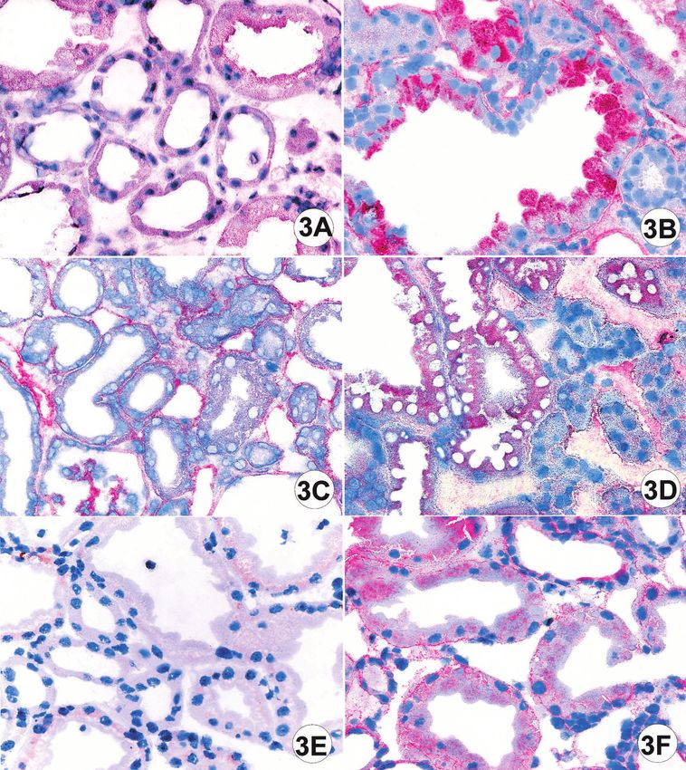
52 Gross et al: Intraperitoneal protein injection of axolotl
lated tubular cell protein loading in vivo in the absence (06.00 to 18.00 hours) and 12-hour dark cycle (18.00 to
of glomerular injury. 06.00 hours).
In the late thirties Randerath and others had used One week prior to the study, animals were randomly
the amphibian kidney to document that the histological allotted to two groups. The first group received daily
abnormalities of proximal tubular cells in proteinuric 0.5 mL fetal bovine serum (FBS) by intraperitoneal in-
patients must be the result of increased glomerular fil- jection (endotoxin free FBS, cc pro; S 14 M, Neustadt,
tration of proteins [11–14]. To this end he made use of an Germany). The second group received 0.5 mL isotonic
anatomical peculiarity of the urodelic amphibian kidney, NaCl solution as a control.
that is, the existence of distinct nephrons that drain the The FBS was assured of being endotoxin free by using
peritoneal cavity by a nephrostoma. Consequently, if the Limulus amebocyte lysate assay (LAL assay; cour-
proteins are injected into the peritoneal cavity, protein tesy of Dr. Klaus-Peter Becker, Institute for Microbiol-
uptake and storage is seen only in the tubular epithelial ogy, Mannheim, Germany) [24]. Concentrations of oxi-
cells of these nephrons. We re-assessed this model to dized lipids were not significantly higher compared to
examine whether selective protein loading of proximal normal human serum by high-pressure liquid chromatog-
tubular epithelial cells is associated with evidence of raphy (HPLC; courtesy of Karin Beumann, Dept. of
epithelial cell activation and local interstitial fibrosis. Pediatrics, Heidelberg, Germany) [25].
To assess uptake of FBS by proximal epithelial cells, a
monoclonal antibody against bovine serum albumin was
METHODS
used for the immunohistological studies (␣BSA; Sigma
The axolotl kidney B2901, 1:25, Lot 129 H 4874, at a dilution of 1:25; Sigma
Several former studies used Salamandra maculatum, Aldrich, Deisenhofen, Germany; Fig. 1F). Negative con-
a close relative of the axolotl (Amblystoma mexicanum) trols were performed by omitting the primary antibody
[11–13]; however, because of animal protection laws, (Fig. 1E).
Salamandras are no longer available. Consequently, we In a pilot experiment we investigated the tubular up-
chose the axolotl, a primitive neotenic amphibium, as take after intraperitoneal injection of human albumin
the experimental animal. (Albumin, Human, glycated A 8301; Sigma) in a concen-
The kidney of the axolotl represents an amphibian tration of 4.5 g/dL for six days. The results showed no
opisthonephros in which ciliated peritoneal funnels, significant differences between experimental animals
called nephrostomes, have access to the peritoneal fluid and saline injected controls (data not shown).
(Fig. 1, A–D). In urodelic amphibians to which the axo- In another pilot experiment we investigated the tubu-
lotl belongs, nephrostomes connect to the proximal tu- lar uptake after intraperitoneal injection of FBS (endo-
bule in close vicinity to the glomerulus [15, 16]. A further toxin free FBS, cc pro; S 14 M) for two days. Only
peculiarity of the nephron of lower vertebrates including marginal protein accumulation was found in tubular epi-
Amphibia is the absence of a loop of Henle so that thelial cells in experimental animals. No interstitial fi-
production of hyperosmotic urine is not possible [17]. brosis was noted.
The axolotl is a well known model species for studies The main study comprised two series. The first series
on neurologic and dermal pathology. The cross reactivity of daily intraperitoneal injection of 0.5 mL bovine serum
and specificity of a variety of human, rat and goat anti- was terminated after four days (referred to as short-
bodies for transforming growth factor- (TGF-), fibro- term study). In the second series, the experiment was
nectin and collagen with axolotl tissue has been well terminated after ten days (long-term study).
documented in several studies [18–23]. Under general anesthesia (3-aminobenzotic acid ethyl
ester, A-5040; 10 g/L water in the tank; Sigma) blood
Experimental protocol was obtained and retrograde perfusion was performed
Eighteen-month-old neotenic axolotls of both sexes, via the main heart ventricle. For light microscopy 3%
weighing between 80 and 120 g, were reared in the animal glutaraldehyde was used as fixative. The kidneys were
rooms of the Department of Zoology II, University of excised and embedded in paraffin or Epon-Araldite. For
Karlsruhe. Eggs were obtained from the parental animals immunohistochemistry, animals were perfused with ice-
of the stock. Hatched larvae were fed with Artemia and cold isotonic saline. The kidneys were then excised. One
Tubifex until they were able to take pellets of fish food. part was snap-frozen and the other part was fixed with
In a pilot study kidneys of male and female axolotl 4% formalin.
differed in size and appearance, but not with respect to
the presence and morphology of nephrostomes; thus, the Measurement of absorbed bovine serum in
results of male and female axolotls were pooled. The amphibian blood
animals were maintained in tanks of aerated tap water After the injection of FBS the serum of the axolotls
at a constant temperature of 18⬚C with a 12-hour light yielded at best a very faint band by the OuchterlonyGross et al: Intraperitoneal protein injection of axolotl 53
double gel diffusion test (antiserum against bovine pro- RESULTS
tein from Riedel-de Haën, Germany, Lot 45258; courtesy Normal anatomy of the axolotl kidney by
of Dr. Lohneis, Chemisches und Veterinäruntersuchung- light microscopy
samt Karlsruhe, Germany) [26], indicating that the peri-
In the one-year-old axolotls the kidney had a mean
toneal cells absorbed only a small amount of serum. The
length of 1.45 ⫾ 0.67 cm and a mean weight of 0.41 ⫾
test was negative in saline-injected control animals.
0.05 g. Glomeruli were located in a semi-circular pattern
Light and electron microscopy in the kidney. The mean diameter of the glomeruli was
201 ⫾ 23.6 m. The unique feature was the presence of
Two-micrometer paraffin sections were stained with
ciliated funnels, located preferentially in the cranial and
a connective tissue stain (Ladewig stain) and examined
middle portion of the kidney (Fig. 1). Proximal and distal
using light microscopy at a magnification of ⫻100. The tu-
tubules could easily be differentiated by the presence
bulointerstitial changes were quantified by two “blinded”
examiners who were unaware of the assignment to treat- and absence of a brush border.
ment, using a score system for each individual structural Intraperitoneal injection of bovine serum:
characteristic, that is, tubular dilation, protein droplets Light and electron microscopy
in tubular epithelial cells and interstitial fibrosis: score
0 ⫽ no change; score 1 ⫽ minimal change; score 2 ⫽ Saline control axolotls. In control kidneys, almost no
moderate change; score 3 ⫽ marked change; score 4 ⫽ interstitial tissue, as defined by staining for collagen, was
very pronounced change. The tubular dilation score 0 noted and mononuclear cells were scarce.
corresponded to an average diameter of 50 , score 1 Axolotls with intraperitoneal protein loading. After
to 100 , score 2 to 150 , score 3 to 200 , and score protein loading, clusters of closely adjacent dilated tu-
4 to 250 or more. In two randomly selected animals bules were seen (Fig. 2 A, B, D). The tubular lumen
per group ultrathin kidney sections were cut, stained contained protein sludge. In the short-term experiment
with lead citrate and uranyl acetate, and assessed using cell detritus was not seen in the tubular lumen, but abun-
a Zeiss EM 10 at various magnifications. dant detritus was noted in the long-term experiment.
The tubules containing protein sludge then comprised
Immunohistochemistry approximately 20 to 30% of the tubular cross sections
For immunohistochemistry the following antibodies in the cranial and middle portion of the kidney. The
were used: anti-TGF- (TGF-1, polyclonal rabbit, SC- proximal epithelial cells were massively swollen and ho-
146, 1:50; Santa Cruz Biotechnology, Santa Cruz, USA), mogenously filled by protein droplets approximately 2
and pan-TGF- (EO-13; 1:50; R&D Systems, Minneapo- in diameter with no preferential location at the luminal
lis, MN, USA), anti-fibronectin (polyclonal rabbit, or abluminal side, respectively. Loading with protein
F 3648, 1:200; Sigma) and anti-collagen I (rabbit ␣ rat col- droplets was progressive with time, that is, more pro-
lagen I, AB 755, Lot 131 DDM; 1:200; Chemicon Interna- nounced in the long-term compared to the short-term
tional Inc. Temecula, CA, USA). Cryostat sections of 5 experiment. Necrosis or mitosis of tubular cells was not
m thickness were used. The concentration that was seen, but occasionally atrophic tubuli were noted in the
optimal for staining with the above-mentioned antibod- long-term experiment. Distal tubules also showed pro-
ies was evaluated testing different dilution series in a tein droplets, but less than proximal tubules.
pilot study. Negative controls were performed by omit- Marked accumulation of interstitial tissue around the
ting the primary antibody. As detection system Fast Red tubules with protein droplets in epithelial cells (and
(K 0699; Dako, Hamburg, Germany) was used. sparse interstitial tissue around tubules without protein
Glomerular, tubular and interstitial structures were droplets) was noted in the short-term and particularly
assessed using a score system. Tubular epithelial cells and in the long-term experiment (Fig. 3). Occasionally, focal
interstitial cells were separately quantified, evaluating infiltrates of mononuclear cells were seen. In control
the area and the intensity of staining. Two investigators animals no interstitial fibrosis was noted.
who evaluated the scores were masked as to the treat- The scores of the histological parameters in the short
ment of the animals. The scores were defined as: 0 ⫽ no term and the long-term experiment are given in Tables 1
staining; 1 ⫽ minimal staining; 2 ⫽ moderate staining; and 2. The results document that the changes increased
3 ⫽ marked staining; 4 ⫽ very pronounced staining. with time.
The light microscopic findings obtained with the intra-
Statistics peritoneal injection of bovine serum were reproduced
Data are given as mean ⫾ SD. After testing for nor- in additional experiments in cohorts of five animals, each
mality (Lillefors test) either the t test or Mann-Whitney injected for 10 days with 1 mL of human transferrin (8
U test was used as appropriate. The zero hypothesis was mg/mL), human low-density lipoprotein (LDL) (10
rejected at P ⬍ 0.05. mg/mL), and human IgG (2 mg/mL), respectively.54 Gross et al: Intraperitoneal protein injection of axolotl Fig. 1. (A) Schema of the axolotl kidney with a “closed nephron” (above) and a nephron with nephrostoma (below) [13], which drains the peritoneal cavity. The nephrostomes communicate with the peritoneum via a wide funnel decorated by long cilia (see also panel D); after Gérard and Cordier [14]. Abbreviations are: Ao, aorta; C.n., nephrostoma; coll, proximal tubule, C.W. Wolff canal; GL, glomerulus; N, funnel; S.bat, proximal part of distal tubule; SIII, Segment III corresponding part to mammalian loop of Henle; S.ex., distal part of distal tubule. (B) Normal axolotl kidney (saline-injected controls), low power (⫻40; Ladewig stain). Note subcapsular arrangement of glomeruli in one row, surrounded by distal tubules and at some distance proximal tubuli (➜) with the characteristic brush border of the epithelial cells. (C ) Distal tubules at higher magnification (⫻400, Ladewig stain). (D) Ciliated funnel on the peritoneal surface of the cranial portion of the axolotl kidney (⫻400, Ladewig stain). Note cilia on the peritoneal surface and in the funnel (➜). (E ) Axolotl, saline injection, long term experiment, high power (⫻400, BSA immunohistochemistry): note absence of staining. (F ) Axolotl, protein injection, long-term experiment, high power (⫻400, BSA immunohistochemis- try). Note marked staining of BSA positive droplets within the epithelial cells (➜).
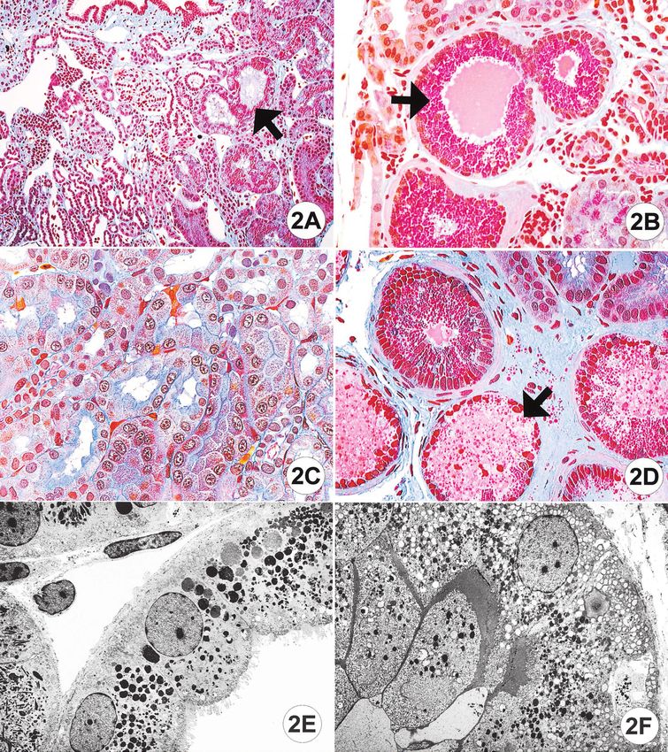
Gross et al: Intraperitoneal protein injection of axolotl                                            55

Fig. 2. Light and electron microscopy of the axolotl kidney under different experimental conditions. (A) Axolotl, protein injection, long-term
experiment, low power (⫻100, Ladewig stain). At the surface (on top) one can observe ciliated funnels. Note proximal tubular epithelial cells
with massive protein droplets (➜). (B) Protein-injected axolotl, long-term experiment, high power (⫻400, Ladewig stain). Note several tubules
with protein-laden epithelial cells (➜) and dilated lumen filled with sludge. Note thickening of visible tubular basement membrane and staining
for collagen (blue color) in the interstitium. (C ) Axolotl, protein injection, short term experiment, high power (⫻400, Ladewig stain). Only delicate
interstitial fibrosis is seen. (D) Axolotl, protein injection, long-term experiment, high power (⫻400, Ladewig stain). Note marked interstitial fibrosis
and beginning necrosis of tubular epithelial cells (➜). (E ) Axolotl, saline injection, long-term experiment, Electron microscopy (1700:1), proximal
tubule: normal epithelial cell with brush border. (F ) Axolotl, protein injection, long-term experiment, electron microscopy (1700:1), proximal
tubule. Note enlarged epithelial cells containing numerous droplets with abundant lipids.
56 Gross et al: Intraperitoneal protein injection of axolotl Fig. 3. Interstitial and epithelial cells after injection with saline or protein. (A) Axolotl, saline injection, long-term experiment, high power (⫻400, TGF- immunohistochemistry). Almost no staining of tubular epithelial cells. (B) Axolotl, protein injection, long-term experiment, high power (⫻400, TGF- immunohistochemistry). Marked staining of epithelial and interstitial cells. (C ) Axolotl, saline injection, long-term experiment, high power (⫻400, fibronectin immunohistochemistry). Only minor staining of epithelial and interstitial cells. (D) Axolotl, protein injection, long- term experiment, high power (⫻400, fibronectin immunohistochemistry). Marked staining of epithelial and interstitial cells. (E ) Axolotl, saline injection, long-term experiment, high power (⫻400, collagen I immunohistochemistry). No staining of epithelial and interstitial cells. (F ) Axolotl, protein injection, long-term experiment, high power (⫻400, collagen I immunohistochemistry). Marked staining of epithelial cells.
Gross et al: Intraperitoneal protein injection of axolotl 57
Table 1. Morphological features of renal damage Table 3. Immunohistochemical staining (long term experiment),
(short term experiment), evaluated by a score system evaluated by a score system
Saline injected Protein injected Saline injected Protein injected
animals (N ⫽ 5) animals (N ⫽ 5) animals (N ⫽ 6) animals (N ⫽ 6)
Protein droplets in tubular Fibronectin
epithelial cells 0 1.43 ⫾ 0.42b Tubuli 0.90 ⫾ 0.68 2.4 ⫾ 0.47a
Tubular dilation 0 1.49 ⫾ 0.43b Interstitium 1.61 ⫾ 0.72 2.49 ⫾ 0.49a
Interstitial fibrosis 0.26 ⫾ 0.24 1.05 ⫾ 0.06a TGF-1
Scores are 0 to 4 as defined in the Methods section.
Tubuli 0.34 ⫾ 0.27 0.95 ⫾ 0.31a
a
P ⬍ 0.05 (Mann-Whitney U) Interstitium 0.11 ⫾ 0.16 0.55 ⫾ 0.32a
b
P ⬍ 0.05 (t test) Collagen I
Interstitium 1.38 ⫾ 0.19 2.27 ⫾ 0.45a
Scores are 0 to 4 as defined in the Methods section.
a
P ⬍ 0.05 (t test)
Table 2. Morphological features of renal damage
(long term experiment), evaluated by a score system
Saline injected Protein injected DISCUSSION
animals (N ⫽ 5) animals (N ⫽ 7)
The present study confirms for the axolotl kidney the
Protein droplets in tubular
epithelial cells 1.31 ⫾ 0.81 3.14 ⫾ 0.54a findings obtained by Randerath [12] and others [14] in
Tubular dilation 0.04 ⫾ 0.08 2.59 ⫾ 0.86a salamanders: that after injection of bovine fetal serum
Interstitial fibrosis 0.09 ⫾ 0.17 2.36 ⫾ 0.31b into the peritoneal cavity, only the nephrons that drain
Scores are 0 to 4 as defined in the Methods section. the coelomic cavity contain protein droplets. In Rander-
a
P ⬍ 0.05 (Mann-Whitney U)
b
P ⬍ 0.05 (t test) ath’s study, such selectivity was lost when low molecular
weight proteins were injected that were stored in both
types of nephrons, presumably because low molecular
weight proteins are filtered in the glomeruli and thus
Intraperitoneal injection of bovine reach the tubules without nephrostomes.
serum: Immunohistochemistry The novel finding in the present study is the demon-
Two TGF- antibodies were tested the specificity of stration that after daily injection of fetal bovine serum,
which had been documented in earlier studies [18–23] protein-loaded tubuli not only showed luminal dilation
and comparable results were found. No TGF- staining and deposition of protein sludge, but also massive diffuse
was seen in saline-injected control animals (Table 3). In progressive accumulation of droplets containing proteins
animals injected with bovine fetal serum, TGF- staining and lipids in the proximal tubular epithelial cells. This
was not seen in tubular epithelial cells that failed to was accompanied by rapidly evolving fibrosis in the inter-
exhibit protein droplets. In contrast, TGF- immune stitium surrounding the groups of protein-loaded neph-
staining was strikingly positive in some, but not all, pro- rons. Protein loading and interstitial fibrosis were further
tein-loaded proximal tubular epithelial cells. The propor- accompanied with pronounced immunohistochemical
staining for TGF-, fibronectin and collagen I. These
tion of positive tubuli was approximately 30%. The stain-
observations are consistent with the paradigm that expo-
ing was specific, since no staining was seen in the negative
sure of tubular epithelial cells to proteins causes intersti-
controls without primary antibody.
tial cell activation and interstitial fibrosis in the kidney
In the interstitium, focally grouped interstitial cells
[5, 6, 27, 28].
showed strongly positive TGF- staining in the vicinity
The classical studies of Metchnikoff on phagocytosis
of protein-loaded tubules. No interstitial TGF- staining
clearly demonstrated that for specific issues, studies in
was seen in saline-injected control animals and in the non-mammalian species are more convenient than stud-
interstitium surrounding tubules that failed to exhibit pro- ies in mammalian species [29]. What are the potential
tein droplets. Fibronectin staining showed marked ex- advantages of using the axolotl kidney? We believe that
pression in the tubulointerstitial tissue in the experimen- the major advantage is the absence of glomerular pathol-
tal animals. In addition protein injected animals showed ogy, so that artifacts resulting from confounding factors
marked interstitial staining for collagen I. cannot occur, such as alterations of post-glomerular he-
The immunohistological finding of increased expres- modynamics and spillover of glomerular pathology [30].
sion of pan-TGF-, TGF-, and platelet-derived growth Alteration of post-glomerular pressure and flow, partial
factor (PDGF) expression followed by intraperitoneal pressure of oxygen, local activation of effector systems,
injection of bovine serum was reproduced in additional such as coagulation, fibrinolysis or complement system,
experiments in cohorts of five animals, each injected for spillover of cytokines causing cell activation and modifi-
ten days with human transferrin, IgG and LDL, respec- cation of trans-capillary cell traffic are excluded in this
tively. amphibian kidney model.58 Gross et al: Intraperitoneal protein injection of axolotl
In view of recent interesting observations that hypoxia terstitial fibrosis by protein loading in vivo. In this model
in the interstitium is a factor in the genesis of interstitial these processes occur independently of alterations of glo-
damage, the present model of the amphibian kidney may merular function that might have potential repercussions
provide the opportunity to control for this confounding on the tubulointerstitial space, such as peritubular hemo-
factor as well [31]. This model also excludes another inter- dynamics, pO2 and spillover of glomerular pathology.
esting confounding mechanism recently proposed by Kriz
et al, that of misdirected filtration [32]. According to ACKNOWLEDGMENTS
their concept, at sites where the glomerular tuft adheres A. Koch and M.L. Groß are recipients of a grant in the Graduierten-
to Bowman’s capsule, misdirected filtration through kolleg “Nieren-und Kreislaufregulation” of the Deutsche Forschungs-
leaks in the basement membrane may permit filtrate to gemeinschaft. The study was supported by a research grant of the Med-
ical Faculty Heidelberg (GR 24/98) and Deutsche Forschungsgemein-
escape into the interstitium, thus causing local interstitial schaft (SFB 423, project B 8). The skillful technical assistance of Z.
cell activation and fibrosis. Antoni, G. Gorsberg, D. Lutz, P. Rieger, S. Söllner, M. Weckbach
A distinct advantage of the present model is the life and S. Wessels is gratefully acknowledged. H. Derks, John Moyers
and U. Burkhard organized photographs and the layout of the color
span of the axolotl, which is two to five years. This may plates.
be an advantage for long-term studies, for instance in
studies on potential reversibility of interstitial fibrosis Reprint requests to Professor Dr. Dr. h.c. mult. Eberhard Ritz, De-
partment Nephrology, University of Heidelberg, Bergheimer Straße 58,
and specific aspects of its long-term evolution. It is also 69115 Heidelberg, Germany.
advantageous that many commercially available reagents E-mail: Prof.E.Ritz@T-online.de
showed remarkable cross-reaction with the renal tissue
of axolotl [18–23]. Potential disadvantages may be the REFERENCES
difficulty of obtaining hemodynamic measurements and 1. Schainuck LI, Striker GE, Cutler RE, et al: Structural-functional
the relative difficulty to obtain urine in this animal as it correlations in renal disease. II. The correlations. Hum Pathol 1:
drains urine into a cloaca. 631–640, 1970
2. Risdon RA, Sloper JC, DeWardener HE: Relationship between
In the present study we tried to avoid several con- renal function and histological changes found in renal biopsy speci-
founders. An effort was made to exclude the possibility mens from patients with persistent glomerular nephritis. Lancet
that the injected FBS was contaminated by endotoxin or 8:363–366, 1968
3. Bohle A, Glomb D, Grund KE, et al: Correlations between rela-
oxidative damage to lipids by performing the respective tive interstitial volume of the renal cortex and serum creatinine
tests. Since we injected non-axolotl proteins, immuno- concentration in minimal changes with nephrotic syndrome and
logical reactions to FBS should not pose any problems in in focal sclerosing glomerulonephritis. Virchows Arch Path Anat
Histol 376:221–232, 1977
short-term experiments, but for long-term investigations 4. Bohle A, Strutz F, Muller GA: On the pathogenesis of chronic
species-specific serum protein might be preferable. renal failure in primary glomerulopathies: A view from the intersti-
In the saline injected controls some protein droplets tium. Exp Nephrol 2:205–210, 1994
5. Strutz F, Müller GA: Interstitial pathomechanisms underlying
were seen in the long-term study. It might be that daily progressive tubulointerstitial damage. Kidney Blood Press Res 22:
handling with the associated increase in the coelomic fluid 71–80, 1999
by saline injection causes a moderate increase of protein 6. Remuzzi G, Ruggenenti P, Benigni A: Understanding the nature
of renal disease progression. Kidney Int 51:2–15, 1997
in the tubular cells. Further studies must clarify the sig- 7. Zoja C, Benigni A, Verroust P, et al: Indomethacin reduces
nificance of this effect in long-term treated animals. proteinuria in passive Heymann nephritis in rats. Kidney Int 31:
It is of note that the injected albumin was not able to 1335–1343, 1987
8. Cameron JS: The enigma of focal segmental glomerulosclerosis.
induce the tubular cell damage seen with bovine serum. Kidney Int 57(Suppl 12):119–131, 1996
Further studies must be performed to clarify whether 9. The GISEN Group: Randomised placebo controlled trial of effect
pure proteins such as IgG and transferrin are responsible of ramipril on decline in glomerular filtration rate and risk of
terminal renal failure in proteinuric, non-diabetic nephropathy.
for interstitial fibrosis. Lancet 349:1857–1863, 1997
The time course of protein uptake by the epithelial 10. Eddy AA, Kim H, Lopez-Guisa J, et al: Interstitial fibrosis in mice
cells of aglomerular nephrons was remarkably rapid for with overload proteinuria: Deficiency of TIMP-1 is not protective.
Kidney Int 58:618–628, 2000
a poikilotherm animal as was the appearance of peritu- 11. Kleier A: Experimentelle Untersuchungen über den Abbau der
bular fibrosis. Consequently, the model is certainly suit- hyalinen Tropfen nach Eiweispeicherung in der Niere von Sala-
able for more detailed mechanistic studies. mandra maculosa. Beitr Path Anat Allg Pathol 103:559–567, 1939
12. Randerath E: Die Entwicklung der Lehre von den Nephrosen
The present study was designed as proof of the princi- in der Pathologischen Anatomie, in Ergebnisse der allgemeinen
ple that the model provides useful information on tubular Pathologie und Pathologischen Anatomie des Menschen und der
epithelial cell protein loading. We are aware of the non- Tiere (vol 32), edited by Hueck W, Frei W, Munich, JF Bergmann,
1937, pp 124–128
random distribution of the two different nephrons and 13. Waldherr R, Ritz E: Edmund Randerath (1899–1961): Experi-
aware that in future studies more sophisticated methods mental proof for glomerular origin of proteinuria. Kidney Int 56:
of stereological analysis would be appropriate. 1591–1596, 1999
14. Gérard P, Cordier R: Sur l’interprétation des alterations morpho-
We conclude that the axolotl kidney provides a novel logiques caractéristiques observées dans le rein au cours de la
in vivo model to study tubulointerstitial activation and in- néphrose lipoı̈dique. CR de L’Assoc des Anat 29:225–232, 1934Gross et al: Intraperitoneal protein injection of axolotl 59
15. Frick H: Vergleichende Anatomie. Hamburg, Parey Verlag, 1991 24. Urbaschek R, McCuskey RS, Rudi V, et al: Endotoxin, endotoxin-
16. Mǿbjerg N, Laresen EH, Jespersen Å: Morphology of the neph- neutralizing-capacity, sCD14, sICAM-1, and cytokines in patients
ron in the mesonephros of bufo bufo (Amphibia, Anura, Bufoni- with various degrees of alcoholic liver disease. Alcohol Clin Exp
dae). Acta Zoologica 79:31–50, 1998 Res 2:261–268, 2001
17. Hanke W, Kaltenhäuser U, Maser-Gluth C, et al: Regulation 25. Sommerburg O, Sostman K, Grune T, et al: Oxidative stress in
of interrenal activity by electrolytes and peptides hormones, in hemodialysis patients treated with a dialysis membrane which has
Biology and Physiology of Amphibians - Fortschritte der Zoologie alphatocopherol bonded to its surface. Biofactors 10:121–124, 1999
(vol 38), edited by Hanke W, Stuttgart, G Fischer, 1990, pp 241–255 26. Winterhoff D, Mehdorn E: Soluble antigens by Ouchterlony
18. Koniski A, Cohen N: Axolotl (Ambystoma mexicanum) lympho- immunodiffusion and immunoelectrophoresis. Arch Hyg Bacteriol
cytes produce and are growth-inhibited by transforming growth 153:447–456, 1969
factor-beta. Dev Comp Immunol 22:91–102, 1998 27. Okada H, Strutz F, Danoff TM, et al: Possible pathogenesis of
19. Muslin AJ, Williams LT: Well-defined growth factors promote renal fibrosis. Kidney Int 54(Suppl 5):37–38, 1996
cardiac development in axolotl mesodermal explants. Development 28. Brunskill NJ: Albumin handling by proximal tubular cells: Mech-
112:1095–1101, 1991 anisms and mediators. Nephrol Dial Transplant 15(Suppl 6):39–
40, 2000
20. Christensen RN, Tassava RA: Apical epithelial cap morphology
29. Petrov RV, Ulyankina TI: The genius of E.E. Metchnikoff—
and fibronectin gene expression in regenerating axolotl limbs. Dev
discoveries over the centuries. Biosci Rep 16:189–205, 1996
Dyn 217:216–224, 2000 30. Kang DH, Hughes J, Mazzali M, et al: Impaired angiogenesis in
21. Johnson KE, Darribere T, Boucaut JC: Amblystoma maculatum the remnant kidney model: II. Vascular endothelial growth factor
gastrulae have an oriented fibronectin-containing extracellular ma- administration reduces renal fibrosis and stabilizes renal function.
trix. J Exp Zool 216:458–471, 1992 J Am Soc Nephrol 7:1448–1457, 2001
22. Chernoff EA, O’Hara CM, Bauerle D, et al: Matrix metallopro- 31. Norman JT, Clark JM, Garcia PL: Hypoxia promotes fibrogenesis
teinase production in regenerating axolotl spinal cord. Wound Re- in human renal fibroblasts. Kidney Int 58:2351–2366, 2000
pair Regen 8:282–291, 2000 32. Kriz W, Hosser H, Hahnel B, et al: From segmental glomerulo-
23. Tonge DA, Golding JP, Edblath M, et al: Effects of extracellular sclerosis to total nephron degeneration and interstitial fibrosis: A
matrix components on axonal outgrowth from peripheral nerves histopathological study in rat models and human glomerulosclero-
of adult animals in vitro. Exp Neurol 146:81–90, 1997 sis. Nephrol Dial Transplant 13:2781–2798, 1998You can also read