Jumping on the Edge-First Evidence for a 2 6-meric Hemocyanin in Springtails - MDPI
←
→
Page content transcription
If your browser does not render page correctly, please read the page content below
biomolecules

Communication
Jumping on the Edge—First Evidence for a 2 × 6-meric
Hemocyanin in Springtails
Juliane Schmidt 1, * , Heinz Decker 1 and Michael T. Marx 2
 1    Institute for Molecular Physiology, Johannes Gutenberg-University Mainz (JGU), 55128 Mainz, Germany
 2    Institute for Zoology, Johannes Gutenberg-University Mainz (JGU), 55128 Mainz, Germany
 *    Correspondence: julschm@uni-mainz.de

 Received: 22 July 2019; Accepted: 18 August 2019; Published: 22 August 2019

 Abstract: Hemocyanins are respiratory dioxygen carrier proteins found in many arthropods
 including ancient terrestrial species such as spiders and scorpions as well as marine horseshoe
 crabs. As hemocyanins are highly conserved in this lineage, it is possible to observe an evolutionary
 descent through its subunits and their overall structure. Unfortunately, little is known about the
 structure and function of hexapod hemocyanins. Using recent springtail taxa (Collembola) as models
 for basal hexapods, and the help of electron microscopy, light scattering, SDS PAGE, and Western blot,
 we could demonstrate for the first time the presence of 2 × 6-meric hemocyanins in the hemolymph
 of hexapods. The quaternary structure is composed of at least two different subunits and looks
 nearly identical to the hemocyanin found in decapod crustaceans. In addition, homology modeling
 and western blotting suggest a close structural relationship between collembolan and crustacean
 hemocyanin. Such a respiratory protein was possibly helpful in the early terrestrialization process
 of ancient Collembola. In addition, physiological adaptations to hypoxic or temporarily anoxic
 conditions could be a possible explanation for the presence of this respiratory protein. Nevertheless,
 it has to be concluded that the primary benefit of hemocyanin for springtails remains unclear.

 Keywords: hemocyanin; springtails; Collembola; Crustacea; terrestrialization; hexapods

1. Introduction
      Hexapoda comprise of the well-known class Insecta with a second one called Entognatha.
Entognatha contain three of the five formerly apterygote taxa: Springtails (Collembola), two-pronged
bristletails (Diplura), and coneheads (Protura). It is unknown when ancient Collembola first occurred
but the fossil springtail Rhyniella praecursor [1] is considered to be one of the oldest terrestrial arthropod
taxa from about 420 to 400 Myr ago [2–4]. Collembola inhabit every soil layer and the soil surface
in very high abundances, making them a key group among the soil arthropods. Over their long
evolutionary history, they were able to colonize different specialized and sometimes extreme habitats
(e.g., water surfaces, sand coasts, deserts, the Arctic, and the Antarctic) [5–12], which makes them one
of the most ecologically, diversified, and distributed arthropod groups [13].
      Hemocyanins (hc) are highly conserved respiratory proteins in the lineage of arthropods and
molluscs. They occur freely dissolved in the hemolymph (hl) and especially for arthropods, they consist
of subunits of ca. 72–74 kDa which connect themselves to hexamers. Depending on the species, they
can be found as single hexamers or as multiples of hexamers [14,15]. Looking at the quarternary
structure of arthropod hemocyanins, they are always composed of a multiple of hexamers. Horseshoe
crabs, spiders, and crustaceans usually form 8 × 6-mers, 4 × 6-mers, and 2 × 6-mers, respectively [15].
Insects or hexapods are thought to possess a single hexamer, which, although functional, is very
poorly distinguished from non-respiratory hexamerin [16]. Recent investigations also focused on the
immunological properties of hemocyanin in arthropods. The antibacterial activity of malacostracean

Biomolecules 2019, 9, 396; doi:10.3390/biom9090396                          www.mdpi.com/journal/biomolecules
Biomolecules 2019, 9, 396 2 of 9
hemocyanin against clinical pathogens or molecular diversity changes of the white shrimp hemocyanin
related to pathogen infection are current examples [17,18].
Recently, hemocyanin was identified in Collembola hemolymph by RT-PCR and RACE techniques,
leading to a full-length sequence for Sinella curviseta hemocyanin (termed ScuHc1) and a partial sequence
from Folsomia candida hemocyanin (FcaHc1) [19]. Both were classified as hexapod hemocyanin subunit
type 1 based on about 60% sequence identity with the subunit type 1 hemocyanin from the stonefly
Perla marginata [20], which was shown to be a biologically active 1 × 6-meric hemocyanin during the
stonefly’s larval stadium [20].
It has not been possible, so far, to present the structure of a hexapod hemocyanin. With this
work on a basal and very old hexapod clade, we want to suggest the collembolan hemocyanin
quaternary structure. Some ideas concerning the function of this respiratory protein in Collembola will
be discussed.
2. Materials and Methods
2.1. Animals and Hemolymph Collection
In order to gain structural information about springtail hemocyanin, we searched for it in the
hemolymph of the tiny springtail species Sinella curviseta, Folsomia candida, Coecobrya tenebricosa,
Isotoma viridis, Orchesella cincta, and Orchesella villosa. The hemolymph samples were freshly collected
before performing the experiments. The animals were anesthetized in the freezer at −24 ◦ C for 5 min.
The protein samples were immediately obtained by decapitation of the anesthetized animals with
a scalpel and immersion of the two body parts in a 15 µL drop of a 0.1 M Tris/HCl buffer (pH 7.8,
10 mM CaCl2, and MgCl2 each) in the presence of protease inhibitors (Protease Inhibitor Cocktail,
Sigma) for 20 s. The same 15 µL drop was used for the next 15 individuals as well. This process
was repeated with new individuals and new drops until the amount of protein was sufficient for
the planned experiment. Only the electron microscopic images were be performed with all listed
Collembola species. Since the sequence of ScuHc1 is known, we focused on the hemocyanin from
Sinella curviseta for the other experiments.
2.2. Protein Determination
Due to the impurity of the samples, the protein concentration could only be roughly estimated.
A UV/Vis spectrum was recorded on a NanoDrop ND-1000 photo spectrometer (PEQLAB Biotechnologie,
Erlangen, Germany). The concentration was determined under the assumption that an impure protein
solution of 1 mg/mL has an absorption of 1 at 280 nm. For a 15 µL protein drop obtained from
15 individuals as described above, this resulted in an approximate concentration of 0.1 mg/mL. Due to
the very low protein yield and the small sample volume, a more accurate calculation of the protein
concentration was not performed.
2.3. Electron Microscopy
For the negative staining electron microscopy, the hemolymph of 15 individuals of each species
was used and processed as previously described [21]. Larger particles were removed from the samples
by centrifugation for 20 min and at 22,000 g. The hemolymph was applied to glowing continuous carbon
carrier layers and stained with aqueous 2% (w/v) uranyl format using the single-drop method [22].
A Technai12 electron microscope and a connected MegaView III camera were used for the electron
microscopic images. Using 13 different 2 × 6-meric objects with exactly the same position and orientation
on the EM images, the software EM-Menu (TVIPS, Gauting, Germany) was used to create a 2D average
image. This class sum was one of 184, which were calculated out of 1258 picked 2 × 6-meric objects on
100 images. The class sum of this orientation was chosen because it best reflects the typical structure of
2 × 6-meric hemocyanin.Biomolecules 2019, 9, 396 3 of 9
2.4. SDS-PAGE and Immunoblotting
Denaturing SDS-PAGE was carried out as 7.5% gels under standard conditions [23]. For each
sample, the hemolymph of 15 individuals from Sinella curviseta and 15 µL buffer were used and
denaturing buffer was added 1:1. The gels were stained with Coomassie dyeing after Kang [24].
For western blotting, the proteins were transferred onto nitrocellulose membranes. Non-specific
binding sites were blocked for 45 min using 4% non-fat dry milk in TBST (10 mM Tris/HCl, pH 7.5,
140 mM NaCl, 0.3% Tween 20). Primary anti-hemocyanin or anti-hemolymph antibody serum, diluted
1:10,000 in blocking solution was carried out overnight. The membrane was then washed four times in
a row with TBST. Afterwards, two hours of incubation with secondary antibodies (goat-anti-rabbit
IgGs coupled with alkaline phosphatase, diluted 2:20,000 with TBST) was performed. The membrane
was finally washed four times with TBST, before staining with NBT and BCIP. All steps, except the
detection, were performed at a temperature of 4 ◦ C.
2.5. Multi-Angle Laser Light Scattering
The hemolymph of 300 individuals of Sinella curviseta in 100 µL buffer was used for this method
because it requires a very high protein concentration. With this concentration, we just reached a credible
detection range to calculate the molecular weight of ScuHc. After the purification by size exclusion gel
chromatography (Superose 6 10/300 GL SEC, GE Healthcare, Chicago, IL, USA), the Multi-Angle Laser
Light scattering (MALLS, Wyatt Technology, Santa Barbara, CA, USA) of the proteins was performed.
BSA monomer (Sigma-Aldrich, St. Louis, MO, USA) was used to calibrate the MALLS instrument by
normalization of the different detector signals with respect to the 90◦ detector signal. The flow rate was
0.4 mL/min and the molecular weight was calculated using the Exzess-Rayleigh ratio by the software
ASTRA (Wyatt Technology, Santa Barbara, CA, USA). A correction factor containing the molecular
weight and the extinction coefficient of the subunits was used for calculation. Both were determined
with Protparam from the existing Sinella curviseta hemocyanin primary sequence [19,25].
2.6. Mass Spectrometry Sample Preparation
The protein identification was performed by the IMB proteomics core facility (Mainz, Germany).
Protein lanes were cut out of the SDS-PAGE gel, crushed, destained in 50% ethanol in 25 mM ammonium
hydrogen carbonate (room temperature, rotated for 15 min), and dehydrated in 100% acetonitrile
(room temperature, rotated for 10 min). Dehydrated samples were trypsin-digested (1 µg trypsin/sample
in 50 mM Triethylammonium bicarbonate buffer pH 8.0) at 37 ◦ C for 12 h. Stepwise peptide extraction
was done as follows: Twice extraction solution (30% acetonitrile) and 100% acetonitrile centrifuged for
15 min (25 ◦ C, 1400 rpm). After purification and desalting using C18 StageTips (Empore, Sigma-Aldrich,
St. Louis, MO, USA) [26], 3.5 µL peptides were loaded and separated on C18 column (75 µm inner
diameter, New Objective) The column was self-packed with 1.9 µm Reprosil beads (Dr. Maisch) and
mounted to an Easy LC 1000 HPLC (Thermo Fisher Scientific, Waltham, MA, USA).
2.7. Mass Spectrometry Measurement and Data Analysis
Reverse phase chromatography was performed with 0.1% formic acid (buffer A), 80% acetonitrile,
and 0.1% formic acid (buffer B). Peptides were eluted under the following gradient: 0–21 min 0–22%
buffer B, 21–28 min 22–40% buffer B, 28–32 min 40–95% buffer B; and directly sprayed into a Q Exactive
Plus mass spectrometer (Thermo Fisher Scientific, Waltham, MA, USA). The mass spectrometer was
operating in positive scan mode with a full scan resolution of 70,000; AGC target 3 × 10⁶ ; max IT = 20 ms;
scan range 300–1650 m/z; and a Top10 MSMS method. Normalized collision energy was set to 25 and
MSMS scan mode operated with resolution of 17,000; AGC target 1 × 10⁵ ; max IT = 120 ms.
A database search was performed using MaxQuant Version 1.5.2.8 [27] against Uniprot Collembola,
Manduca secxta, Carausius morosus, and insecticyanin database (downloaded 15th October 2015,
2315 entries, FASTAfile attached in the supplementary) with Trypsin/P as a digestion enzyme (2 missedBiomolecules 2018, 8, x 4 of 9
Biomolecules 2019, 9, 396 4 of 9
October 2015, 2315 entries, FASTAfile attached in the supplementary) with Trypsin/P as a digestion
enzyme (2 missed cleavages allowed). The following settings were used: The variable modifications
cleavages allowed).
were; Acetyl The following
(Protein N-term) and settings were used:
Oxidation (M),Theandvariable
the modifications were; Acetyl
fixed modifications were;
(Protein N-term) and Oxidation (M), and the fixed modifications
Carbamidomethyl (C), FDR of 1% on peptide, and protein level. were; Carbamidomethyl (C), FDR of
1% onProteins
peptide,were
and protein level.to be identified if they matched at least two peptides (one of them
considered
Proteins were considered to be identified if they matched at least two peptides (one of them unique).
unique).
Proteins
Proteins that
that correspond
correspond to to the
the reverse
reverse database
database or
or the
the common
common contamination
contamination list
list and
and proteins
proteins
with
with peptides
peptides identified
identified only
only by
by modified
modifiedpeptides
peptides were
werefiltered
filteredout.
out. Further
Further bioinformatic
bioinformatic analyses
analyses
were
wereperformed
performedin inRR[28]
[28]using
usingthethefollowing
followinglibraries:
libraries: Knitr,
Knitr,plyr,
plyr,reshape,
reshape,ggplot2,
ggplot2,and
andpsych.
psych.
3.
3. Results
Results and
and Discussion
Discussion
Analysis
Analysis ofofthe
thehemolymph
hemolymphbyby transmission
transmission electron
electronmicroscopy
microscopydemonstrated
demonstrated hemocyanin-like
hemocyanin-
structures in every species (Figure 1). They were characterized as 2 × 6-mer
like structures in every species (Figure 1). They were characterized as 2 × 6-mer and and strongly reminiscent
strongly
to the hemocyanins
reminiscent to the found in Astacusfound
hemocyanins leptodactylus (crayfish),
in Astacus Homarus (crayfish),
leptodactylus americanus (lobster),
Homarus and Cancer
americanus
paragurus (crab) [29]. Here, two hexamers dimerize, being rotated by 90 ◦ against each other (Figure 1g).
(lobster), and Cancer paragurus (crab) [29]. Here, two hexamers dimerize, being rotated by 90° against
Since
each the sequence
other (Figureof1g).
ScuHc1
Sinceisthe
known, we focused
sequence on theishemocyanin-like
of ScuHc1 known, we focused protein, from
on the Sinella curviseta.
hemocyanin-like
Multi-Angle
protein, fromLaser Light
Sinella scattering
curviseta. of the proteins
Multi-Angle purified
Laser Light by sizeof
scattering exclusion gel chromatography
the proteins purified by size
revealed a molecular mass of app. 850 kDa (Supplementary Figure
exclusion gel chromatography revealed a molecular mass of app. 850 kDa (Supplementary S1) which is in theFigure
order S1)
of
× 6-meric
2which is inhemocyanins
the order ofbeing built uphemocyanins
2 × 6-meric by subunits with typical
being builtmasses
up byranging between
subunits 73–83 kDa
with typical [30].
masses
Smaller particles were also found which could not be assigned to 1 × 6-meric hemocyanins
ranging between 73–83 kDa [30]. Smaller particles were also found which could not be assigned to 1 due to their
form and size.
× 6-meric hemocyanins due to their form and size.
Figure 1.1. Electron
Figure Electron microscopical
microscopical images
images of of hemolymph
hemolymph proteins
proteins ofofvarious
variousspringtails.
springtails. The
The “single
“single
droplet negative staining“ technique and 2% (v/v) aqueous uranyl formate
droplet negative staining“ technique and 2% (v/v) aqueous uranyl formate was applied for was applied for the
the
preparation of the sample. (a) Overview of the hemolymph proteins of Orchesella
preparation of the sample. (a) Overview of the hemolymph proteins of Orchesella villosa. A huge amount villosa. A huge
amount
of the 2 ×of6-meric
the 2 ×protein
6-meric protein in
is present is the
present in the hemolymph.
hemolymph. Inserts showInserts show proteins
2 × 6-meric 2 × 6-meric
found proteins
in the
found in thefrom
hemolymph hemolymph from(b),
Isotoma viridis Isotoma viridis
Folsomia (b),(c),
candida Folsomia candida
Coecobrya (c), Coecobrya
tenebricosa tenebricosa
(d), Orchesella cincta (d),
(e),
Orchesella
and Sinella cincta (e),(f)
curviseta and Sinella
[29]. curviseta (f) [29].
(g) Superposition of 13(g) Superposition
images of 13 images
of Sinella curviseta of Sinella
hemocyanin for curviseta
gaining
ahemocyanin for gaining a better contrast.
better contrast.
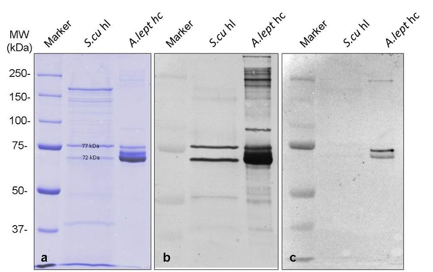
Twodifferent
Two differentbands
bandsfrom
fromthetheSDS-PAGE
SDS-PAGE(Figure
(Figure2a)
2a)with
withabout
about7272and
and 77
77 kDa
kDa were
were investigated
investigated
by LC-MS/MS
by LC-MS/MStotoidentify
identify
thethe proteins.
proteins. Unique
Unique peptides
peptides (six (six of77
of the the
kDa77 and
kDatwo
andoftwo of kDa
the 72 the 72 kDa
bands)
bands)the
match match the sequence
ScuHc1 ScuHc1 sequence
(Figures (Figures
S2 and S3)S2[19].
and Thus,
S3) [19].
theThus, the 2 ×protein
2 × 6-meric 6-mericmolecules
protein molecules
of Sinella
of Sinellaare
curviseta curviseta are hemocyanins,
hemocyanins, built by twobuilt by two
different different hemocyanin
hemocyanin subunit typessubunit types
with 72 kDawith 72 kDa.
and 77 kDaBiomolecules 2018, 8, x 5 of 9
and 77 kDa.
Biomolecules The
2019, latter one correlates the known ScuHc1, while we annotated the other one as ScuHc2,
9, 396 5 of 9
which has not been detected in springtails before. The question arises whether and how its sequence
differs from the previously known subunit type 2 [19] and whether the ScuHc2 subunit is responsible
The latter one correlates the known ScuHc1, while we annotated the other one as ScuHc2, which has not
for the formation of 2 × 6-mers, as it has already been shown for some crustaceans hemocyanins
been detected in springtails before. The question arises whether and how its sequence differs from the
[15,29]. Also of great interest would be how this missing sequence is placed in the phylogenetic
previously known subunit type 2 [19] and whether the ScuHc2 subunit is responsible for the formation
studies in order to further elucidate the evolution of hemocyanins. Further investigations are
of 2 × 6-mers, as it has already been shown for some crustaceans hemocyanins [15,29]. Also of great
necessary to determine the complete sequence of subunit ScuHc2 and to answer this question. For
interest would be how this missing sequence is placed in the phylogenetic studies in order to further
example, the complete amino acid sequence and the glycosylation of the hemocyanins from the
elucidate the evolution of hemocyanins. Further investigations are necessary to determine the complete
malacostracean taxa Carcinus aestuarii and Eriphia verrucosa helped to understand the quaternary
sequence of subunit ScuHc2 and to answer this question. For example, the complete amino acid
structure of the molecules [31,32]. These analyses could also help to gain a better understanding of
sequence and the glycosylation of the hemocyanins from the malacostracean taxa Carcinus aestuarii and
the quaternary structure of hexapod hemocyanins.
Eriphia verrucosa helped to understand the quaternary structure of the molecules [31,32]. These analyses
To obtain information about the immunological relationship, antibodies against the 2 × 6-mer
could also help to gain a better understanding of the quaternary structure of hexapod hemocyanins.
hemocyanins of crayfish Astacus leptodactylus and the 4 × 6-meric hemocyanins of tarantula Eurypelma
To obtain information about the immunological relationship, antibodies against the 2 × 6-mer
californicum were used. These references were chosen because both animals are well studied model
hemocyanins of crayfish Astacus leptodactylus and the 4 × 6-meric hemocyanins of tarantula Eurypelma
organisms in hemocyanin research [14,15,29]. Antibodies raised against hemolymph (hl) proteins
californicum were used. These references were chosen because both animals are well studied model
from A. leptodactylus strongly recognize Sinella curviseta hemocyanins (Figure 2b), while almost no
organisms in hemocyanin research [14,15,29]. Antibodies raised against hemolymph (hl) proteins
cross-reactivity was observed with antibodies raised against purified 4 × 6-meric E. californicum
from A. leptodactylus strongly recognize Sinella curviseta hemocyanins (Figure 2b), while almost no
hemocyanins (Figure 2c). Thus, Western blots reveal a close immunological relationship of the two
cross-reactivity was observed with antibodies raised against purified 4 × 6-meric E. californicum
subunit types from Sinella curviseta with crustacean hemocyanins. The bands of S.cu hl always
hemocyanins (Figure 2c). Thus, Western blots reveal a close immunological relationship of the two
showed a significantly broader expression compared to other samples on the SDS-PAGE. This is
subunit types from Sinella curviseta with crustacean hemocyanins. The bands of S.cu hl always showed
probably due to the fact that these samples had to be applied unpurified and therefore contained
a significantly broader expression compared to other samples on the SDS-PAGE. This is probably
tissue fluid, small cell debris, and other macromolecules in addition to the different proteins of the
due to the fact that these samples had to be applied unpurified and therefore contained tissue fluid,
hemolymph.
small cell debris, and other macromolecules in addition to the different proteins of the hemolymph.
Figure 2.2.Identification
Figure Identificationofoftwotwo hemocyanin
hemocyanin subunit
subunit typestypes in S. curviseta
in S. curviseta hemolymph
hemolymph by Western
by Western blotting.
blotting. (a) SDS-PAGE (pH: 8.8, 7.5%). Marker: Biorad Precision Plus Unstained
(a) SDS-PAGE (pH: 8.8, 7.5%). Marker: Biorad Precision Plus Unstained Standards. S.cu hl = SinellaStandards. S.cu hl =
curviseta hemolymph. A. lep hc = Astacus leptodactylus hemocyanin. (b) Western blot with antibodies
Sinella curviseta hemolymph. A. lep hc = Astacus leptodactylus hemocyanin. (b) Western blot with
antibodies
against against hemolymph
hemolymph proteins fromproteins
crab from
Astacuscrab Astacus leptodactylus.
leptodactylus. Two bands Two at bands
about at
72about
and 7772kDa
and
77 kDa
from fromcurviseta
Sinella Sinella curviseta
hemolymph hemolymph are strongly
are strongly recognized,
recognized, while proteins
while various various proteins from semi-
from semi-purified
purified
A. A. leptodactylus
leptodactylus hemocyanin hemocyanin are observed.
are observed. (c) Western(c)blot
Western blot with antibodies
with antibodies againsttarantula
against purified purified
tarantula hemocyanin. Two strong bands of semi-purified A. leptodactylus
hemocyanin. Two strong bands of semi-purified A. leptodactylus hemocyanin can be observed hemocyanin canbut
be
observed
only but only
two very weaktwo very
bands weak
from bands
Sinella from Sinella
curviseta curviseta
hemolymph hemolymph
bands bands can
can be detected. be detected.
Marker: Biorad
Marker: Biorad
Precision Precision
Plus Dual Color Plus Dual Color
Standards. Standards.
Blot was scanned Blot wasand
black scanned
white.black and white.Biomolecules 2019, 9, 396 6 of 9
To summarize, negative staining electron microscopic images, light scattering, Western plots, and
the protein identification by LC-MS/MS prove that the observed proteins are 2 × 6-meric hemocyanins,
composed of two different subunits, occur in the hemolymph of the springtail Sinella curviseta.
Furthermore, the EM images of the hemolymph of other springtail species (Figure 1) imply that the
2 × 6-meric hemocyanin is dominant throughout different families of Collembola.
Hemocyanin is the main respiratory protein in Pancrustacea and it is discussed that the ancestors of
terrestrial hexapods used hemocyanin in addition to their gills and tegumentary respiration to establish
their life outside of water [30]. We suggest that collembolan hemocyanin has a cooperative oxygen
affinity due to its 2 × 6-meric structure as it has been shown for their malacrustacean equivalents [15],
and that springtails retained the 2 × 6-meric hemocyanin of their crustacean ancestor possibly as
a plesiomorph feature, which was helpful in the early terrestrialization phase of this taxon. The function
of hemocyanin as a respiratory protein could be linked to physiological adaptations in Collembola
species inhabiting deeper layers of the soil, sand coasts, or tidal zones. Different collembolan species
showed a metabolic shift under anaerobic conditions (anoxia) [33]. Folsomia candida has distinctly
elevated lactate levels following artificially induced anoxia [34–36]. Additionally, an increased heart
rate was measured in this species during hypoxic conditions. This blood circulation modulation helps
to maintain the partial pressure between the medium, the blood, and the tissue [37]. Both adaptations
allow individuals of F. candida a fully functional respiration performance even during extreme low
partial pressures of oxygen (down to 6.666 Pa) [36]. In Anurida maritima (Guérin-Méneville, 1836)
(Neanuridae, Collembola), the partial pressure of oxygen under extreme conditions can be even
lower [38]. This species prefers habitats in tidal zones and should be well adapted to periodic flooding
events. At low tides, these animals feed on fine substrates such as the algae and suspended sediments
of the surface. With increasing water levels, individuals gather in aggregations (“nests”) under stones
in order to survive the flooded period in a commonly used air pocket [39]. Exposed to such changing
environmental conditions, the possibility to use very low partial pressures of oxygen (as low as 1000 Pa)
makes this species highly competitive. A critical oxygen level for these animals was not achievable
during experiments performed by Zinkler et al. [38]. However, at 1000 Pa, a small reduction of the
oxygen uptake could be measured, but without causing serious problems for the animals concerning
the regulation of the oxygen uptake. These features coupled with an effective respiratory protein as
an oxygen reservoir could increase the survival abilities of these species in partially hypoxic or even
anoxic conditions as can occur in deeper soil layers or tidal zones.
The ability to survive longer periods of flooding in the egg stage was documented for Isotomiella
minor (Schäffer, 1896) (Isotomidae, Collembola) and several members of the taxon Symphypleona
(Collembola) [40,41]. Some of these species have the ability to survive embryonic and post-embryonic
development as well as the first molt submerged (semiaquatic lifestyles) [42]. All these adaptations to
low oxygen environments could also imply the need for a respiratory protein like hemocyanin.
Nevertheless, despite these assumptions it remains unclear why hemocyanins are present in
Collembola. They do not seem to be needed as respiratory proteins in the majority of species, as the
supply with oxygen by diffusion (known as tegumentary respiration [15]) is sufficient to meet the
respiratory demands. This is also supported by the lack of trachea in Sinella curviseta [43]. However,
the mRNA sequence of ScuHc1 [19] contains all six histidines coordinating the two copper atoms at
the active site of type 3 copper proteins. As we could not detect the typical absorbance peak at 340 nm
for bound dioxygen (Supplementary Figure S4), it seems likely that Collembola hemocyanin occurs in
the met state, as often observed for hemocyanins [44]. Further experiments could address this question
if it is possible to gain enough intact hemolymph from these extremely small animals.
In summary, this study showed the occurrence of a 2 × 6-meric quaternary hemocyanin structure
consisting of at least two different subunits in all investigated species of the taxon Collembola.
The benefit of hemocyanin for springtails remains unclear, but the early terrestrialization process or
physiological adaptation to hypoxic or transient anoxic conditions may be possible explanations for
the presence of this respiratory protein.Biomolecules 2019, 9, 396 7 of 9
Supplementary Materials: The following are available online at http://www.mdpi.com/2218-273X/9/9/396/s1,
Figure S1: Absorbance of size exclusion gel chromatography and diffraction of the Multi Angle Laser Light
scattering (MALLS) and SDS-PAGE of the collected Peaks, Figure S2: Protein identification of the 72 and 77
kDa bands out of the SDS-PAGE of Sinella curviseta hemolymph (Figure 2) by mass spectroscopy, Figure S3:
MSMS spectrum masses of the Protein identification of S.cu hemocyanin by mass spectroscopy, Figure S4: UV/Vis
absorption spectrum of Sinella curviseta hemolymph. Data S6: Detailed data of the performed protein identification
as compressed data incl.FASTA file of the databases which were used for the protein identification by mass
spectroscopy. Uniprot Collembola, Manduca secxta, Carausius morosus and insecticyanin database (downloaded
15th October 2015, 2315 entries.
Author Contributions: Conceptualization, J.S., H.D. and M.M.; Methodology, J.S. and H.D.; Software, J.S.;
Investigation, J.S.; Resources, H.D. and M.M.; Data curation, J.S.; Writing—J.S., H.D. and M.M.; Writing—review
and editing, J.S. and M.M.; Visualization, J.S.; Supervision, H.D.; Funding acquisition, H.D.
Funding: This work was granted by the DFG Graduate School of Immunotherapy 1043. Support by IMB
Proteomics Core Facility instrument funded by DFG INST 247/766-1 FUGG.
Acknowledgments: In grateful memory of Prof. Dr. Heinz Decker, without whose knowledge, support and
commitment this research would never have taken place. We also thank Dr. C. Leake, Dr. J. Markl, Dr. S. Schenk
and Dr. D. Hemmerich for their critical discussion and Dr. P. Arnold for his help to obtain the electron microscopical
images. Proteomics Core Facility is gratefully acknowledged. In particular, we wish to thank Anja Freiwald and
Dr. Mario Dejung for sample measurement and data analysis.
Conflicts of Interest: The authors declare no conflict of interest.
References
1. Hirst, S.; Maulik, S. On some arthropod remains from the Rhynie Chert (Old Red Sandstone). Geol. Mag.
1926, 63, 69–71. [CrossRef]
2. Scourfield, D.J. The oldest known fossil insect. Nature 1940, 145, 799–801. [CrossRef]
3. Whalley, P.; Jarzembowski, E.A. A new assessment of Rhyniella, the earliest known insect, from the Devonian
of Rhynie, Scotland. Nature 1982, 291, 317. [CrossRef]
4. Habgood, K.S.; Hass, H.; Kerp, H. Evidence for an early terrestrial food web: Coprolites from the early
Devonian Rhynie chert. Trans. R. Soc. Edinb. 2004, 94, 371–389. [CrossRef]
5. Bauer, R.; Christian, E. Adaptations of three springtail species to granite boulder habitats (Collembola).
Pedobiologia 1993, 37, 280–290.
6. Brand, R.H. The effect of prescribed burning on epigeic springtails (Insecta: Collembola) of woodland litter.
Am. Midl. Nat. 2002, 148, 383–393. [CrossRef]
7. Elnitsky, M.A.; Benoit, J.B.; Denlinger, D.L.; Lee, R.E., Jr. Desiccation tolerance and drought acclimation in
the Antarctic collembolan Cryptopygus antarcticus. J. Insect Physiol. 2008, 54, 1432–1439. [CrossRef]
8. Greenslade, P. Survival of Collembola in arid environments: Observations in South Australia and the Sudan.
J. Arid Environ. 1981, 4, 219–228. [CrossRef]
9. Hawes, T.C.; Couldridge, C.E.; Bale, J.S.; Worland, M.R.; Convey, P. Habitat temperature and the temporal
scaling of cold hardening in the high Arctic collembolan, Hypogastrura tullbergi (Schäffer). Ecol. Entomol.
2006, 31, 450–459. [CrossRef]
10. Hawes, T.C.; Worland, M.R.; Convey, P.; Bale, J.S. Aerial dispersal of springtails on the Antarctic Peninsula:
Implications for local distribution and demography. Antarct. Sci. 2007, 19, 3–10. [CrossRef]
11. Palissa, A. Collembola. In Süßwasserfauna von Mitteleuropa 10, 1st ed.; Schwoerbel, J., Zwick, P., Eds.; Spektrum
Verlag: Heidelberg/Berlin, Germany, 2000; pp. 1–166.
12. Shaw, P.C.A.; Ozanne, C.; Speight, M.; Palmer, I. Edge effects and arboreal Collembola in coniferous
plantations. Pedobiologia 2007, 51, 287–293. [CrossRef]
13. Rusek, J. Biodiversity of Collembola and their functional role in the ecosystem. Biodivers. Conserv. 1998,
7, 1207–1219. [CrossRef]
14. Van Holde, K.E.; Miller, K.I.; Decker, H. Hemocyanins and invertebrate evolution. J. Biol. Chem. 2001,
276, 15563–15566. [CrossRef] [PubMed]
15. Markl, J.; Decker, H. Molecular Structure of the Arthropod Hemocyanins. Adv. Comp. Environ. Physiol. 1992,
13, 325–376.
16. Telfer, W.H.; Kunkel, J.G. The function and evolution of insect storage hexamers. Annu. Rev. Entomol. 1991,
36, 205–228. [CrossRef] [PubMed]Biomolecules 2019, 9, 396 8 of 9
17. Kizheva, Y.K.; Rasheva, I.K.; Petriova, M.N.; Milosheva-Ivanova, A.V.; Velkova, L.G.; Dolashka, P.A.;
Dolashki, A.K.; Hristova, K. Antibacterial activity of crab hemocyanin against clinical pathogens. Biotechnol.
Biotechnol. Equip. 2019, 33, 873–880. [CrossRef]
18. Fan, J.; Li, X.; Lu, H.; Aweya, J.J.; Zhang, Y. N-terminal diversity of Litopenaeus vannamei hemocyanin and
immunity. Mol. Immunol. 2019, 112, 360–368. [CrossRef] [PubMed]
19. Pick, C.; Schneuer, M.; Burmester, T. The occurrence of hemocyanin in Hexapoda. FEBS J. 2009, 276, 1930–1941.
[CrossRef]
20. Hagner-Holler, S.; Schoen, A.; Erker, W.; Marden, J.H.; Rupprecht, R.; Decker, H.; Burmester, T. A respiratory
hemocyanin from an insect. Proc. Natl. Acad. Sci. USA 2004, 101, 871–874. [CrossRef]
21. Brenner, S.; Horne, R.W. A negative staining method for high resolution electron microscopy of viruses.
Biochim. Biophys. Acta 1959, 34, 103–110. [CrossRef]
22. Harris, J.R. Negative staining of thinly spread biological particulates. Methods Mol. Biol. 1999, 117, 13–30.
[CrossRef] [PubMed]
23. Laemmli, U.K. Cleavage of structural proteins during the assembly of the head of bacteriophage T4. Nature
1970, 227, 680–685. [CrossRef] [PubMed]
24. Kang, D.; Gho, Y.S.; Suh, M.; Kang, C. Highly Sensitive and Fast Protein Detection with Coomassie Brilliant Blue
in Sodium Dodecyl Sulfate-Polyacrylamide Gel Electrophoresis. Bull. Korean Chem. Soc. 2002, 23, 1511–1512.
[CrossRef]
25. Gasteiger, E.; Hoogland, C.; Gattiker, A.; Duvaud, S.; Wilkins, M.R.; Appel, R.D.; Bairoch, A. Protein
Identification and Analysis Tools on the ExPASy Server. In The Proteomics Protocols Handbook; Walker, J.M., Ed.;
Humana Press: Totowa, NJ, USA, 2005; pp. 571–607. [CrossRef]
26. Rappsilber, J.; Mann, M.; Ishihama, Y. Protocol for micro-purification, enrichment, pre-fractionation and
storage of peptides for proteomics using StageTips. Nat. Protoc. 2007, 2, 1896–1906. [CrossRef] [PubMed]
27. Cox, J.; Mann, M. MaxQuant enables high peptide identification rates, individualized p.p.b.-range mass
accuracies and proteome-wide protein quantification. Nat. Biotechnol. 2008, 26, 1367–1372. [CrossRef]
[PubMed]
28. R Core Team. R: A Language and Environment for Statistical Computing. R Foundation for Statistical
Computing. 2016. Available online: https://www.R-project.org/ (accessed on 19 October 2015).
29. Stöcker, W.; Raeder, U.; Bijlholt, M.M.C.; Wichertjes, T.; van Bruggen, E.F.J.; Markl, J. The quaternary structure
of four crustacean 2 × 6 hemocyanins: Immunocorrelation, stoichiometry, reassembly and topology of
individual subunits. J. Comp. Physiol. 1988, 158, 271–289. [CrossRef]
30. Burmester, T. Evolution of Respiratory Proteins across the Pancrustacea. Integr. Comp. Biol. 2015, 55, 792–801.
[CrossRef] [PubMed]
31. Dolashka-Angelova, P.; Dolashki, A.; Savvides, S.N.; Hristova, R.; Van Beeumen, J.; Voelter, W.; Devreese, B.;
Weser, U.; Di Muro, P.; Salvato, B.; et al. Structure of hemocyanin subunit CaeSS2 of the crustacean
Mediterranean crab Carcinus aestuarii. J. Biochem. 2005, 138, 303–312. [CrossRef]
32. Dolashki, A.; Radkova, M.; Todorovska, E.; Ivanov, M.; Stevanovic, S.; Molin, L.; Traldi, P.; Voelter, W.;
Dolashka, P. Structure and characterization of Eriphia verrucosa Hemocyanin. Mar. Biotechnol. 2015, 17, 743–752.
[CrossRef]
33. Zinkler, D.; Platthaeus, J. Tolerance of soil-dwelling Collembola to high carbon dioxide concentrations.
Eur. J. Entomol. 1996, 93, 443–450.
34. Marx, M.T.; Wild, A.-K.; Knollmann, U.; Kamp, G.; Wegener, G.; Eisenbeis, G. Responses and adaptations of
collembolan communities (Hexapoda: Collembola) to flooding and hypoxic conditions. Pesqui. Agropecu.
Bras. 2009, 44, 1002–1010. [CrossRef]
35. Zinkler, D. Vergleichende Untersuchungen zur Atmungsphysiologie von Collembolen (Apterygota) und
anderen Kleinarthropoden. Z. Vgl. Physiol. 1966, 52, 99–144. [CrossRef]
36. Zinkler, D.; Rüssbeck, R. Ecophysiological adaptations of Collembola to low oxygen concentrations.
In Proceedings of the 2nd International Seminar on Apterygota, Siena, Italy, 4–6 September 1986; pp. 123–127.
37. Paul, R.J.; Colmorgen, M.; Hüller, S.; Tyroller, F.; Zinkler, D. Circulation and respiration control in
millimeter-sized animals (Daphnia magna, Folsomia candida) studied by optical methods. J. Comp. Physiol.
1997, 167, 399–408. [CrossRef]
38. Zinkler, D.; Rüssbeck, R.; Biefang, M.; Baumgärtl, H. Intertidal respiration of Anurida maritima (Collembola:
Neanuridae). Eur. J. Entomol. 1999, 96, 205–209.Biomolecules 2019, 9, 396 9 of 9
39. Joosse, E.N.G. Some observations on the biology of Anurida maritima (Collembola). Zeitschrift für Morphologie
und Ökologie der Tiere 1966, 57, 320–328. [CrossRef]
40. Blancquaert, J.P.; Coessens, R.; Mertens, J. Life history of some Symphypleona (Collembola) under experimental
conditions. I. Embryonal development and Diapause. Revue d’Écologie et de Biologie de Sol 1981, 18, 115–126.
41. Gauer, U. Collembola in Central Amazon inundation forests—Strategies for surviving floods. Pedobiologia
1997, 41, 69–73.
42. Thibaud, J.M. Biologie et écologie des Collemboles Hypogastruridae édaphiques et cavernicoles. Mémoires
du Muséum National d‘Histoire Naturelle 1970, 51, 86–201.
43. Marx, M.T.; Guhmann, P.; Decker, P. Adaptations and Predispositions of Different Middle European
Arthropod Taxa (Collembola, Araneae, Chilopoda, Diplopoda) to Flooding and Drought Conditions. Animals
2012, 2, 564–590. [CrossRef]
44. Rolff, M.; Schottenheim, J.; Decker, H.; Tuczek, F. Copper-O2 reactivity of tyrosinase models towards external
monophenolic substrates: Molecular mechanism and comparison with the enzyme. Chem. Soc. Rev. 2011,
40, 4077–4098. [CrossRef]
© 2019 by the authors. Licensee MDPI, Basel, Switzerland. This article is an open access
article distributed under the terms and conditions of the Creative Commons Attribution
(CC BY) license (http://creativecommons.org/licenses/by/4.0/).You can also read