Root samples provide early and improved detection of Candidatus Liberibacter asiaticus in Citrus - Nature
←
→
Page content transcription
If your browser does not render page correctly, please read the page content below
www.nature.com/scientificreports
OPEN Root samples provide early
and improved detection
of Candidatus Liberibacter asiaticus
in Citrus
W. Evan Braswell1,4, Jong‑Won Park2,4, Philip A. Stansly3,5, Barry Craig Kostyk3,
Eliezer S. Louzada2, John V. da Graça2 & Madhurababu Kunta2*
Huanglongbing (HLB), or Citrus Greening, is one of the most devastating diseases affecting agriculture
today. Widespread throughout Citrus growing regions of the world, it has had severe economic
consequences in all areas it has invaded. With no treatment available, management strategies focus
on suppression and containment. Effective use of these costly control strategies relies on rapid
and accurate identification of infected plants. Unfortunately, symptoms of the disease are slow to
develop and indistinct from symptoms of other biotic/abiotic stressors. As a result, diagnosticians
have focused on detecting the pathogen, Candidatus Liberibacter asiaticus, by DNA-based detection
strategies utilizing leaf midribs for sampling. Recent work has shown that fibrous root decline occurs
in HLB-affected trees before symptom development among leaves. Moreover, the pathogen, Ca.
Liberibacter asiaticus, has been shown to be more evenly distributed within roots than within the
canopy. Motivated by these observations, a longitudinal study of young asymptomatic trees was
established to observe the spread of disease through time and test the relative effectiveness of leaf-
and root-based detection strategies. Detection of the pathogen occurred earlier, more consistently,
and more often in root samples than in leaf samples. Moreover, little influence of geography or host
variety was found on the probability of detection.
Huanglongbing (HLB) disease is ravaging the Citrus industry around the world. Known as Citrus Greening
in many areas, due to the incomplete ripening of fruit in affected trees, the disease has been detected in over
58 countries in the tropical and subtropical regions of Africa, Asia, Oceania, Americas and the C aribbean1. By
reducing fruit quality, fruit yield, and tree l ifespan2–5 in affected trees, HLB is causing severe economic losses.
Within the United States, HLB was first detected in Florida in 20056, a year after the detection of the disease
in Brazil7. Over the subsequent ten growing seasons (i.e., from 2006–2007 to 2015–2016), Florida is estimated
to have lost $4.6 billion in industry output, $2.77 billion in value added impacts, $1.8 billion in labor income,
and 34,124 jobs as a result of HLB8. The disease has since spread to most Citrus producing states with detections
in Louisiana and Mississippi in 2008, South Carolina and Georgia in 2009, California and Texas in 2012; and
Alabama in 20171,9,10.
Although Koch’s postulates have not been met, due to the unculturable nature of the bacteria, extensive
evidence supports the conclusion that Candidatus Liberibacter asiaticus (CLas)11, Ca. Liberibacter africanus
(CLaf)11, and Ca. Liberibacter americanus (CLam)12 are the causative agents of Huanglongbing disease. These
bacteria are vector-borne and phloem-limited within the plant. Inoculation occurs during feeding by one of two
highly mobile psyllid species, Diaphorina citri Kuwayama (Asian citrus psyllid) for CLas and CLam and Trioza
erytreae Del Guercio (African citrus triozid) for CLaf13–15. Both vectors prefer to feed and oviposit on newly
developing flushes of young l eaves14,16–19. As a result, young flush represents the site of pathogen i noculation20.
Most work on HLB has focused on CLas as this species is the most widespread, reaching most Citrus
producing areas in Asia, Africa, and the Americas3, and appears to cause more severe symptoms21,22. CLas
invades plants during feeding by D. citri13. Following inoculation, disease detection may take between 3 and
6 months for a greenhouse tree and more than a year for field trees17,23–25.
1
Mission Laboratory, USDA APHIS PPQ S&T, Edinburg, TX 78541, USA. 2Texas A&M University-Kingsville Citrus
Center, 312 N. International Blvd., Weslaco, TX 78599, USA. 3Southwest Florida Research and Education Center,
University of Florida-IFAS, Immokalee, FL 34142, USA. 4These authors contributed equally: W. Evan Braswell and
Jong-Won Park. 5Philip A. Stansly is deceased. *email: madhura.kunta@tamuk.edu
Scientific Reports | (2020) 10:16982 | https://doi.org/10.1038/s41598-020-74093-x 1
Vol.:(0123456789)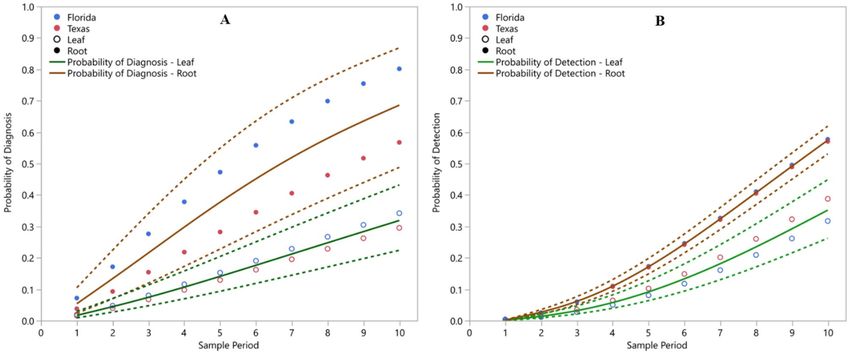
www.nature.com/scientificreports/
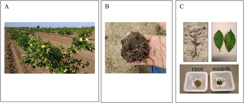
Figure 1. Images depicting Citrus leaf and root samples typical of those used in this study. (A) Grapefruit trees
in the field in Texas. (B) Fibrous roots as collected from the field. (C) Fibrous root cleaned of soil (upper left),
symptomatic leaf (upper right), and root and midrib samples chopped for DNA extraction (bottom).
Although inoculation occurs in young growing leaves, it is clear that CLas and CLam move through the plant
vascular system21,22,26–30. However, disease progression within the plant after inoculation is poorly understood.
In an effort to identify the plant organ most likely to provide reliable sources of CLas for real-time PCR-based
detection, Li et al.28 sampled from petioles, leaf mid-ribs, leaf blade, green stem bark, mature bark, roots, and
fruit from symptomatic plants. They found the highest bacterial titers within petioles and leaf mid-ribs. Similarly,
significantly higher CLas titers were found in peduncle, columella, and leaf midribs compared to seeds, young
shoots, flower buds, flowers, and bark. These results led to a focus on foliar tissue for diagnostic analysis that has
provided years of successfully implemented diagnostics.
Although detection of CLas from symptomatic leaves is s traightforward30, symptoms can be slow to develop
with this d isease23. Methods for early detection have been called for by growers and systematic reviews of the
disease management process31,32. While PCR-based detection of CLas in leaf midribs are highly sensitive, the
distribution of pathogen within the tree canopy is patchy27,33. Efforts to overcome the potential for false negative
diagnostic tests due to the uneven distribution of CLas in the tree canopy have focused on pooling midribs from
multiple leaves31, thereby improving the probability of including infected tissue in the diagnostic sample.
The earliest evidence that alternative sampling locations may be profitable, came from Graham et al.34 and
Johnson et al.29. Their data showed that the earliest symptom of HLB was found in the roots. Following that
observation, Louzada et al.33 demonstrated that CLas was consistently and evenly distributed throughout fibrous
roots of infected trees. This result suggested that the use of root samples for diagnostics could alleviate difficulties
caused by the patchy distribution of CLas within the canopy. Park et al.35 then demonstrated that CLas can be
detected in the roots of asymptomatic trees. Despite these advances, each of these studies27–29,33–36 were based on
limited sample sizes and could not evaluate the probabilities of detection using different samples or determine
which sample provided earliest detection. Here, we report on the first large-scale, longitudinal study of CLas
detection to address these questions.
Materials and methods
Field sites and sample collection. To evaluate the relative utility of sampling roots and leaves for the
detection of CLas, we established a longitudinal study to track the fate of individual trees in Florida and Texas.
We searched for young Citrus trees (4 to 5 years old) lacking visible symptoms of HLB but that had a high
likelihood of becoming infected during the course of the study. A ca. 4-hectare block of young sweet orange
(Hamlin) trees on Cleopatra mandarin rootstock was identified in an orchard in Immokalee, FL. This block of
sweet orange trees was surrounded by ca. 31.5 hectares of 10–15-year-old mature trees with a high incidence of
HLB. A ca. 2-hectare block of young grapefruit trees on sour orange rootstock in Donna, TX was also identified.
This block was bordered on the north and south by ca. 33 acres of 10–15-year-old mature trees, where routine
HLB survey showed a gradual increase of HLB incidence (data not shown).
The sweet orange and grapefruit trees in these two blocks were intensively surveyed for visible symptoms of
HLB (Fig. 1a,c). Those trees that lacked visible symptoms, 99 sweet orange trees in Florida and 112 grapefruit
trees in Texas, were selected for continued study. Leaf samples were collected and processed according to USDA
protocol37. Eight to ten leaves were collected from throughout the canopy of each tree with preference given to
symptomatic leaves when present. Root samples were collected from two locations for each tree, approximately
two feet from the trunk and perpendicular to the row direction on opposite sides of the tree, by digging one to
five inches deep into the soil with a small hand trowel (Fig. 1b). Approximately, one to two grams of small fibrous
roots (≤ 1 mm in diameter, Fig. 1c) were collected from larger feeder roots and stored in paper bags for transport
to the laboratory. Leaf and root samples were collected from each tree on a monthly basis from November 2015
through September 2016 in Florida (excluding December), and from January through November 2016 in Texas.
Scientific Reports | (2020) 10:16982 | https://doi.org/10.1038/s41598-020-74093-x 2
Vol:.(1234567890)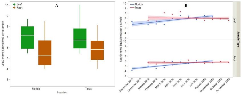
www.nature.com/scientificreports/
In total, 1980 samples (990 leaf and 990 root samples) in Florida and 2464 samples (1232 leaf and 1232 root
samples) were collected and examined in the study.
Nucleic acid extraction and real‑time PCR analysis. In the laboratory, leaf petioles and midribs
were separated from leaf blades and chopped, each with a new sterile razor blade, to 1–2 mm in length to aid
maceration prior to DNA extraction (Fig. 1c). Two hundred milligrams of pooled, chopped petiole and mid-rib
tissue was used for DNA extraction with the Qiagen BioSprint 96 workstation and BioSprint 96 DNA Plant kit
following the manufacturer protocol (Qiagen, Hilden, Germany). The leaf DNA fraction was eluted in 200 µl
of the kit elution buffer. Root samples were air dried for 24 h at 25 °C, thoroughly dusted to remove excess soil
(Fig. 1c) and chopped, each with a new sterile razor blade, to 1–2 mm in length to aid maceration prior to DNA
extraction (Fig. 1c). Chopped roots from a single tree were mixed, and a 150 mg subsample was used for DNA
extraction with the DNeasy PowerPlant Pro HTP96 kit (Qiagen, Hilden, Germany). The root DNA fraction was
eluted in 100 µl of the kit elution buffer. Potential PCR inhibitors were removed from the root DNA fraction
with the OneStep-96 PCR inhibitor removal kit (Zymo Research, Irvine, CA, USA), after which the root DNA
fraction was diluted 1:1 with 100 µl of nuclease-free water (Ambion, Austin, TX, USA) as described by Park
et al.35. DNA concentration was measured on NanoDrop 2000 (Thermo Scientific, Waltham, MA, USA).
Extracted DNA from leaf and root samples were tested for the presence of CLas using real-time PCR protocols
specific to the organ from which the DNA originated as described below. Leaf DNA samples were tested using
real-time PCR with the HLBaspr primer/probe s ystem30 to detect a region of the CLas 16S rDNA. This marker
system provides highly sensitive detection from leaf-based DNA extracts but binds to and amplifies non-
specific targets found within root-based DNA extracts38. Thus, to test root samples for the presence of CLas, we
employed the TXCChlb primer/probe system35, which targets the same gene using primers that avoid off-target
amplification. Nuclease-free water and a plasmid DNA that contains CLas 16s rDNA target were used as a
negative and positive control for real-time PCR reaction, respectively.
Detection of CLas was defined as acquisition of a real-time PCR quantification cycle (Cq) value less than
37 following USDA protocol37. Reactions that produced a Cq of 37 or greater were scored as negative. Upon
identification of at least one sample from either organ (root or leaf) producing a detection of CLas, trees were
classified as “diagnosed" with HLB. Cq values were also used to estimate bacterial titer (i.e., L og10(Genome
Equivalents) per gram of sample) using the amplification efficiency equations published by Li et al.39 for leaf
and Park et al.35 for root. The estimated detection limit of both real-time PCR methods with a Cq cutoff < 37 is
approx. 102 copies of a plasmid DNA containing a partial CLas 16s rDNA (data not shown). After developing
the real-time PCR protocol for CLas detection in fibrous roots using TXCChlb primer–probe set35, we have
extensively evaluated the consistency of CLas detection between TXCChlb- and HLBaspr-based assay s ystems39
against leaf DNA fractions prepared from Citrus trees growing in Texas.
Statistical analyses. The influence of sample type, location, and month on bacterial titer (as measured
by number of genome equivalents) from trees diagnosed with HLB was determined using factorial Analysis of
Variance (ANOVA). Since not all samples from a tree diagnosed with HLB produce positive detections, we tested
for the influence of negative real-time PCR reactions with a second factorial ANOVA using only those real-time
PCRs that produced positive CLas detections.
Contingency analysis was used to evaluate differences in the frequency of tree diagnosis by each sample
type (i.e., the sample in which CLas was first detected). Similarly, we compared all samples (i.e., regardless of
detection order) in which CLas was detected using contingency analysis. To evaluate the consistency of detection
(i.e., detections in repeated samples through time) for each sample type, we compared the average number of
detections per tree between leaf and root samples using t-tests. Since earlier detections in one sample type over
another could bias the number of detections, we compared the proportion of detections in each sample type
following initial detection within that organ using contingency analysis. To evaluate whether use of leaf or root
samples resulted in earlier detection, we used contingency analysis to compare the number of trees diagnosed
(i.e., first detected) by each sample type.
Parametric survival analysis was used to estimate the probabilities of diagnosis and of detection from each
sample type and for each location. We scored diagnosis of trees and detection of CLas within samples as events
and measured the time until each event occurred. Trees that lacked detection within either sample type by the
end of the study were censored. Sample collection dates in Florida and Texas did not align so for this analysis
we used sample period, rather than the month of sample collection, to allow joint analysis of Florida and Texas
trees (however, independent analyses produced similar results). Goodness of fit testing along with corrected
Akaike Information Criterion (AICc) and Bayesian Information Criterion (BIC) were used to choose the best
fit distribution to model the time until detection for each tree. We used parametric survival analysis to compare
the time until detection for each tree between leaf- and root-based samples and to evaluate how the probability
of detection changed through time. We used goodness-of-fit tests to evaluate the influence of sample type,
location, and their interaction on time until detection. Finally, we used t-tests to evaluate whether there was a
significant time savings from testing one sample type over the other. All data was analyzed in JMP ver. 13.1.0
(SAS Institute, Cary, NC, 1989–2019).
Results
Pattern of CLas detection. Patterns of CLas detection were quite similar among the sweet orange trees
from Florida and the grapefruit trees from Texas. Using real-time PCR on DNA extracts from each sample, CLas
was detected in 385 (19%) of the Florida samples and 410 (17%) of the Texas samples. This corresponded to
diagnosis of 89 (90%) of the Florida trees and 82 (73%) of the Texas trees as infected with CLas by the end of the
Scientific Reports | (2020) 10:16982 | https://doi.org/10.1038/s41598-020-74093-x 3
Vol.:(0123456789)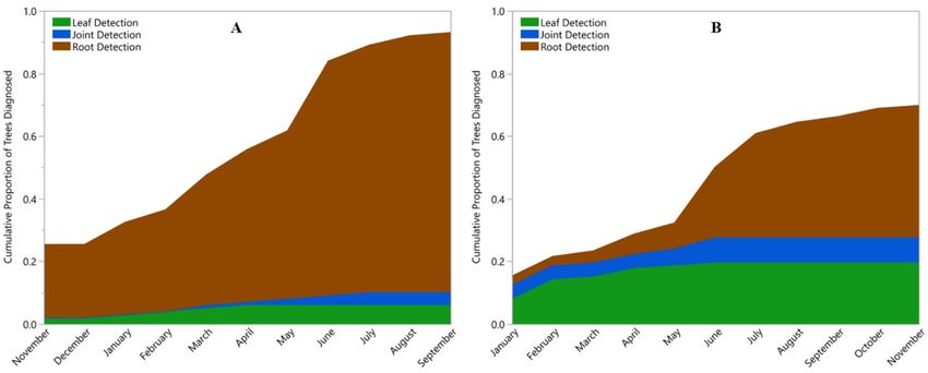
www.nature.com/scientificreports/
Figure 2. Cumulative proportion of Citrus trees diagnosed with Huanglongbing by real-time PCR detection
of Candidatus Liberibacter asiaticus (CLas) in Florida (A) and Texas (B). The sample type by which each tree
was diagnosed with HLB are indicated by color: Brown represents trees in which CLas was first detected in a
root sample; Green represents trees in which CLas was first detected in a leaf sample; and Blue represents trees
with simultaneous detection in roots and leaves.. Note, no samples were collected in December 2015 in Florida.
study (Fig. 2a,b). Although we did not continue recording data on symptomology, all trees in the first sampling
period lacked visible symptoms. Despite this, we detected CLas in 25 Florida trees (23 among root samples and
2 among leaf samples) and 17 Texas trees (3 among root samples, 9 among leaf samples, and 5 in both sample
types) in the first sample period.
CLas titer. Beyond detection, real-time PCR can be used to estimate bacterial titer (i.e., the estimated
number of target molecules divided by the number of copies of the target in the CLas genome) in one gram
of sample. The estimate of bacterial titer, L og10 (Genome Equivalents) per g, varied significantly between these
groups (F(41,753) = 8.38, p < 0.0001). Sample type and the interaction between sample location and sample period
significantly impacted differences in titer while location alone had little impact. The average titer found among
leaves was greater than that found among roots ( X ± SE = 7.01 ± 0.09 and 5.67 ± 0.05 copies, respectively; Fig. 3a).
Sample location, on its own, had little influence on bacterial titer with mean (± SE) genome equivalents in Florida
and Texas of 5.86 ± 0.07 and 6.07 ± 0.06, respectively (Fig. 3a). However, the interaction between sample location
and sample period revealed a significant and interesting pattern (F(8,753) = 3.16, p < 0.0016; Fig. 3b). Bacterial titer
increased through time in Florida (Leaf: F(1,77) = 20.11, p < 0.0001; Root: F(1,304) = 40.74, p < 0.0001) but remained
stable in Texas (Leaf: F(1,98) = 0.09, p = 0.76; Root: F(1,308) = 0.42, p = 0.52).
Frequency and consistency of detection. Sample type impacted the frequency with which the bacteria
was detected. Contingency analysis among trees showed that leaf samples identified infected trees less often than
expected whereas root samples diagnosed infected trees more often than expected by chance. In Florida, CLas
was detected among leaf samples of 37 trees (42% of diagnosed trees) and from root samples of 88 trees (99% of
diagnosed trees; χ2(1) = 60.74, p < 0.0001). Similarly, in Texas CLas was detected among leaf samples of 37 trees
(45% of diagnosed trees) and from root samples of 74 trees (90% of diagnosed trees: χ2(1) = 24.91, p < 0.0001). In
all, more than twice as many trees produced positive root samples than leaf samples.
Similar patterns were observed when individual samples, rather than trees, were compared. Contingency
analysis of samples showed that leaf samples contained detectable CLas less often than expected whereas roots
contained detectable CLas more often than expected. Of the 385 samples from which CLas was detected in
Florida, 306 (79%) were root samples (χ2(1) = 221.34, p < 0.0001). The same pattern held in Texas where 310
(76%) of the 410 CLas detections were found among roots (χ2(1) = 172.43, p < 0.0001). Across all samples, there
were more than three times as many positive root samples as leaf samples.
The increased frequency of detection of CLas among root samples corresponded to an increase in the
consistency of detection within individual trees (Fig. 4). Not only was CLas detected among roots more often
than among leaves, but it was detected more consistently among roots as well. Each tree was sampled repeatedly
resulting in ten and eleven sample periods per tree for Florida and Texas trees, respectively. The number of
detections per tree, from trees found to contain CLas, was significantly greater for roots than leaves with an
average of 3.4 detections per tree for roots and 0.89 for leaves from Florida trees ( t(176) = 9.72, p < 0.0001) and
3.8 for roots and 1.2 for leaves from Texas trees ( t(162) = 5.79, p < 0.0001; Fig. 4). Moreover, contingency analysis
showed that the proportion of samples from which CLas was detected after the initial detection was significantly
greater for root than leaf samples in both Florida (χ2(1) = 225.9, p < 0.0001) and Texas (χ2(1) = 172.43, p < 0.0001).
Regardless of the sample type from which CLas was first detected, it was detected again in 15% of the subsequent
Scientific Reports | (2020) 10:16982 | https://doi.org/10.1038/s41598-020-74093-x 4
Vol:.(1234567890)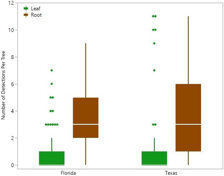
www.nature.com/scientificreports/
Figure 3. Bacterial titer per milligram sample, as estimated by the log of the number of Candidatus Liberibacter
asiaticus genome equivalents, in Citrus. Panel A depicts the central tendency and variation in titer in the form of
box plots for leaf (green) and root (brown) samples from Florida (left) and Texas (right). The data are presented
as log10 values to better approximate a normal distribution. The horizontal line within the box represents the
median number of genomes, the box spans the interquartile range (25% to 75% quantile), and the whiskers
reach from these points to 1.5 times the interquartile range. Panel B depicts the average titer for each month in
Florida (blue) and Texas (red). The lines represent the best fit linear regression and the shaded areas represent
the 95% confidence intervals of the lines. Note, no samples were collected in December 2015 in Florida, and the
September 2016 data points for Florida and Texas overlap.
Figure 4. The consistency of detecting Candidatus Liberibacter asiaticus (CLas) within infected Citrus trees
is shown by the number of detections per tree from monthly samples of trees infected with CLas. Green and
brown box plots indicate leaf and root samples, respectively. The horizontal line within the box represents the
median number of detections, the box represents the interquartile range (25% to 75% quantile), and whiskers
reach from these points to 1.5 times the interquartile range and define statistical outliers, which are represented
by dots.
leaf samples compared to 60% of the subsequent root samples in Florida. Similarly, after CLas was first detected
in a Texas tree, 17% of the subsequent leaf samples detected it compared to 53% of the subsequent root samples.
Timing of detection. Detection of CLas was achieved earlier with root samples than with leaf samples. In
79 Florida trees (89% of trees diagnosed as infected), CLas was first detected in root samples while in only six
Scientific Reports | (2020) 10:16982 | https://doi.org/10.1038/s41598-020-74093-x 5
Vol.:(0123456789)www.nature.com/scientificreports/
Figure 5. The probability functions for diagnosing trees with HLB (i.e., the first detection regardless of tissue)
(A) and detecting Candidatus Liberibacter asiaticus among samples (i.e., including repeated detections) (B)
using DNA extractions from Citrus leaves (solid green line) and roots (solid brown line). Dashed lines represent
the upper and lower 95% confidence intervals of the estimate of the corresponding color. Geographic variation
exists in these functions as depicted by probability estimates from Florida (blue circles) and Texas (red circles).
While the estimated probability of detection from leaves (open circles) varies between Florida and Texas to a
greater degree than do roots (closed circles), these differences are not statistically different from one another.
Sample period represents the order in which the monthly samples were collected.
trees (7% of trees diagnosed as infected) were leaf samples the first to produce detectable CLas. Four Florida trees
had detections of CLas in root and leaf samples simultaneously (Fig. 2a). In Texas, the first detection of CLas
was from root samples in 51 trees (62% of diagnosed trees) and from leaf samples in 22 trees (27% of diagnosed
trees). In nine Texas trees, CLas was detected simultaneously in roots and leaves (Fig. 2b). Contingency analysis
found these differences to be statistically significant (χ2(1) = 14.89, p < 0.0001).
We compared the time until diagnosis (i.e., first detection of CLas regardless of sample type) for each tree
using parametric survival analysis of leaf and root samples. The distribution of times to diagnosis were best fit by
the Weibull distribution (AICc = 1695.80, BIC = 1715.89) and the fit was statistically significant (χ2(3) = 84.03, p
< 0.0001). Sample type (leaf or root) and location had significant impacts on time until diagnosis (χ2(1) = 59.29,
p < 0.0001 and χ2(1) = 9.16, p = 0.0025, respectively). The interaction between sample type and location was not
significant (χ2(1) = 3.02, p = 0.08). Using these relationships, we estimated the probability of diagnosing trees with
HLB from leaf samples and from root samples. The probability of diagnosing trees with HLB changed through
time, as shown in Fig. 5a, and was significantly higher in roots than in leaves at all time points (p < 0.05 for each
time point).
To evaluate the probability of detection from any given sample, we compared the time until detection for
each tree using parametric survival analysis of leaf and root samples. The distribution of times to detection
were best fit by the Weibull distribution (AICc = 5292.52, BIC = 5317.99) and the fit was statistically significant
(χ2(3) = 72.42, p < 0.0001). Using this model, a significant impact of sample type (leaf or root) on time until
detection (χ2(1) = 70.52, p < 0.0001) was found. However, neither location (Florida or Texas), nor the interaction
between sample type and location had a significant impact on time until detection (χ 2(1) = 1.67, p = 0.17;
χ2(1) = 2.50, p = 0.11, respectively). Using these relationships, we estimated the probability of detecting CLas
from leaf samples and from root samples. The probability of detecting CLas changed through time, as shown
in Fig. 5b, but was significantly higher in roots than in leaves at all time points, from the beginning of the study
through the end (p < 0.05 for each time point).
The improvement in the probability of diagnosing trees and detecting CLas among samples gained through
the use of root samples was greater in Florida than in Texas. Diagnosis of trees (i.e., first detections) improved
by more 275% in Florida and 136% in Texas through the use of root samples. While still substantial, this
improvement declined through time to 134% and 92%, respectively, by the end of the study (Fig. 6a). Similarly,
the probability of detecting CLas among individual samples was improved through the use of roots by more than
125% in Florida and by nearly 75% in Texas. The improvement declined to 82% and 47% for Florida and Texas,
respectively, over the duration of the study (Fig. 6b). Survival models estimated that 50% of the trees would be
diagnosed 9 months earlier in Florida and 8 months earlier in Texas through the use of root samples (Fig. 7).
Scientific Reports | (2020) 10:16982 | https://doi.org/10.1038/s41598-020-74093-x 6
Vol:.(1234567890)www.nature.com/scientificreports/
Figure 6. Percent improvement in the probability of diagnosing trees with HLB (i.e., the first detection
regardless of tissue) (A) and detecting Candidatus Liberibacter asiaticus among samples (i.e., including repeated
detections) (B) using DNA extractions from Citrus root samples over those from Citrus leaf samples. The
percent improvement observed in Florida is shown in blue while that in Texas is shown in red. Sample period
represents the order in which the monthly samples were collected.
Figure 7. Earlier detection of Candidatus Liberibacter asiaticus in Citrus root samples is shown by the time
to diagnosis of 50% of trees estimated by the survival model. The estimated median time (± 95% confidence
intervals) to diagnosis trees is shown for Florida (left) and Texas (right) using leaf (green) and root (brown)
samples.
Discussion
HLB poses a significant threat to citriculture around the world due to the lack of commercial Citrus cultivars
tolerant or resistant to HLB. Currently available measures to limit the spread of HLB include control of the insect
vector population, removal and destruction of infected trees, and the exclusive use of pathogen-free b udwoods40.
The utility of tree removal and destruction relies on rapid and accurate diagnostics.
Early detection of CLas has long been a goal of the Citrus community31,32. A number of researchers have
focused efforts on improving the rate of detection using a wide variety of methods including: differences in
spectra41 or polarization of light reflected from leaves using portable d evices42,43 or satellite imagery44; the
presence of CLas secreted proteins45; and the presence of salivary sheaths from Asian citrus psyllid (ACP)46.
Scientific Reports | (2020) 10:16982 | https://doi.org/10.1038/s41598-020-74093-x 7
Vol.:(0123456789)www.nature.com/scientificreports/
However, commercial growers have been hesitant to adopt methods not approved by regulators, and regulators
require direct evidence of the pathogen’s presence (i.e., its genome).
The “gold standard” for detecting CLas is real-time PCR-based methods targeting various regions of CLas
genome30,35,47,48. However, pre-symptomatic detection from leaf samples has proven difficult due to the uneven
(patchy) distribution of CLas within the tree canopy27,33. This study was conducted to evaluate if diagnostic tests
using Citrus root samples could improve upon early detection.
After collecting, isolating DNA, and using real-time PCR to test for the presence of CLas from 4,444 samples
in Florida and Texas across 11 months, we found that most of the samples (82%) failed to detect CLas. However,
the frequency of detection increased over time presumably as disease spread among these trees. By the end of
the study, 90% of trees in Florida and 73% of trees in Texas had been diagnosed with HLB. Interestingly, we
detected CLas in 42 trees that lacked visible symptoms at the beginning of the study. Most of these detections
were among root samples (n = 31) as opposed to leaf samples (n = 16). However, 11 trees produced detections
in leaves but not roots at this time point. One hypothesis is that these samples were taken prior to pathogen
migration to the roots. Moreover, the relative dearth of such trees in Florida (2 trees), where the incidence of HLB
is higher, compared to Texas (9 trees) would support such a hypothesis. However, such limited sample sizes make
it difficult to differentiate from random chance. Regardless, these results indicate that both leaf- and root-based
diagnostics can detect CLas prior to the initiation of symptoms but suggest that roots may be the more efficient
sample for pre-symptomatic detection.
As reported previously28, CLas titers were higher in leaves than roots. This pattern appears to be widespread
as we observed little difference between Florida and Texas samples. However, an interesting pattern emerged
through time and across space. CLas titer increased through time in Florida, but not in Texas (Fig. 3b). Several
hypotheses could explain this pattern. First, sampling began earliest in Florida and may have captured the
early invasion of CLas into these trees. Captured early enough, one would expect to see titer increase within
individual plants while incidence increases among plants. This hypothesis would suggest that titer should level
off in Florida as most trees become infected. This hypothesis seems unlikely as more trees were diagnosed on the
first sampling period and a greater proportion of trees became infected by the end of the study in Florida than
in Texas (Fig. 2a,b). Alternatively, psyllid-driven re-inoculation could explain the increase in titer through time
in Florida while lower psyllid population sizes in Texas explain the lack of this increase. Finally, CLas titer may
respond to environmental variables. There is substantial evidence from both g reenhouse21 and field s tudies49,50 to
support this conclusion. This hypothesis would suggest that the variation about the regression line the data from
Texas is not mere error, but rather a non-linear relationship. There is insufficient data at this time to distinguish
between these hypotheses.
Among the trees diagnosed with HLB, there was a dramatic difference in the frequency with which we
detected CLas in each sample type. More than twice as many trees diagnosed with CLas produced positive root
detections than produced positive leaf detections and more than three times as many root samples produced
positive detections of CLas than leaf samples. The results were remarkably similar between sweet orange trees
in Florida and grapefruit trees in Texas suggesting the result is general among Citrus varieties and across
environments.
The increased frequency of detections using root samples was mirrored within individual trees. The average
number of detections per tree was over three times higher in root samples than in leaf samples. This improvement
in consistency of detection may be due to the even distribution of CLas within roots and the patchy distribution
of CLas within the c anopy27,33. Louzada et al.33 found 96% of horizontally growing roots just under the soil surface
contained detectable quantities of CLas. It is possible that the consistency of detection in the canopy improves
through time as more leaves become infected (see below).
By asking which sample (leaf or root) provided earliest evidence of CLas infections for each tree, the current
study found that CLas was first detected in root samples for more than 75% of the infected trees. Moreover,
survival analysis documented significantly higher probabilities of diagnosis (Fig. 5a) and detection (Fig. 5b) at
each sample period. These results support the hypothesis posed by Louzada et al.33 and Park et al.35.
Although geographical location did not have a statistically significant impact on the probability of detecting
CLas nor a significant interaction effect with sample type, interesting patterns emerged when comparing the
impact of location between leaf and root samples. The probability of diagnosing trees with HLB among root
samples, but not leaf samples, varied between Florida and Texas. Florida had higher probabilities of diagnosing
with roots than Texas (Fig. 5a). Conversely, the probability of detecting CLas among leaf samples, but not
root samples, varied between Florida and Texas. In this case, Texas leaf samples had a higher probability of
detecting CLas than those from Florida (Fig. 5b). Additionally, the probability of diagnosing trees with HLB
and detecting CLas within samples changed over time. For both sample types, these probabilities increased with
time. However, the degree of improvement gained by using root samples declined and differed between Florida
and Texas (Fig. 6a,b). The difference in rates of improvement were likely an artifact due to the fewer number of
trees remaining to be detected by root sampling. However, it is possible that canopy inoculation rate increased
as incidence rate increased among surrounding trees, that establishment rate increased as tree health declined,
or that transport of CLas from roots to leaves increased as infection spread throughout the root system.
The TXCChlb- and HLBaspr-based assay systems produce highly consistent results using leaf samples (data
not shown). A small number of discrepancies, however, do occur among samples with Cq values near the Cq
cutoff (e.g. 36.5 < Cq < 37) due to the slightly lower amplification efficiency of TXCChlb (data not shown). Despite
this possibility of underestimating detections using TXCChlb, our marker for roots detections, the improved
probability of diagnosing trees with HLB using root samples was apparent. This improvement was likely due to
the higher consistency of detection and more even distribution of CLas within r oots33. We suspect that further
improvement would be seen through the use of root samples and assays built on CLas genic regions with higher
amplification efficiencies such as the nrdB gene, which has five copies in the CLas g enome51. Adaptation of such
Scientific Reports | (2020) 10:16982 | https://doi.org/10.1038/s41598-020-74093-x 8
Vol:.(1234567890)www.nature.com/scientificreports/
markers to root samples would be beneficial as survivorship models suggest that root-based diagnostics may alert
growers to the presence of CLas within their orchards months earlier than leaf-based diagnostics.
Current management strategies generally rely on visual inspection of the tree canopy for distinct HLB
symptoms followed by collection of symptomatic leaves for HLB diagnostic test by real-time PCR in the lab. This
approach effectively offers confirmation of visual hypotheses. As previously reported by Coletta-Filho et al.26 and
Lee et al.17, HLB spread from pre-symptomatic trees is likely taking place in the field. Pre-symptomatic diagnosis
offers a route to limiting spread by triggering earlier deployment of HLB control measures. The current study
clearly indicates the benefit of using fibrous root samples for HLB diagnosis. In regions where HLB and/or its
insect vector have not yet been reported, or in the areas where HLB incidence is localized, surveying the trees
at the edge of the field52,53 using root-based diagnostics, may provide growers early insight into disease presence
even in the absence of visible symptoms.
Received: 14 February 2020; Accepted: 23 September 2020
References
1. CABI. Citrus Huanglongbing (Greening) Disease (Citrus Greening). In Invasive Species Compendium. (CABI International,
Wallingford, 2020).
2. Aubert, B. Citrus greening disease, a serious limiting factor for citriculture in Asia and Africa. In Proceedings of the 4th Congress
of the International Society of Citrus Nurserymen, South Africa, pp. 134–142 (1993).
3. Bové, J. M. Huanglongbing: a destructive, newly-emerging, century-old disease of citrus. J. Plant Pathol. 88, 7–37 (2006).
4. Bassanezi, R. B., Montesino, L. H. & Stuchi, E. S. Effects of Huanglongbing on fruit quality of sweet orange cultivars in Brazil. Eur.
J. Plant Pathol. 125, 565–572 (2009).
5. Dagulo, L. et al. Chemical characterization of orange juice from trees infected with citrus greening (Huanglongbing). J. Food Sci.
75, C199–C207 (2010).
6. Halbert, S. E. The discovery of Huanglongbing in Florida. In Proceeding of Second International Citrus Canker and Huanglongbing
Research Workshop, Nov 2005, Florida Citrus Mutual, Orlando, FL, p. H-3 (2005).
7. Coletta-Filho, H. D. et al. First report of the causal agent of Huanglongbing (“Candidatus Liberibacter asiaticus”) in Brazil. Plant
Dis. 88, 1382 (2004).
8. Court, C. D., Hodges, A. W., Rahmani, M. & Spreen, T. H. Economic Contributions of the Florida Citrus Industry in 2015–2016
(University of Florida, Food and Resource Economics Department, Gainesville, 2017).
9. Kunta, M. et al. First report of citrus Huanglongbing in Texas. Phytopathology 102, 466 (2012).
10. Kumagai, L. B. et al. First report of Candidatus Liberibacter asiaticus associated with Citrus Huanglonbing in California. Plant
Dis. 97, 283 (2013).
11. Jagoueix, S., Bove, J. M. & Garnier, M. The phloem-limited bacterium of greening disease of citrus is a member of the subdivision
of the Proteobacteria. Int. J. Syst. Bacteriol. 44, 379–386 (1994).
12. Teixeira, C. D. et al. ‘Candidatus Liberibacter americanus’, associated with citrus huanglongbing (greening disease) in São Paulo
State, Brazil. Int. J. Syst. Evol. Microbiol. 55, 1857–1862 (2005).
13. Capoor, S. P., Rao, D. G. & Viswanath, S. M. Diaphorina citri Kuway., a vector of the greening disease of citrus in India. Ind. J. Agric.
Sci. 37, 572–576 (1967).
14. Hall, D. G., Richardson, M. L., Ammar, E. D. & Halbert, S. E. Asian citrus psyllid, Diaphorina citri, vector of citrus huanglongbing
disease. Entomol. Exp. Appl. 60, 207–223 (2012).
15. McClean, A. P. D. & Oberholzer, P. C. J. Citrus psylla, a vector of greening disease of sweet orange. S. Afr. J. Agric. Sci. 8, 297–298
(1965).
16. Chiyaka, C., Singer, B. H. B., Halbert, S. E., Morris, J. G. & van Bruggen, A. H. C. Modeling huanglongbing transmission within a
citrus tree. Proc. Natl. Acad. Sci. 109, 12213–12218 (2012).
17. Lee, J. A. et al. Asymptomatic spread of huanglongbing and implications for disease control. Proc. Natl. Acad. Sci. 112, 7605–7610
(2015).
18. Sétamou, M., Alabi, O. J., Kunta, M., Jifon, J. L. & da Graҫa, J. Enhanced acquisition rates of ‘Candidatus Liberibacter asiaticus’ by
the Asian citrus psyllid (Hemiptera:Liviidae) in the presence of vegetative flush growth in citrus. J. Econ. Entomol. 109, 1973–1978
(2016).
19. Cifuentes-Arenas, J. C., de Goes, A., de Miranda, M. P., Beattie, G. A. C. & Lopes, S. A. Citrus flush shoot ontogeny modulates
biotic potential of Diaphorina citri. PLoS ONE 13(1), e019056 (2018).
20. McCollum, G., Kunta, M. & Braswell, W. E. Improving early detection of HLB-affected trees. Citrograph 9, 50–54 (2018).
21. Lopes, S. A. et al. Graft transmission efficiencies and multiplication of ‘Candidatus Liberibacter americanus’ and ‘Ca. Liberibacter
asiaticus’ in citrus plants. Phytopathology 99, 301–306 (2009).
22. Lopes, S. A. et al. Liberibacter associated with Citrus Huanglongbing in Brazil: ‘Candidatus Liberibacter asiaticus’ is heat tolerant,
‘Ca. L. americanus’ is heat sensitive. Plant Dis. 93, 257–262 (2009).
23. Gottwald, T. R., da Graҫa, J. V. & Bassanezi, R. B. Citrus Huanglongbing: the pathogen and its impact. Plant Health Prog. 8, 31–67
(2007).
24. Manjunath, K. L., Halbert, S. E., Ramadugu, C., Webb, S. & Lee, R. F. Detection of ‘Candidatus Liberibacter asiaticus’ in Diaphorina
citri and its importance in the management of Citrus Huanglongbing in Florida. Bacteriology 9, 387–396 (2008).
25. Pelz-Stelinski, K. S., Brlansky, H. R., Ebert, T. A. & Rogers, M. E. Transmission parameters for Candidatus Liberibacter asiaticus
by Asian Citrus psyllid. J. Econ. Entomol. 103, 1531–1541 (2010).
26. Coletta-Filho, H. D., Daugherty, M. P., Ferreira, C. & Lopes, J. R. S. Temporal progression of ‘Candidatus Liberibacter asiaticus’
infection in Citrus and acquisition efficiency by Diaphorina citri. Phytopathology 104, 416–421 (2014).
27. Tatineni, S. et al. In planta distribution of ‘Candidatus Liberibacter asiaticus’ as revealed by polymerase chain reaction (PCR) and
real-time PCR. Phytopathology 98, 592–599 (2008).
28. Li, W., Levy, L. & Hartung, J. S. Quantitative distribution of ‘Candidatus Liberibacter asiaticus’ in citrus plants with citrus
huanglongbing. Phytopathology 99, 139–144 (2009).
29. Johnson, E. G., Wu, J., Bright, D. B. & Graham, J. H. Association of ‘Candidatus Liberibacter asiaticus’ root infection, but not
phloem plugging with root loss on huanglongbing-affected trees prior to appearance of foliar symptoms. Plant Pathol. 63, 290–298
(2014).
30. Li, W., Hartung, J. S. & Levy, L. Quantitative real-time PCR for detection and identification of Candidatus Liberibacter species
associated with citrus huanglongbing. J. Microbiol. Methods 66, 104–115 (2006).
31. National Research Council. Strategic Planning for the Florida Citrus Industry: Addressing Citrus Greening Disease (The National
Academies Press, Washington, DC, 2010).
Scientific Reports | (2020) 10:16982 | https://doi.org/10.1038/s41598-020-74093-x 9
Vol.:(0123456789)www.nature.com/scientificreports/
32. National Academies of Sciences, Engineering, and Medicine. A Review of the Citrus Greening Research and Development Efforts
Supported by the Citrus Research and Development Foundation: Fighting a Ravaging Disease (The National Academies Press,
Washington, DC, 2018).
33. Louzada, E. S. et al. Distribution of ‘Candidatus Liberibacter asiaticus’ above and below ground in Texas citrus. Phytopathology
106, 702–709 (2016).
34. Graham, J. H., Johnson, E. F., Gottwald, T. R. & Irey, M. S. Pre-symptomatic fibrous root decline in citrus trees caused by
huanglongbing and potential interaction with Phytophthora spp. Plant Dis. 97, 1195–1199 (2013).
35. Park, J. W. et al. A new real-time PCR method for huanglongbing detection in citrus root tissue. J. Gen. Plant Pathol. 84, 359–367
(2018).
36. Kunta, M., da Graça, J. V., Malik, N., Louzada, E. S. & Sétamou, M. Quantitative distribution of Candidatus Liberibacter asiaticus
in the aerial parts of the HLB-infected citrus trees in Texas. HortScience 49, 65–68 (2014).
37. USDA. Plant sample extraction for use in Citrus Greening (Huanglongbing, HLB) molecular diagnostic assays. Revision 2.
Document control number: WI-B-T-1–17 (2015).
38. Shin, K. & van Bruggen, A. H. C. Bradyrhizobium isolated from huanglong (HLB) affected citrus trees reacts positively with primers
for Candidatus Liberibacter asiaticus. Eur. J. Plant Pathol. 151, 291–306 (2018).
39. Li, W., Li, D., Twieg, E., Hartung, J. S. & Levy, L. Optimized quantification of unculturable Candidatus Liberibacter spp. in host
plants using real-time PCR. Plant Dis. 92, 854–861 (2008).
40. Li, J. et al. Developing citrus Huanglonbing (HLB) management strategies based on severity of symptoms in HLB-endemic Citrus-
producing regions. Dis. Control. Pest Manag. 109, 582–592 (2019).
41. do Brasil Cardinali, M. C. et al. Infrared spectroscopy: a potential tool in huanglongbing and citrus variegated chlorosis diagnosis.
Talanta 91, 1–6 (2012).
42. Pourreza, A., Lee, W. S., Ehsani, R., Schueller, J. K. & Raveh, E. An optimum method for real-time in-field detection of
Huanglongbing disease using a vision sensor. Comput. Electron. Agric. 110, 221–232 (2015).
43. Pérez, M. R. V. et al. Raman spectroscopy an option for the early detection of Citrus Huanglongbing. Appl. Spectrosc. 70, 829–839
(2016).
44. Li, X. et al. Feasibility study on Huanglongbing (Citrus greening) detection based on WorldView-2 satellite imagery. Biosyst. Eng.
132, 28–38 (2015).
45. Pagliaccia, D. et al. A pathogen secreted protein as a detection marker for Citrus Huanglongbing. Front. Microbiol. 8, 2041 (2017).
46. Pandey, S. S. & Wang, N. Targeted early detection of citrus huanglongbing causal agent “Candidatus Liberibacter asiaticus” before
symptom expression. Phytopathology 109, 952–959 (2019).
47. Teixeira, D. C. et al. Distribution and quantification of Candidatus Liberibacter americanus, agent of huanglongbing disease of
citrus in São Paulo State, Brasil, in leaves of an affected sweet orange tree as determined by PCR. Mol. Cell Probes 22, 139–150
(2008).
48. Wang, Z. et al. Development and application of molecular-based diagnosis for ‘Candidatus Liberibacter asiaticus’, the causal
pathogen of citrus huanglongbing. Plant Pathol. 55, 630–638 (2006).
49. Sétamou, M., Alabi, O. J., Kunta, M., Dale, J. & da Graça, J. V. Distribution of Candidatus Liberibacter asiaticus in citrus and the
Asian citrus psyllid in Texas over a decade. Plant Dis. 104, 1118–1132 (2020).
50. Lopes, S. A., Luiz, F. Q. B. F. & Oliveira, H. T. Seasonal variation of ‘Candidatus Liberibacter asiaticus’ titers in new shoots of citrus
in distinct environments. Plant Dis. 101, 583–590 (2017).
51. Zheng, Z., Xu, M., Bao, M., Wu, F. & Chen, J. Deng X (2016) Unusual five copies and dual forms of nrdB in “Candidatus Liberibacter
asiaticus”: biological implications and PCR detection application. Sci Rep. 6, 39020 (2016).
52. Gottwald, T., Irey, M. & Gast, T. The Plantation Edge Effect of HLB: A Geostatistical Analysis. Proc. IRCHLB, p. 305 (2008).
53. Sétamou, M. & Bartels, D. W. Living on the edges: spatial niche occupation of Asian citrus psyllid, Diaphorina citri Kuwayama
(Hemiptera: Liviidae), in citrus groves. PLoS ONE 10(7), e0131917 (2015).
Acknowledgements
The authors would like to thank Norman Barr, David Bartels, Raul Ruiz, Terrance Todd, and anonymous reviewers
for comments on a previous draft of the manuscript. This research was supported by a cooperative agreement
15-8130-0489-CA between USDA-Animal and Plant Health Inspection Service—Science and Technology and
Texas Agricultural and Mechanical University at Kingsville. Mention of trade names or commercial products in
this publication is solely for the purpose of providing specific information and does not imply recommendation
or endorsement by the U.S. Department of Agriculture, an equal opportunity employer.
Author contributions
J-.W.P and W.E.B. contributed equally to this project. M.K. and W.E.B. conceived and designed the experiments.
M.K., E.S.L., J.V.G., and P.A.S. identified the field sites. B.C.K., P.A.S., J-.W.P., and M.K. collected the samples.
J-.W.P. conducted the experiments. W.E.B. analyzed the data and wrote the manuscript with input from all other
authors.
Competing interests
The authors declare no competing interests.
Additional information
Correspondence and requests for materials should be addressed to M.K.
Reprints and permissions information is available at www.nature.com/reprints.
Publisher’s note Springer Nature remains neutral with regard to jurisdictional claims in published maps and
institutional affiliations.
Scientific Reports | (2020) 10:16982 | https://doi.org/10.1038/s41598-020-74093-x 10
Vol:.(1234567890)www.nature.com/scientificreports/
Open Access This article is licensed under a Creative Commons Attribution 4.0 International
License, which permits use, sharing, adaptation, distribution and reproduction in any medium or
format, as long as you give appropriate credit to the original author(s) and the source, provide a link to the
Creative Commons licence, and indicate if changes were made. The images or other third party material in this
article are included in the article’s Creative Commons licence, unless indicated otherwise in a credit line to the
material. If material is not included in the article’s Creative Commons licence and your intended use is not
permitted by statutory regulation or exceeds the permitted use, you will need to obtain permission directly from
the copyright holder. To view a copy of this licence, visit http://creativecommons.org/licenses/by/4.0/.
© The Author(s) 2020
Scientific Reports | (2020) 10:16982 | https://doi.org/10.1038/s41598-020-74093-x 11
Vol.:(0123456789)You can also read