Analysis and Detection of Nephrolithiasis using Imaging Techniques - NAUN
←
→
Page content transcription
If your browser does not render page correctly, please read the page content below
INTERNATIONAL JOURNAL OF BIOLOGY AND BIOMEDICAL ENGINEERING
      DOI: 10.46300/91011.2021.15.6                                                                      Volume 15, 2021

     Analysis and Detection of Nephrolithiasis using
                  Imaging Techniques

                             Smiti Tripathy1, *Sivakumar R1, Simran Nair1, Inbamalar TM2
          1
           School of Electronics Engineering, Vellore Institute of Technology, Vellore, Tamilnadu, India
                        2
                          RMK College of Engineering and Technology, Tamilnadu, India
                                                  *rsivakumar@vit.ac.in

                   Received: January 6, 2020. Revised: February 3, 2021. Accepted: February 12, 2021.
                                             Published: February 17, 2021.

Abstract: Nephrolithiasis (kidney stone) is a disease              urinary tract. Nephrolithiasis or Renal calculus is the
which affects 7% of females and 11% of males at some               scientific term for kidney stone. In the medical field, this
stage in their life. Early identification of                       condition is called Nephrolithiasis or urinary stones.
Nephrolithiasis is necessary to avoid complications.               Usually, stones originate from kidneys under the age
Imaging techniques form the basis for the detection of             group of 20 -50 years or in premature infants. Though
kidney stones and aid in locating the position, size, and          anyone can face kidney stone problem, there are a few
the number of stones present in the renal structure.               conditions which increase the chance of stone
This paper reports an extensive analysis of recent                 development such as intake of insufficient water,
trends in the detection of Nephrolithiasis using                   consumption of food rich in sodium, sugar, and protein,
Imaging techniques. Since Computed Tomography                      genetic (presence of stones in the family) factors,
(CT) and ultrasound imaging are commonly used in                   overweight, intake of water-pills (diuretic) or calcium
the medical field, analysis of both the methods is                 based antacids, gastro-intestinal or gastric-bypass surgery,
considered in this paper. The detailed study on                    and the presence of polycystic kidney disease. A sample
various methodologies and algorithms that have been                image of the kidney with stones is shown in figure 1.
adopted on CT and ultrasound images in recent years                There are different types of kidney stones based on the
in locating kidney stones, finding the exact size of the           significant constituents they are formed from,
stones based on pixel count, enhancing image quality,              1) Calcium oxalate stones - These types of stones are
obtaining better de-speckling, faster segmentation,                formed when urine contains a high amount of calcium,
and pre-processing of the renal images has been                    oxalate, and low levels of citrate. The majority of the
carried out. Based on the analysis, an artificial                  renal stone is due to this calcium oxalate [28]. Diet rich in
intelligence-based approach is proposed that will aid              oxalates such as chocolates, nuts, and spinach are
the medical practitioner for faster, accurate detection            responsible for stone formation.
of Nephrolithiasis and a technique to reduce the                   2) Calcium phosphate stones - These types of stones are
exposure of radiation in Computed Tomography                       produced due to the malfunctioning of the Urinary
Imaging. Further, it is concluded that ultrasound                  system, often present with calcium oxalate stones.
techniques can be employed subsequently for                        3) Struvite stones - These are the types of stones formed
preliminary diagnosis through CT if the medical                    due to infections in the urinary tract. They grow fast and
practitioner recommends.                                           occupy a large area. These stones are sometimes called as
Keywords: Computed Tomography, Kidney stone,                       the stag horn calculi when they form hefty enough [29].
Nephrolithiasis, Ultrasound Imaging                                These are most commonly found in women.
                                                                   4) Uric acid stones - These are formed due to insufficient
                                                                   intake of water, a diet rich in animal protein, gout (a type
              I.        INTRODUCTION                               of arthritis developed due to high amount of uric acid in
                                                                   the blood), or can develop after chemotherapy most
Kidneys are the filtering organs of the body. They are             commonly found in men.
located at the sides of the abdomen responsible for the            5) Cystine stones - These are formed due to the high
purification of blood through nephrons, and they produce           amount of cystine (amino acid) present in urine. Most
urine (extra water containing urea, ions, toxic ammonia,           commonly, it is a hereditary disorder called cystinuria,
carbon dioxide, sodium) as a waste by-product. The                 which can result in stone formation at any part of the
presence of less fluid in the body, the ions, and the other        urinary tract (kidney, bladder, and urethra).
waste product deposits hard masses in the kidney known
as kidney stones. They can be present anywhere in the

      Ε-ISSN: 1998-4510                                       36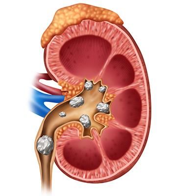
INTERNATIONAL JOURNAL OF BIOLOGY AND BIOMEDICAL ENGINEERING
      DOI: 10.46300/91011.2021.15.6                                                                      Volume 15, 2021

                                         Fig1: Image of stones present in the kidney
                                              (Source: Medscape article, 2018)

Stones in kidneys generally do not give rise to warning                  A. Ultrasound Imaging
signs and may pass undiagnosed. On the other hand, if a             Ultrasound technology uses sound waves to create a
stone obstructs the stream of urine out of the kidney, it           picture of kidneys and bladder. Primarily ultrasound
may lead to severe pain. Pain in the abdomen or at the              imaging has been in use as a diagnostic technique. Due to
sides is the main indication of kidney stones. Symptoms             later advancements, it is also used for the treatment of
like - fever, chills, vomiting, nausea, blood in urine, bad-        kidney stones and therapy guidance aid [1]. Ultrasound is
odour, and reduction in the amount of urine and frequent            a non-invasive test and it is fast and simple to carry out.
urge for urination are other most common symptoms of                These machines are smaller, portable, and are capable of
kidney stones, which should not be ignored. When the                real-time imaging. Hence urologists use ultrasound to
size of the stone is small, no symptoms or pain would be            locate stone as well as the removal of stones. Moreover,
visible; however, as the mass of the stone increases pain           for paediatric and pregnant patients, ultrasound is widely
in the abdomen or groin area develops. Proper hydration             accepted as the diagnostic tool.
(intake of water), more consumption of citrate juices,
oxalate food, and low intake of animal proteins are a few                B. X rays
ways to prevent the formation of kidney stones.                     In standard X-rays, a beam of energy is aimed at the body
Approximately 11 percent of male and 6 percent of the               part being studied. A plate behind the body part captures
female have kidney stones at least once all through their           variations of the energy beam after it passes through the
life span. If kidney stones are not taken care of, they can         skin, bone, muscle, and other tissues. Nephrolithiasis in
cause haematuria, kidney infections and the loss of                 the urinary tract can be detected using abdominal x-rays.
kidney function.                                                    It can show the location of Kidney stones in the urinary
Detection of kidney stones can be done in several ways:             tract. Not all stones are visible on abdominal x-ray.
urinalysis (test for blood, white cell, bacteria, and
crystals), BUN (blood urea nitrogen) and creatinine to                  C.    Kidney, Ureter, Bladder Radiography and
diagnose urinary tract function, test for calcium,                            Fluoroscopy
electrolytes, and phosphorus in the blood stream. There             Normally X-ray units consist of both radiographic and
are also other detection methods available for kidney               fluoroscopic facilities. It contains a high voltage power
stones such as Ultrasound Imaging of kidney, X-ray, and             supply, an X-ray tube, a collimating device, and an X-ray
Computerized Tomography (CT) of the abdomen,                        detector or film. Fluoroscopy technique makes use of X-
Magnetic resonance imaging (MRI) of a kidney, and                   rays to acquire instantaneous moving images of the
abdomen, and Intravenous Pyelogram (IVP).                           interior of the body. It includes an electronic image
In this paper, various imaging techniques have been taken           intensifier and an image display system. In Kidney,
into consideration. A review of the imaging techniques              ureter, bladder (KUB) plain film radiography and
and their performance analysis are also presented. A                fluoroscopy, photons produced by a single energy source
detailed analysis of various algorithms adopted in                  pass through tissues. This technique uses the same
Ultrasound and CT images for kidney stone is discussed.             fundamental concepts as CT but in a single plane.
Furthermore artificial intelligence and deep learning               Advantages of KUB radiography include relatively low
techniques are presented in section III. The proposed AI            ionizing radiation exposure compared with CT and low
based approach for f detection of renal stone is presented          cost compared to ultrasonography. However, KUB
in the Section IV.                                                  radiography views stones at one angle and, therefore,
                                                                    accuracy are decreased leading to reduced sensitivity and
       II. REVIEW OF IMAGING TECHNIQUES                             specificity and, consequently, limiting its utility.

Imaging tests have been used to show kidney stones in                    D. Intravenous Pyelogram (IVP)
the human urinary tract. Ultrasound Imaging, X-ray                  IVP or intravenous urogram is a radiological procedure
Imaging,     Computerized       Tomography,   Magnetic              that uses an injection of contrast material to detect
resonance imaging techniques in use in the medical field            Nephrolithiasis. Contrast materials may cause undesirable
for the diagnosis of Nephrolithiasis.                               allergic effects in some people. IVP tests are rarely used
                                                                    in infants and children. Instead, ultrasound tests are
                                                                    advised for paediatric patients

      Ε-ISSN: 1998-4510                                        37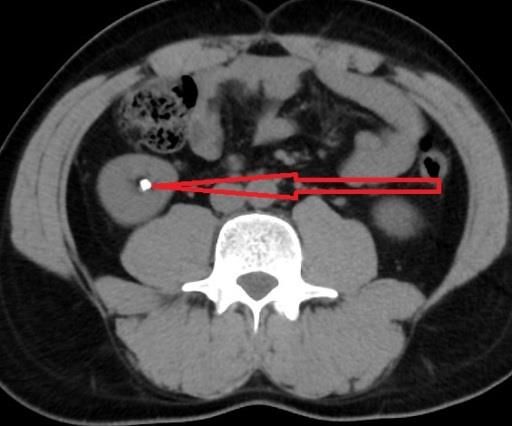
INTERNATIONAL JOURNAL OF BIOLOGY AND BIOMEDICAL ENGINEERING
DOI: 10.46300/91011.2021.15.6 Volume 15, 2021
E. Computerized Tomography general, MRI costs about three times more than a CT scan
CT scans use is amalgamation of x-rays and computer and has lower accuracy and much longer image
technology to generate images of the urinary tract. In acquisition times. MRI is probably the most appropriately
computed tomography, the X-ray beam moves in a circle used as an adjunctive to ultrasonography in patients that
around the body. The X-ray information is sent to a are pregnant.
computer that interprets the X-ray data and displays it in
two-dimensions (2D) on a monitor. CT scans may be G. Summary
done with or without "contrast." Contrast refers to a High-speed or dual energy computerized tomography
substance taken by mouth or injected into an intravenous (CT) may reveal even tiny stones. Simple abdominal X-
(IV) line that causes the particular organ or tissue under rays are used less frequently because this kind of imaging
study to be seen more clearly. CT scans of the kidneys test can miss small kidney stones. During a CT scan, the
can provide more detailed information about the kidneys patient is briefly exposed to ionizing radiation. The
than standard kidney, ureter, and bladder (KUB) X-rays. amount of radiation is greater than one would get during a
CT scans can show the size and location of a kidney stone plain X-ray because the CT scan gathers more detailed
if the stone is blocking the urinary tract, and can also information. The low doses of radiation used in CT scans
show conditions that may have caused the kidney stone to have not been shown to cause long-term harm, although,
form. at much higher doses, there may be a small increase in the
risk of cancer. MRI scans which do not use radiation are
F. Magnetic resonance Imaging not typically used to evaluate kidney stones. But, it helps
MRI uses a magnetic field to align the patient’s free water to safely diagnose kidney stones in pregnant women.
protons along a magnetic field axis. A radiofrequency
antenna, referred to as a coil, is placed over the area to be III. ANALYSIS OF TECHNIQUES FOR
imaged and releases pulses of energy that disrupt the NEPHROLITHIASIS DETECTION
alignment of the protons. When the pulses stop, protons
release energy as they realign with the magnetic field — In the following sections, various algorithms and
this released energy can be captured as an image. The techniques that have been adopted for kidney stone
sensitivity of MRI is higher than that of ultrasonography detection are discussed. The general steps in the image
and KUB radiography but less than that of CT, as stones processing algorithms used in kidney stone detection are
are less easily visible when using MRI. A major explained with a block diagram as shown in figure 2.
advantage of MRI is the ability to provide 3D imaging
without radiation. Unfortunately, the drawbacks of MRI
prevent it from widespread use in stone imaging. In
Renal Image Image Image
Acquisition Preprocessing Segmentation
Detection of Feature Region of
Kidney stone Extraction Interest
Fig2: Illustration of steps in kidney stone detection using Ultrasound Imaging
A. Analysis of Kidney Stone Detection on
Ultrasound Images noise in the Ultrasound images. The difference in
Ultrasound is the diagnostic imaging technique which has travelling path of the coherent acoustic waves adds up
several pros such as no exposure to the ionizing radiation, interference thereby results in introduction of speckle
it’s is a non-invasive procedure, that uses a device called noise in the ultrasound image [22].Therefore; speckle
transducer which is rubbed against the patient’s skin area noise reduction is the first pre-processing step to improve
of examination, provides a clear detailed image of the soft the Ultrasound image quality. To remove the speckle
tissues and is cost-effective .The ultrasonic image noise, the following filtering techniques can be utilized:
properties of body tissues determine the characteristics of Wavelet Filter: It provides better accuracy even
ultrasound image. The two important parameters of the in the presence of noise in the image, but this
ultrasound imaging is the attenuation and the velocity of filter never raises SNR (Signal to noise ratio)
propagation that decides with how much frequency the high.
body tissues can be imaged.It is one of the techniques Lee Filter: This is a special filter, widely used in
used for kidney stone detection where ultrasound image medical Ultrasound imaging This filter reduces
acquired is processed through image processing speckle noise by employing minimum mean
algorithms. The diagnostic ability is reduced by speckle squared error approach
Ε-ISSN: 1998-4510 38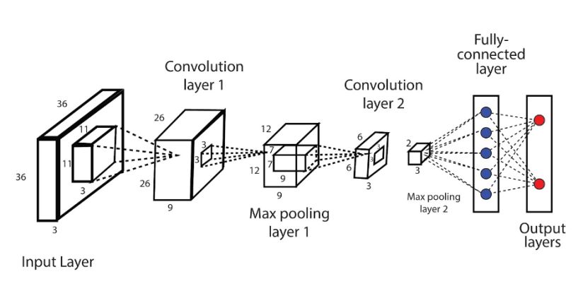
INTERNATIONAL JOURNAL OF BIOLOGY AND BIOMEDICAL ENGINEERING
DOI: 10.46300/91011.2021.15.6 Volume 15, 2021
Diffusion Filter: It is a non-linear filter, which System (ESRANFIS) method detects the renal
utilizes a coefficient of variation to enhance the calculi with the use of Adaptive Neuro-Fuzzy
contrast in the image along with the reduction of Inference System (ANFIS).This technique is
the speckle noise. utilized in two phases. In the first phase, the
Bilateral Filter: It is a non-linear and non- normal and the affected kidney are classified; in
iterative filter, considered as a robust method for the second phase of the segmentation, the kidney
smoothing of noisy images and the preservation stone areas are found in the classified image
of edges. The basic concept of this filtering (Figure 3).
technique is that it replaces the noisy pixel of the RICS Method: Region Indicator with Contour
image with weighted values, which depends on Segmentation (RICS) method involves five
the photometric and the geometric distance. major steps. Computation of renal calculi region
parameters is carried out in the first and second
After pre-processing of the ultrasound image, comes the steps. Histogram equalization is performed for
segmentation of the image; the image segmentation can contrast enhancement of the image, and then k-
be carried out using Level set segmentation methods. mean clustering selects the most desired pixel
Apart from Level Set segmentation methods, several value. The most desired pixel value is utilized to
other Segmentation methodologies which can be locate the stone exactly in the renal image. In the
implemented on the Ultrasound image, the following lists final step, pixel matching and thresholding
some of the significant Segmentation methods: process are performed to locate the calculi. The
ESRANFIS Method: Effective Segmentation of RICS method of segmentation shows high
Renal calculi Adaptive Neuro-Fuzzy Inference efficiency with the minimized error.
Fig3: Ultrasound image showing renal calculi.
An imaging technique for early diagnosis of PallaviVaish et al (2016) have developed a scheme
Nephrolithiasis has been developed by Tamilselvi that detects malfunctions in kidneys. Smartphone
and P Thangaraj (2011) [2]. Here improved Seeded acquires the image data from
Region Growing (SRG) technique has been used for Ultrasound scanners for performing Computer-aided
segmentation and classification. This method diagnosis and generating results [5]. Viola-Jones
depends on image granularity features and the algorithm, texture feature extraction and Support
detection procedure is carried out based on intensity Vector Machine (SVM) classifier are employed for
threshold disparities that are acquired from the automatically detecting the abnormality in the
segmented portions of the image. Bryan Cunitz et al kidney. JyotiVerma et al (2017) have designed an
(2014) proposed an improved method for algorithm for recognizing Nephrolithiasis using Kth
Nephrolithiasis detection technique using Doppler Nearest Neighbour (KNN) and SVM techniques [6].
imaging [3]. Here Doppler output parameters have Here Image enhancement is performed using the
been optimized in vitro to improve the overall Median filter. Gaussian and Median filters are also
performance of ‘twinkling artefact’ (TA) which a applied for filtering besides Un-sharp masking for
kidney stone exhibits under Color Doppler sharpening. Subsequently, erosion and dilation are
ultrasound. This method exhibits improved SNR. carried out. This is followed by entropy-based
Viswanath K and Gunasundari R (2015) have segmentation for locating the region of interest.
proposed a model to detect Nephrolithiasis by Further processing by KNN and SVM techniques
Reaction-diffusion level set segmentation [4]. For results in the final outputs
implementation, Xilinx System generator on FPGA S. M. K. Chaitanya and P. Rajesh Kumar (2019)
is used. Here, the ultrasound image is preprocessed to employedgrey-scale conversion for pre-processing
avoid speckle noise. Then smoothing by using Gabor followed by region-of-interest generation. Followed
filtering is followed by Histogram equalization. by feature extraction by Gabor wavelet followed by
Multilayer Perceptron (MLP) and Back Propagation Cuckoo Search Artificial Neural Network (ANN)
have been employed to categorize the form of stone. algorithm for optimization [7].To compare the
performance of various methods, the various
Ε-ISSN: 1998-4510 39INTERNATIONAL JOURNAL OF BIOLOGY AND BIOMEDICAL ENGINEERING
DOI: 10.46300/91011.2021.15.6 Volume 15, 2021
performance metrics for the existing methods have
been tabulated in table 1.
Year Authors Methods Accuracy Specificity
2014 Viswanath K Multilayer Perceptron, Back Propagation 98.8% -
&Gunasundari R ANN
2016 PallaviVaish et al Viola-Jones algorithm, SVM Classifier 90.91% -
2017 JyotiVerma et al KNN, SVM 85% -
2019 S. M. K. Chaitanya, P. Cuckoo Search (CS) and 94 100
Rajesh Kumar Artificial Neural Network (ANN)
Table1: Performance metrics for Ultrasound Imaging
B. Analysis of Nephrolithiasis Detection on CT Boolean operation, morphological operation, and
Images localization improve the efficiency in the program. Sujata
CT is a technique that uses x-ray equipment to create a Navratnam et al (2016) have worked on enhanced seed
highly detailed scan of the internal areas of the body. In pixel region growing segmentation and ANN
recent years, the Computed Tomography imaging classification which accurately distinguishes the presence
techniques have been used for Nephrolithiasis detection. and absence of renal calculi [13]. The system uses texture
Several types of studies have been carried out on features which compute the pixels in each slice of the
obtaining better CT image quality; this section provides a ultrasound image. A set of seeds and the image are taken
detailed analysis of the filtering techniques and as the input in the automatic region algorithm. The region
algorithms that have been implemented on CT images. of the kidney to be segmented is marked by seeds which
D.Y. Kim & J.W. Park (2004) has applied a Gray level are taken as the input. The output of the segmentation
threshold method for the segmentation of the CT kidney depends on the selection of seeds. The next step is feature
image along with average and standard deviations to extraction. Feature space can be intensity, texture, and
detect kidney tumor [8]. J. Kolomaznık et al. (2010) shape. The feature extraction process depends on the
presented an algorithm for fast segmentation of kidneys usage of classifiers.
in CT images [9]. Three-Dimensional segmentation on
parametric snakes also called active contour model is Prema T.Akkasaligar et al. (2017) have designed a model
implemented in volume slices to define the organ's for Kidney stone detection in Computed Tomography
volume that is the kidney. Wu Zhou and YaoqinXie (CT) images [14]. This method uses Level set
(2013) have been using the active contour model in the segmentation where the input images are preprocessed
segmentation of volumetric medical images [10]. The and the region of interest is segmented. Fuzzy C-means
snake model algorithm for segmentation of medical clustering is applied to this image to obtain cluster
image has been effective as almost all medical images are centers. This is then segmented using Level set
in 3-dimensional form. Regions are obtained in stacks to segmentation which is then analyzed to detect stone size
and location. NilarThein et al (2018) have proposed an
form volumetric region after images slices are segmented
Image preprocessing method for the segmentation of
individually. Jianfei Liu et al. (2014) have developed a
kidney stones in CT scan images [15]. Here three
total variation (TV) flow methodology to minimize image thresholding algorithms that are based on intensity, size
noise in the renal structure to maintain the characteristic and location are applied for removing unwanted regions
appearance of renal calculi, as image noise is the main in CT images. 30 CT scan images of the digitized
challenge that hinders accurate Nephrolithiasis detection transverse abdomen from the patients with kidney stones
[11]. are included for validation and statistical analysis. This
Maximally stable external regions (MSER) features were algorithm provided experimental results with a sensitivity
used to identify kidney stones. MSER is appropriate to of 95.24%.
detect kidney stones as it exploits extreme image values Stalina S et al (2018) designed an algorithm for detecting
to detect image blobs. The texture and shape features are kidney stones on CT images using Image Processing [16].
computed and are imported to support vector machines The image is preprocessed using filters to smoothen and
for calculus classification. SamanEbrahimi et al. (2015) remove noise. It is followed by Image enhancement with
worked on Kidney urine belly-CT (KUB-CT) where KUB Power-law transformation. The thresholding technique is
is an imaging modality that can improve Nephrolithiasis applied for Image segmentation and thus kidney stones
screening and diagnosis [12]. The image processing are detected.To compare the performance of various
techniques that are employed in the program, namely, methods, the various performance metrics for the existing
Gamma Adjustment, Image Segmentation, Binarization, methods have been tabulated and are listed in table 2.
Ε-ISSN: 1998-4510 40INTERNATIONAL JOURNAL OF BIOLOGY AND BIOMEDICAL ENGINEERING
DOI: 10.46300/91011.2021.15.6 Volume 15, 2021
Researchers show that the stone diagnosis on CT image is algorithms have been developed for the removal of soft
a challenging task while performing segmentation organs and bony skeleton present in the Abdominal CT
because of the complex structure of interest in the image (Figure 4). The figure shows the presence of stone
abdomen CT.Therefore, the pre-processing algorithm in the kidney region.
plays an essential role in improvising the performance of
3D image segmentation. Along with these techniques,
Fig4: CT Image showing renal calculi
(Source: Newyork Urology Specialist)
Year Authors Methods Performance Measures
2015 SamanEbrahimi& Thresholding, Accuracy: 84.61%
Vladimir Y Boundary defining
Mariano[12]
2018 NilarThein et al Three Thresholding Sensitivity:95.25%
[15] algorithms based on
Intensity, Size &
Location
2018 L Prisilla et al Contrast limited Accuracy:87.41%
[24] adaptive histogram
equalization
2020 John Brandon Kidney Dice scores*: 0.9620
Graham-Knight et al Segmentation using
[25] deep transfer
learning model
nnU-net
2020 Riya Mishr etal Classification using Accuracy:98.8 %
[23] Back Propagation
Network , Fuzy C-
Mean clustering
algorithm for
segmentation
* Dice scores is a kind of metrics for evaluating the segmentation of image
Table2: Performance metrics for CT Imaging
C. Soft Computing and Artificial Intelligence classification techniques K - Nearest neighbour (KNN)
algorithms in Nephrolithiasis Detection and Support Vector Machine (SVM for the analysis of
Nephrolithiasis images [18]. Artificial intelligence (AI)
Soft Computing and Artificial Intelligence algorithms are algorithms can be used to accurately detect
increasingly employed in Nephrolithiasis detection. Nephrolithiasis detection. Considering artificial
Verma J et al (2017) have used the soft computing-based intelligence methodologies, the neural network provides
Ε-ISSN: 1998-4510 41INTERNATIONAL JOURNAL OF BIOLOGY AND BIOMEDICAL ENGINEERING
DOI: 10.46300/91011.2021.15.6 Volume 15, 2021
more than 90% accuracy for both types of detections of accuracy of the model [29]. For Nephrolithiasis detection
Nephrolithiasis detection. Deep Learning Technology is a and analysis CNN, SVM and ANN are most commonly
concept of Machine learning which plays a vital role in used machine learning algorithms.
medical imaging diagnosis. Convolution Neural Support Vector Machine: It was introduced in
Networks (CNN) is one of the deep learning which can be the year 1992, is a supervised learning network
used to classifying detected kidney diseases. CNN which works as a classifier by analysing the
architecture is capable of processing a large number of data, recognizing pattern and solves linear and
hidden layers as compared to the conventional layered non linear classification problems.SVM is
architecture. Kristian M et al (2020) have used Deep considered as a non probabilistic binary linear
Convolution Neural Networks to detect Nephrolithiasis classifier which takes input pattern as feature
from digital images with better performance [19].G vector and identifies to which class the dataset
Sumana et al proposed a model which uses Artificial
belongs [31]. The figure 5 shows the architecture
Neural Network as the classifier for diagnosis, GLCM
of the SVM [32].
algorithm for feature and Forward Back propagation
algorithm to reduce the diagnosis time and increase the
Fig5: Architecture of SVM
Artificial Neural Network: It’s a neural network in between known as the hidden layer and final
model and data processing technique that is layer is the output layer. The main advantage of
based on our human brain more like the neuron neural network is that it works swiftly even
pathway or the nervous system route.ANN is a when there is high noise present in the dataset
network of the processing elements (biological [29].The basic architecture of the ANN is shown
neurons) which work in a parallel manner. The in figure 6 [30].
network has input layer (first layer), extra layers
Fig6: ANN Architecture
Convolution Neural Network: It is a deep the colors, gradients and edges and with addition
learning algorithm which takes images as the of extra layers the convolution layer can extract
input and is capable of differentiating the images the high level features. The main role of the
from one another by passing it through several pooling layer is to reduce the computational
layers of network. These layers include power and increase the efficiency of the training
Convolution layer and the pooling layer. The and testing dataset. Figure 7 shows the basic
convolution layer performs the convolution architecture of the CNN [33].
operation which extracts low level features as
Ε-ISSN: 1998-4510 42INTERNATIONAL JOURNAL OF BIOLOGY AND BIOMEDICAL ENGINEERING
DOI: 10.46300/91011.2021.15.6 Volume 15, 2021
Fig7: CNN Architecture
D. Comparative analysis of the performance of Nephrolithiasis (Fulgham PF et al) such as KUB, Low-
various Nephrolithiasis detection techniques dose CT, Ultrasound Imaging, Intravenous pyelography,
Comparative analysis of the performance and Radiation CT and MRI are presented in Table 3 [17, 20].
exposure of various techniques in the diagnosis of
Modality Sensitivity Specificity Radiation Exposure in
millisieverts (mSv)[17]
Low-dose CT 95%[26] 97%[26] ~3
Ultrasound 61% 97% None
Intravenous pyelogram(IVP) 70% 95% 3.0
MRI 82% 98.3% None
CT 98% 97% 10.0
KUB 57% 76% 0.7
Table3: Comparative analysis of the performance of various Nephrolithiasis detection technique
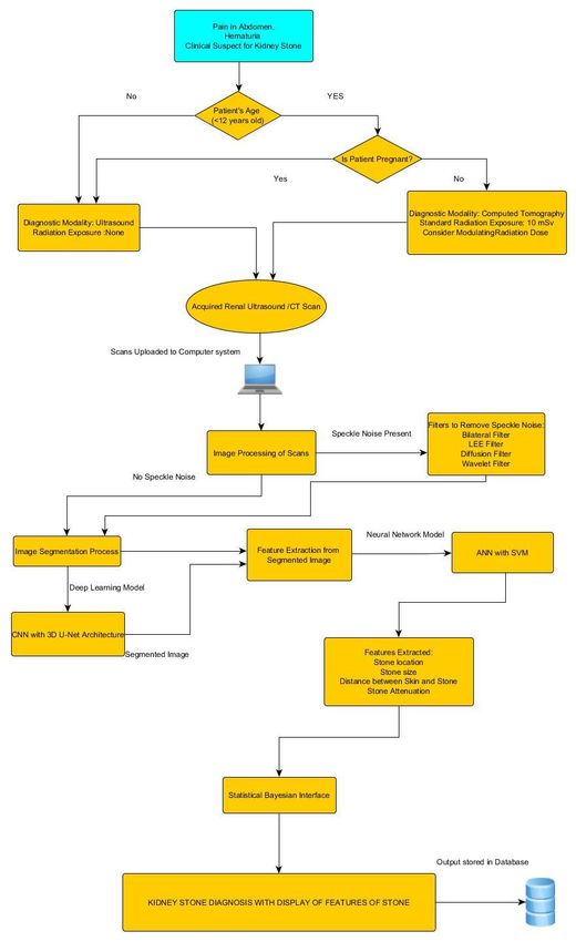
IV. Proposed Approach involves using two CNNs, in which one network trains
An approach is proposed for imaging the subjects with and targets the areas of kidney and the other CNN
renal stones and an Artificial Intelligence based network targets the non-kidney area, and then combining
algorithms and models for processing the Ultrasound or the two networks to generate clean, high quality and
CT scans in order to increase the efficiency and accuracy resolution and also increases the processing time.
of diagnosis/prognosis of kidney stones and also to aid The images obtained from the Ultrasound and CT is
the physicians and medical practioners in their decision uploaded to the computer system for processing. The first
management for treatment and medications. The initial step of the processing of image is removing the speckle
step is decision making for choosing the right imaging noise. The filters mentioned in Section III A are effective
modality based on the clinical symptoms of renal stone, in removing the speckle noise from the image. If there is
patient’s age, health condition of patient that is if she is no speckle noise present, the image is sent for the next
pregnant or not. For the patients suffering from step that is Segmentation and if there is noise present then
Nephrolithiasis under the age of 12 and pregnant women, based on the speckle noise characteristics any of the filter
the Ultrasound is to be chosen as the diagnostic modality. mentioned in Section III A can be used to clean the
Ultrasound imaging modality is used as there is no risk of image. For Image Segmentation process, the CNN with
radiation exposure and the diagnosis can be carried out 3D U-net architecture is considered to be best. The U-net
safely for pregnant woman and children under 12.The architecture is capable of working with few training
Computed Tomography imaging modality is to be chosen datasets and samples and can segment images with better
for patients above the age of 12 .CT involves the efficiency and accuracy. The efficiency of the
standard radiation exposure of 10 mSv. However, the segmentation process can be checked using the
radiation dose can be modulated as per the requirement performance parameters such as dice similarity
and health conditions of the individual. The Low-dose CT coefficient and weighted loss function (dice loss). Once
has radiation exposure less than 3mSv, but the the image is segmented, it is then passed to the neural
disadvantage of this is that it results in low quality and network model which is combination of ANN and SVM
resolution. Low quality and poor resolution of the scans for feature extraction and classification of the segmented
obtained from Low-dose CT can be resolved by images. The algorithm designed by Priyanka Chak et al is
processing the scans using deep learning techniques. promising in better feature extraction and classification,
From the research works, the CNN seems promising in in which ANN designed, has 12 input nodes, 10 hidden
improving the quality of the images. This basically nodes and 2 output nodes and a combination with SVM
Ε-ISSN: 1998-4510 43INTERNATIONAL JOURNAL OF BIOLOGY AND BIOMEDICAL ENGINEERING
DOI: 10.46300/91011.2021.15.6 Volume 15, 2021
gives accuracy of 99% with all the features [27]. This interface to obtain the probability rank differential
algorithm can be used for the feature extraction and diagnosis for further analysis, comparison and study. The
segmentation process. The features such as stone location, basic architecture and brief details of the neural network
stone size, stone attenuation and distance between skin models and deep learning models proposed in the
and stone extracted from the neural network model will approach is discussed in section III C. The results
aid the Physician in understanding the Nephrolithiasis obtained can be stored in a database for future reference
condition of the patient better. Further, the features and study. The flowchart (figure 8) gives an insight of the
extracted can be combined to statistical Bayesian proposed AI-based system discussed above.
Fig8: Proposed AI-Based System for Detection of Nephrolithiasis
Comparative analysis of the performance of various
V. DISCUSSION techniques for the diagnosis of Nephrolithiasis is
An analysis of various techniques and algorithms that presented. It is inferred that direct detection of most
have been employed to obtain better image quality, better stones is not possible with MRI. MRIs are generally more
de-speckling, improvisation in image contrast, and faster expensive than other techniques, such as CT scans. But,
segmentation of the image has been carried out. A MRI produces no dangerous radiation and it can readily
comparative analysis of research works on CT and reveal urinary obstruction. It has been learnt that both CT
Ultrasound images has also been carried out. imaging and ultrasound imaging techniques detect the
Ε-ISSN: 1998-4510 44INTERNATIONAL JOURNAL OF BIOLOGY AND BIOMEDICAL ENGINEERING
DOI: 10.46300/91011.2021.15.6 Volume 15, 2021
majority of Nephrolithiasis conditions.In future, there image thereby enhancing the diagnostic efficiency of
may be a decision support system utilizing the concepts Nephrolithiasis detection on CT and Ultrasound
of deep learning for easy and faster processing of the Images[21]
VI. CONCLUSION Applications and Services, Munich, Germany, pp.1-6,
It has been learnt that Kidney stone detection using 2016
Ultrasound Imaging is independent of the composition of [6] Verma J, Nath M, Tripathi P andSaini KK, “Analysis
stones. This technique is capable of identifying uric-acid and identification of kidney stone using Kth nearest
stones as well as calcium stones. In the case of neighbour (KNN) and support vector machine (SVM)
Ultrasound imaging, it is not always possible to detect classification technique,” Pattern Recognition and Image
small stones. Unlike ultrasound, CT exposes patients to Analysis, vol.27, no.3, pp.574–580, 2017.
significant amounts of radiation. The low dose-CT in [7] S. M. K. Chaitanyaand P. Rajesh Kumar,
conjugation with deep learning technique can help “Classification of Kidney Images Using Cuckoo Search
improve the quality and resolution of the scan. The Algorithm and Artificial Neural Network”, International
ultrasound imaging technique is favoured in the case of Journal of Engineering and Advanced Technology, Vol.8,
pregnant women and people sensitive to IV contrast. No.3, 2019.
Hence, it is recommended that Ultrasounds have to be [8] D. Y. Kim and J. W. Park, “Computer-Aided
used for preliminary diagnosis subsequently through CT detection of kidney tumor on abdominal computed
if the medical practitioner senses it is necessary. tomography scans”, ActaRadiologica, vol. 45, no. 7, pp.
Advances in CT, ultrasonography, KUB radiography and 791-795, 2004
MRI technologies are continuing and are likely to [9] J. Kolomaznık, “Fast Segmentation of Kidneys in CT
improve all modalities in the future. At present Artificial Images, Proceedings of Contributed Papers”, Part I,
Intelligence based algorithms are evolving which could pp.70–75, 2010.
improve efficiency. The present trend is the usage of deep [10] Wu Zhou and YaoqinXie, “Interactive Medical
learning algorithms for radiographic and sonogram Image Segmentation Using Snake and Multiscale Curve
images for Nephrolithiasis detection. The proposed AI Editing”, Hindawi Mathematical Methods and
based technique discussed in the section 4 when Applications in Medical Imaging, Vol. 2013, pp.1-13.
implemented will be promising for faster detection and [11] Liu, J., Wang, S., Turkbey, E. B., Linguraru, M. G.,
analysis of kidney stone and serve as the decision support Yao, J., andSummers, R. M., “Computer-aided detection
system for the Physicians as well as the radiologists. of renal calculi from non- contrast CT images using TV-
In future deep learning can be employed for detecting flow and MSER features”, Medical Physics, Vol.42,
kidney stone composition during endoscopy. This paves No.1, pp.144–153, 2014.
the way to integrated endoscopic and laser systems that [12] SamanEbrahimi and Vladimir Y. Mariano, “Image
automatically provide laser settings based on stone Quality Improvement in Kidney Stone Detection on
composition recognition which could improve surgical Computed Tomography Images, Journal of Image and
efficiency.Soon, algorithms may be developed to assist in Graphics”, Vol. 3, No. 1, 2015.
deciding on the best approach for imaging patients. [13] SujataNavratnam, SitiFazilahValliappan Raman and
SundaresanPerumal.“Seed Pixel Region Growing
REFERENCES Segmentation and Artificial Neural Network Classifier
[1]David T. Tzou, ManintUsawachintachit, Kazumi for Detecting the Renal Calculi in Ultrasound Images for
Taguchi, and Thomas Chi, “Ultrasound Use in Urinary Urologist Decisions”, International Journal of Computer
Stones: Adapting Old Technology for a Modern-Day Science Issues, Vol. 13, No. 5, pp. 1694-0814, 2016.
Disease”, Journal of Endourology, Vol. 31, 2017. [14] Akkasaligar, P. T., Biradar, S. and Kumbar, V.
[2] Tamilselvi P R andThangaraj P, “Computer Aided “Kidney stone detection in computed tomography
Diagnosis System for Stone Detection and Early images”, Proceedings of the International Conference on
Detection of Kidney Stones,” Journal of Computer Smart Technologies For Smart Nation, 2017.
Science, vol.7, no.2, pp.250–254, 2011. [15] Thein N, Nugroho H A, Adji T B andHamamoto K,
[3] Cunitz B, Dunmire B, Paun M, Sapozhnikov O, “An image preprocessing method for kidney stone
Kucewicz J, Hsi R, Lee F, Sorensen M, Harper J and segmentation in CT scan images,” International
Bailey M, “Improved detection of kidney stones using an Conference on Computer Engineering, Network and
optimized Doppler imaging sequence,” 2014 IEEE Intelligent Multimedia, Surabaya, Indonesia, pp.147-150,
International Ultrasonics Symposium Proceedings, 2018.
Chicago, United States, pp. 452-455, 2014. [16] Stalina S, Aditi S, Anuja R and Prof. Pooja L Gohel,
[4] Viswanath K andGunasundari R, “Design and “Kidney Stone Detection Using Image Processing On CT
analysis performance of Kidney Stone Detection from Images,” International Journal of Management,
Ultrasound Image by Level Set Segmentation and ANN Technology And Engineering, vol.8, no.9, pp. 2257-2260,
Classification,” 2014 International Conference on 2018.
Advances in Computing, Communications and [17] Fulgham PF, Assimos DG, Pearle MS, and
Informatics, India, pp. 407-414, 2014. Preminger GM. “Clinical effectiveness protocols for
[5] Vaish P, Bharath R, Rajalakshmi P and Desai U B, imaging in the management of ureteral calculous disease:
“Smartphone based automatic abnormality detection of AUA technology assessment”, J Urol. 2013;189:1203–
kidney in ultrasound images,” 2016 IEEE 18th 1213.
International Conference on e-Health Networking,
Ε-ISSN: 1998-4510 45INTERNATIONAL JOURNAL OF BIOLOGY AND BIOMEDICAL ENGINEERING
DOI: 10.46300/91011.2021.15.6 Volume 15, 2021
[18] Verma, J., Nath, M., Tripathi, P, “ Analysis and [26] Coursey CA, et al. ACR Appropriateness Criteria(R)
identification of kidney stone using Kth nearest neighbour acute onset flank pain-suspicion of stone
(KNN) and support vector machine (SVM) classification disease. Ultrasound Q. 2012;28:227–
techniques”, Pattern Recognit. Image Anal. 27, 574–580 233. [PubMed] [Google Scholar]
(2017). [27] Chak P., Navadiya P., Parikh B., Pathak K.C.
[19] Kristian M. Black Hei Law Ali AldoukhiJia Deng (2020) Neural Network and SVM Based Kidney Stone
Khurshid R. Ghani, “Deep learning computer vision Based Medical Image Classification. In: Nain N.,
algorithm for detecting kidney stone composition”, BJU Vipparthi S., Raman B. (eds) Computer Vision and
International, Volume125, Issue 6, 2020, pp. 920-924. Image Processing. CVIP 2019.Communications in
[20] Wayne Brisbane, Michael R. Bailey and Mathew D. Computer and Information Science, vol 1147.
Sorensen, “An overview of kidney stone imaging Springer, Singapore.
techniques”, Nat Rev Urol. 2016 November; 13(11): 654– [28] https://www.webmd.com/kidney-
662. stones/understanding-kidney-stones-basics#1
[21] Saxena, Ashish, Eddie Yin Kwee Ng, and SooTeik [29] G. Sumana ,G. Anjan Babu, Prediction of
Lim. "Imaging modalities to diagnose carotid artery Nephrolithiasis Based on Extracted Features of CT-Scan
stenosis: progress and prospect", Biomedical engineering Images using Artificial Neural Networks, International
online 18.1 (2019): 66. Journal of Advanced Research in Computer Science,
[22] Cincotti G, Loi G, Pappalardo M. Frequency Volume 8, No. 5, May-June 2017
decomposition compounding of ultra-sound medical [30] http://www.neural-
images with wavelet packets. IEEE Trans Med Imag forecasting.com/support_vector_machines.htm
2001;20(8):764–71. [31] Abhishek, Gour Sundar Mitra Thakur, Dolly Gupta,
[23] Riya Mishr,Avik Bhattacharjee,M.Gayathri, Proposing Efficient Neural Network Training Model for
C.Malathy,Kidney Stone Detection with CT Images Kidney Stone Diagnosis, (IJCSIT) International Journal
Using Neural Network,International Journal of of Computer Science and Information Technologies, Vol.
Psyshosocial Rehabilitation,Vol.24,Issue 08,2020 3 (3) , 2012,3900-3904
[24] L. Prisilla ,I. Laurance Aroqiaraj, Kidney Stone [32] http://www.neural-
Detection using Contrast Limited Adaptive Histogram forecasting.com/support_vector_machines.htm
Equalization (CLAHE) on CT Scan Images, International [33]https://www.analyticssteps.com/blogs/convolutional-
Journal of Computational Intelligence and Informatics, neural-network-cnn-%20graphical-visualization-code-
Vol. 7: No. 4, March 2018 explanation
[25] Graham-Knight J.B. et al. (2020) Accurate Kidney
Segmentation in CT Scans Using Deep Transfer
Learning. In: McDaniel T., Berretti S., Curcio I., Basu A.
(eds) Smart Multimedia. ICSM 2019. Lecture Notes in
Computer Science, vol 12015. Springer, Cham.
Ε-ISSN: 1998-4510 46You can also read