Postnatal Imaging of the Microcephalic Pediatric Patient
←
→
Page content transcription
If your browser does not render page correctly, please read the page content below

MedDocs Publishers

                                                                                                                     ISSN: 2637-885X

                                               Journal of Radiology and Medical Imaging
                                                                                                           Open Access | Review Article

               Postnatal Imaging of the Microcephalic
                          Pediatric Patient
Saurabh Verma; Gordon Heller*
Department of Radiology, Icahn School of Medicine at Mount Sinai, New York.

  *Corresponding Author(s): Gordon Heller                                      Abstract
 Department of Radiology, Icahn School of Medicine at                          The purpose of this article is to review the clinical and
 Mount Sinai, 1000 10th ave NY, NY 10019. New York.                        imaging manifestations of microcephaly, with an empha-
 Tel: 212-636-3364, Fax:212-523-7050;                                      sis on syndromic causes. Defined as a head circumference
                                                                           greater than 2 standard deviations below the mean, mi-
 Email: gordon.heller@mountsinai.org
                                                                           crocephaly can manifest from primary causes during fetal
                                                                           development, or can occur postnatally from postnatal in-
                                                                           jury or genetic syndromes. Imaging plays an integral role in
Received: Feb 03, 2021                                                     the evaluation of microcephaly and can hint at contributory
Accepted: Mar 10, 2021                                                     gene or external causes; most importantly, imaging can help
                                                                           detect signs of nonaccidental trauma. While ultrasound re-
Published Online: Mar 13, 2021                                             mains the primary diagnostic tool for prenatal evaluation,
Journal: Journal of Radiology and Medical Imaging                          Magnetic Resonance Imaging is the preferred imaging mo-
Publisher: MedDocs Publishers LLC                                          dality following birth.
Online edition: http://meddocsonline.org/
Copyright: © Heller G (2021). This Article is
distributed under the terms of Creative Commons
Attribution 4.0 International License

Keywords: TORCH: Toxoplasmosis; Other (Syphilis, Varicella-
Zoster, Parvovirus B19), Rubella, Cytomegalovirus (CMV), And
Herpes Infections, CNS: Central Nervous System; DTI: Diffusion
Tensor Imaging; SWI: Susceptibility Weighted Imaging; HIE: Hy-
poxic Ischemic Encephalopathy; PVL: Periventricular Leukoma-
lacia; IQ: Intelligence Quotient.

   Introduction
    Microcephaly is a result of abnormal brain development and           velopment, whereas the majority of dendritic connections and
is defined as a head circumference greater than 2 standard de-           myelination occur after birth [2]. Therefore, prenatal injuries
viations below the mean. The term “severe microcephaly” is re-           result in a failure of neurogenesis, whereas postnatal injuries
served for head circumference exceeding 3 standard deviations            result in abnormal development of previously-normal neurons.
[1]. The abnormal development can result from either prenatal
                                                                            The etiologies for microcephaly can also be categorized
or postnatal injury, and the time of clinical presentation divides
                                                                         based on extent of the disease manifestation, whether the
microcephaly into primary or secondary (acquired) categories
                                                                         anomalies are limited to cerebral development, or occur in con-
[2]. The majority of neurogenesis occurs by week 21 of fetal de-

                 Cite this article: Verma S, Heller G. Postnatal Imaging of the Microcephalic Pediatric Patient. J Radiol Med Imag-
              ing. 2021: 4(1); 1043.

                                                                     1
MedDocs Publishers
junction with other extracerebral anomalies as part of a syn-             formations, and/or facial dysmorphism. Causes include chro-
drome. The non-syndromic causes are relatively common, and                mosomal abnormalities and gene deletion syndromes. Utilizing
are typically limited to cerebral development [1]. The syndromic          the “Online Mendelian Inheritance In Man”, a comprehensive
causes are relatively rare and are associated with extracerebral          online database categorizing human genes and genetic pheno-
involvement including visceral malformations, skeletal malfor-            types, a database search for “microcephaly [clinical synopsis]”
mations, and/or facial dysmorphisms. Syndromic and nonsyn-                yields 795 genetic phenotypes with a component of microceph-
dromic causes may also co-exist in some etiologies such as fan-           aly [5]. For the purpose of this review, however, we will discuss
coni anemia [3]. For the purpose of this review, we will discuss          select syndromes including, Cri-du-chat Syndrome, mitochon-
microcephaly with respect to syndromic and non-syndromic                  drial abnormalities, such as Leigh’s syndrome and Wilsons
causes, with an emphasis on select syndromes. Furthermore,                disease, Craniosynostosis in a patient with Aperts syndrome,
we will concentrate on postnatal patients, for whom MRI is the            Sturge Weber Encephalotrigeminal Angiomatosis, Trisomy 13,
primary modality.                                                         Cockayne Syndrome and Pelizaeus-Merzbacher Disease.
   Clinical and imaging assessment                                           Cri-du-chat syndrome
    The evaluation of microcephaly relies on both clinical and                After the discovery of the normal human chromosome
imaging assessment, and is initiated when a head circumfer-               number in 1956, Cri du chat syndrome was first recognized by
ence measurement is greater than 2 standard deviations below              Lejeune in 1963 [6]. This karyotype contains the normal number
the mean. Careful history, including maternal factors, is crucial         of chromosomes, but results from deletions of the short arm of
in the workup of primary microcephaly. Etiologies, including a            chromosome 5p. A cat-like cry, facial dysmorphism, microceph-
maternal history of diabetes, phenylketonuria, medication use,            aly, and severe psychomotor and mental retardation are clini-
alcohol or substance use, radiation exposure, and prenatal in-            cally evident [7]. Additional findings such as congenital heart
fections such as the TORCH infections or fetal alcohol syndrome           disease and renal anomalies are present. Characteristic radio-
are important considerations. Therefore, the history and the              graphic findings initially described include microcephaly and
clinical assessment of the parents as well as the infant is invalu-       hypertelorism [7]. Further assessment of brain morphometry
able in the assessment of the microcephalic infant [4]. Genetic           utilizing MRI revealed a characteristic brainstem atrophy, pre-
testing with microcephaly panels are also a valuable component            dominantly at the pontine level, with associated small cerebel-
in the assessment of a microcephalic patient with a suspected             lum, cerebellar peduncles, and cerebellar white matter (Figure
syndrome.                                                                 1) [8,9]. In(b)addition, the literature suggests a lack of myelination
                                                                          in the anterior limbs of the internal capsule in this disease en-
    Clinically, the prognosis of microcephaly varies, with some           titiy [10,11].
of the worst outcomes seen in syndromic cases such as Trisomy
13, which is nearly fatal by the first year of life. Another impor-          Mitochondrial chromosomal abnormalities
tant clinical association is cognitve and developmental delay,
the severity of which has been directly linked to the severity of             Mitochondrial diseases are an established etiology for mi-
microcephaly. In a study of 212 patients, decreasing head cir-            crocephaly. These are a myriad of disease processes that result
cumference directly correlated to decreasing IQ, with the high-           from abnormal oxidative phosphorylation, and can be inherited
est IQs documented in conditions typically arising after the first        in germline mutations or acquired from environmental factors
trimester, and the lowest IQs documented in children with chro-           such as infections. They are categorized as primary when direct-
mosomal abnormalities, inborn errors of metabolism, or mul-               ly involving mutations in the electron transport chain proteins,
tiple congenital anomalies. Other predictors of poor cognitive            such as in Leigh’s Syndrome (Figure 2). Mitochondrial disease
performance include atrophy, cortical dysplasia, and myelina-             are termed secondary when accompanying another pathologic
tion abnormalities [4,5,45].                                              process that affects mitochondrial function, such as in Wilson’s
                                                                          disease or hepatocellular degeneration (Figure 3) [12].
    Similarly, imaging plays an integral role in the evaluation
of microcephaly and can hint at contributory gene or external                Due to the pervasive nature of oxidative phosphorylation,
causes. Imaging can help to assess for the sequalea of non ac-            a defect can affect any tissue, leading to progressive multi-or-
cidental injury by demonstrating subdural hematoma accompa-               gan system involvement, typically beginning with the central
nied by post traumatic encephalomalacia. MR imaging is used to            and/or peripheral nervous system. Progressive myopathy, ne-
identify gyral, migrational, or myelination abnormalities, while          phropathy, hepatic dysfunction, and cardiomyopathy are often
CT is generally restricted to confirming calcifications, which            demonstrated [11]. Patients can present either in childhood or
may be present in the TORCH syndromes, our institution MR                 adulthood, but approximately 45% of childhood presentations
protocol will include T1, T2, and Inversion Recovery sequences            begin with neuromuscular symptoms of weakness, atrophy,
in multiple planes on a high field magnet. Gradient echo and              hypotonia, peripheral neuropathy, cerebellar ataxia, and leuko-
susceptibility weighted imaging is performed to assess for calci-         dystrophy [13].
fications and can preclude the need for CT evaluation. We note               Neuroimaging findings include cerebral and cerebellar at-
that the fluid attenuated inversion recovery (FLAIR) sequence             rophy, white matter changes, and signal abnormality involving
may be a limited value in the unmyelinated brain. Furthermore,            the basal ganglia and thalami which are believed to represent
intravenous contrast agent is not generally administered. MR              necrosis in a pattern resembling hypoxic-ischemic encephalopa-
spectroscopy is utilized, especially if metabolic disease is sus-         thy, but without a history of birth asphyxia [14,15]. MR Spec-
pected.                                                                   troscopy has proven to be a useful adjunct in diagnosis and
   Syndromic microcephaly                                                 can demonstrate decrease in NAA, representing neuronal loss
                                                                          as well as elevation of the lactate doublet, suggesting necrosis,
   Syndromic microcephaly presents with cerebral and extrace-             in the affected areas, as noted in Leigh’s Syndrome (Figure 2)
rebral anomalies including visceral malformations, skeletal mal-          [16,17].

Journal of Radiology and Medical Imaging                              2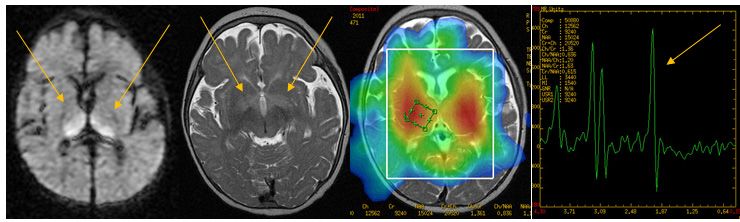
MedDocs Publishers
   Craniosynostosis – Apert syndrome                                         ate, cyclopia, hypoplastic face, and microphthalmia. Of the CNS
                                                                             anomalies, holoprosencephaly is the most prevalent (39%), but
   Apert syndrome is an autosomal dominant mutation in the                   the absence of holoprosencephaly does not exclude Trisomy 13
FGFR2 gene. Characteristic clinical features include craniosyn-              [23]. Few case reports of cross sectional imaging in postnatal
ostosis, facial dysmorphism, abnormal skull development, and                 infants over the age of 1 have documented milder morphologi-
syndactyly [17]. The CNS anomalies in Apert are a result of ei-              cal CNS manifestations of trisomy 13, which may be an indicator
ther primary malformations, or secondary to the osseous de-                  for better outcomes [24-26].
formity.
                                                                                Additional syndromes with microcephaly
    In a review of 30 patients with Apert syndrome, Quintero-
Rivera et al. documented the most common intracranial find-                     Two additional rare syndromes which have as a main fea-
ings as ventriculomegaly with cerebral volume loss (87% of pa-               ture microcephaly are Cockayne’s Syndrome and Pelizaeus-
tients), partial or complete absence of the septum pellucidum                Merzbacher disease. Cockayne’s Syndrome is a rare autosomal
(40%), malformations of the septum pellucidum (23%), and pro-                recessive condition comprising microcephaly, photosensitivity,
gressive hydrocephalus (13%) (Figure 4) [18]. The cranial mal-               cachectic dwarfism, progeria, and progressive neurological de-
formation can subsequently lead to tonsillar herniation, tem-                generation. Imaging findings include diffuse cerebral atrophy
poral lobe deformity, hydrocephalus, and encephaloceles [19].                and basal ganglia calcifications (Figure 7) [27,28].
Though not known to be associated with microcephaly, the sub-
mitted patient was noted to have the diagnoses. Microcephaly                     Pelizaeus-Merzbacher is an X linked autosomal recessive
is associated with craniosynostosis in the MCPH1 mutation [20].              syndrome presenting with microcephaly and a near complete
                                                                             lack of myelin. The disease can be categorized into three types;
   Sturge Weber angiomatosis                                                 classical, connatal, and transitional, which differ in their time of
                                                                             onset and clinical severity. The classical form is noted at infancy
   Sturge Weber is a neurocutaneous disorder characterized                   with lack of control of the head and nystagmus. Patients experi-
by facial port-wine stains, ocular choroidal hemangiomas, and                ence cognitive decline by age 5 or 6 with death in most patients
pial angiomatosis, with clinical manifestations of glaucoma,                 in late adolescence or young adulthood (Figure 8) [29].
seizures, and possible hemiparesis. Facial port-wine stains are
not specific for Sturge Weber, and are most often an isolated                   Non-syndromic microcephaly
uncomplicated finding. The underlying mechanism is impaired
cortical venous drainage due to an anomalous venous plexus                      Differential concerns for non-syndromic microcephaly in-
over the cerebral surface, leading to venous stasis and progres-             clude intracranial malformations, environmental factors such as
sive venous outflow obstruction. Collaterals form in the deep                fetal alcohol syndrome, hypoxic ischemic encephalopathy (HIE),
venous system to help provide cortical drainage, but are in-                 intrauterine infections, teratogens, as well as maternal disease,
adequate. CT and MRI will demonstrate abnormal intracranial                  such as phenylketonuria and poorly controlled maternal diabe-
calcifications, enlarged choroid plexus, cortical enhancement                tes [1]. It is important to recognize that microcephaly can be the
due to disruption of the blood brain barrier, and abnormal deep              sequelae of non accidental injury.
venous system (Figure 5) [21]. Characteristic white matter sig-                  The environmental causes have been extensively described
nal abnormality and atrophy are best noted on the T2 weighted                in other reviews, therefore we will focus primarily on HIE, a
and Fluid Attenuated Inversion Recovery (FLAIR) images.                      potentially devastating condition, and its manifestations. Fol-
   Susceptibility Weighted Imaging (SWI) and Diffusion Tensor                lowing this, microcephaly as the result TORCH infections will be
Imaging (DTI) also offer an additional benefit of assessing micro-           illustrated, with examples of HIV and Pneumococcus as well as
structural abnormalities in the early stages of Sturge Weber, al-            Herpes Simplex Two. Finally microcephaly, seen in non-acciden-
lowing for earlier interventions. SWI can detect transmedullary              tal injury, will be briefly discussed.
collateral veins in patients that have not yet developed cortical               Hypoxic ischemic encephalopathy
functional abnormality, as assessed by PET imaging. DTI in these
patients can demonstrate abnormal diffusion in the white mat-                    The pattern of brain injury by a perinatal ischemic event de-
ter tracts surrounding these transmedullary veins which drain                pends on the severity and duration of the ischemic event, as
the functionally normal cortex. Therefore, while the cortical                well as the stage of brain development during the event. A mild
integrity may not yet be compromised in this early stage, the                to moderate hypoxic-ischemic event can result in periventricu-
presence of transmedullary veins and the loss of integrity of the            lar leukomalacia or germinal matrix hemorrhage in preterm ne-
surrounding white matter surrounding veins may lead to future                onates. On the other hand, watershed infarcts can be expected
cognitive impairment [22].                                                   in full-term neonates.

   Trisomy 13 with holoprosencephaly                                            Cranial ultrasound is the initial imaging of choice and is highly
                                                                             sensitive for detecting intracranial hemorrhage, hydrocephalus,
   Trisomy 13 (Patau syndrome) is a chromosomal abnormality                  and cystic Periventricular Leukomalacia (PVL). However, ultra-
associated with CNS, facial/ocular, renal, and cardiac anomalies,            sound has lower sensitivity for detecting cortical involvement,
as well as polydactyly, rocker-bottom feet, and a single umbili-             and has marked inter observer and operator variability [30].
cal artery. It is nearly always fatal within the first year, therefore       MRI offers the most sensitive and specific evaluation of HIE. Dif-
postnatal imaging is sparse and the documented findings are                  fusion weighted imaging is excellent for detecting white mat-
predominantly based on prenatal ultrasound.                                  ter injury, while MR spectroscopy has shown high sensitivity for
   Based on a prenatal ultrasound study of 33 fetuses with Tri-              predicting the severity of brain injury in full term infants, per-
somy 13, the CNS abnormalities include holoprosencephaly,                    formed within 24 hours after birth. An elevated lactate/creatine
lateral ventricular dilatation, enlarged cisterna magna, and                 ratio on day 1 predicts a poor neurological outcome, whereas
microcephaly (Figure 6). Facial anomalies include cleft lip/pal-             the absence of lactate on day 1 predicts a normal outcome. MR
                                                                             spectroscopy is unfortunately not recommended in preterm
Journal of Radiology and Medical Imaging                                 3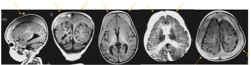
MedDocs Publishers
neonates as they show elevated lactate peaks [31,32]. CT can               the cortex and subcortical white matter, with parieto-occipital
also be useful since it does not require sedation, but offers less         and posterior temporal lobes typically more affected than the
sensitivity and specificity than MRI, with the adverse effect of           anterior regions. As the area of ischemia becomes infarcted,
radiation exposure [33,34].                                                cortical thinning with laminar necrosis and diminutive subcorti-
                                                                           cal white matter is demonstrated, with resultant ex vacuo dila-
   Periventricular leukomalacia                                            tation of the adjacent lateral ventricles (Figure 11).
   The periventricular white matter is the most vulnerable to                 The basal ganglia-thalamus pattern occurs due to inadequate
injury in preterm neonates with an immature brain, and mild to             shunting to the vital structures, and is seen with severe hypoxia
moderate hypoxia-ischemia will lead to periventricular leuko-              or cardiopulmonary arrest. Signal abnormality is present in the
malacia (PVL) [34]. Studies have demonstrated the distribution             ventrolateral thalami, posterior putamina, corticospinal tracts,
of watershed infarcts differs in preterm and term neonates as              and hippocampi. An additional finding is the loss of the normal
the watershed regions of the brain change as the fetus matures.            hyperintense focus seen in the posterior limb of the internal
Therefore, in preterm infants, watershed infarcts occur in the             capsule on T1-weighted images, called the absent posterior
periventricular regions, specifically adjacent to the trigone and          limb sign. It is important to note that this sign is useful after 72
frontal horns of the lateral ventricles. As the gestational age            hours from the onset of a severe ischemic event, as the normal
increases, watershed ischemia move peripherally to lie in the              signal may not be lost within the first 72 hours [40,41].
cortical and subcortical areas, in an adult like pattern [35].
                                                                              TORCH infections
    The predominant neuroimaging finding is loss of periven-
tricular white matter with cerebral volume loss. (Figure 9) [36].              The sequelae of neonatal infection and the effect on the
Increased T2 and FLAIR signal is typical in those fetus which are          developing brain is well documented. Furthermore, the age
able to mount a gliotic response (Figure 10) [37]. Impairment              of the fetus is the greatest predictor of insult type and degree.
of cerebral cortical gray matter development in premature in-              For example, if the fetus is exposed to the pathogen during the
fants with PVL has also been documented [38]. Periventricular              first and second trimester, there will be a propensity to develop
cavitation and cyst formation is seen in end-stage PVL, and cysts          congenital malformations. If the exposure is during the 3rd tri-
>3mm often carry a poorer prognosis [35].                                  mester and at birth, the result will be brain destruction with cal-
                                                                           cification and leukoencephalomalacia. For example, in neonatal
   Watershed infarcts in term neonates                                     Herpes Simplex Encephalitis, the fetal exposure occurs during
    Watershed infarcts are typically due to a perinatal acute              birth with symptoms manifesting within the first month of life
cerebral hypoperfusion event, leading to bilateral infarcts and            (Figure 12). Babies born with HIV can have additional infections,
subsequent brain atrophy [38]. As cerebral blood flow declines,            such as Pneumococcus (Figure 13). Since 2015, Zika infection
circulation is shunted from the anterior to the posterior circu-           has been a well documented etiology for microcephaly [42,43].
lation, to maintain supply to the vital structures such as the                Non accidental injury
brainstem, basal ganglia, and cerebellum. This leads to two pre-
dominant imaging patterns: A peripheral pattern (also known as                Children who have sustained non accidental injury can have
parasagittal, watershed, or borderzone), which is more common              a myriad of intracranial manifestations. Classically, the complex
and occurs with mild to moderate hypoxia, and a basal ganglia-             subdural hematoma is the hallmark, although neglect, resulting
thalamus pattern, which is less common and occurs with more                in brain shrinkage can result in microcephaly as well (Figure 14).
severe hypoxic episodes [39].                                              The prevalence of microcephaly following abusive head trauma
                                                                           was noted to be 93% in a study of 15 children who sustained
   The peripheral pattern demonstrates restricted diffusion in             abusive head injury and subsequently developed microcephaly
                                                                           [44].

   Figure 1: Cri-du-chat:
  Serial axial images demonstrate cerebral volume loss and left orbital retinal detachment. There is brain stem hypoplasia and a lack of
  myelination of the anterior limbs of the internal capsules (arrows).

Journal of Radiology and Medical Imaging                               4
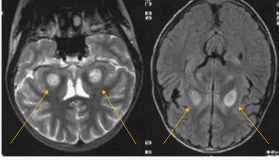
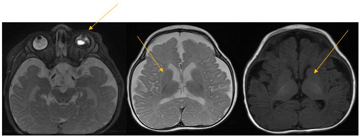
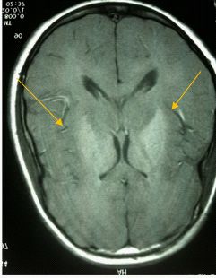
MedDocs Publishers

  Figure 2: Mitochondrial chromosomal abnormalities-Leigh’s Syndrome:
 Axial Diffusion, T2 weighted, and MR spectroscopy images demonstrate cortical atrophy, restricted diffusion in the thalamus (arrow), and a
 broad elevated lactate peak (arrow).

  Figure 3: Mitochondrial chromosomal abnormalities-Wilson’s Disease:
 Patient 1: Coronal T2 and axial flair images demonstrate bilateral symmetric increased signal in the putamen,
 globus pallidus and midbrain (arrow).
 Patient 2: Axial T1 weighted image reveals increased T1 signal in the basal ganglia (arrow).

  Figure 4: Craniosynostosis – Aperts Syndrome:
 Three dimensional reconstructed and axial images demonstrate bilateral coronal synostosis and microcephaly. There is ridging of the coronal
 sutures (arrow).

Journal of Radiology and Medical Imaging                              5
MedDocs Publishers Figure 5: Sturge Weber Angiomatosis: Axial Gradient echo and axial and coronal post-gadolinium T1 images demonstrate calcifications and associated pial enhance- ment typical of Sturge Weber encephalotrigeminal angiomatosis (arrows). Figure 6: Trisomy 13 with Holoprosencephaly: Axial and coronal T2 weighted images demonstrating forebrain fusion anomaly in this patient with trisomy 13 (arrows). There is microphthalmia on the first image (arrow). Figure 7: Cockayne Syndrome: CT: arrows demonstrate calcification in the cerebellum. Calcification is present in the frontal lobes as well. sagittal T1, coronal T2 and axial T2 reveal cerebral atrophy in this microcephalic patient (arrow). Figure 8: Pelizaeus-Merzbacher Disease: Sagittal T1 and axial T2 weighted images reveal a reduction in cerebral white matter which is not myelinated in a 9 year old patient (arrow demonstrates unmyelinated white matter in the right forceps minor and left forceps major). Journal of Radiology and Medical Imaging 6
MedDocs Publishers Figure 9: Non gliotic periventricular leukomalacia: Sagittal T1, axial T2 and coronal T2 weighted images reveal cerebral volume loss and reduction of white matter volume (arrows), including the corpus callosum in this 9 month preterm patient with microcephaly, developmental delay and hypertonicity. Figure 10: Periventricular Leukomalacia: Axial T2, coronal T2 and sagittal T1 images demonstrate hypoplasia of the splenium corpus callosum as well as gliosis in the coronal radiata (arrows). Figure 11: Watershed Infarcts: Coronal T2, axial T2, and axial T1 weighted images demonstrate sulcal and ventricular enlargement with leukoen- cephalomalacia involving the cerebral hemispheres in this patient with chronic watershed infarcts (arrows). The coronal T2 weighted image demonstrates severe atrophy of the cerebrum with relative preservation of cerebellar volume. Figure 12: Herpes Simplex Two: Serial axial T1 weighted images demonstrate extensive cerebral atrophy, cystic encephalomalacia and laminar necrosis, as represented as intrinsic increase T1 signal (arrow). Journal of Radiology and Medical Imaging 7
MedDocs Publishers

      Figure 13: HIV and Pneumococcus:
     Coronal T2 weighted images (left, center) and contrast enhanced axial t1 weighted images demonstrate extensive
     cerebral atrophy and cerebral white matter gliosis (arrows).

       Figure 14: Non accidental trauma:
     Sagittal, coronal and axial T1 weighted, axial T2 and axial T1 weighted images demonstrate complex subdural hematomas containing dif-
     ferent ages of blood products (arrows) seen in conjunction with cerebral atrophy.

      Conclusion
    Assessment of microcephaly requires a multidisciplinary                    7.    Everette James A, Atkins L, Feingold M, & Janower ML. The Cri
approach with important clinical and imaging components.                             du Chat Syndrome. Radiology. 1969; 92: 50-52.
Careful history taking can help exclude common environmen-
                                                                               8.    Tamraz J, Rethoré MO, Lejeune J, et al. Morphométrie
tal factors such as fetal alcohol syndrome or TORCH infections,                      encéphalique en irm dans la maladie du chat. A propos de sept
and imaging can help detect important causes such as non-acci-                       patients, avec revue de la littérature. Association pour la recher-
dental injury. Imaging also offers clues to diagnosing syndromic                     che sur la sclérose en plaques [Brain morphometry using MRI in
causes of microcephaly, and is integral to the evaluation of the                     Cri-du-Chat Syndrome. Report of seven cases with review of the
microcephalic pediatric patient.                                                     literature]. Ann Genet. 1993; 36: 75-87.

      References                                                               9.    Ninchoji T, Takanashi J. Pontine hypoplasia in 5p-syndrome: A
                                                                                     key MRI finding for a diagnosis. Brain Dev. 2010; 32: 571-573.
1.       Abuelo D. Microcephaly Syndromes. Semin. Pediatr. Neurol.
         2007; 14: 118–127.                                                    10.   Lee H, You S, Lee S, Cho H. Lack of Myelination in the Anterior
                                                                                     Limbs of the Internal Capsule Associated with Cri-du-Chat Syn-
2.       Woods CG. Human microcephaly. Current Opinion in Neurobiol-                 drome: Case Report Investig Magn Reson Imaging. 2015; 19:
         ogy. 2004; 14: 112–117.                                                     114-116.
3.       Passemard S, Kaindl AM, Verloes A. Microcephaly. Handb. Clin.         11.   Hong JH, Lee HY, Lim MK, et al. Brain stem hypoplasia associated
         Neurol. 2013; 111: 129–141.                                                 with Cri- du-Chat syndrome. Korean J Radiol. 2013; 14: 960-962.
4.       von der Hagen M, Pivarcsi M, Liebe J, von Bernuth H, et al. Di-       12.   Niyazov DM, Kahler SG, Frye RE. Primary Mitochondrial Disease
         agnostic approach to microcephaly in childhood: a two-center                and Secondary Mitochondrial Dysfunction: Importance of Dis-
         study and review of the literature. Dev Med Child Neurol. 2014;             tinction for Diagnosis and Treatment. Mol Syndromol. 2016; 7:
         56: 732-741.                                                                122-137.
5.       Hamosh A, Scott AF, Amberger J, Bocchini C, Valle D, McKusick         13.   Munnich A, Rötig A, Chretien D. et al. Clinical presentations and
         VA. Online Mendelian Inheritance in Man (OMIM), a knowl-                    laboratory investigations in respiratory chain deficiency. Eur J
         edgebase of human genes and genetic disorders. Nucleic Acids                Pediatr. 1996; 155: 262–274.
         Res. 2002; 30: 52-55.
                                                                               14.   Poretti A, Blaser SI, Lequin MH, et al. Neonatal neuroimaging
6.       Cerruti Mainardi P. Cri du Chat syndrome. Orphanet J Rare Dis.              findings in inborn errors of metabolism. J Magn Reson Imaging.
         2006; 1: 33.                                                                2013; 37: 294-312.
Journal of Radiology and Medical Imaging                                   8
MedDocs Publishers
15.   Gire C, Girard N, Nicaise C, Einaudi MA, Montfort MF, Dejode JM.        31.   Barkovich AJ, Westmark KD, Bedi HS, Partridge JC, Ferriero DM,
      Clinical features and neuroradiological findings of mitochondrial             et al. Proton spectroscopy and diffusion imaging on the first day
      pathology in six neonates. Childs Nerv Syst. 2002; 18: 621-628.               of life after perinatal asphyxia: preliminary report. AJNR Am J
                                                                                    Neuroradiol. 2001; 22: 1786-1794.
16.   Bianchi MC, Tosetti M, Battini R, et al. Proton MR spectroscopy
      of mitochondrial diseases: analysis of brain metabolic abnor-           32.   Zarifi MK, Astrakas LG, Poussaint TY, Plessis Ad Ad, Zurakowski
      malities and their possible diagnostic relevance. AJNR Am J Neu-              D, Tzika AA. Prediction of adverse outcome with cerebral lactate
      roradiol. 2003; 24: 1958-1966.                                                level and apparent diffusion coefficient in infants with perinatal
                                                                                    asphyxia. Radiology. 2002; 225: 859-870.
17.   Kutkowska-Kaźmierczak A, Gos M, Obersztyn E. Craniosynos-
      tosis as a clinical and diagnostic problem: molecular pathology         33.   Barkovich AJ, Miller SP, Bartha A, et al. MR imaging, MR spec-
      and genetic counseling. J Appl Genet. 2018; 59: 133-147.                      troscopy, and diffusion tensor imaging of sequential studies in
                                                                                    neonates with encephalopathy. AJNR Am J Neuroradiol. 2006;
18.   Quintero-Rivera F, Robson CD, Reiss RE, et al. Intracranial anom-             27: 533-547.
      alies detected by imaging studies in 30 patients with Apert syn-
      drome. Am J Med Genet A. 2006; 140: 1337-1338.                          34.   Bano S, Chaudhary V, Garga UC. Neonatal Hypoxic-ischemic En-
                                                                                    cephalopathy: A Radiological Review. J Pediatr Neurosci. 2017;
19.   Tan AP, Mankad K. Apert syndrome: Magnetic Resonance Imag-                    12: 1-6.
      ing (MRI) of associated intracranial anomalies. Childs Nerv Syst.
      2018; 34: 205-216.                                                      35.   Barkovich AJ, Truwit CL. Brain damage from perinatal asphyxia:
                                                                                    correlation of MR findings with gestational age. AJNR Am J Neu-
20.   Farooq M, Baig S, Tommerup N, Kjaer KW. Craniosynostosis-mi-                  roradiol. 1990; 116: 1087-1096.
      crocephaly with chromosomal breakage and other abnormali-
      ties is caused by a truncating MCPH1 mutation and is allelic to         36.   Flodmark O, Lupton B, Li D, et al. MR imaging of periventricu-
      premature chromosomal condensation syndrome and primary                       lar leukomalacia in childhood. AJR Am J Roentgenol. 1989; 152:
      autosomal recessive microcephaly type 1. Am J Med Genet A.                    583-590.
      2010; 152: 495-497.
                                                                              37.   Bodensteiner JB, Johnsen SD. Magnetic Resonance Imaging
21.   Vogl T, Stemmler J, Bergman C, Pfluger T, E. et al. MR and MR An-             (MRI) Findings in Children Surviving Extremely Premature Deliv-
      giography of Sturge-Weber Syndrome. AJNR Am. J. Neuroradiol.                  ery and Extremely Low Birthweight With Cerebral Palsy. Journal
      1993; 14: 417–425.                                                            of Child Neurology. 2006; 21: 743-747.

22.   Juhász C, Haacke EM, Hu J, et al. Multimodality imaging of corti-       38.   Inder TE, Huppi PS, Warfield S, et al. Periventricular white mat-
      cal and white matter abnormalities in Sturge-Weber syndrome.                  ter injury in the premature infant is followed by reduced cere-
      AJNR Am J Neuroradiol. 2007; 28: 900-906.                                     bral cortical gray matter volume at term. Ann Neurol. 1999; 46:
                                                                                    755-760.
23.   Lehman CD, Nyberg DA, Winter TC 3rd, Kapur RP, Resta RG, Lu-
      thy DA. Trisomy 13 syndrome: prenatal US findings in a review of        39.   Rutherford MA. MRI of the Neonatal Brain. Gulf Professional
      33 cases. Radiology. 1995; 194: 217-222.                                      Publishing. 2002.

24.   Goff RD, Soares BP. Neuroradiological findings of trisomy 13 in a       40.   Ghei SK, Zan E, Nathan JE, Choudhri A, Tekes A, et al. MR imaging
      rare long-term survivor. Neuroradiol J. 2018; 31: 412-414.                    of hypoxic-ischemic injury in term neonates: Pearls and pitfalls.
                                                                                    Radiographics: A review publication of the Radiological Society
25.   Imataka G, Yamanouchi H, Hirato J, et al. Autopsy report of a                 of North America, Inc. 34; 1047-1061.
      7-year old patient with the mosaic trisomy 13. Cell Biochem Bio-
      phys. 2013; 67: 813-817.                                                41.   Rutherford MA, Pennock JM, Schwieso JE, Cowan FM, Dubowitz
                                                                                    LM. Hypoxic ischaemic encephalopathy: Early magnetic reso-
26.   Zoll B, Wolf J, Lensing-Hebben D, Pruggmayer M, Thorpe B. Tri-                nance imaging findings and their evolution. Neuropediatrics.
      somy 13 (Patau syndrome) with an 11-year survival. Clin Genet.                1995; 26: 183-191.
      1993; 43: 46-50.
                                                                              42.   Hanzlik E, Gigante J. Microcephaly. Children (Basel). 2017; 4: 47.
27.   Tan W, Baris H, Robson C, Kimonis V. Cockayne syndrome: The
      developing phenotype. Am. J. Med. Genet. 2005; 135: 214-216.            43.   Faizan M, I, Abdullah M, Ali S, Naqvi I, H, Ahmed A, Parveen
                                                                                    S: Zika Virus-Induced Microcephaly and Its Possible Molecular
28.   Koob M, Laugel VM. Durand MH. Fothergill H, et al. Neuroimag-                 Mechanism. Intervirology. 2016; 59: 152-158.
      ing In Cockayne Syndrome. American Journal of Neuroradiology.
      2010; 31: 1623-1630.                                                    44.   Lo TY, McPhillips M, Minns RA, Gibson RJ. Cerebral atrophy fol-
                                                                                    lowing shaken impact syndrome and other non-accidental head
29.   van der Knaap M, Breiter S, Naidu S, Hart A, et al. Defining and              injury (NAHI). Pediatr Rehabil. 2003; 6: 47-55.
      Categorizing Leukoencephalopathies of Unknown Origin: MR
      Imaging Approach. Radiology. 1999; 213:121-133.                         45.   Pryor HB, Thelander H. Abnormally small head size and intellect
                                                                                    in children. The Journal of Pediatrics. 1968; 73: 593-598.
30.   Benson JE, Bishop MR, Cohen HL. Intracranial neonatal neuro-
      sonography: an update. Ultrasound Q. 2002; 18: 89-114.

Journal of Radiology and Medical Imaging                                  9
You can also read