A novel diagnostic signature based on three circulating exosomal mircoRNAs for chronic obstructive pulmonary disease
←
→
Page content transcription
If your browser does not render page correctly, please read the page content below
EXPERIMENTAL AND THERAPEUTIC MEDICINE 22: 717, 2021

                  A novel diagnostic signature based on three
                 circulating exosomal mircoRNAs for chronic
                        obstructive pulmonary disease
              YAHUI SHEN1,2, LINA WANG1, YUNHUI WU1, YINGWEI OU1, HUIYU LU2 and XIN YAO1

     1
      Department of Respiratory and Critical Care Medicine, The First Affiliated Hospital of Nanjing Medical University,
      Nanjing, Jiangsu 210029; 2Department of Respiratory and Critical Care Medicine, Taizhou Clinical Medical School of
                             Nanjing Medical University, Taizhou, Jiangsu 225300, P.R. China

                                       Received October 31, 2020; Accepted March 18, 2021

                                                     DOI: 10.3892/etm.2021.10149

Abstract. Exosomal microRNAs (exo‑miRNAs or miRs) have                 Of these, nine were then selected for subsequent analysis,
demonstrated diagnostic value in various diseases. However,            five of which were found to be upregulated (miR‑23a, miR‑1,
their diagnostic value in chronic obstructive pulmonary                miR‑574, miR‑152 and miR‑221) and four of which were down‑
disease (COPD) has yet to be fully established. The purpose            regulated (miR‑3158, miR‑7706, miR‑685 and miR‑144). The
of the present study was to screen differentially expressed            results of Gene Ontology and KEGG pathway analysis revealed
exo‑miRNAs in the plasma of patients with COPD and healthy             that these miRNAs were mainly involved in certain biological
individuals and to evaluate their potential diagnostic value in        functions, such as metabolic processes, such as galactose
COPD. Differentially expressed exo‑miRNAs in the plasma                metabolism and signaling pathways (PI3K‑AKT) associated
of patients with COPD and controls were identified using               with COPD. The expression levels of three exo‑miRNAs
high‑throughput sequencing and confirmed using reverse                 (miR‑23a, miR‑221 and miR‑574) were found to be nega‑
transcription‑quantitative PCR (RT‑qPCR). Bioinformatics               tively associated with the forced expiratory volume in the 1st
analysis was then performed to predict the function of the             second/forced vital capacity. Furthermore, the area under the
selected exo‑miRNAs and their target genes in COPD.                    curve values of the three exo‑miRNAs (miR‑23a, miR‑221 and
After a network model was constructed, linear regression               miR‑574) for COPD diagnosis were 0.776 [95% confidence
analysis was performed to determine the association between            interval (CI), 0.669‑0.882], 0.688 (95% CI, 0.563‑0.812) and
exo‑miRNA expression and the clinical characteristics of               0.842 (95% CI, 0.752‑0.931), respectively. In conclusion, the
subjects in a validated cohort (46 COPD cases; 34 matched              three circulating exosomal miRNAs (miR‑23a, miR‑221
healthy controls). Receiver operating characteristic curve was         and miR‑574) may serve as novel circulating biomarkers for
subsequently plotted to test the diagnostic value of the candi‑        the diagnosis of COPD. These results may also enhance our
date biomarkers. The top 20 significantly aberrantly expressed         understanding and provide novel potential treatment options
COPD‑associated exo‑miRNAs were verified using RT‑qPCR.                for patients with COPD.

                                                                       Introduction

Correspondence to: Professor Xin Yao, Department of Respiratory        Chronic obstructive pulmonary disease (COPD) is a
and Critical Care Medicine, The First Affiliated Hospital of Nanjing   common respiratory disease with a high rate of morbidity
Medical University, 300 Guangzhou Road, Nanjing, Jiangsu 210029,       and mortality (1‑3). As the rates of COPD incidence have
P.R. China                                                             increased, so too has its social and economic burden. It is
E‑mail: groupyaoxin@163.com                                            estimated that by 2020, COPD will rank fifth in the list of
                                                                       diseases that most impacts the world's economy (4). The
Abbreviations: COPD, chronic obstructive pulmonary disease;            pathological characteristics of COPD include chronic inflam‑
miRNAs, microRNAs; Exo‑miRNA, exosomal microRNA;
                                                                       mation of the lung parenchyma and the surrounding airways,
TSG101, tumor susceptibility gene 101; RT‑qPCR, reverse
                                                                       emphysema and small airway stenosis or remodeling (5‑7). A
transcription‑quantitative PCR; KEGG, Kyoto Encyclopedia of
Genes and Genomes; GO, Gene Ontology; ROC, receiver operating          number of processes, including oxidative stress, inflammation
characteristic; AUC, area under the curve; FEV1, forced expiratory     and mitochondrial dysfunction have all been reported to serve
volume in the 1st second; FVC, forced vital capacity                   mechanistical roles (8‑11). However, additional research is
                                                                       required to determine the molecular mechanisms underlying
Key words: exosome, microRNA, chronic obstructive pulmonary            COPD.
disease, biomarkers, diagnosis                                             MicroRNAs (miRNA or miRs) are a group of non‑coding
                                                                       RNAs that consist of 18‑25 nucleotides and serve to increase
                                                                       mRNA degradation or repress target‑specific mRNA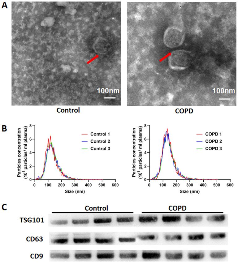
2                                    SHEN et al: CIRCULATING EXOSOMAL miRNAs FOR COPD

translation (12). miRNAs can modulate almost all biological       forced vital capacity (FVC) value,
EXPERIMENTAL AND THERAPEUTIC MEDICINE 22: 717, 2021                                           3

plasma exosomes. Brownian particles were tracked, following         oxyhemoglobin was then determined via spectrophotometry
which their hydrodynamic diameters and concentrations               at 414 nm. Once hemolysis occurred, the specimen was
were calculated using the Stockes‑Einstein equation (36).           discarded. Total RNA was extracted from plasma exosomes
NTA assay was performed using the ZetaView PMX 110                  using TRIzol® reagent (Invitrogen; Thermo Fisher Scientific,
(Particle Metrix GmbH). The data obtained with the ZetaView         Inc.). The primers (One RT primer and a pair of qPCR primers
instrument were analysed using the corresponding software           for each set) specific for miRNAs were designed by RiboBio
ZetaView v.8.02.28 (Particle Metrix GmbH).                          (Guangzhou RiboBio Co., Ltd.). The primer sequences were
                                                                    patented. cDNA was synthesized from 500 ng RNA using
Western blotting. Western blotting was used to detect the           a reverse transcriptase kit (cat. no. R10031.8; Guangzhou
specific marker proteins (CD9, CD63, TSG101) on the                 RiboBio Co., Ltd.) with the following temperature protocol:
surface of plasma exosomes. Exosome samples were lysed              42˚C for 60 min, and followed by 70˚C for 10 min. qPCR
using RIPA lysis buffer (cat. no. 20‑188; Sigma‑Aldrich,            was carried out in accordance with the protocols of the
Merck KGaA) on ice for 30 min. A bicinchoninic acid (BCA)           Bulge‑Loop™ miRNA qRT‑PCR Starter kit (cat. no. c10211‑1;
Protein Assay kit (cat. no. 23225; Pierce; Thermo Fisher            Guangzhou RiboBio Co., Ltd.) and ABI Prism 7900HT
Scientific, Inc.) was used to detect protein concentrations, and    (Applied Biosystems; Thermo Fisher Scientific, Inc.). The
a 12% sodium dodecyl sulfate‑polyacrylamide gel was used            following thermocycling conditions were used: Initial dena‑
for total protein (30 µg/lane) separation. The proteins in the      turation at 95˚C for 20 sec, followed by 40 cycles of 95˚C for
gel were transferred to a 0.45‑µM pore size PVDF membrane           10 sec, 60˚C for 20 sec and 70˚C for 10 sec. U6 small nuclear
(cat. no. IPVH00010; EMD Millipore) via wet electrophoretic         RNA and cel‑miR‑39 were used as endogenous (39) and exog‑
transfer. The membranes were blocked with 5% skimmed milk           enous controls, respectively. The expression of exo‑miRNAs
powder for 1 h at room temperature and incubated overnight          was calculated using the 2‑ΔΔCq method (40).
at 4˚C with anti‑CD9 (cat. no. ab92726; Abcam), anti‑CD63
(cat. no. ab216130), anti‑TSG101 (cat. no. ab125011) antibodies,    GO and KEGG pathway analysis. TargetScan (version 7.2,
all diluted to 1:1,000 in TBS‑1% Tween‑20. Subsequently,            https://www.targetscan.org/), miRDB (version 5.0, http://www.
the membranes were incubated with a horseradish perox‑              targetscan.org/), miRTarBase (version 7.0, http://starbase.sysu.
idase‑conjugated goat anti rabbit (1:3,000; cat. no. A0208;         edu.cn/) and miRWalk (version 7.0, http://mirwalk.umm.
Beyotime Institute of Biotechnology) or goat anti mouse IgG         uni‑heidelberg.de/) databases were used to predict the target
secondary antibodies (1:3,000; cat. no. A0216; Beyotime             genes of selected miRNAs. Those simultaneously predicted by
Institute of Biotechnology) for 1 h at room temperature.            ≥ two of the tools aforementioned were selected as candidate
The membranes were visualized using LumiBest enhanced               target genes. KOBAS release number 3.0 (http://kobas.cbi.
chemiluminescence (cat. no. SB‑WB011; Shanghai Shenger              pku.edu.cn/) was used for further GO and KEGG pathway
Biotechnology Co., Ltd.).                                           analyses (41). The significance of GO and KEGG pathway
                                                                    analysis was then determined using Fisher's exact and
Exosomal miRNA sequencing. A total of 4 ml plasma was mixed         χ2 tests. The subsequent P‑value was corrected using the false
with Ribo™ Exosome Isolation Reagent (Guangzhou RiboBio             discovery rate (FDR). GO and KEGG terms with an adjusted
Co., Ltd.), after which exosomes were isolated. Exosomal RNA        P‑value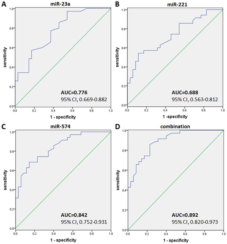
4                                       SHEN et al: CIRCULATING EXOSOMAL miRNAs FOR COPD

Table I. Characteristics of patients.

Parameters                                             Control                              COPD                              P‑value

Number of subjects                                        34                                  46                                NA
Male/Female                                              27/7                               36/10                               NA
Age, years (range)                                     61.2±6.3                            62.3±5.6                            >0.05
Smoking, pack years                                      N/A                              52.6±12.5
EXPERIMENTAL AND THERAPEUTIC MEDICINE 22: 717, 2021 5
Figure 1. Isolation of plasma‑derived exosomes from patients with COPD and healthy individuals. (A) Representative transmission electron microscopy images
of plasma‑derived exosomes isolated using the ExoQuick method. Scale bar, 100 nm. (B) Nanoparticle tracking analysis of the plasma‑derived exosomes.
Representative graphs showing the concentration and size of the isolated particles are presented. n=3/group. (C) Western blotting was performed to detect
TSG101, CD63 and CD9 expression, protein markers of exosomes. TSG101, CD63 and CD9, were highly expressed in the exosomes separated from the plasma
samples of patients with chronic obstructive pulmonary disease and healthy individuals. n=5/group. COPD, chronic obstructive pulmonary disease; TSG101,
tumor susceptibility gene 101.
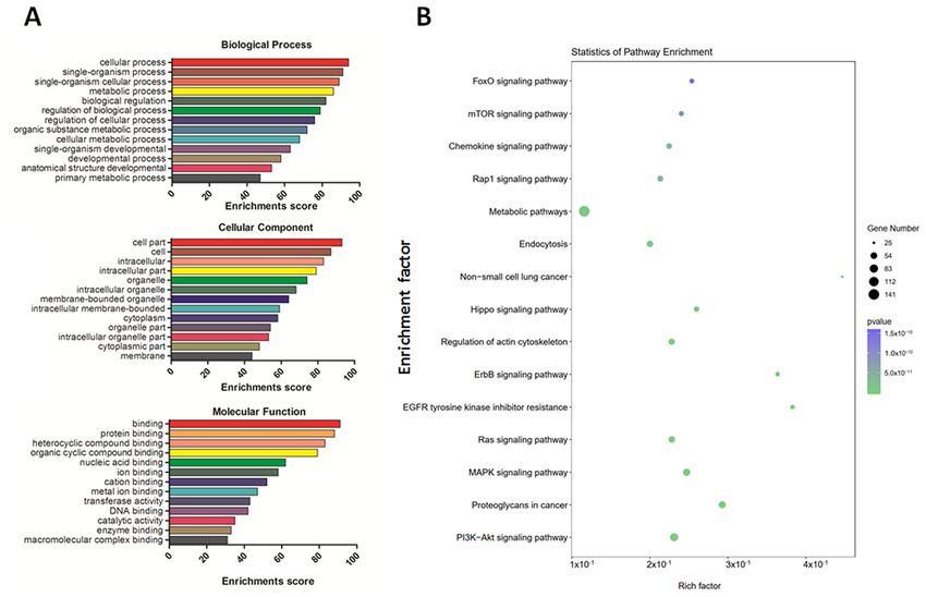
‘heterocyclic compound binding’, ‘nucleic acid binding’ and and 98 mRNAs (Fig. 5). This network confirmed that one
‘transferase activity’ (Fig. 3A). The primarily enriched path‑ exo‑miRNA targeted one or two mRNAs, and one mRNA
ways included the mTOR, chemokine, MAPK and PI3K‑AKT was regulated by multiple miRNAs simultaneously, which
signaling pathways (Fig. 3B). suggested that a regulatory mechanism existed between
exo‑miRNAs and mRNAs in COPD.
Validation of dysregulated miRNAs in patients with COPD.
The 20 exo‑miRNAs with the most significant differential Correlation between exo‑miRNA expression with clinical
expression in patients with COPD were further validated via parameters. The correlation between the exo‑miRNA expres‑
RT‑qPCR. Nine of the exo‑miRNAs were determined to be sion levels and the FEV1/FVC value was further assessed
significantly differentially expressed by exosome sequencing, using linear regression. Potential confounding factors were
including five that were upregulated (miR‑23a, miR‑1, adjusted, including age, sex, smoking status and corticosteroid
miR‑574, miR‑152 and miR‑221) and four that were downregu‑ therapy were adjusted using Bonferronis correction. The
lated (miR‑3158, miR‑7706, miR‑685 and miR‑144) in patients results indicated that the expression levels of three upregulated
with COPD compared with those in healthy individuals. These exo‑miRNAs (miR‑23a, miR‑221 and miR‑574) correlated
results were consistent with data obtained using RT‑qPCR significantly with the FEV1/FVC values, even after adjusting
(Fig. 4). However, expression of the other 11 exo‑miRNAs for the confounding factors. The correlation analysis between
did not significantly differ between patients with COPD and exo‑miRNA and FEV1/FVC is presented in Fig. 6. miR‑23a
healthy controls. (R 2 =0.706; P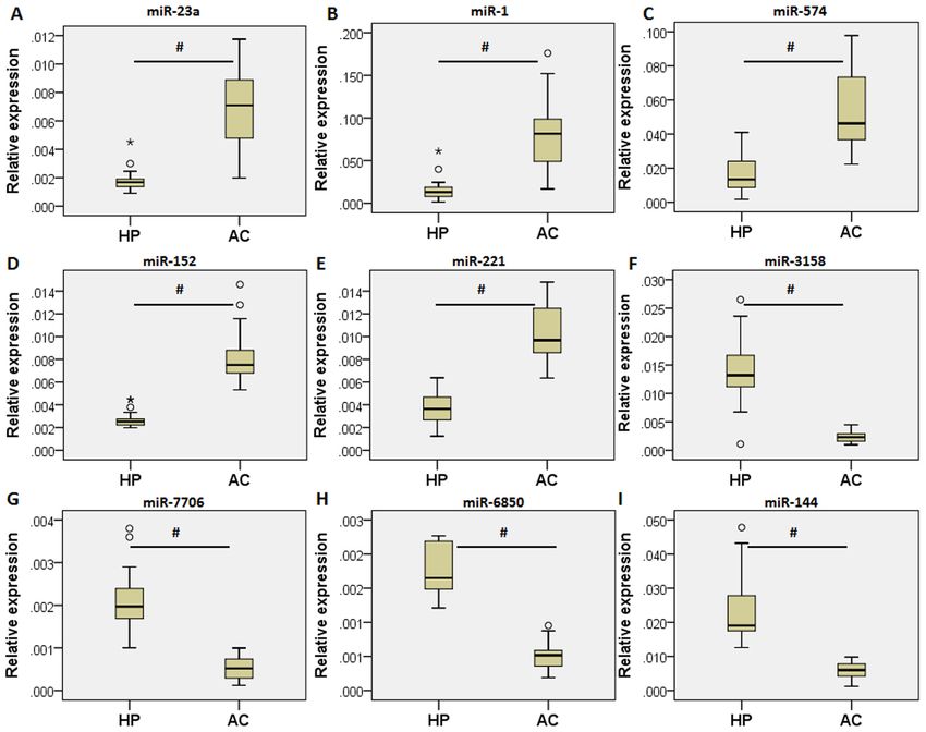
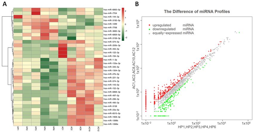
6                                          SHEN et al: CIRCULATING EXOSOMAL miRNAs FOR COPD

Figure 2. Hierarchical clustering (made with R language package) and scatter plot results of differentially expressed exo‑miRNAs in the plasma of
patients with COPD and healthy individuals. (A) Hierarchical clustering images of the exo‑miRNA expression of pooled RNA samples from the plasma
of patients with COPD. Brick‑red indicates upregulated miRNA and green indicates downregulated miRNAs. (B) Scatter plot of miRNA expression.
Values on the x‑ and y‑axis represent the normalized signal values of the samples (log2 scaled). Red and green dots represent up‑ and downregulated
miRNAs, respectively. Exo, exosomal; miRNA, microRNA; COPD, chronic obstructive pulmonary disease; HP, healthy individuals; AC, patients with
COPD.

Figure 3. Functional analysis of exo‑miRNAs. (A) Gene Ontology enrichment histogram of the differentially expressed exo‑miRNAs. (B) Pathway
analysis based on exo‑miRNAs target genes. P‑values represent the significance level of target gene enrichment in this pathway. Exo, exosomal; miRNAs,
microRNAs.
EXPERIMENTAL AND THERAPEUTIC MEDICINE 22: 717, 2021                                                           7

Figure 4. Validation of the differentially expressed exosomal‑miRNAs (41 patients with chronic obstructive pulmonary disease and 29 healthy controls).
Relative expression of (A) miR‑23a, (B) miR‑1, (C) miR‑574, (D) miR‑152, (E) miR‑221, (F) miR‑3158, (G) miR‑7706, (H) miR‑6850 and (I) miR‑144 in the
validation cohort. Data are presented as median. #P
8 SHEN et al: CIRCULATING EXOSOMAL miRNAs FOR COPD
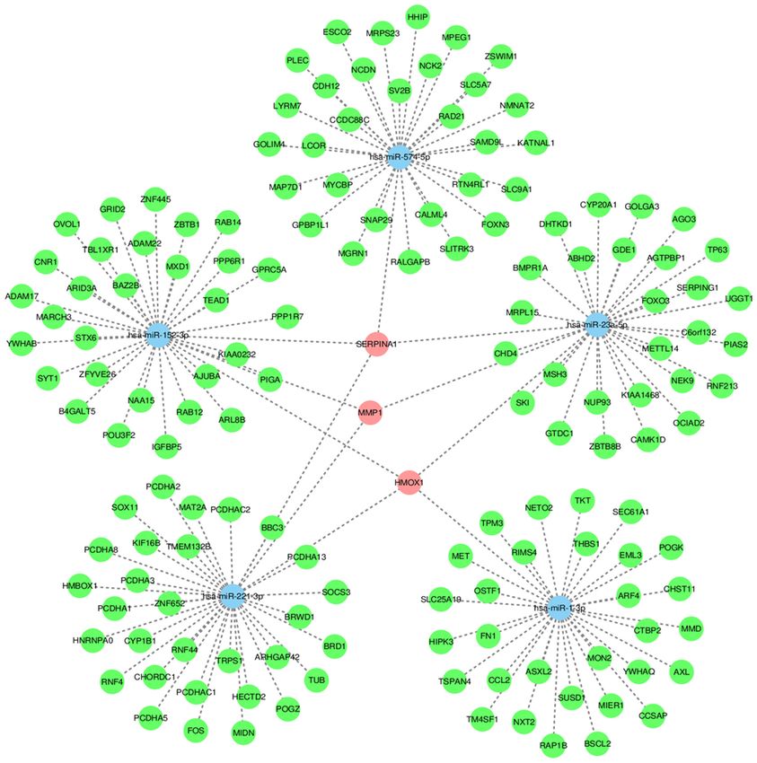
Figure 5. Regulatory network between exosomal‑miRNAs and mRNAs. The miRNA‑mRNA co‑expression interaction network was constructed using the
Cytoscape software. The network consists of five nodes (miRNA). Blue and green circles represent miRNAs and mRNAs, respectively. The pink nodes signify
that the genes have a targeted regulatory relationship with two or more miRNAs. The interaction between two factors is represented by one edge. miRNA,
microRNA.
eight miRNAs in smokers with COPD was significantly lower Furthermore, 59 differentially expressed exo‑miRNAs were
compared with that in non‑smokers. The results of the present identified, including 39 that were upregulated and 20 that were
study differed from the aforementioned studies. This was downregulated. GO enrichment analysis provides a unified
mainly as miRNAs were extracted from different samples, vocabulary to elaborate gene and gene product properties in
namely airway epithelial cells, serum or lung tissues, diffi‑ various organisms (56). GO analysis in the present study indi‑
culty remains in obtaining a comprehensive comparison with cated that the identified exo‑miRNAs were enriched in various
previous experimental results. In addition, these results may annotations associated with COPD, as demonstrated by the
demonstrate discrepancy, arising from other factors, including top enriched biological processes and molecular functions,
different sample sizes, analytical tools and statistical methods. including ‘metabolic process’, ‘biological regulation’, ‘intra‑
Results from the current study suggested that exosomal cellular membrane‑bounded organelle’, ‘transferase activity’,
miRNAs were differentially expressed in patients with COPD. ‘catalytic activity’ and ‘enzyme binding’.EXPERIMENTAL AND THERAPEUTIC MEDICINE 22: 717, 2021 9
Figure 6. Correlation analysis of exo‑miRNA expression and FEV1/FVC%. Correlation between the expression of exo‑miRNAs (A) miR‑23a, (B) miR‑221
and (C) miR‑574 independently and FEV1/FVC% are presented as scatter plots, combined with the regression line. Data are derived from the validation
cohort (n=70). Exo‑miRNA, exosomal microRNA; FEV1, forced expiratory volume in the 1st second; FVC, forced vital capacity; COPD, chronic obstructive
pulmonary disease.
High‑throughput sequencing, such as small RNA metalloproteinase 1 (MMP1), heme oxygenase‑1 (HOMX1)]
sequencing technology makes it possible to measure the of the 5 miRNAs, which are closely related to the pathogenesis
expression of almost all coding genes, which assists in of COPD. SERPINA1 has been shown to affect the suscepti‑
identifying genes and pathways related to the development bility of COPD (57). Dysregulation in the production of MMP
of diseases (51). In the present study, based on the KEGG has been associated with lung matrix destruction and small
pathway result, the target genes of dysregulated exo‑miRNAs airways disease in COPD (58). HOMX1 (induction attenu‑
were involved in pathways that are closely associated with the ated senescence in chronic obstructive pulmonary disease
pathophysiology of COPD, such as the mTOR, chemokine, lung fibroblasts by protecting against mitochondria dysfunc‑
MAPK and PI3K‑Akt signaling pathways. Additionally, the tion (59).
present network analysis indicated a potential association Exo‑miRNAs that were differentially expressed between
between miRNAs and their target genes, suggesting that patients with COPD and healthy controls were screened and
exo‑miRNAs and their target genes cooperate to regulate analyzed in an independent validation cohort. Through this
the pathogenesis of COPD. Furthermore, to determine the method, nine differentially expressed exo‑miRNAs were
function of the identified exo‑miRNAs, interactions between identified, of which five were upregulated and four were
exo‑miRNAs and their target mRNAs were theoretically downregulated. To remove various confounding factors, linear
predicted using conserved seed‑matching sequences with regression analysis was performed to assess the relationship
software for miRNA target prediction, such as TargetScan and between plasma miRNA expression and FEV1/FVC. As a
miRDB. This network suggested the potential associations result, three exo‑miRNAs (miR‑23a, miR‑221 and miR‑574)
between exo‑miRNAs and their target genes. The network were significantly correlated with FEV1/FVC after adjusting
also provided an important reference value for studying the for age, sex and treatment with corticosteroids. These results
interaction of other differentially expressed exo‑miRNAs suggested that these 3 exo‑miRNAs may be used to assess the
with their potential targets. The current study predicted that severity of COPD lung function.
the interaction of exo‑miRNAs and their target genes was Exosomal molecules have the potential to serve as disease
associated with COPD. The present study found 3 common biomarkers for a number of reasons. Exosomes contain
target genes [encoding α1‑antitrypsin (SERPINA1), matrix specific proteins and nucleic acids that carry information10 SHEN et al: CIRCULATING EXOSOMAL miRNAs FOR COPD
Figure 7. Receiver operating characteristic curves for three candidate exosomal‑microRNAs. (A) miRNA‑23a, (B) miR‑221, (C) miR‑574 and (D) Combination
of three miRNAs. miR, microRNA; AUC, area under the curve.
regarding the physiology and microenvironment of their cells number of limitations. A single center, small sample size,
of origin (60‑62). In addition, exosomes can exist in various various forms of bias in the present study may lead to inac‑
biological fluids, including blood (25), urine (63), sputum, curate conclusions. Therefore, a large‑scale, multi‑center and
bronchoalveolar lavage (26), synovial fluid (64), pleural fluid prospective validation study should be performed in future
and ascites (65). Due to the bilayer structure of phospholipids, studies. Additionally, the underlining mechanism of the asso‑
exosomes are highly stable in the extracellular environ‑ ciation between exosomal miRNAs and COPD remain unclear
ment (66). Therefore, many exosomal proteins and miRNAs and should be investigated further.
have been reported to be potential biomarkers of multiple In summary, to the best of our knowledge, the present
diseases, particularly in cancer (22,23). There is considerable study determined for the first time that miR‑23a, miR‑221 and
evidence to support the notion that exo‑miRNAs can serve miR‑574 may serve as novel diagnostic biomarkers of COPD.
an important role in multiple pulmonary diseases, such as The results indicated that the evaluation of exosomal miRNA
COPD (67). ROC analysis in the present study revealed that expression could provide useful information regarding the
three candidate miRNAs (miR‑23a, miR‑221 and miR‑574) diagnosis of patients with COPD.
can be used as new biomarkers of COPD. Furthermore, when
the three exo‑miRNAs were combined together, the diagnostic Acknowledgements
efficiency was improved further.
The present study provided genome‑wide profiles of Not applicable.
exosomal miRNAs from human blood samples, demonstrating
the feasibility of identifying COPD biomarkers based on Funding
exosomal miRNA profiling. Additionally, due to the double
membrane structure of exosomes, exosomal miRNAs can The present study was supported by grants from the National
overcome certain limitations of current biomarkers due to Natural Science Foundation of China (grant no. 81870039),
increased stability. However, the present study also had a the Jiangsu Provincial Medical Youth Talent (grantEXPERIMENTAL AND THERAPEUTIC MEDICINE 22: 717, 2021 11
no. QNRC2016512) and the Taizhou Municipal Science and 9. Demedts IK, Demoor T, Bracke KR, Joos GF and Brusselle GG:
Role of apoptosis in the pathogenesis of COPD and pulmonary
Technology Bureau (grant no. TS201729). emphysema. Respir Res 7: 53, 2006.
10. Adcock IM, Tsaprouni L, Bhavsar P and Ito K: Epigenetic regu‑
Availability of data and materials lation of airway inflammation. Curr Opin Immunol 19: 694‑700,
2007.
11. Wan ES and Silverman EK: Genetics of COPD and emphysema.
The datasets used and/or analyzed during the current study are Chest 136: 859‑866, 2009.
available in the National Center for Sequence Read Archive 12. Bartel DP: MicroRNAs: Genomics, biogenesis, mechanism, and
function. Cell 116: 281‑297, 2004.
(SRA) data, (SRA; https://www.ncbi.nlm.nih.gov/sra/; acces‑ 13. Hough KP, Chanda D, Duncan SR, Thannickal VJ and
sion no. PRJNA703816). Deshane JS: Exosomes in immunoregulation of chronic lung
diseases. Allergy 72: 534‑544, 2017.
14. Murgoci AN, Duhamel M, Raffo‑Romero A, Mallah K,
Authors' contributions Aboulouard S, Lefebvre C, Kobeissy F, Fournier I, Zilkova M,
Maderova D, et al: Location of neonatal microglia drives small
YS and XY designed the study and drafted the manuscript. YS extracellular vesicles content and biological functions in vitro.
J Extracell Vesicles 9: 1727637, 2020.
and LW performed the experiments. YW and YO performed 15. Nana‑Sinkam SP, Acunzo M, Croce CM and Wang K:
the statistical analysis. YO and HL performed sample collec‑ Extracellular vesicle biology in the pathogenesis of lung disease.
tion. HL diagnosed the patients and revised the manuscript Am J Respir Crit Care Med 196: 1510‑1518, 2017.
16. Stolzenburg LR and Harris A: The role of microRNAs in chronic
for important intellectual content. YS and XY confirm all the respiratory disease: Recent insights. Biol Chem 399: 219‑234,
authenticity of the raw data. All authors read and approved the 2018.
final manuscript. 17. De Smet EG, Mestdagh P, Vandesompele J, Brusselle GG and
Bracke KR: Non‑coding RNAs in the pathogenesis of COPD.
Thorax 70: 782‑791, 2015.
Ethics approval and consent to participate 18. Molina‑Pinelo S, Pastor MD, Suarez R, Romero‑Romero B,
González De la Peña M, Salinas A, García‑Carbonero R,
De Miguel MJ, Rodríguez‑Panadero F, Carnero A and
The present study was approved by the research ethics Paz‑Ares L: MicroRNA clusters: Dysregulation in lung adeno‑
committee of Taizhou People's Hospital (Taizhou clinical carcinoma and COPD. Eur Respir J 43: 1740‑1749, 2014.
Medical School of Nanjing Medical university; Taizhou, 19. Robbins PD and Morelli AE: Regulation of immune responses
by extracellular vesicles. Nat Rev Immunol 14: 195‑208, 2014.
China). All individuals provided informed consent for the use 20. Colombo M, Raposo G and Théry C: Biogenesis, secretion, and
of their samples for clinical research. intercellular interactions of exosomes and other extracellular
vesicles. Annu Rev Cell Dev Biol 30: 255‑289, 2014.
21. Yoshioka Y, Konishi Y, Kosaka N, Katsuda T, Kato T and
Patient consent for publication Ochiya T: Comparative marker analysis of extracellular vesicles
in different human cancer types. J Extracell Vesicles 2, 2013.
Not applicable. 22. Mirzaei H, Sahebkar A, Jaafari MR, Goodarzi M and Mirzaei HR:
Diagnostic and therapeutic potential of exosomes in cancer: The
beginning of a new tale? J Cell Physiol 232: 3251‑3260, 2017.
Competing interests 23. Saadatpour L, Fadaee E, Fadaei S, Nassiri Mansour R,
Mohammadi M, Mousavi SM, Goodarzi M, Verdi J and
Mirzaei H: Glioblastoma: Exosome and microRNA as novel
The authors declare that they have no competing interests. diagnosis biomarkers. Cancer Gene Ther 23: 415‑418, 2016.
24. Sun L, Zhu W, Zhao P, Wang Q, Fan B, Zhu Y, Lu Y, Chen Q,
References Zhang J and Zhang F: Long noncoding RNA UCA1 from
hypoxia‑conditioned hMSC‑derived exosomes: A novel molec‑
ular target for cardioprotection through miR‑873‑5p/XIAP axis.
1. Wang C, Xu J, Yang L, Xu Y, Zhang X, Bai C, Kang J, Ran P, Cell Death Dis 11: 696, 2020.
Shen H, Wen F, et al: Prevalence and risk factors of chronic 25. Caby MP, Lankar D, Vincendeau‑Scherrer C, Raposo G and
obstructive pulmonary disease in China [the China pulmonary Bonnerot C: Exosomal‑like vesicles are present in human blood
health (CPH) study]: A national cross‑sectional study. Lancet 391: plasma. Int Immunol 17: 879‑887, 2005.
1706‑1717, 2018. 26. Admyre C, Grunewald J, Thyberg J, Gripenbäck S, Tornling G,
2. GBD 2015 Chronic Respiratory Disease Collaborators: Global, Eklund A, Scheynius A and Gabrielsson S: Exosomes with major
regional, and national deaths, prevalence, disability‑adjusted histocompatibility complex class II and co‑stimulatory molecules
life years, and years lived with disability for chronic obstruc‑ are present in human BAL fluid. Eur Respir J 22: 578‑583, 2003.
tive pulmonary disease and asthma, 1990‑2015: A systematic 27. Porro C, Lepore S, Trotta T, Castellani S, Ratclif L, Battaglino A,
analysis for the global burden of disease study 2015. Lancet Di Gioia S, Martínez MC, Conese M and Maffione AB: Isolation
Respir Med 5: 691‑706, 2017. and characterization of microparticles in sputum from cystic
3. Fang X, Wang X and Bai C: COPD in China: The burden fibrosis patients. Respir Res 11: 94, 2010.
and importance of proper management. Chest 139: 920‑929, 2011. 28. Salimian J, Mirzaei H, Moridikia A, Harchegani AB, Sahebkar A
4. Neumeier A and Keith R: Clinical guideline highlights for the and Salehi H: Chronic obstructive pulmonary disease:
hospitalist: The GOLD and NICE guidelines for the management MicroRNAs and exosomes as new diagnostic and therapeutic
of COPD. J Hosp Med 15: 240‑241, 2020. biomarkers. J Res Med Sci 23: 27, 2018.
5. Rabe KF, Hurd S, Anzueto A, Barnes PJ, Buist SA, Calverley P, 29. Lener T, Gimona M, Aigner L, Börger V, Buzas E, Camussi G,
Fukuchi Y, Jenkins C, Rodriguez‑Roisin R, van Weel C, et al: Chaput N, Chatterjee D, Court FA, Del Portillo HA, et al:
Global strategy for the diagnosis, management, and prevention Applying extracellular vesicles based therapeutics in clinical
of chronic obstructive pulmonary disease: GOLD executive trials‑an ISEV position paper. J Extracell Vesicles 4: 30087,
summary. Am J Respir Crit Care Med 176: 532‑555, 2007. 2015.
6. Pauwels RA and Rabe KF: Burden and clinical features of 30. Dang X, Qu X, Wang W, Liao C, Li Y, Zhang X, Xu D, Baglole CJ,
chronic obstructive pulmonary disease (COPD). Lancet 364: Shang D and Chang Y: Bioinformatic analysis of microRNA
613‑620, 2004. and mRNA regulation in peripheral blood mononuclear cells
7. Lopez AD and Mathers CD: Measuring the global burden of of patients with chronic obstructive pulmonary disease. Respir
disease and epidemiological transitions: 2002‑2030. Ann Trop Res 18: 4, 2017.
Med Parasitol 100: 481‑499, 2006. 31. Kara M, Kirkil G and Kalemci S: Differential expression of
8. Ito K and Barnes PJ: COPD as a disease of accelerated lung MicroRNAs in chronic obstructive pulmonary disease. Adv Clin
aging. Chest 135: 173‑180, 2009. Exp Med 25: 21‑26, 2016.12 SHEN et al: CIRCULATING EXOSOMAL miRNAs FOR COPD
32. Wang L and Zhang L: Circulating exosomal miRNA as diag‑ 52. Ezzie ME, Crawford M, Cho JH, Orellana R, Zhang S, Gelinas R,
nostic biomarkers of neurodegenerative diseases. Front Mol Batte K, Yu L, Nuovo G, Galas D, et al: Gene expression networks
Neurosci 13: 53, 2020. in COPD: microRNA and mRNA regulation. Thorax 67: 122‑131,
33. Albitar HAH and Iyer VN: Adherence to global initiative for 2012.
chronic obstructive lung disease guidelines in the real world: 53. Akbas F, Coskunpinar E, Aynaci E, Oltulu YM and Yildiz P:
Current understanding, barriers, and solutions. Curr Opin Pulm Analysis of serum micro‑RNAs as potential biomarker in chronic
Med 26: 149‑154, 2020. obstructive pulmonary disease. Exp Lung Res 38: 286‑294, 2012.
34. Sundar IK, Li D and Rahman I: Small RNA‑sequence analysis 54. Schembri F, Sridhar S, Perdomo C, Gustafson AM, Zhang X,
of plasma‑derived extracellular vesicle miRNAs in smokers and Ergun A, Lu J, Liu G, Zhang X, Bowers J, et al: MicroRNAs
patients with chronic obstructive pulmonary disease as circu‑ as modulators of smoking‑induced gene expression changes
lating biomarkers. J Extracell Vesicles 8: 1684816, 2019. in human airway epithelium. Proc Natl Acad Sci USA 106:
35. Théry C, Amigorena S, Raposo G and Clayton A: Isolation and 2319‑2324, 2009.
characterization of exosomes from cell culture supernatants and 55. Van Pottelberge GR, Mestdagh P, Bracke KR, Thas O, van
biological fluids. Curr Protoc Cell Biol Chapter 3: Unit 3 22, Durme YM, Joos GF, Vandesompele J and Brusselle GG:
2006. MicroRNA expression in induced sputum of smokers and patients
36. Mehdiani A, Maier A, Pinto A, Barth M, Akhyari P and with chronic obstructive pulmonary disease. Am J Respir Crit
Lichtenberg A: An innovative method for exosome quantification Care Med 183: 898‑906, 2011.
and size measurement. J Vis Exp: 50974, 2015. 56. Ashburner M, Ball CA, Blake JA, Botstein D, Butler H,
37. Mortazavi A, Williams BA, McCue K, Schaeffer L and Wold B: Cherry JM, Davis AP, Dolinski K, Dwight SS, Eppig JT, et al:
Mapping and quantifying mammalian transcriptomes by Gene ontology: Tool for the unification of biology. The gene
RNA‑Seq. Nat Methods 5: 621‑628, 2008. ontology consortium. Nat Genet 25: 25‑29, 2000.
38. Robinson MD, McCarthy DJ and Smyth GK: edgeR: A biocon‑ 57. Nakamura H: Genetics of COPD. Allergol Int 60: 253‑258,
ductor package for differential expression analysis of digital gene 2011.
expression data. Bioinformatics 26: 139‑140, 2010. 58. Ostridge K, Williams N, Kim V, Bennett M, Harden S,
39. Parker VL, Cushen BF, Gavriil E, Marshall B, Waite S, Pacey A Welch L, Bourne S, Coombs NA, Elkington PT, Staples KJ and
and Heath PR: Comparison and optimisation of microRNA Wilkinson TM: Relationship between pulmonary matrix metal‑
extraction from the plasma of healthy pregnant women. Mol Med loproteinases and quantitative CT markers of small airways
Rep 23: 1, 2021. disease and emphysema in COPD. Thorax 71: 126‑132, 2016.
40. Livak KJ and Schmittgen TD: Analysis of relative gene expres‑ 59. Even B, Fayad‑Kobeissi S, Gagliolo JM, Motterlini R,
sion data using real‑time quantitative PCR and the 2(‑Delta Delta Boczkowski J, Foresti R and Dagouassat M: Heme oxygenase‑1
C(T)) method. Methods 25: 402‑408, 2001. induction attenuates senescence in chronic obstructive pulmo‑
41. Xie C, Mao X, Huang J, Ding Y, Wu J, Dong S, Kong L, Gao G, nary disease lung fibroblasts by protecting against mitochondria
Li CY and Wei L: KOBAS 2.0: A web server for annotation and dysfunction. Aging Cell 17: e12837, 2018.
identification of enriched pathways and diseases. Nucleic Acids 60. de Jong OG, Verhaar MC, Chen Y, Vader P, Gremmels H,
Res 39 (Web Server Issue): W316‑W322, 2011. Posthuma G, Schiffelers RM, Gucek M and van Balkom BW:
42. Shannon P, Markiel A, Ozier O, Baliga NS, Wang JT, Ramage D, Cellular stress conditions are reflected in the protein and
Amin N, Schwikowski B and Ideker T: Cytoscape: A software RNA content of endothelial cell‑derived exosomes. J Extracell
environment for integrated models of biomolecular interaction Vesicles 1, 2012.
networks. Genome Res 13: 2498‑2504, 2003. 61. Beninson LA and Fleshner M: Exosomes: An emerging factor
43. Barabási AL and Oltvai ZN: Network biology: Understanding the in stress‑induced immunomodulation. Semin Immunol 26:
cell's functional organization. Nat Rev Genet 5: 101‑113, 2004. 394‑401, 2014.
44. Yáñez‑MóM, Siljander PR, Andreu Z, Zavec AB, Borràs FE, 62. Iraci N, Leonardi T, Gessler F, Vega B and Pluchino S: Focus on
Buzas EI, Buzas K, Casal E, Cappello F, Carvalho J, et al: extracellular vesicles: Physiological role and signalling proper‑
Biological properties of extracellular vesicles and their physi‑ ties of extracellular membrane vesicles. Int J Mol Sci 17: 171,
ological functions. J Extracell Vesicles 4: 27066, 2015. 2016.
45. Fujita Y, Kosaka N, Araya J, Kuwano K and Ochiya T: 63. Pisitkun T, Shen RF and Knepper MA: Identification and
Extracellular vesicles in lung microenvironment and pathogen‑ proteomic profiling of exosomes in human urine. Proc Natl Acad
esis. Trends Mol Med 21: 533‑542, 2015. Sci USA 101: 13368‑13373, 2004.
46. Wang C, Song C, Liu Q, Zhang R, Fu R, Wang H, Yin D, Song W, 64. Song JE, Kim JS, Shin JH, Moon KW, Park JK, Park KS and
Zhang H and Dou K: Gene expression analysis suggests immuno‑ Lee EY: Role of synovial exosomes in osteoclast differentiation
logical changes of peripheral blood monocytes in the progression in inflammatory arthritis. Cells 10: 120, 2021.
of patients with coronary artery disease. Front Genet 12: 641117, 65. Hu Y, Qi C, Liu X, Zhang C, Gao J, Wu Y, Yang J, Zhao Q,
2021. Li J, Wang X and Shen L: Malignant ascites‑derived exosomes
47. Hou Z, Qin X, Hu Y, Zhang X, Li G, Wu J, Li J, Sha J, Chen J, promote peritoneal tumor cell dissemination and reveal a distinct
Xia J, et al: Longterm exercise‑derived exosomal miR‑342‑5p: miRNA signature in advanced gastric cancer. Cancer Lett 457:
A novel exerkine for cardioprotection. Circ Res 124: 1386‑1400, 142‑150, 2019.
2019. 66. Sun L, Zhu W, Zhao P, Zhang J, Lu Y, Zhu Y, Zhao W,
48. Han Y, Chen J, Zhao X, Liang C, Wang Y, Sun L, Jiang Z, Liu Y, Chen Q and Zhang F: Down‑regulated exosomal
Zhang Z, Yang R, Chen J, et al: MicroRNA expression signatures MicroRNA‑221‑3p derived from senescent mesenchymal stem
of bladder cancer revealed by deep sequencing. PLoS One 6: cells impairs heart repair. Front Cell Dev Biol 8: 263, 2020.
e18286, 2011. 67. Chen J, Hu C and Pan P: Extracellular vesicle MicroRNA transfer
49. Wang LP, Peng XY, Lv XQ, Liu L, Li XL, He X, Lv F, Pan Y, in lung diseases. Front Physiol 8: 1028, 2017.
Wang L, Liu KF and Zhang XM: High throughput circRNAs
sequencing profile of follicle fluid exosomes of polycystic ovary This work is licensed under a Creative Commons
syndrome patients. J Cell Physiol, Feb 18, 2019 (Epub ahead of Attribution-NonCommercial-NoDerivatives 4.0
print). doi: https://doi.org/10.1002/jcp.28201. International (CC BY-NC-ND 4.0) License.
50. El‑Mogy M, Lam B, Haj‑Ahmad TA, McGowan S, Yu D,
Nosal L, Rghei N, Roberts P and Haj‑Ahmad Y: Diversity and
signature of small RNA in different bodily fluids using next
generation sequencing. BMC Genomics 19: 408, 2018.
51. ‘t Hoen PA, Ariyurek Y, Thygesen HH, Vreugdenhil E,
Vossen RH, de Menezes RX, Boer JM, van Ommen GJ and den
Dunnen JT: Deep sequencing‑based expression analysis shows
major advances in robustness, resolution and inter‑lab portability
over five microarray platforms. Nucleic Acids Res 36: e141, 2008.You can also read