MicroRNA profiles in five pairs of early gastric cancer tissues and adjacent non cancerous tissues
←
→
Page content transcription
If your browser does not render page correctly, please read the page content below
ONCOLOGY LETTERS 22: 595, 2021

               MicroRNA profiles in five pairs of early gastric cancer
                    tissues and adjacent non‑cancerous tissues
                YUAN LIANG1‑3*, YONGXUN ZHAO4*, LONGQUAN LI1‑3*, HUI WEI1‑3, TAOBI HUANG1‑3,
                 HUIYUN ZHANG1‑3, XIA CHEN1‑3, HENG YUN5, WEIMING SUN6 and YUPING WANG2,3

                    1
                    The First School of Clinical Medicine, Lanzhou University; 2Department of Gastroenterology,
             The First Hospital of Lanzhou University; 3Key Laboratory for Gastrointestinal Diseases of Gansu Province,
                            The First Hospital of Lanzhou University; 4Department of Oncology Surgery,
                The First Hospital of Lanzhou University, Lanzhou, Gansu 730000; 5Department of General Surgery,
                        Baiyin First People's Hospital, Baiyin, Gansu 730900; 6Department of Endocrinology,
                           The First Hospital of Lanzhou University, Lanzhou, Gansu 730000, P.R. China

                                      Received November 10, 2020; Accepted April 13, 2021

                                                    DOI: 10.3892/ol.2021.12856

Abstract. Approximately half of the world's gastric cancer         and via Gene Ontology analysis, Kyoto Encyclopedia of Genes
cases and deaths occur in China. In addition, the incidence        and Genomes analysis and Gene Set Enrichment Analysis of
and mortality rates of gastric cancer in Gansu province in         the target genes. Finally, survival analyses of DEMs, which
China are much higher than the average nationwide levels. The      were in the miRNA‑gene network, was performed. The results
present study investigated microRNA (miRNA/miR) profiles           suggested that a number of miRNAs, including hsa‑let‑7a‑5p,
in early gastric cancer (EGC) without specific symptoms.           hsa‑miR‑27a‑3p, hsa‑miR‑126‑5p and hsa‑miR‑424‑5p, may
miRNA expression levels in five pairs of EGC tissues and           serve critical roles in EGC. The present study could provide
adjacent non‑cancerous mucosa tissues of patients from Gansu       a basis for the identification of EGC screening biomarkers.
province in China were analyzed using a miRNA microarray.          Furthermore, the present study may provide a basis for the
A total of 47 differentially expressed miRNAs (DEMs) were          exploration of the cause of the high incidence of gastric cancer
identified. Subsequently, mRNA expression profiles of three        in Gansu province from the perspective of miRNAs.
pairs of cancer tissues and adjacent non‑cancerous tissues
from 3 Asian patients with stage I or stage II gastric cancer      Introduction
(stage I/II; American Joint Committee on Cancer classifica‑
tion, Eighth Edition) were obtained from The Cancer Genome         Gastric cancer was the third most common cause of
Atlas database, and differentially expressed genes (DEGs)          cancer‑associated mortality in Asia in 2018 (1). Asia has a
were identified. The target genes of DEMs were filtered from       high incidence of gastric cancer globally, and approximately
the DEGs using the miRDB database and a miRNA‑gene                 half of the world's gastric cancer cases and deaths occur in
network was constructed. The functions of DEMs were evalu‑         China (1). In China, gastric cancer is the second most common
ated using the tool for annotations of human miRNAs database,      malignancy and the third leading cause of death from malig‑
                                                                   nant neoplasms (1). In 2012, Gansu province in China had a
                                                                   gastric cancer age‑standardized incidence rate by Chinese
                                                                   standard population and a mortality rate of 62.34/100,000 and
                                                                   36.94/100,000 (2), respectively, which is much higher than the
Correspondence     to: Dr Yuping Wang, Department of               average levels in China of 22.06/100,000 and 15.16/100,000 (3).
Gastroenterology, The First Hospital of Lanzhou University,
                                                                   The prevalence of gastric cancer in Wuwei, Gansu, China is
1 West Donggang Road, Lanzhou, Gansu 730000, P.R. China
E‑mail: wangyuping@lzu.edu.cn                                      almost five times higher than that nationwide (4). Our previous
                                                                   study established a large‑scale natural population cohort
Dr Weiming Sun, Department of Endocrinology, The First             involving 24,115 individuals from Wuwei, Gansu, China, and
Hospital of Lanzhou University, 1 West Donggang Road, Lanzhou,
                                                                   has conducted research regarding various aspects to explore
Gansu 730000, P.R. China
                                                                   the causes of the high incidence of gastric cancer in this region
E‑mail: swm77@163.com
                                                                   and to provide a theoretical basis for the formulation of control
*
    Contributed equally                                            policies (5).
                                                                       Great efforts, such as the development of more effective
Key words: early gastric cancer, microRNA, microarray, The         biomarkers for diagnosis, prognosis, monitoring and predic‑
Cancer Genome Atlas, Gansu province, China                         tion, have been made regarding the clinical management of
                                                                   patients with gastric cancer (6). Gastric cancer has atypical
                                                                   symptoms in the early stage and lacks effective early screening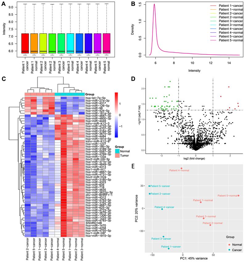
2            LIANG et al: miRNA PROFILES IN FIVE PAIRS OF EARLY GASTRIC CANCER TISSUES AND NORMAL TISSUES

markers; therefore, most patients have entered the advanced        Department of Gastroenterology, The First Hospital of Lanzhou
stage when they are detected (7). A retrospective study revealed   University (Lanzhou, China) between November 2014 and
that the 5‑year rate of relative survival of patients with early   April 2015. The inclusion criteria were: i) Patients who agreed
gastric cancer (EGC) with treatment is 105.0% compared with        to participate in the study and signed the informed consent
the expected survival of individuals from the general popula‑      were included; ii) all patients were pathologically diagnosed as
tion matched for age and sex (8). By contrast, few EGCs are        EGC. The tumor stage was confirmed according to the Eighth
discovered in China and the West, leading to 5‑year relative       Edition of the The American Joint Committee on Cancer
survival rates of 10‑40% (9‑11). In Japan, the male mortality      (AJCC) TNM staging system guidelines (22); and iii) the
rate of gastric cancer has fallen by more than half since a        patients did not receive radiotherapy or chemotherapy before
mass screening program was introduced in the early 1970s (9).      surgery. The exclusion criteria were: i) Patients who did not
Therefore, early diagnosis and treatment of gastric cancer,        agree to participate in the study; ii) patients who had other
and screening are important. Gastroscopy is a valuable tool        malignancies; and iii) patients who had other diseases, such
for reducing the mortality associated with gastric cancer (12).    as diabetes mellitus and hypertension. The patients included
However, due to its acceptability and cost, large‑scale            3 men and 2 women. Clinicopathological features of the
screening and early detection with gastroscopy might not be        patients are listed in Table I. The present study was approved
easy. At present, available tumor markers for gastric cancer,      by the Ethics Committee of The First Hospital of Lanzhou
including carcinoembryonic antigen and cancer antigen 19‑9,        University (approval no. LDYYLL2012001; Lanzhou, China).
are useful for detecting recurrence and distant metastasis or      All patients and their family members signed an informed
predicting patient survival; however, these are inadequate for     consent form. The tissue samples were stored and transported
gastric cancer screening due to their low sensitivity, particu‑    at ‑80˚C.
larly for EGC (13). Therefore, there is an urgent requirement
for novel non‑invasive methods for the screening of patients       RNA extraction. Total RNA from 10 tissue samples was
with gastric cancer, and microRNAs (miRNAs/miRs) have              isolated using TRIzol® (Invitrogen; Thermo Fisher Scientific,
been increasingly studied for this.                                Inc.) and purified using a RNeasy mini kit (Qiagen, Inc.)
    miRNAs are endogenous 18‑24 nucleotide RNAs, which             according to the manufacturer's protocols. RNA quality and
can serve critical regulatory roles in animals and plants (14).    quantity were measured using a NanoDrop spectrophotometer
miRNAs can combine with other associated proteins to form          (ND‑1000; NanoDrop Technologies; Thermo Fisher Scientific,
an active RNA‑induced silencing complex (RISC), and RISC           Inc.). RNA integrity was determined by formaldehyde agarose
combines with the 5' untranslated region, open reading frames      gel electrophoresis in 3‑(N‑morpholino)propanesulfonic acid
or 3' untranslated region of a target gene mRNA to suppress its    buffer with a 1.2% gel. Ethidium bromide was used as a fluo‑
translation or to induce its degradation (15,16). An increasing    rescent dye, and the GelDoc Go Gel Imaging System (Bio‑Rad
number of studies have reported that miRNAs can be used as         Laboratories, Inc.) was used for imaging.
biomarkers for gastric cancer diagnosis, as well as targets for
disease treatment (17‑19). For example, a study of 682 partici‑    miRNA labeling and array hybridization. miRNA microarray
pants examined the expression levels of 578 miRNAs in serum        assays were performed by Aksomics, Inc. The miRCURY™
and demonstrated that the combination of 12 miRNAs in              Hy3™/Hy5™ Power labeling kit (Exiqon; Qiagen, Inc.) was
serum has excellent diagnostic value for gastric cancer (13).      used with total RNA samples according to the manufacturer's
Microarray technology has been widely used to investigate          guidelines for miRNA labeling. After termination of the
miRNA expression in multiple tumor types, such as gastric          labeling procedure, the Hy3™‑labeled samples were hybrid‑
cancer and lung cancer (20,21).                                    ized on the array in miRCURY LNATM microRNA Array
    The present study, as a part of the aforementioned Wuwei       Kit, 7th generation‑hsa, mmu and rno (Exiqon; Qiagen, Inc.).
cohort research project, utilized samples collected from           Following hybridization, the slides were washed several times
patients diagnosed with EGC during the screening of this           using Wash buffer kit (Exiqon; Qiagen, Inc.). The slides were
disease in Wuwei. miRNA profiles in five pairs of EGC tissues      scanned using an Axon GenePix 4000 B microarray scanner
and adjacent non‑cancerous tissues were explored using a           (Molecular Devices, LLC).
miRNA microarray. Bioinformatics methods were used to
analyze the functions and mechanisms of the dysregulated           Microarray data processing. Scanned images were subse‑
miRNAs, as well as their potential as prognostic factors. The      quently imported into GenePix Pro 6.0 (Molecular Devices,
present study aimed to provide a basis for the identification      LLC) for grid alignment and data extraction. Data analyses
of EGC screening biomarkers and to discuss the particularity       were performed using R software (v3.6.3; https://cran.r‑project.
of gastric cancer in Gansu province from the perspective of        org/src/base/R‑3/). The ‘limma’ package (v3.42.2) (23) in
miRNAs.                                                            R was used for background correction and normalization
                                                                   between arrays. The robust multi‑array average algorithm
Materials and methods                                              was selected when performing background correction, and
                                                                   the ‘offset’ parameter was set to 50. Expression data were
Patients and samples. EGC tissues and adjacent non‑cancerous       normalized using median normalization. The landing lights
mucosa tissues (5 cm from the cancer tissues) were obtained        (probe ID 13138; annotated as Hy3™) were only included for
from 5 patients with EGC who were treated with endoscopic          gal‑file orientation, and their corresponding data points were
submucosal dissection at the Department of Gastroenterology,       removed prior to the normalization of the dataset, according to
The Gansu Wuwei Tumor Hospital (Wuwei, China) and                  the manual of the miRCURY LNATM microRNA Array Kit, 7th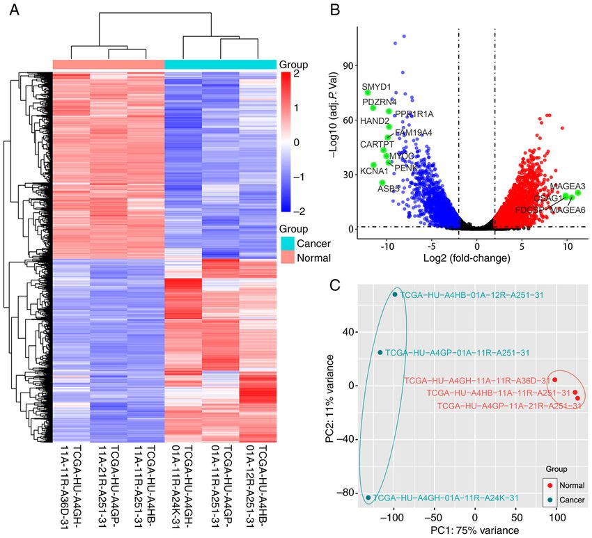
ONCOLOGY LETTERS 22: 595, 2021                                                  3

Table I. Clinicopathological features of patients with early gastric cancer whose tissues underwent microRNA microarray testing.

									 AJCC
		          Age,					                                          N     M pathologic stage
Patient Sex years Location Lesion location Histology T stage stage stage (8th edition)

Patient 1   Female   65    Wuwei, Gansu, China        Cardia           Adenocarcinoma      T1a       0      0           IA
Patient 2   Male     72    Wuwei, Gansu, China        Gastric antrum   Adenocarcinoma      Tis       0      0            0
Patient 3   Male     46    Wuwei, Gansu, China        Gastric body     Adenocarcinoma      T1b       0      0           IA
Patient 4   Male     45    Wuwei, Gansu, China        Cardia           Adenocarcinoma      T1a       0      0           IA
Patient 5   Female   67    Lanzhou, Gansu, China      Gastric antrum   Adenocarcinoma      Tis       0      0            0

AJCC, American Joint Committee on Cancer.

generation‑hsa, mmu and rno (Exiqon; Qiagen, Inc.). For the        html) (30), ‘ggplot2’ (v3.3.2) (31) and ‘enrichplot’ (v1.6.1;
same probes, the expression data of all samples corresponding      http://bioconductor.org/packages/devel/bioc/html/enrichplot.
to each probe were averaged. The probe with the maximum            html) packages were utilized to find the enriched GO terms
average value was retained among the same probes. The              and KEGG pathways, where a false discovery rate
4            LIANG et al: miRNA PROFILES IN FIVE PAIRS OF EARLY GASTRIC CANCER TISSUES AND NORMAL TISSUES

Table II. Clinicopathological features of patients with gastric cancer at stage I/II whose gene expression profiles were obtained
from TCGA.

									 AJCC
		          Age,				                                    T     N     M   pathologic stage
Patient Sex years Ethnicity Lesion location Histology stage stage stage  (8th Edition)

TCGA‑HU‑A4GH         Male        75      Asian     Gastric body      Adenocarcinoma         T1b      N0       M0             IA
TCGA‑HU‑A4GP         Female      62      Asian     Gastric antrum    Adenocarcinoma         T2       N1       M0             IIA
TCGA‑HU‑A4HB         Male        68      Asian     Gastric antrum    Adenocarcinoma         T2       N2       M0             IIB

AJCC, American Joint Committee on Cancer; TCGA, The Cancer Genome Atlas.

the miRNA‑gene network, the ‘survminer’ package (v0.4.8;
https://cran.r‑project.org/web/packages/survminer/index.html)
was used to find a best separation cutoff value, and then
patients were divided into two groups according to the cutoff
value. The prognostic values of partial DEMs were identi‑
fied by Kaplan‑Meier analysis with the ‘survival’ package
(v3.2.3) (36) in R software, and P
ONCOLOGY LETTERS 22: 595, 2021 5
Figure 2. Data analysis of the miRNA microarray using five early gastric cancer tissues and five adjacent non‑cancerous tissues, and the identification of
DEMs. (A) Box plot after normalization, which shows the maximum, upper quartile, median, lower quartile and minimum of different datasets. (B) Density
plot after normalization, which shows a probability distribution at different miRNA expression levels. (C) Heat map generated by unsupervised hierarchical
clustering for DEMs between two groups. The color varies from red to blue, indicating upregulated or downregulated expression of DEMs, respectively.
(D) Volcano plot of the DEMs in the miRNA microarray. Red and green dots represent upregulated and downregulated molecules (54 miRNAs and one
small nuclear RNA), respectively. (E) Principal component analysis result of the tissue samples. adj. P. Val., adjusted P‑value; DEMs, differentially expressed
miRNAs; miRNA/miR, microRNA; PC, principal component.
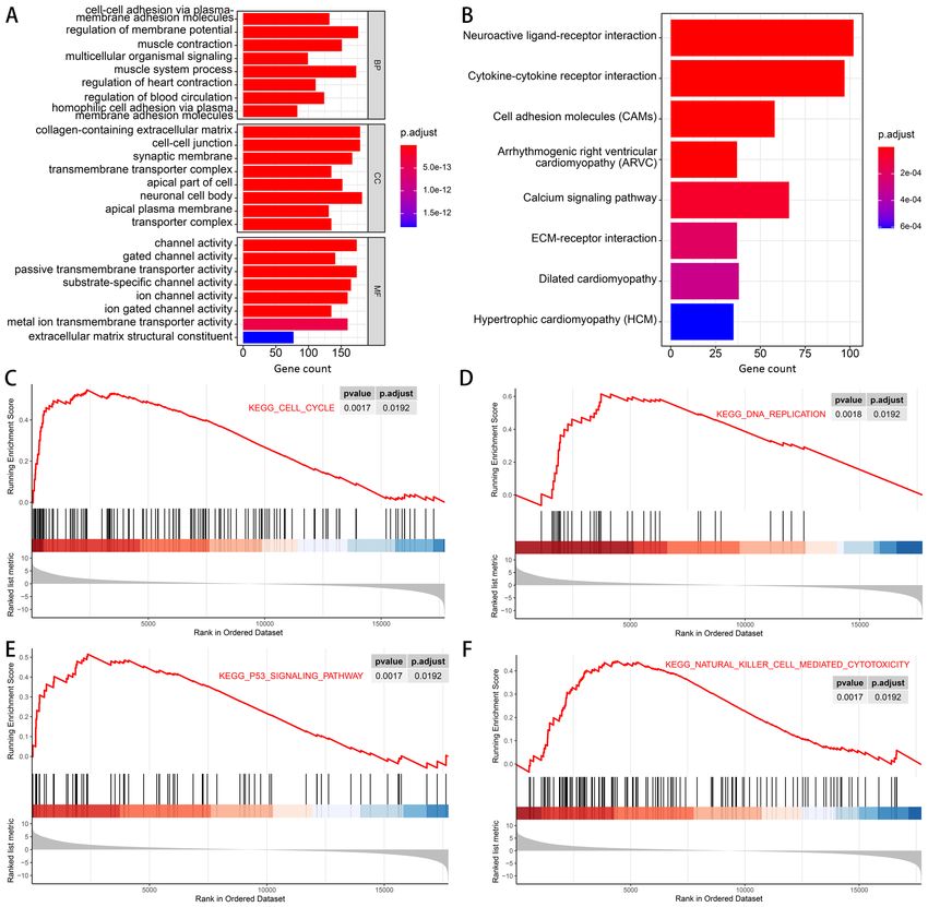
also enriched (Fig. 4B). Finally, GSEA results suggested that them. For the upregulated miRNAs, target genes were
pathways of the ‘cell cycle’, ‘DNA replication’, ‘P53 signaling predicted among the downregulated DEGs and vice versa.
pathway’ and ‘natural killer cell‑mediated cytotoxicity’ Subsequently, a miRNA‑gene network was constructed
tended to be activated in the cancer group (Fig. 4C‑F). (Fig. 5A).
GO and KEGG analyses of the predicted target genes
miRNA‑gene network construction and functions of target were performed to learn more about the functions of the
genes. After the DEMs and DEGs were identified, miRDB DEMs. In the MF category of GO analysis, the terms with
was used to predict the possible target relationships between the highest gene counts were ‘Metal ion transmembrane6 LIANG et al: miRNA PROFILES IN FIVE PAIRS OF EARLY GASTRIC CANCER TISSUES AND NORMAL TISSUES
Figure 3. Gene expression profiles of three pairs of gastric cancer tissue samples and the adjacent normal mucosa tissue samples from 3 Asian patients with
gastric cancer at stage I/II based on data obtained from TCGA. (A) Heat map generated by unsupervised hierarchical clustering for DEGs between the two
groups. The color varies from red to blue, indicating upregulated or downregulated expression of DEGs, respectively. (B) Volcano plot of the DEGs. Red dots
represent upregulated genes, and blue dots represent downregulated genes. Green dots represent genes with |log2 (fold change)| ≥9.5 (C) Principal component
analysis plot of the six samples. DEGs, differentially expressed genes; adj. P. Val., adjusted P‑value; TCGA, The Cancer Genome Atlas; PC, principal component.
transporter activity’, ‘Actin binding’ and ‘Ion channel activity’ Discussion
(Fig. 5B). KEGG terms, such as ‘Neuroactive ligand‑receptor
interaction’, ‘Pathways in cancer’ and ‘Focal adhesion’, were A total of seven upregulated and 40 downregulated human
significantly enriched (Fig. 5C). miRNAs were identified in EGC tissues compared with
The results of PPI analysis are shown in Fig. 5D. A total adjacent non‑cancerous tissues. The functions of the DEMs
of 13 genes, including ADRB3, ADCYAP1, POMC, CALCB, were annotated using the TAM webtool. ‘Hormones regula‑
PTHLH, ADCY5, GNB4, VIP, PTGDR, LHCGR, ADRB2, tion’ was the most enriched function term, which indicated
GPR83 and GIPR were filtered as hub genes in the network, that hormones may be regulated in EGC by miRNAs. A
which indicated that the DEMs may mainly work by regulating population‑based matched cohort study in Sweden suggested
these hub genes. that menopausal hormone therapy users are at a decreased
risk of gastric adenocarcinoma (37). In addition, gonad‑
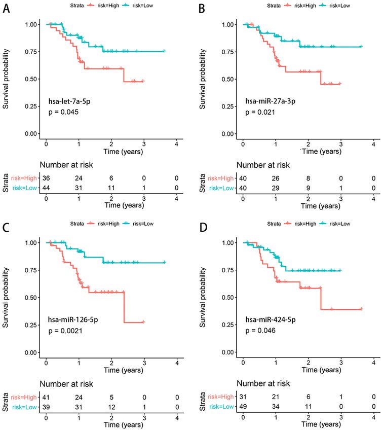
Survival analysis validation. Clinical data and RNA expres‑ otropin‑releasing hormone (38), corticotropin‑releasing
sion data of Asian patients with gastric cancer (irrespective hormone (39), steroid hormones (40) and growth hormone (41)
of the stage) from TCGA were used for survival analysis. have been reported to be associated with gastric cancer.
A total of four miRNAs (hsa‑let‑7a‑5p, hsa‑miR‑27a‑3p, However, more evidence is required to verify the role of
hsa‑miR‑126‑5p and hsa‑miR‑424‑5p) were identified to be hormone regulation in EGC.
significantly associated with the prognosis of patients with Expression profiles of three pairs of cancer tissues
gastric cancer (Fig. 6). and adjacent non‑cancerous tissues from patients withONCOLOGY LETTERS 22: 595, 2021 7 Table III. Functional annotation of differentially expressed miRNAs using the tool for annotations of human miRNAs. Function term Count Percent Fold P‑value Hormones regulation 9 0.14516129 3.27217742 0.00062950 Human embryonic stem cell (hESC) regulation 8 0.09411765 2.12156863 0.02291040 Immune response 8 0.16666667 3.75694444 0.00052898 Inflammation 8 0.19512195 4.39837398 0.00016338 Cell cycle related 7 0.10606061 2.39078283 0.01856736 Epithelial‑mesenchymal transition 5 0.12195122 2.74898374 0.02820963 Cell death 5 0.09090909 2.04924242 0.08525479 Hematopoiesis 5 0.16129032 3.63575269 0.00867962 MiRNA tumor suppressors 5 0.13513514 3.04617117 0.01854272 Lipid metabolism 4 0.20000000 4.50833333 0.00898287 Angiogenesis 3 0.12500000 2.81770833 0.08400821 Cell proliferation 3 0.10714286 2.41517857 0.12100311 HIV latency 3 0.14285714 3.22023810 0.06027038 Adipocyte differentiation 3 0.11111111 2.50462963 0.11122976 Onco‑miRNAs 3 0.09677419 2.18145161 0.15217690 Apoptosis 2 0.04545455 1.02462121 0.59774380 Bone regeneration 2 0.05882353 1.32598039 0.45394525 Folliculogenesis 2 0.28571429 6.44047619 0.03458783 Anti‑cell proliferation (Hwang etal BJC2006) 2 0.18181818 4.09848485 0.08125429 Cell division 2 0.11764706 2.65196078 0.17103576 Immune system (Xiao's Cell2009) 2 0.11111111 2.50462963 0.18736736 Cell differentiation 1 0.05882353 1.32598039 0.54311212 Chemosensitivity of tumor cells 1 0.25000000 5.63541667 0.16641762 DNA repair 1 0.10000000 2.25416667 0.36724517 Granulopoiesis 1 0.10000000 2.25416667 0.36724517 Carbohydrate metabolism 1 0.14285714 3.22023810 0.27345270 Cell fate determination 1 0.03846154 0.86698718 0.70138621 Heart development 1 0.14285714 3.22023810 0.27345270 miRNA, microRNA. stage I/II gastric cancer were downloaded. Gastric cancer neurodegenerative events are considered to be associated exhibits biological and epidemiological differences between with gastric cancer, according to previous reports (46‑48). Asian and non‑Asian populations (42,43). To improve the Based on the target gene results, ‘pathways in cancer’ and understanding of the roles of DEMs, the present study aimed other pathways associated with cancer, such as ‘Focal adhe‑ to identify target mRNAs among DEGs in Asian patients. sion’ (49) and ‘Melanoma’, were also affected. Since the Since only 1 Asian patient with EGC has been included results obtained by this method are closely associated with in the TCGA database to date, the scope was expanded the gene set, the two methods of detecting functional enrich‑ to patients at stage I/II (AJCC, Eighth Edition). Notably, ment complement each other. ‘Neuroactive ligand‑receptor interaction’ and ‘Calcium The miRNA‑gene network was constructed. However, signaling pathway’ were enriched KEGG terms for DEGs. >90% of the target genes were identified to be poten‑ ‘Neuroactive ligand‑receptor interaction’, ‘Axon guidance’ tially modulated by hsa‑let‑7a‑5p, hsa‑miR‑27a‑3p, and ‘Neurotrophin signaling pathway’ were enriched KEGG hsa‑miR‑126‑5p, hsa‑miR‑424‑5p, hsa‑miR‑181a‑5p and terms for the target genes of DEMs. Furthermore, ‘muscle hsa‑miR‑1915‑3p in the network. Survival analysis of Asian contraction’ was an enriched biological process term in patients demonstrated that four of the miRNAs (hsa‑let‑7a‑5p, GO analysis of DEGs. These results suggest that pathways hsa‑miR‑27a‑3p, hsa‑miR‑126‑5p and hsa‑miR‑424‑5p) were associated with nerves, muscle contraction and calcium significantly associated with the prognosis of patients with signaling may serve a role in gastric cancer. There is much gastric cancer. Multiple studies have demonstrated that evidence regarding the association between the calcium hsa‑miR‑27a‑3p (50‑53) and hsa‑miR‑424‑5p (54‑56) are signaling pathway and gastric cancer (44,45). Regarding upregulated in gastric cancer and act as tumor activators. nerve and muscle contraction, certain proteins involved in The present results demonstrated that hsa‑miR‑27a‑3p and
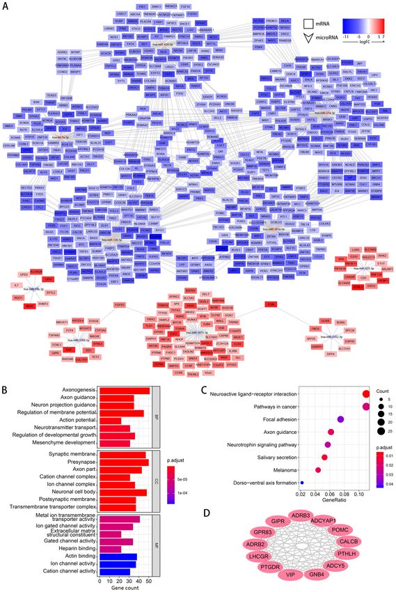
8              LIANG et al: miRNA PROFILES IN FIVE PAIRS OF EARLY GASTRIC CANCER TISSUES AND NORMAL TISSUES

Figure 4. GO analysis, KEGG analysis and GSEA of the differentially expressed genes between the cancer tissues and non‑cancerous tissues of 3 Asian
patients with stage I/II gastric cancer. (A) Results of GO analysis, including BP, CC and MF aspects. (B) Results of KEGG analysis. (C‑F) Results of GSEA
revealing that the KEGG pathways of (C) ‘cell cycle’, (D) ‘DNA replication’, (E) ‘P53 signaling pathway’ and (F) ‘natural killer cell‑mediated cytotoxicity’
were activated in the gastric cancer tissues at stage I/II. BP, biological process; CC, cellular component; GO, Gene Ontology; GSEA, Gene Set Enrichment
Analysis; KEGG, Kyoto Encyclopedia of Genes and Genomes; MF, molecular function; p.adjust, adjusted P‑value.

has‑miR‑424‑5p could be upregulated in EGC and may affect                       hsa‑miR‑126‑5p in gastric cancer and EGC need to be verified.
the prognosis of patients with gastric cancer. Liang et al (57)                 Experiments have demonstrated that hsa‑miR‑181a‑5p can
reported that the hsa‑let‑7 family inhibits tumor invasion                      promote the progression of gastric cancer via Ras associa‑
and metastasis by targeting myosin heavy chain 9 in human                       tion domain family member 6‑mediated mitogen activated
gastric cancer, which is different from the results of the                      kinase‑like protein signaling activation (61) or by regulating
present study. Since the sample size included in the present                    protein tyrosine phosphatase non‑receptor type 9 (62).
study was small, the results should be verified with larger                     Furthermore, hsa‑miR‑1915‑3p inhibits Bcl‑2 expression
sample sizes. To the best of our knowledge, there have been                     in the development of gastric cancer (63). hsa‑miR‑1915‑3p
no studies on hsa‑miR‑126‑5p in gastric cancer. Although a                      serves a role in breast cancer inhibition (64) and increases
study using expression profile analysis of miRNAs in pros‑                      drug sensitivity in colorectal cancer (65). The present results
tate cancer by next‑generation sequencing demonstrated that                     support the role of hsa‑miR‑181a‑5p as a tumor activator and
hsa‑miR‑126‑5p is highly expressed in tumor tissues (58),                       the role of hsa‑miR‑1915‑3p as a tumor suppressor. Overall,
other studies came to different conclusions and have suggested                  the studies of miRNAs in EGC are still limited, and the
that it may be a tumor suppressor (59,60). The functions of                     present study provided some evidence for this. There are also
ONCOLOGY LETTERS 22: 595, 2021                                                                    9

Figure 5. Construction of the miRNA‑gene network and functions of the target genes. (A) Predicted miRNA‑gene network in gastric cancer tissues at stage I/II.
(B) Gene Ontology analysis and (C) Kyoto Encyclopedia of Genes and Genomes analysis of the predicted target genes of miRNAs. (D) Hub genes that interact
closely with each other, which were filtered from the protein‑protein interaction results. BP, biological process; CC, cellular component; FC, fold change;
MF, molecular function; miRNA, microRNA; p.adjust, adjusted P‑value.

differentially expressed viral miRNAs and snRNAs. Further                          The present study intended to perform preliminary
research is required to clarify their functions.                                screening of miRNAs and the results need to be verified in
10             LIANG et al: miRNA PROFILES IN FIVE PAIRS OF EARLY GASTRIC CANCER TISSUES AND NORMAL TISSUES

Figure 6. Survival analysis of the differentially expressed miRNAs. Prognostic value of (A) hsa‑let‑7a‑5p, (B) hsa‑miR‑27a‑3p, (C) hsa‑miR‑126‑5p and
(D) hsa‑miR‑424‑5p in Asian patients with gastric cancer based on data from The Cancer Genome Atlas database. The x‑axis indicates survival time in years,
and the y‑axis indicates survival probability. P‑values, which are shown in each figure, refer to the comparison between the high and low miRNA expression
groups, which were divided according to the best separation cutoff value. miR/miRNA, microRNA.

a larger cohort. Additionally, the present study was based on                  mechanisms and abilities as prognostic factors of DEMs
individuals in Gansu province and Asia, which is helpful to                    were assessed using bioinformatics methods. The present
explore the particularity of gastric cancer in these regions.                  study revealed that certain miRNAs, including hsa‑let‑7a‑5p,
However, the population is also a limitation of the present                    hsa‑miR‑27a‑3p, hsa‑miR‑126‑5p and hsa‑miR‑424‑5p, were
study when the results need to be extended to other regions or                 upregulated in EGC, and were associated with the prognosis
other high incidence areas of gastric cancer. Comparing the                    of gastric cancer based on analysis of the miRNA expression
results with other population cohorts should be a direction of                 profiles of patients from Gansu province, China. The present
future research.                                                               study could provide a basis for the identification of EGC
     In conclusion, in the present study, the miRNA profiles in                screening biomarkers and for exploring the cause of the high
five pairs of EGC tissues and adjacent non‑cancerous tissues                   incidence of gastric cancer in Gansu province, China from the
were investigated using a miRNA microarray. The possible                       perspective of miRNAs.
ONCOLOGY LETTERS 22: 595, 2021 11
Acknowledgements 4. Guo Q, Liu X, Lu L, Yuan H, Wang Y, Chen Z, Ji R and Zhou Y:
Comprehensive evaluation of clinical efficacy and safety of cele‑
coxib combined with chemotherapy in management of gastric
Not applicable. cancer. Medicine (Baltimore) 96: e8857, 2017.
5. Ji R: Gastric cancer screening and related risk factors analysis in
the Wuwei Natrual population cohort. Lanzhou University, 2018.
Funding 6. Pasechnikov V, Chukov S, Fedorov E, Kikuste I and Leja M:
Gastric cancer: Prevention, screening and early diagnosis.
The present study was supported by the National Key World J Gastroenterol 20: 13842‑13862, 2014.
7. Everett SM and Axon AT: Early gastric cancer in Europe. Gut 41:
R&D Program of China (grant no. 2016YFC1302201), the 142‑150, 1997.
Natural Science Foundation of Gansu Province, China 8. Suzuki H, Oda I, Abe S, Sekiguchi M, Mori G, Nonaka S,
(grant nos. 18JR3RA336 and 17JR5RA272) and the Yoshinaga S and Saito Y: High rate of 5‑year survival among
patients with early gastric cancer undergoing curative endoscopic
Fundamental Research Funds for the Central Universities submucosal dissection. Gastric Cancer 19: 198‑205, 2016.
(grant no. lzujbky‑2020‑kb16). 9. Crew KD and Neugut AI: Epidemiology of gastric cancer.
World J Gastroenterol 12: 354‑362, 2006.
10. Gong W, Luo S, Hu R, Wang H, Pan J, Fei F, Wu H and Yu M:
Availability of data and materials Survival analysis of gastric cancer patients during 2005‑2010
in Zhejiang Province, China. Zhonghua Zhong Liu Za Zhi 36:
The datasets generated and analyzed during the current 636‑639, 2014 (In Chinese).
11. Zheng L, Wu C, Xi P, Zhu M, Zhang L, Chen S, Li X, Gu J and
study are available in the Gene Expression Omnibus Zheng Y: The survival and the long‑term trends of patients with
repository (https://www.ncbi.nlm.nih.gov/geo/query/acc. gastric cancer in Shanghai, China. BMC Cancer 14: 300, 2014.
cgi?&acc=GSE158315). 12. Hamashima C, Shabana M, Okada K, Okamoto M and Osaki Y:
Mortality reduction from gastric cancer by endoscopic and radio‑
graphic screening. Cancer Sci 106: 1744‑1749, 2015.
Authors' contributions 13. So JBY, Kapoor R, Zhu F, Koh C, Zhou L, Zou R, Tang YC,
Goo PCK, Rha SY, Chung HC, et al: Development and valida‑
tion of a serum microRNA biomarker panel for detecting gastric
YL, WS and YW conceived the study, and YZ and LL cancer in a high‑risk population. Gut 70: 829‑837, 2021.
designed the study. YZ, HW and HY were involved in the 14. Bartel DP: MicroRNAs: Genomics, biogenesis, mechanism, and
sample and data collection. YL, LL and TH performed the function. Cell 116: 281‑297, 2004.
15. Calin GA, Sevignani C, Dumitru CD, Hyslop T, Noch E,
RNA extraction experiments and the quality control. YL, LL Yendamuri S, Shimizu M, Rattan S, Bullrich F, Negrini M and
and HW performed the bioinformatics analysis and prepared Croce CM: Human microRNA genes are frequently located at
the figures. YZ, HZ, HY and XC performed the statistical fragile sites and genomic regions involved in cancers. Proc Natl
Acad Sci USA 101: 2999‑3004, 2004.
analysis. YL, YZ and TH drafted the manuscript. HZ, XC and 16. Han J, Lee Y, Yeom KH, Kim YK, Jin H and Kim VN: The
WS contributed substantially to the revision of the manuscript. Drosha‑DGCR8 complex in primary microRNA processing.
The authenticity of all the raw data has been assessed by Genes Dev 18: 3016‑3027, 2004.
17. Zheng GD, Xu ZY, Hu C, Lv H, Xie XH, Huang T, Zhang YQ,
YL, WS and YW. All authors read and approved the final Chen GP, Fu YF and Cheng XD: Exosomal miR‑590‑5p in serum
manuscript. as a biomarker for the diagnosis and prognosis of gastric cancer
front. Mol Biosci 8: 636566, 2021.
18. Shi Y, Wang Z, Zhu X, Chen L, Ma Y, Wang J, Yang X and Liu Z:
Ethics approval and consent to participate Exosomal miR‑1246 in serum as a potential biomarker for early
diagnosis of gastric cancer. Int J Clin Oncol 25: 89‑99, 2020.
T he st udy protocol was approved by the Et h ics 19. Xia L, Zhang D, Du R, Pan Y, Zhao L, Sun S, Hong L, Liu J and
Fan D: miR‑15b and miR‑16 modulate multidrug resistance by
Committee of The First Hospital of Lanzhou University targeting BCL2 in human gastric cancer cells. Int J Cancer 123:
(approval no. LDYYLL2012001; Lanzhou, China). Written 372‑379, 2008.
informed consent forms were signed by the patients and their 20. Shin VY and Chu KM: MiRNA as potential biomarkers and
therapeutic targets for gastric cancer. World J Gastroenterol 20:
family members. 10432‑10439, 2014.
21. Lu M, Hu C, Wu F, Shu L, Pan Y, Liu X, Liu P, Ma F, Deng C
Patient consent for publication and Huang M: MiR‑320a is associated with cisplatin resistance
in lung adenocarcinoma and its clinical value in non‑small cell
lung cancer: A comprehensive analysis based on microarray
Not applicable. data. Lung Cancer 147: 193‑197, 2020.
22. Amin MB, Edge SB, Greene FI, Byrd DR, Brookland RK,
Washington MK, Gershenwald JE, Compton CC, Hess KR,
Competing interests Sullivan DC, et al: AJCC cancer staging manual. Springer‑Verlag.
New York, the United States, 2016.
The authors declare that they have no competing interests. 23. Ritchie ME, Phipson B, Wu D, Hu Y, Law CW, Shi W and
Smyth GK: Limma powers differential expression analyses for
RNA‑sequencing and microarray studies. Nucleic Acids Res 43:
References e47, 2015.
24. Barrett T, Wilhite SE, Ledoux P, Evangelista C, Kim IF,
Tomashevsky M, Marshall KA, Phillippy KH, Sherman PM,
1. Bray F, Ferlay J, Soerjomataram I, Siegel RL, Torre LA and Holko M, et al: NCBI GEO: Archive for functional genomics
Jemal A: Global cancer statistics 2018: GLOBOCAN estimates data sets‑update. Nucleic Acids Res 41: D991‑D995, 2013.
of incidence and mortality worldwide for 36 cancers in 185 25. Li J, Han X, Wan Y, Zhang S, Zhao Y, Fan R, Cui Q and Zhou Y:
countries. CA Cancer J Clin 68: 394‑424, 2018. TAM 2.0: Tool for MicroRNA set analysis. Nucleic Acids Res 46:
2. Liu Y, Zhang X, Chen L, Zhao Q and Xia X: Cancer incidence W180‑W185, 2018.
and mortality in Gansu province, 2012. Chin J Cancer Res 28: 26. Love MI, Huber W and Anders S: Moderated estimation of fold
301‑310, 2016. change and dispersion for RNA‑seq data with DESeq2. Genome
3. Chen W, Zheng R, Zhang S, Zeng H, Zuo T, Jia M, Xia C, Zou X Biol 15: 550, 2014.
and He J: Report of cancer incidence and mortality in China, 27. Gaudet P and Dessimoz C: Gene ontology: Pitfalls, biases, and
2012. China Cancer 25: 1‑8, 2016. remedies. Methods Mol Biol 1446: 189‑205, 2017.12 LIANG et al: miRNA PROFILES IN FIVE PAIRS OF EARLY GASTRIC CANCER TISSUES AND NORMAL TISSUES
28. Kanehisa M, Furumichi M, Tanabe M, Sato Y and Morishima K: 48. Park T, Lee YJ, Jeong SH, Choi SK, Jung EJ, Ju YT, Jeong CY,
KEGG: New perspectives on genomes, pathways, diseases and Park M, Hah YS, Yoo J, et al: Overexpression of neuron‑specific
drugs. Nucleic Acids Res 45: D353‑D361, 2017. enolase as a prognostic factor in patients with gastric cancer.
29. Subramanian A, Tamayo P, Mootha VK, Mukherjee S, Ebert BL, J Gastric Cancer 17: 228‑236, 2017.
Gillette MA, Paulovich A, Pomeroy SL, Golub TR, Lander ES and 49. Eke I and Cordes N: Focal adhesion signaling and therapy resis‑
Mesirov JP: Gene set enrichment analysis: A knowledge‑based tance in cancer. Semin Cancer Biol 31: 65‑75, 2015.
approach for interpreting genome‑wide expression profiles. Proc 50. Du M, Zheng R, Ma G, Chu H, Lu J, Li S, Xin J, Tong N, Zhang G,
Natl Acad Sci USA 102: 15545‑15550, 2005. Wang W, et al: Remote modulation of lncRNA GCLET by risk
30. Yu G, Wang LG, Han Y and He QY: clusterProfiler: An variant at 16p13 underlying genetic susceptibility to gastric
R package for comparing biological themes among gene clusters. cancer. Sci Adv 6: eaay5525, 2020.
OMICS 16: 284‑287, 2012. 51. Yang F, Chen X, Li X, Chen J, Tang Y, Cai Y, Wang Y, Chen Z,
31. Wickham H: ggplot2: Elegant Graphics for Data Analysis. Li L, Li R and Deng Z: Long intergenic non‑protein coding
Springer‑Verlag, New York, NY, 2016. RNA 1089 suppresses cell proliferation and metastasis in gastric
32. Chen Y and Wang X: miRDB: An online database for predic‑ cancer by regulating miRNA‑27a‑3p/epithelial‑mesenchymal
tion of functional microRNA targets. Nucleic Acids Res 48: transition (EMT) axis. Cancer Manag Res 12: 5587‑5596, 2020.
D127‑D131, 2020. 52. Geng G, Liu X, Xu A, Lu Z, Chen K, He J, Qi D and Yuan X:
33. Shannon P, Markiel A, Ozier O, Baliga NS, Wang JT, Ramage D, Low abundance of TFPI‑2 by both promoter methylation and
Amin N, Schwikowski B and Ideker T: Cytoscape: A software miR‑27a‑3p regulation is linked with poor clinical outcome in
environment for integrated models of biomolecular interaction gastric cancer. J Gene Med 22: e3166, 2020.
networks. Genome Res 13: 2498‑2504, 2003. 53. Zhou L, Liang X, Zhang L, Yang L, Nagao N, Wu H, Liu C,
34. Szklarczyk D, Gable AL, Lyon D, Junge A, Wyder S, Lin S, Cai G and Liu J: MiR‑27a‑3p functions as an oncogene in
Huerta‑Cepas J, Simonovic M, Doncheva NT, Morris JH, gastric cancer by targeting BTG2. Oncotarget 7: 51943‑51954,
Bork P, et al: STRING v11: Protein‑protein association networks 2016.
with increased coverage, supporting functional discovery in 54. Zhang J, Liu H, Hou L, Wang G, Zhang R, Huang Y, Chen X
genome‑wide experimental datasets. Nucleic Acids Res 47: and Zhu J: Circular RNA_LARP4 inhibits cell proliferation and
D607‑D613, 2019. invasion of gastric cancer by sponging miR‑424‑5p and regu‑
35. Bader GD and Hogue CW: An automated method for finding lating LATS1 expression. Mol Cancer 16: 151, 2017.
molecular complexes in large protein interaction networks. BMC 55. Wei S, Li Q, Li Z, Wang L, Zhang L and Xu Z: Correction:
Bioinformatics 4: 2, 2003. miR‑424‑5p promotes proliferation of gastric cancer by targeting
36. Therneau TM and Grambsch PM: Modeling Survival Data: Smad3 through TGF‑β signaling pathway. Oncotarget 8: 34018,
Extending the Cox Model. Springer‑Verlag, New York, NY, 2000. 2017.
37. Brusselaers N, Maret‑Ouda J, Konings P, El‑Serag HB and 56. Wei S, Li Q, Li Z, Wang L, Zhang L and Xu Z: miR‑424‑5p
Lagergren J: Menopausal hormone therapy and the risk of promotes proliferation of gastric cancer by targeting Smad3
esophageal and gastric cancer. Int J Cancer 140: 1693‑1699, 2017. through TGF‑β signaling pathway. Oncotarget 7: 75185‑75196,
38. Lu M, Zhu J, Ling Y, Shi W, Zhang C and Wu H: The lower 2016.
expression of gonadotropin‑releasing hormone receptor associ‑ 57. Liang S, He L, Zhao X, Miao Y, Gu Y, Guo C, Xue Z, Dou W,
ated with poor prognosis in gastric cancer. Int J Clin Exp Med 8: Hu F, Wu K, et al: MicroRNA let‑7f inhibits tumor invasion
13365‑13370, 2015. and metastasis by targeting MYH9 in human gastric cancer.
39. Yang S, Liu W, Wen J, Zhu M and Xu S: Corticotropin releasing PLoS One 6: e18409, 2011.
hormone is correlated with tumorigenesis of gastric cancer. 58. Song C, Chen H, Wang T, Zhang W, Ru G and Lang J:
Cancer Invest 31: 167‑171, 2013. Expression profile analysis of microRNAs in prostate cancer by
40. Frycz BA, Murawa D, Borejsza‑Wysocki M, Wichtowski M, next‑generation sequencing. Prostate 75: 500‑516, 2015.
Spychała A, Marciniak R, Murawa P, Drews M and 59. Queiroz AL, Zhang B, Comstock DE, Hao Y, Eriksson M,
Jagodziński PP: mRNA expression of steroidogenic enzymes, Hydbring P, Vakifahmetoglu‑Norberg H and Norberg E:
steroid hormone receptors and their coregulators in gastric miR‑126‑5p targets Malate Dehydrogenase 1 in non‑small cell
cancer. Oncol Lett 13: 3369‑3378, 2017. lung carcinomas. Biochem Biophys Res Commun 499: 314‑320,
41. Gan J, Ke X, Jiang J, Dong H, Yao Z, Lin Y, Lin W, Wu X, Yan S, 2018.
Zhuang Y, et al: Growth hormone‑releasing hormone receptor 60. Wang C, Zhou B, Liu M, Liu Y and Gao R: miR‑126‑5p resto‑
antagonists inhibit human gastric cancer through downregulation ration promotes cell apoptosis in cervical cancer by targeting
of PAK1‑STAT3/NF‑κ B signaling. Proc Natl Acad Sci USA 113: Bcl2l2. Oncol Res 25: 463‑470, 2017.
14745‑14750, 2016. 61. Mi Y, Zhang D, Jiang W, Weng J, Zhou C, Huang K, Tang H,
42. Yamada T, Yoshikawa T, Taguri M, Hayashi T, Aoyama T, Yu Y, Liu X, Cui W, et al: miR‑181a‑5p promotes the progres‑
Sue‑Ling HM, Bonam K, Hayden JD and Grabsch HI: The sion of gastric cancer via RASSF6‑mediated MAPK signalling
survival difference between gastric cancer patients from the activation. Cancer Lett 389: 11‑22, 2017.
UK and Japan remains after weighted propensity score analysis 62. Liu Z, Sun F, Hong Y, Liu Y, Fen M, Yin K, Ge X, Wang F, Chen X
considering all background factors. Gastric Cancer 19: 479‑489, and Guan W: MEG2 is regulated by miR‑181a‑5p and functions as
2016. a tumour suppressor gene to suppress the proliferation and migra‑
43. Kerckhoffs KGP, Liu DHW, Saragoni L, van der Post RS, Langer R, tion of gastric cancer cells. Mol Cancer 16: 133, 2017.
Bencivenga M, Iglesias M, Gallo G, Hewitt LC, Fazzi GE, et al: 63. Cui HW, Han WY, Hou LN, Yang L, Li X and Su XL: miR‑1915‑3p
Mucin expression in gastric‑ and gastro‑oesophageal signet‑ring inhibits Bcl‑2 expression in the development of gastric cancer.
cell cancer: Results from a comprehensive literature review and a Biosci Rep 39: BSR20182321, 2019.
large cohort study of Caucasian and Asian gastric cancer. Gastric 64. Jin ML, Kim YW, Jin HL, Kang H, Lee EK, Stallcup MR and
Cancer 23: 765‑779, 2020. Jeong KW: Aberrant expression of SETD1A promotes survival
44. Zhang ZL, Li ZR, Li JS and Wang SR: Calcium‑sensing receptor and migration of estrogen receptor α‑positive breast cancer cells.
antagonist NPS‑2143 suppresses proliferation and invasion of Int J Cancer 143: 2871‑2883, 2018.
gastric cancer cells. Cancer Gene Ther 27: 548‑557, 2020. 65. Xu K, Liang X, Cui D, Wu Y, Shi W and Liu J: miR‑1915
45. Tang B, Wu J, Zhu MX, Sun X, Liu J, Xie R, Dong TX, Xiao Y, inhibits Bcl‑2 to modulate multidrug resistance by increasing
Carethers JM, Yang S and Dong H: VPAC1 couples with TRPV4 drug‑sensitivity in human colorectal carcinoma cells. Mol
channel to promote calcium‑dependent gastric cancer progression Carcinog 52: 70‑78, 2013.
via a novel autocrine mechanism. Oncogene 38: 3946‑3961, 2019.
46. Ge C, Li Q, Wang L and Xu X: The role of axon guidance factor This work is licensed under a Creative Commons
semaphorin 6B in the invasion and metastasis of gastric cancer. Attribution-NonCommercial-NoDerivatives 4.0
J Int Med Res 41: 284‑292, 2013. International (CC BY-NC-ND 4.0) License.
47. Pan G, Zhang X, Ren J, Lu J, Li W, Fu H, Zhang S and Li J:
Semaphorin 5A, an axon guidance molecule, enhances the inva‑
sion and metastasis of human gastric cancer through activation of
MMP9. Pathol Oncol Res 19: 11‑18, 2013.You can also read